Università Politecnica delle Marche
Dottorato di Scienze
Microbial biodiversity and viral impact in benthic
deep-sea ecosystems
Tesi di dottorato: Elisabetta Manea Tutor: Prof.ssa Cinzia Corinaldesi
Co-tutor: Prof. Antonio Dell’Anno
Dottorato di ricerca –Anno accademico 2015/2016 – XV° ciclo
Curriculum Biologia ed Ecologia Marina
Index
State of art
pag.7Microbiome of deep-sea sediments: abundance, distribution and diversity pag.7
Virus-prokaryote interactions in deep-sea sediments pag.9 Hadal trenches and microbial assemblages pag.10 Deep-sea mining impact on the seafloor assemblages pag.14
References
pag.17Chapter 1
Impact of viral infection on prokaryotic assemblages in hadal benthic
ecosystems of the Pacific Ocean
1.1 Introduction
pag.261.2 Materials and Methods
pag.271.2.1 Site description and sampling pag.27 1.2.2 Sedimentary organic matter pag.29 1.2.3 Prokaryotic abundance and biomass pag.30 1.2.4 Extracellular enzymatic activities pag.31 1.2.5 Viral abundance pag.32 1.2.6 Viral production, virus-induced prokaryotic mortality (VIPM) and turnover pag.32 1.2.7 Carbon contribution to microbial C cycle due to viral shunt pag.33 1.2.8 Statistics pag.33
1.3 Results
pag.34 1.3.1 Biochemical composition of organic matter content and photosynthetic pigments pag.34 1.3.2 Prokaryotic abundance and biomass pag.36 1.3.3 Extracellular enzymatic activity and efficiency pag.37 1.3.4 Viral abundance and virus to prokaryote ratio pag.39 1.3.5 Viral production and turnover pag.401.4 Discussion
pag.441.5 Conclusions
pag.46References
pag.47Chapter 2
Virus-prokaryote interactions and prokaryotic diversity in the deepest
ecosystem on Earth: the Mariana Trench
2.1 Introduction
pag.512.2 Materials and Methods
pag.52 2.2.1 Sampling areas pag.52 2.2.2 Biochemical composition of organic matter pag.54 2.2.3 Virus abundance pag.55 2.2.4 Viral production, turnover and virus-induced prokaryotic mortality (VIPM) pag.55 2.2.5 Prokaryotic abundance and biomass pag.56 2.2.6 Extracellular enzymatic activities pag.57 2.2.7 DNA extraction pag.57 2.2.8 Sequencing and bioinformatics pag.58 2.2.9 Statistical analyses pag.582.3 Results
pag.59 2.3.1 Trophic resources inside and outside the Mariana Trench pag.59 2.3.2 Viral abundance, production and turnover rates pag.60 2.3.3 Prokaryotic abundance and biomass pag.61 2.3.4 Virus to prokaryote ratio and virus-induced mortality pag.62 2.3.5 Extracellular enzymatic activity pag.64 2.3.6 Prokaryotic diversity pag.65 2.3.7 Taxonomy composition of the prokaryotic assemblages pag.692.4 Discussion
pag.73References
pag.77Chapter 3
Patterns of microbial biodiversity in abyssal and hadal systems
3.1 Introduction
pag.813.2 Materials and Methods
pag.82 3.2.1 Sampling areas pag.82 3.2.2 Environmental and trophic characteristics and virus-induced prokaryoticmortality pag.84 3.2.3 Bacteria and Archaea abundances pag.85 3.2.4 DNA extraction pag.86 3.2.5 Sequencing and bioinformatics pag.86 3.2.6 Statistics pag.87
3.3 Results
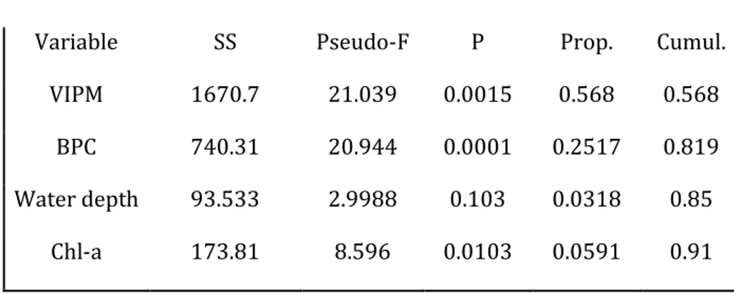
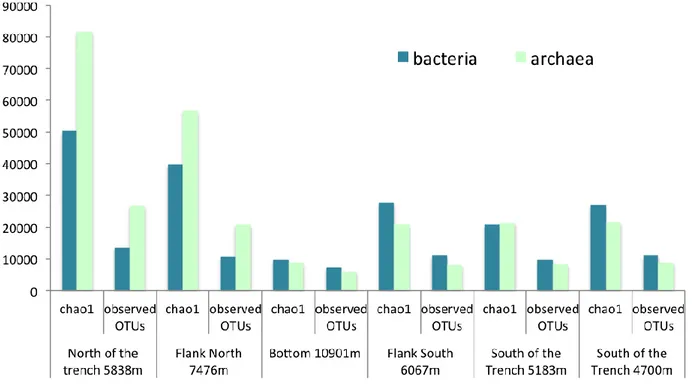
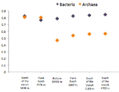
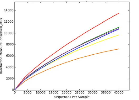
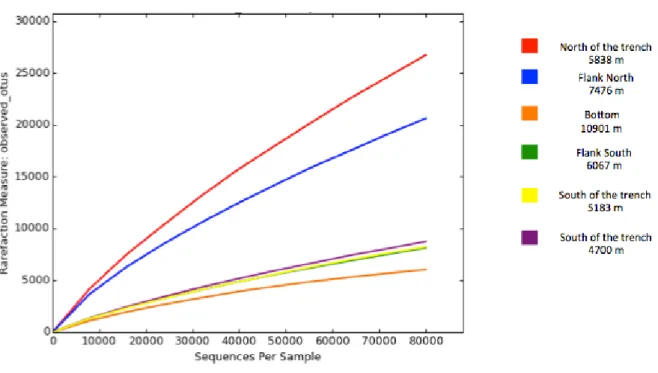
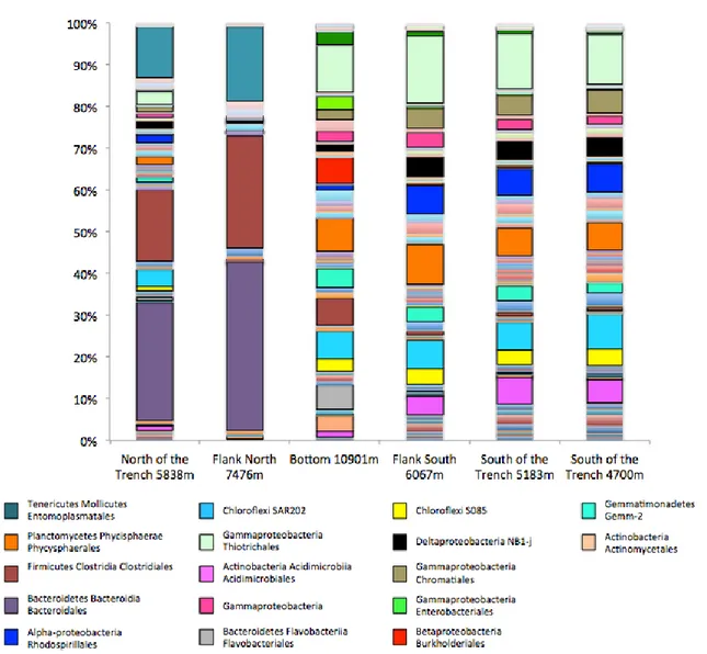
pag.87 3.3.1 Environmental variables and virus-induced prokaryotic mortality pag.87 3.3.2 Bacterial and archaeal abundances pag.88 3.3.3 Estimates of bacterial richness and diversity and rarefaction curves pag.90 3.3.4 Taxonomy of bacterial assemblages pag.92 3.3.5 Estimates of archaeal richness and diversity and rarefaction curves pag.94 3.3.6 Taxonomy of archaeal assemblages pag.96 3.3.7 Influence of environmental variables on hadal microbial assemblages pag.983.4 Discussion
pag.1003.5 Conclusions
pag.102Chapter 4
Anthropogenic impact due to mining activity on virus-prokaryote
interactions and prokaryotic diversity in abyssal ecosystems of the
Pacific Ocean
4.1 Introduction
pag.1084.2 Materials and Methods
pag.110 4.2.1 Study area and sampling pag.110 4.2.2 Biochemical composition of organic matter pag.112 4.2.3 Prokaryotic abundance and biomass pag.113 4.2.4 Extracellular enzymatic activities pag.113 4.2.5 Virus abundance pag.114 4.2.6 Viral production, turnover and virus-induced prokaryotic mortality (VIPM) pag.115 4.2.7 DNA extraction suitable for molecular analysis pag.115 4.2.8 Sequencing and bioinformatics pag.116 4.2.9 Statistical analyses pag.1174.3 Results
pag.117 4.3.1 Impact on trophic resources pag.117 4.3.2 Impact on benthic deep-sea prokaryotes pag.118 4.3.3 Impact on extracellular enzymatic activities pag.119 4.3.4 Impact on viral abundance, production and turnover times pag.121 4.3.5 Impact of deep-sea mining on virus-prokaryote interactions pag.122 4.3.6 Impact on bacterial richness and diversity pag.124 4.3.7 Bacterial diversity pag.1274.4 Discussion
pag.1374.5 Conclusions
pag.140References
pag.141Supplementary material
pag.147Acknowledgements
This PhD study was carried out in the framework of the European project MIDAS (Managing Impacts of Deep-seA reSources exploitation). I want to thank Dr. Pedro Martinez Arbizu from the Deutsches Zentrum für Marine Biodiversitätsforschung Senckenberg am Meer Südstrand (Germany), for the logistical support in collecting sediment samples in the CCFZ (Pacific Ocean) during the oceanographic cruise carried out in March-April 2015 on board of the R/V Sonne. I want to thank Dr. Takuro Nunoura from the Japan Agency for Marine-Earth Science and Technology, JAMSTEC, for the logistical support in collecting additional sediment samples in different locations of the Pacific Ocean including trenches and abyssal plains.
In particular, I want to thank Prof. Cinzia Corinaldesi, Prof. Antonio Dell’Anno and Prof. Roberto Danovaro for the opportunity they gave me to conduct my PhD study.
State of art
Microbiome of deep-sea sediments: abundance, distribution and
diversity of prokaryotes and viruses
Deep-sea environments (the waters and sediments of the ocean interior beneath 200m depth) are the largest biome of the world, representing more than 65% of the Earth’s surface and more than 95% of the global biosphere (Herring, 2001; Corinaldesi, 2015). In general, abyssal (3,000–6,000 m) and hadal (>6,000 m) environments are characterized by low temperature, high hydrostatic pressure and the absence of solar radiation (Bartlett, 1992; Lauro & Bartlett, 2008), and their exploration is very challenging because of their extreme conditions. In fact, despite the technological advancement, current estimates indicate that only 5% of the deep oceans have been explored in detail, and less than 0.001% has been studied in terms of microbiology and biodiversity (Rex et al., 2006; Wei et al., 2010; Danovaro et al., 2015). However, there is evidence that microbes in the deep-sea represent the ‘hidden majority’ of all life forms, representing 50-80% of the Earth’s total microbial biomass and 10-30% of the Earth’s total living biomass (Whitman et al., 1998, Teske, 2005).
Prokaryotic abundance ranges from ca. 108 to 109 cells cm-3 in deep-sea surface sediments (Orcutt
et al., 2011). Typically the dominant group of Bacteria inhabiting deep-sea sediments are Gammaproteobacteria, Deltaproteobacteria, Planctomycetes, Actinobacteria, and Acidobacteria (Zinger et al., 2011; Bienhold et al., 2016). Recent investigations highlighted the presence of a core microbiome consisting of 18 OTUs, with two predominantly cosmopolitan OTUs affiliated with the JTB255 clade of the order Xanthomonadales, and one with the OM1 clade of the order Acidomicrobiales. The exact environmental functions of these two clades are actually unknown, even though it has been hypothesized a sulphur-oxidizing activity related with the JTB255 clade
(Bowman & McCuaig, 2003; Dyksma, 2016). A number of studies provide evidence of the important role of Archaea in benthic deep-sea systems (Karner et al., 2001; Schippers et al., 2005; Biddle et al., 2006; Schippers & Neretin, 2006; Lipp et al., 2008; Lloyd et al., 2013; Xie et al., 2013; Danovaro et al., 2015). Different lineages of Euryarchaeota affiliated with Methanosarcinales (e.g., ANME-1, ANME-2, and ANME-3) and phylotypes of Crenarchaeota (e.g., Marine Benthic Group B and Miscellaneous Group) have been detected in benthic sediments of different marine systems (Boetius et al., 2000; Orphan et al., 2001; Orphan et al., 2002; Schleper et al., 2005; DeLong & Pace, 2001; Jørgensen & Boetius, 2007). Especially, ANME lineages perform the sulphate-dependent anaerobic methane oxidation process syntrophically with sulphate reducing bacteria (Dong et al., 2015), being also able to fix N2 thus presenting an important role in regulating biogeochemical
cycles in deep-sea sediments (Dekas et al., 2009). As regard Crenarchaeota, the Marine Benthic Group B occurs widely in methanogenic and anaerobic sediments, while the Miscellaneous Crenarchaeotic Group are hypothesized to be heterotrophic anaerobes able to use and assimilate complex organic substrates (Teske & Sørensen, 2008).
Viruses are the most abundant life forms in the ocean (Weinbauer et al., 2011) showing abundances in the order of 1030–1031 and exceeding those of Bacteria and Archaea by approximately 15-fold
(Suttle, 2007). Previous studies highlighted high virus abundances, in the order of 108 per gram of
sediment in different benthic marine ecosystems (Danovaro et al., 2002; Danovaro et al., 2008). Such high viral abundances in deep-sea sediments could be due to the supply of virioplankton adsorbed onto sinking particles (Mari et al., 2007; Zhang et al., 2014) and/or to the in situ viral production rates (Danovaro et al., 2008; Siem-Jørgensen et al., 2008), even though there is evidence that they are mostly produced in situ (Danovaro & Serresi, 2000; Danovaro et al., 2008). Information on viral diversity in benthic deep-sea ecosystems is very limited (Yoshida et al., 2013; Anderson et al., 2013; Tangherlini et al. 2016). However, available studies allowed hypothesizing that deep-sea viral metagenomes could be highly diversified and characterized by a large portion of new genes and potential functions, even involved in microbial adaptation (Tangherlini et al., 2016; Yoshida et al., 2015; Danovaro et al., 2016, Rohwer & Thurber, 2009; Rohwer & Youle, 2011). Viral
diversity in benthic deep-sea ecosystems is characterized by the presence of either double-stranded DNA phages, mainly belonging to the order Caudovirales (i.e., Myoviridae, Podoviridae, Siphoviridae) that are believed to infect prokaryotes (Angly et al., 2006; Rosario and Breitbart, 2011; Hurwitz and Sullivan, 2013), or by single-stranded DNA viruses belonging to Circoviridae and infecting eukaryotes (Labonté and Suttle, 2013; Yoshida et al., 2013).
Recognizing the fundamental role of microbes (prokaryotes and viruses) in influencing all of the biogeochemical cycles in each benthic marine habitat, appears to be of fundamental importance to expand and deepen the actual knowledge on their distribution, functions and diversity for a better understanding of the deep-sea ecosystem functioning.
Virus-prokaryote interactions in deep-sea sediments
Every second, approximately 1023 viral infections occur in the ocean influencing the composition of
marine communities (Suttle, 2007). Growing evidence suggests that viruses are important players in microbial food webs, not only as agents of mortality but also as mediators of carbon and nutrient cycling (Bonilla-Findji et al., 2008) thus contributing importantly to functioning of the deep-sea trophic webs. Viral infections in deep-sea surface sediments are responsible for the abatement from 40 to up to >80% of the overall heterotrophic carbon production by bacteria and archaea (below 1000-m depth), causing the release of ∼0.37–0.63 Gt C year-1 on a global scale (Danovaro et
al., 2008). By killing their hosts through lytic infections, viruses can transform the living biomass into organic detritus (mostly dissolved organic matter; DOM), which can then be used again by other microbes stimulating their growth (Corinaldesi et al., 2007; Corinaldesi et al., 2012). Viral infections may play also key roles in modifying prokaryotes abundance and in shaping their diversity by laterally transferring DNA between microbes in different biomes, altering their phenotypic characteristics and modifying their fitness (Hewson et al., 2003; Sano et al., 2004; Paul, 2008; Weitz & Wilhelm, 2012). Previous studies, indeed, found that in different investigated benthic systems, >80% of the total variance of the prokaryotic assemblage composition was
explained not only by the availability of trophic resources (in term of phytopigments, biopolymeric C and C released by the viral shunt) but also by the ‘viral predatory pressure’ (Corinaldesi et al., 2012).
Different models have been developed aiming at describing the interactions between prokaryotes and viruses. One of the main models describing the possible influence of viral infection on prokaryotic diversity is the concept ‘Killing the Winner’ (KtW; Thingstad and Lignell, 1997), which indicates that lytic viruses can keep in check competitive dominants allowing the co-existence of less competitive populations (Weinbauer & Rassoulzadegan, 2004). Recently, a new model (Piggyback-the-Winner, PtW) has been proposed. It predicts that lytic dynamics can be suppressed at high host density and the lytic to temperate switching of viral communities is promoted (Knowles et al., 2016).
Recent studies reported that viral infection in deep-seabed can affect selectively Bacteria and Archaea, having, on average, a significantly higher impact on the Archaea component, especially on specific taxa belonging to MG-I Thaumarchaeota, the most represented archaeal group in surface deep-sea sediments, being one of the main drivers of chemoautotrophic production (Danovaro et al, 2016).
Virus-prokaryote interactions and their understanding are fundamental keys to comprehend the ecological processes that establish the basis for the sustainment of the ocean, even in its remote and unknown deep-sea systems.
Hadal trenches and microbial assemblages
The deepest areas of the ocean, commonly defined as ultra-abyssal or hadal zones (> 6500 m depth), represent ~1.5% of the global deep-sea floor (Belyaev, 1989). They are almost exclusively represented by trenches, which are one of the most remote and least explored ecosystems on Earth (Jamieson et al., 2010). To date 37 deep-sea trenches, 28 of which are localized in the Pacific Ocean
including the 9 deepest one (9-11 Km depth), have been discovered (Belyaev, 1989; Watling et al., 2013). Trenches origin at convergent margins, where one plate sub-ducts beneath another one (Orcutt et al. 2011). They are characterised by absence of light, high hydrostatic pressures (up to 1100 bar or approximately 1.1 tonnes per cm2) and, regarding the Pacific trenches, temperatures
ranging from ca. 1 to 2.5 °C (Jamieson, 2010). There are bottom currents that flow trough the trenches (e.g., up to 9.9 cm s-1 within the Izu-Ogasawara Trench and 8.1 cm s-1 within the Mariana
Trench; Taira, 1987; Taira et al., 2004) exhibiting tidal cycles with semi-lunar and lunar periodicity, comparable to those observed on abyssal plains (Gould & McKee, 1973). Dissolved oxygen concentrations can change widely, not only among the different trenches (with the lowest reported from the Banda trench 2.03-2.38 ml l-1), but also within the same trench as in the case of the
Cayman and Puerto Rico trenches (from 4.9 to 6.9 ml l-1, Belyaev, 1989). In general, trench habitats
are thought to be food-rich but physically unstable compared to the most of the abyssal seafloor (Smith & Demopoulos, 2003). A number of investigations carried out in the Mariana, Atacama and the northern Japan trenches, provided evidence that these deep-sea habitats represent active bioreactors due to the elevated rates of deposition of organic matter of photoautotrophic origin resulting in high microbial remineralization rates (Danovaro et al., 2003; Oguri et al., 2013; Glud et al., 2013; Turnewitsch et al., 2014). However, the organic matter inputs can be extremely different between trenches located close to the continental shelves (e.g., Atacama Trench; Danovaro et al., 2003) and open-ocean trenches. Indeed, recent studies conducted in the Kermadec Trench (New Zealand), based on a mathematical model of gravitational lateral sediment transport, showed that the topographic characteristics of these hadal ecosystems may play a fundamental role in driving regional and local scale patterns in food availability and, as a consequence, in benthic biomass distribution in trench environment.
Past studies indicate prokaryotic abundances in the order of 106-108 cells cm-3 from superficial
sediments of hadal trenches (Danovaro et al. 2003, Glud et al. 2013), abundance that falls in the range of values found in other deep-sea ecosystems (Wei et al., 2010; Middelboe et al 2006; Danovaro et al. 2005). Higher microbial biomass and more elevated diagenetic activity in hadal
trenches compared those of the abyssal plain were detected within the Japan, the Tonga and the Mariana trenches probably sustained by the higher food supply that characterizes these systems and also thanks to the presence of tectonic activity thought to be able to favour biological hot spots based on chemosynthetic communities (Wenzhöfer et al., 2016). In fact, trenches are regions of high tectonic activity and therefore possibly places able to support chemosynthesis where sulfide or methane escapes are present (Blankenship-Williams & Levin, 2009). Due to their isolation, distribution and geomorphology, trenches represent an important realm of speciation and may contribute significantly to deep-sea biodiversity (Levin and Dayton, 2009; Blankenship-Williams and Levin, 2009). These hadal systems potentially host endemism and species highly specialized in living in the extreme environmental conditions characterizing these habitats. Past studies of microbial isolates origin from the Mariana Trench observed microbial genetic adaptations to high-pressure conditions (Takami et al., 1997; Kato et al., 1997; Tamegai et al., 1997) as well as diverse and indigenous marine actinobacteria (Pathom-aree et al., 2006). León-zayas et al. (2015) revealed novel metabolic capabilities, including those associated with nitrogen, sulphur, carbon, and energy acquisition mechanisms, related with isolates from water samples and amphipods collected in the Puerto Rico Trench. Especially, they identified the potential for the isolated Thaumarchaeota to synthesize fatty acids and utilize alternate ways to recover ammonia, CO2, and methyl groups.
Despite their isolation, several studies suggest that part of the microbial assemblages inhabiting hadal trenches origin from other ocean habitats. Past analysis of psychrophilic bacteria isolated from the deep-sea water of the Japan Trench identified the same phylogenetic and phenotypic group of bacteria dominated in extremely cold and remote environments such as Antarctic surface waters, hypothesizing that such microorganisms originate in polar regions and are transported by cooled seawater and/or fecal pellets driven by deep-current directed toward this trench (Maruyama et al., 2000).
The hypothesis that hadal trenches may host microorganisms supplied from other habitats was also sustained by Takai et al. (1999) that isolated a novel extremely thermophilic bacterium from the Mariana Trench sediments at 10897 m depth, named Thermaerobacter marianensis, thought to
have originated from shallow marine hydrothermal environments. A recent study on bacterial diversity inhabiting the first 10 cm of sediment of the Mariana Trench bottom revealed a predominance of Archaea Marine Group I, Planctomycetes, Chloroflexi and Firmicutes (Yoshida et al., 2013). However, studies on prokaryotic diversity in hadal trenches are mainly limited to the water column, leading to a constant lack of knowledge on benthic microbial communities. The predominance of Gammaproteobacteria, Bacteroidetes, SAR406, Planctomycetes and Thaumarchaeota in water within the Mariana and the Japan trenches was recently described (Nunoura et al., 2015; Nunoura et al., 2016). Bottom water samples within the Challenger Deep, 1 m above the seafloor, were also collected by Tarn et al. (2016) and sequence-based analysis were carried out observing that the most abundant microbial groups belonged to Gammaproteobacteria. They found also Alpha-, Beta- and Deltaproteobacteria, Chlorobi, Atribacteria, Chloroflexi, Marinimicrobia, Cyanobacteria, Gemmatimonadetes and Nitrospirae, as well as previously cultured piezophiles such as Colwellia piezophila and Moritella abyssi. Archaea were dominated by Nitrosopumilus, methane-oxidizing archaea (ANME-1 and 2D), Marine Group II and III (Euryarchaeota) and Marine Benthic Group A.
Conversely, data on trench virome are very scant. Benthic viruses were detected to be relatively low in the Izu-Ogasawara and Mariana trenches since they ranged from 106 to 107 viruses cm-3
(Yoshida et al., 2013). It was detected the predominance of ssDNA viruses belonged to the Circo-Nano and Microviridae groups and it was observed the presence of novel viromes, distinct from the viral genotypes previously identified in ocean environments.
If data on distribution, activity and diversity of hadal microbial assemblages are highly limited, information on virus-prokaryote interactions are completely absent. Because of the fundamental importance of such interactions in deep-sea benthic ecosystems, and the potential novel prokaryotic diversity that may be hidden in hadal trenches seabed, it is necessary to deepen the knowledge on microbial assemblages living the ocean’s deepest environments.
Deep-sea mining impact on the seafloor assemblages
Benthic deep-sea ecosystems can be highly vulnerable to anthropogenic impacts including trawling activities, exploitation of resources as methane hydrates and polymetallic nodules, and spill of contaminants and wastes of different nature (Pusceddu et al. 2014, Thiel, 2003 Ramirez-Llodra, 2011). Increasing evidence suggests that the exploitation of mineral resources will be in the near future one of the main threats of deep-sea habitats. In any given year, deep-sea mining could disrupt seafloor communities over areas of 600 to 8,000 km2, and 15 years of such activity could
conceivably affect 120,000 km2 of seafloor (Smith et al., 2009). High coverage of nodules was
recorded in five main ocean regions: between the Clarion and Clipperton Fracture Zones (CCFZ) in the equatorial North Pacific and extending westwards into the northern sector of the Central Pacific Basin, in the abyssal plain area around the Musicians Seamounts in the Northeast Pacific Basin, in the central sector of the Southwestern Pacific Basin, in an E-W trending belt in the Southern Ocean coincident with the Antarctic Convergence, and in the northern sector of the Peru Basin (Glasby, 2006).
Manganese nodules are polymetallic concretions consisting mainly of manganese (26–30%) and iron (6–7%), but also including economically significant amounts (3% combined) of nickel, copper, cobalt, lithium, molybdenum, zirconium, and rare earth elements (Cronan, 1980; Halbach et al., 1988; Rühlemann et al., 2011; Hein et al., 2010, 2013; Mewes et al., 2014). They are end products of chemical precipitation of dissolved Fe and Mn within the marine bottom sediments under oxic or suboxic conditions, which is followed by the incorporation of other metals thanks to the involvement of microbial processes (Wang et al., 2009; Somayajulu, 2000).
Different environmental programs, such as DOMES (Deep Ocean Mining Environmental Study) and MESEDA (Metalliferous Sediments Atlantis II Deep), have been developed since the 1970s in order to study the possible impacts of mining activity on the seabed and the inhabiting communities trying to establish the risk assessments of this exploitation. In fact, manganese nodules can be used as substrate from benthic organisms for their growth, protection and grazing (Mullineaux, 1987,
Veillet et al., 2007, Veillet et al., 2007), therefore their removal represents an un-repairable threat for deep-sea habitats (Oebius et al., 2001). Not only the removal of the nodules but also the destruction of the seabed can lead to biodiversity loss. Past experiments of simulated mining activity revealed an intense decrease of benthic assemblages inhabiting the surface bottom sediments after the mechanical impact of mining devices (Thiel, 2001; Ingole et al., 2001), as also data obtained from a long-term Disturbance and Recolonisation experiment (DISCOL) in the deep Peru Basin demonstrated that meio-, macro- and megafauna community structure were still disturbed also several years after the impact with irregular distribution of species and different diversity patterns compared to the pre-impact condition (Borowski & Thiel, 1998; Borowski, 2001; Bluhm, 2001). Deep-sea mining causes intense sediment resuspension and resedimentation processes other than seabed destruction and dredging. Past experiments to evaluate the impact of such processes on benthic assemblages observed a stronger effect of the forming sediment plume on deposit feeders than on suspension feeders and omnivores (Shirayama, 1999), results that confirm the inevitable shift in species diversity of the impacted communities. Other investigations indicated that phenomena of re-colonisation of species and re-establishment of balanced communities, even though over long time periods, are reasonably foreseen, but the perspective is that the new stable community will be differently structured from the one existing before the collection of the nodules (Thiel & Tiefsee-Umweltschutz, 2001). In addition, there is proof that the impact of mining activity implies also severe modifications of the chemical gradient and of the geochemical structure of seabed sediments that seem not to be restored by any diffusive or bioturbation processes even for long and unknown period of time (Khripounoff et al. 2006). Similar impacts can be caused by a more studied human activity, the deep-sea trawling, which can be considered for comparison to foresee the possible consequences of mining disturbances on benthic habitats. Pusceddu et al. (2014) revealed lower organic C turnover and significant depletion of organic matter in deep-sea trawled sediments with important effects on food availability for benthos and severe implications for the key ecosystem function of C cycling. Conversely, Raghukumar et al. (2001) observed an increase in total organic carbon content after a simulated mining activity through the use of a hydraulic device, probably due to the strong bioturbation
involving the subsuperficial sediment layers, and hypothesized the benefit of such increase for meio- and macrobenthos biomass production.
On the overall, the impact of anthropogenic activities on marine ecosystems has been investigated by assessing changes in different physical-chemical variables and biological components including mega, macro, and meiofauna (Kaneko & Maejima, 1997; Thiel & Tiefsee-Umweltschutz, 2001; Lodge et al., 2014), while the microbial component has often been neglected despite potentially affected by such activities (Nogales et al., 2011). Few studies on the impact of mining activity on microbial assemblages observed a drastic reduction of bacterial number after the nodule removal (Raghukumar et al., 2001; Raghukumar et al., 2006).
Data on prokaryotic diversity in the sediments rich in polymetallic nodules (e.g., containing Mn and Fe) were carried out especially for commercial and technological purposes in order to highlight the diversity of bacteria associated with metals and involved in the sulphur and nitrogen cycling (Wang et al., 2010). Unique microbial assemblages living within the nodules were indeed identified, distinct from those in the surrounding sediments and possibly involved in the formation and/or growth of nodules (Wu et al., 2013).
Despite the increased knowledge acquired during the last fifty years and addressed to assess the impact of mining activity on deep-seas, information on the effects of such human exploitation on virus-prokaryote interactions and on microbial assemblages diversity and structure changes are completely lacking. Their investigation is crucial for a better comprehension of the consequences of such anthropogenic impact on the benthic food webs and biogeochemical cycles of deep-sea ecosystems.
References
1. Anderson, R.E., Brazelton, W.J., Baross, J.A. (2013) The deep viriosphere: assessing the viral impact on microbial community dynamics in the deep subsurface. Rev. Mineral. &
Geochem., 75:649-675.
2. Angly, F.E., Felts, B., Breitbart, M., Salamon, P., Edwards, R.A., Carlson, C., and others (2006) The marine viromes of four oceanic regions. PLoS Biol. 4:e368. doi:
10.1371/journal.pbio.0040368.
3. Bartlett, D.H. (1992) Microbial life at high-pressures. Sci. Prog., 76: 479–496.
4. Belyaev, G.M. (1989) Deep-sea ocean trenches and their fauna. Nauka Publishing House, Moscow, Russia.
5. Biddle, J.F., Lipp, J.S., Lever, M.A., Lloyd, K.G. and others (2006) Heterotrophic Archaea dominate sedimentary subsurface ecosystems off Peru. Proc. Natl. Acad. Sci. USA, 103: 3846−3851.
6. Bienhold, C., Zinger, L., Boetius, A., Ramette, A. (2016) Diversity and biogeography of bathyal and abyssal seafloor bacteria. PLoS ONE, 11(1): e0148016.
7. Blankenship-Williams, L.E. & Levin, L.A. (2009) Living deep: a synopsis of hadal trench ecology. Mar. Tec. Soc. J., 43: 137-143.
8. Bluhm, H. (2001). Re-establishment of an abyssal megabenthic community after
experimental physical disturbance of the seafloor. Deep-Sea Res. Part II, 48: 3841-3868. 9. Boetius, A., Ravenschlag, K., Schubert, C.J., Rickert, D., Widdel, F., and others (2000) A
marine microbial consortium apparently mediating anaerobic oxidation of methane. Nature, 407: 623–626.
10. Bonilla-Findjia, O., Malitsa, A., Lefèvre, D., Rochelle-Newalla, E., Lemée, Weinbauer, M. G., Gattuso, J. P. (2008) Viral effects on bacterial respiration, production and growth efficiency: consistent trends in the Southern Ocean and the Mediterranean Sea. Deep-Sea Res. Part II, 55: 790-800.
11. Borowski, C. (2001) Physically disturbed deep-sea macrofauna in the Peru Basin, southeast Pacific, revisited 7 years after the experimental impact. Deep-Sea Res. Part II, 48: 3809– 3839.
12. Borowski, C., Thiel, H. (1998) Deep-sea macrofaunal impacts of a large-scale physical disturbance experiment in the southeast pacific. Deep-Sea Res. Part II, 45: 55–81. 13. Bowman, J.P., McCuaig, R.D. (2003) Biodiversity, community structural shifts, and
biogeography of prokaryotes within Antarctic continental shelf sediment. Appl. Env. Micorbiol., 69: 2463-2483.
14. Corinaldesi, C. (2015) New perspective in benthic deep-sea microbial ecology. Front. Mar. Sci., 2: 1-12.
15. Corinaldesi, C., Dell’Anno, A., Danovaro, R. (2007) Viral infection plays a key role in extracellular DNA dynamics in marine anoxic systems. Limnol. Oceanogr., 52: 508-516. 16. Corinaldesi, C., Dell’Anno, A., Danovaro, R. (2012) Viral infections stimulate the metabolism
and shape prokaryotic assemblages in submarine mud volcanoes. ISME J., 6:1250-9. 17. Cronan, D.S. (1980) Underwater Minerals. Academic Press, London p. 362.
18. Danovaro, R., Corinaldesi, C., Dell’Anno, A., Fabiano, M., Corselli, C. (2005) Viruses, prokaryotes and DNA in the sediments of a deep-hypersaline anoxic basin (DHAB) of the Mediterranean Sea. Environ. Microbiol., 7: 586-592.
19. Danovaro, R., Corinaldesi, C., Filippini, M., Fischer, U.R., Gessner, M.O., and others (2008) Viriobenthos in freshwater and marine sediments: a review. Freshwat. Biol. 53: 1186–1213. 20. Danovaro, R., Corinaldesi, C., Rastelli, E., Dell’Anno, A. (2015) Towards a better quantitative assessment of the relevance of deep-sea viruses, Bacteria and Archaea in the functioning of the ocean seafloor. Aquat. Microb. Ecol., 75:81-90.
21. Danovaro, R., Dell’Anno, C., Corinaldesi, C., Magagnini, M., Noble, R., Tamburini, C., Weinbauer, M. (2008) Major viral impact on the functioning of benthic deep-sea ecosystems. Nature, 454:1084-1087.
22. Danovaro, R., Dell’Anno, A., Corinaldesi, C., Rastelli, E., Cavicchioli, R. and others (2016) Virus-mediated archaeal hecatomb in the deep seafloor. Sci. Adv., 2:e1600492.
23. Danovaro, R., Della Croce, N., Dell’Anno, A., Pusceddu, A. (2003) A depocenter of organic matter at 7800 m depth in the SE Pacific Ocean. Deep Sea Res. Part I, 50: 1411-1420.
24. Danovaro, R., Manini, E., Dell’Anno, A. (2002) Higher abundance of bacteria than of viruses in deep Mediterranean sediments. Appl. Env. Microbiol., 68: 1468-1472.
25. Danovaro, R., Molari, M., Corinaldesi, C., Dell’Anno, A. (2016) Macroecological drivers of archaea and bacteria in benthic deep-sea ecosystems. Sci. Rep., 2:e1500961.
26. Danovaro R. & Serresi M. (2000) Viral abundance and virus-to-bacterium ratio in deep-sea sediments of the Eastern Mediterranean. Appl. Environ. Microbiol., 66: 1857–1861.
27. Dekas, A.E., Poretsky, R.S., Orphan, V.J. (2009) Deep-sea Archaea fix and share nitrogen in methane-consuming microbial consortia. Science, 326: 422-426.
28. DeLong, E.F., Pace, N.R. (2001) Environmental diversity of Bacteria and Archaea. Syst. Biol., 50: 470-478.
29. Dong, J., Ding, L., Wang, X., Chi, Z., Lei, J. (2015) Vertical profiles of community abundance and diversity of Anaerobic Methanotrophic Archaea (ANME) and Bacteria in a simple waste landfill in Noth China. Appl. Biochem. Biotechnol., 175: 2729-2740.
30. Dyksma, S., Bischof, K., Fuchs, B.M., Hoffmann, K., Meier, D. and others (2016) Ubiquitous Gammaproteobacteria dominate dark carbon fixation in coastal sediments. ISME J., 1-15, doi:10.1038/ISMEJ.2015.257.
31. Glasby, G. (2006) Manganese: predominant role of nodules and crusts. In: Schulz, H.D., Zabel, M. (Eds.), Mar. Geochem.. Springer, Heidelberg, pp. 371–415.
32. Glud, R.N., Wenzhöfer, F., Middelboe, M., Oguri, K., Turnewitsch, R., Canfield, D.E., Kitazato, H. (2013) High rates of microbial carbon turnover in sediments in the deepest oceanic trench on Earth. Nat. Geosci., doi: 10.1038/NGEO1773.
33. Gould, W.J., McKee, W.D. (1973) Vertical structure of semi-diurnal tidal currents in the Bay of Biscay. Nature, 244: 88-91.
34. Halbach, P., Friedrich, G., von Stackelberg, U. (Eds.), (1988) The Manganese Nodule Belt of the Pacific Ocean. Enke, Stuttgart, p. 254
35. Hein, J.R., Conrad, T.A., Staudigel, H. (2010) Seamount mineral deposits a source of rare metals for high-technology industries. Oceanography 23 (1), 184–189.
36. Herring, P. (2001) The Biology of the Deep Ocean. Oxford, UK: Oxford University Press. 37. Hewson, I., Vargo, G.A., Fuhrman, J.A. (2003) Bacterial diversity in shallow oligotrophic
marine benthos and overlying waters: effects of virus infection, containment, and nutrient enrichment. Microb. Ecol., 46: 322-336.
38. Hurwitz, B.L., and Sullivan, M.B. (2013) The Pacific Ocean Virome (POV): a marine viral metagenomic dataset and associated protein clusters for quantitative viral ecology. PLoS ONE 8:e57355. doi: 10.1371/journal.pone.0057355.
39. Ingole, B.S., Ansari, Z.A., Rathod, V., Rodrigues, N. (2001) Response of deep-sea
macrobenthos to a small-scale environmental disturbance. Deep-Sea Res. Part II, 48: 3401-3410.
40. Jamieson, A.J., Fujii, T., Mayor, D.J., Solan, M., Priede, I.G. (2010) Hadal trenches: the ecology of the deepest places on Earth. Trends Ecol. Evol., 25: 190-197.
41. Jørgensen, B.B., Boetius, A. (2007) Feast and famine – microbial life in the deep-sea bed. Nat. Rev. Microbiol., 5: 770-781.
42. Kaneko, T., Maejima, Y., Teishima, H. (1997) The abundance and vertical distribution of abyssal benthic fauna in the Japan deep-sea impact experiment. In The Seventh International Offshore and Polar Engineering Conference. CONF, International Society of Offshore and Polar Engineers.
43. Karner, M.B., DeLong, E.F., Karl, D.M. (2001) Archaeal dominance in the mesopelagic zone of the Pacific Ocean. Nature, 409: 507−510.
44. Kato, C., Li, L., Tamaoka, J., Horikoshi, K. (1997). Molecular analysis of the sediment of the 11000-m deep Mariana Trench. Extremophiles, 1: 117-123
45. Khripounoff, A., Caprais, J-C., Crassous, P., Etoubleau, J. (2006) Geochemical and biological recovery of the disturbed seafloor in polymetallic nodule fields of the Clipperton-Clarion Fracture Zone (CCFZ) at 5,000-m depth. Limnol. Oceanogr., 51: 2033-2041.
46. Knowles, B., Silveira, C.B., Bailey, B.A., Barott, K., Cantu, V.A. and others (2016) Lytic to temperate switching of viral communities. Nature, 531 :533-537.
47. Labonté, J. M., and Suttle, C. A. (2013). Previously unknown and highly divergent ssDNA viruses populate the oceans. ISME J. 7, 2169–2177. doi: 10.1038/ismej.2013.110
48. Lauro, F., Bartlett, D. (2008) Prokaryotic lifestyle in deep sea habitats. Extremophiles: life under extreme conditions, 12: 15-25.
49. León-zayas, R., Novotny, M., Podell, S., Shepard, C.M., Berkenpas, E. and others (2015) Single cells within the Puerto Rico Trench suggest hadal adaptation of microbial lineages. Appl. Env. Microb., 81: 8265-8276.
50. Levin, L.A., Dayton, P.K. (2009) Ecological theory and continental margins: where shallow meets deep. Trends Ecol. Evol., 24: 606-617.
51. Lipp, J.S., Morono, Y., Inagaki, F., Hinrichs, K.U. (2008) Significant contribution of Archaea to extant biomass in marine sub- surface sediments. Nature, 454: 991−994.
52. Lloyd, K.G., May, M.K., Kevorkian, R., Steen, A.D. (2013) Meta analysis of quantification methods shows archaea and bacteria to be similarly abundant in the subseafloor. Appl. Environ. Microbiol., 79: 7790−7799.
53. Lodge, M., Johnson, D., Le Gurun, G., Wengler, M., Weaver, P., Gunn, V. (2014) Seabed mining: International Seabed Authority environmental management plan for the Clarion-Clipperton Zone. A partnership approach. Mar. Pol., 49: 66-72.
54. Mari, X., Kerros, M.-E. & Weinbauer, M.G. (2007) Virus attachment to transparent exopolymeric particles along trophic gradients in the Southwestern Lagoon of New Caledonia. Appl. Environ. Microbiol. 73, 5245–5252.
55. Maruyama, A., Honda, D., Yamamoto, H., Kitamura, K., Higashihara, T. (2000) Phylogenetic analysis of psychrophilic bacteria isolated from the Japan Trench, including a description of the deep-sea species. Int. J. Syst. Evol. Microbiol., 50: 835-846.
56. Mewes, K., Mogollón, J.M., Picard, A., Rühlemann, C., Kuhn, T. and others (2014) Impact of depositional and biogeochemical processes on small scale variations in nodule abundance in the Clarion-Clipperton Fracture Zone. Deep-Sea Res. Part I, 91: 125-141.
57. Middelboe, M., Glud, R., Wenzhöfer, F., Oguri, K., Kitazato, H. (2006) Spatial distribution and activity of viruses in the deep-sea sediments of Sagami Bay, Japan. Deep-Sea Res. Part I, 53: 1-13.
58. Middelburg, J.J. (2011) Chemoautotrophy in the ocean. Geophys. Res. Lett. 38, L24604 32 59. Molari, M. et al. (2013) Dark inorganic carbon fixation sustains the functioning of benthic
60. Mullineaux, L.S. (1987) Organisms living on manganese nodules and crusts: distribution and abundance at three North Pacific sites. Deep-Sea Res., 34: 165–184.
61. Nunoura, T., Hirai, M., Yoshida-Takashima, Y., Nishizawa, M., Kawagucci, S., and others (2016). Distribution and niche separation of planktonic microbial communities in the water column from the surface to the hadal waters of the Japan Trench under the eutrophic ocean. FEMS, 7: 1261, doi: 10.3389/fmicb.2016.01261.
62. Nunoura, T., Takaki, Y., Hirai, M., Shimamura, S., Makabe, A., et al. (2015). Hadal biosphere: insight into the microbial ecosystem in the deepest ocean on Earth. Proc. Natl. Acad. Sci. U.S.A. 112, E1230–E1236. doi: 10.1073/pnas.1421816112
63. Oebius, H. U., Becker, H. J., Rolinski, S., Jankowski, J. A. (2001). Parametrization and evaluation of marine environmental impacts produced by deep-sea manganese nodule mining. Deep-Sea Res. Part II, 48:3453-3467.
64. Oguri, K., Kawamura, K., Sakaguchi, A., Toyofuku, T., Kasaya, T., and others (2013) Hadal disturbance in the Japan Trench induced by the 2011 Tohoku-Oki Earthquake. Sci. Rep., 3: 1915.
65. Orcutt, B.N., Sylvan, J.B., Knab, N.J., Edwards, K.J. (2011) Microbial Ecology of the Dark Ocean above, at, and below the Seafloor. Microbiol. Mol. Biol. R.: MMBR, 75:361-422.
66. Orphan, V. J., House, C. H., Hinrichs, K. U., McKeegan, K. D. & DeLong, E. F. (2001) Methane-consuming archaea revealed by directly coupled isotopic and phylogenetic analysis. Science, 293: 484–487.
67. Orphan, V. J., House, C. H., Hinrichs, K. U., McKeegan, K. D. & Delong, E. F. (2002) Multiple archaeal groups mediate methane oxidation in anoxic cold seep sediments. Proc. Natl Acad. Sci. USA, 99: 7663–7668.
68. Pathom-aree, W., Stach, J.E.M., Ward, A.C., Horikoshi, K., Bull, A.T., Goodfellow, M. (2006) Diversity of actinomycetes isolated from Challenger Deep sediment (10,898 m) from the Mariana Trench. Extremophiles, 10: 181-189.
69. Paul, J. (2008) Prophages in marine bacteria: dangerous molecular time bombs or the key to survival in the sea? ISME J., 2: 579-589.
70. Pusceddu, A., Bianchelli, S., Martin, J., Puig, P., Palanques, A., Masqué, P., Danovaro, R. (2014). Chronic and intensive bottom trawling impairs deep-sea biodiversity and ecosystem functioning. Proc. Natl. Acad. Sci. USA, 111: 8861-8866.
71. Raghukumar, C., Loka Bharathi, P.A., Ansari, Z.A., Nair, S., Ingole, B., and others (2001) Bacterial standing stock, meiofauna and sediment-nutrient characteristics: indicators of benthic disturbance in the Central Indian Basin. Deep-Sea Res. Part II, 48: 3381-3399. 72. Raghukumar, C., Nagender Nath, B., Sharma, R., Loka Bharathi, P.A., Dalal, S.G. (2006)
Long-term changes in microbial and biochemical parameters in the Central Indian Basin. Deep-Sea Res. Part I, 53: 1695-1717.
73. Ramirez-Llodra, E., Tyler, P.A., Baker, M.C., Bergstad, O.A., Clark, M.R., et al. (2011). Man and the last great wilderness: human impact on the deep sea. PLoS ONE, 6: e22588.
74. Rex, M. A., Etter, R. J., Morris, J. S., Crouse, J. and others (2006) Global bathymetric patterns of standing stock and body size in the deep-sea benthos. Mar. Ecol. Prog. Ser., 317: 1−8. 75. Rohwer, F., Thurber, R. V. (2009) Viruses manipulate the marine environment. Nature, 459:
207-212.
76. Rohwer, F., Youle, M. (2011) Consider something viral in your research. Nat. Rev. Microbiol. 9: 308-309.
77. Rosario, K., and Breitbart, M. (2011) Exploring the viral world through metagenomics. Curr. Opin. Virol. 1, 289–297. doi: 10.1016/j.coviro.2011.06.004.
78. Rühlemann, C., Kuhn, T., Wiedicke, M., Kasten, S., Mewes, K., Picard, A. (2011) Current status of manganese nodule exploration in the German license area. In: Proceedings of the ninth (2011) ISOPE Ocean Mining Symposium. International Society of Offshore and Polar Engineers (ISOPE), Maui, pp. 168–173.
79. Sano, E., Carlson, S., Wegley, L., Rohwer, F. (2004) Movement of viruses between biomes. Appl. Env. Microbiol., 70: 5842-5846.
80. Schippers, A., Neretin, L.N. (2006) Quantification of microbial communities in near-surface and deeply buried marine sediments on the Peru continental margin using real- time PCR. Environ. Microbiol., 8: 1251−1260.
81. Schippers, A., Neretin, L.N., Kallmeyer, J., Ferdelman, T.G., Cragg, B.A., Parkes, R.J., Jørgensen, B.B. (2005) Prokaryotic cells of the deep subsurface biosphere identified as living bacteria. Nature, 433: 861−864.
82. Schleper, C., Jurgens, G., Jonuscheit, M. (2005) Genomic studies of uncultivated archaea. Nat. Rev., 3: 479-488.
83. Shirayama, Y., Fukushima, T., Matsu, T., Kuboki, E. (2001) The responses of deep-sea benthic organisms to experimental removal of the surface sediment. In Fourth ISOPE Ocean Mining Symposium. CONF, International Society of Offshore and Polar Engineers.
84. Siem-Jørgensen, M., Glud, R. N. & Middelboe, M. (2008) Viral dynamics in a coastal sediment: seasonal pattern, controlling factors and relations to the pelagic- benthic coupling. Mar. Biol. Res., 4: 165–179.
85. Smith, C.R., Demopoulos, A.W.J. (2003) In The deep Pacific ocean floor. Ecosystems of the World, (Tyler, P.A., ed.), pp. 179–218, Elsevier.
86. Smith, C. R., Levin, L. A., Koslow, A., Tyler, P. A., Glover, A. G. (2009). The near future of the deep seafloor ecosystems. Aquatic Ecosystems: Trends and Global Prospects, pp. 334-351. 87. Somayajulu, B.L.K. (2000) Growth rates of oceanic manganese nodules: implications to their
88. Suttle, C. (2007). Marine viruses – major players in the global ecosystem. Nat. Rev. Microbiol., 5: 801-812.
89. Taira, K. (1987) Direct measurements of bottom current in Izu- Ogasawara Trench. Chigaku-Zasshi, 96, 429–433 (in Japanese).
90. Taira, K., Kitagawa, S., Yamashiro, T., Yanagimoto, D. (2004) Deep and bottom currents in the Challenger Deep, Mariana Trench, measured with super-deep current meters. J. Oceanogr., 60: 919-926.
91. Takai, K., Inoue, A., Horikoshi, K. (1999). Thermaerobacter marianensis gen. nov., sp. nov., an aerobic extremely thermophilic marine bacterium from the 11000 m deep Mariana Trench. Int. J. Syst. Bacteriol., 49: 619-628.
92. Takami, H., Inoue, A., Fuji, F., Horikoshi, K. (1997). Microbial flora in the deepest sea mud of the Mariana Trench. FEMS Microbial Letters, 152: 279-285.
93. Tamegai, K., Li, L., Masui, N., Kato, C. (1997). A denitrifying bacterium from the deep sea at 11000-m depth. Extremophiles, 1: 207-211.
94. Tangherlini, M., Dell’Anno, A., Zeigler Allen, L., Riccioni, G., Corinaldesi, C. (2016) Assessing viral taxonomic composition in benthic marine ecosystems: reliability and efficiency of different bioinformatic tools for viral metagenomic analyses. Sci. Rep., 6:28428 | DOI: 10.1038/srep28428.
95. Tarn, J., Peoples, L.M., Hardy, K., Cameron, J., Bartlett, D.H. (2016). Identification of free-living and particle-associated microbial communities present in hadal regions of the Mariana Trench. Front. Microbiol., 7: 1-15.
96. Teske, A.P. (2005) The deep subsurface biosphere is alive and well. Trends Microbiol., 13: 402−404.
97. Teske, A., Sørensen, K.B. (2008) Unculterd archaea in deep marine subsurface sediments: have we caught them all? ISME J., 2: 3-18.
98. Thiel, H. (2003). Anthropogenic impacts on the deep sea. In; Tyler PA, ed. Ecosystems of the World, Vol. 28, Ecosystems of the Deep Ocean. Amsterdam: Elsevier. pp 427–472. 26. 99. Thiel, H., Tiefsee-Umweltschuz, F. (2001). Evaluation of the environmental consequences of
polymetallic nodule mining based on the results of the TUSCH Research Association. Deep-Sea Res. Part II, 48: 3433-3452.
100. Thingstad, T. F., and Lignell, R. (1997) Theoretical models for the control of bacterial growth rate, abundance, diversity and carbon demand. Aquat. Microb. Ecol. 13: 19–27
101. Turnewitsch, R., Falahat, S., Stehlikova, J., Oguri, K., Glud, R.N., and others (2014) Recent sediment dynamics in hadal trenches: evidence for the influence of higher-frequency (tidal, near-inertial) fluid dynamics. Deep-Sea Res. I, 90: 125-138.
102. Veillette, J., Juniper, S.K., Gooday, A.J., Sarrazin, J. (2007) Influence of surface texture and microhabitat heterogeneity in structuring nodule faunal communities. Deep-Sea Res. Part I, 54: 1936-1943.
103. Veillette, J., Sarrazin, J., Gooday, A. J., Gale´ ron, J.,Caprais,J.-C., Vangriesheim, A., E´ toubleau, J., Christian, J.R., Juniper, S.K. (2007) Ferromanganese nodule fauna in the tropical North Pacific Ocean: species richness, faunal cover and spatial distribution. Deep-Sea Res. Part I, doi:10.1016/j.dsr.2007.06.011
104. Wang, C-S., Liao, L., Xu, H-X., Xu, X-W., Wu, M., Zhu, L-Z. (2010) Bacterial diversity in the sediment from the polymetallic nodule fields of the Clarion-Clipperton Fracture Zone. J. Microbiol., 48: 573-585.
105. Wang, X., Schloβmacher, U., Wiens, M., Schröder, H.C., Müller, W.E.G. (2009) Biogenic origin of polymetallic nodules from the Clarion-Clipperton Zone in the Eastern Pacific Ocean: electron microscopic and EDX evidence. Mar. Biotechnol., 11: 99-108.
106. Watling, L., Guinotte, J., Clark, M.R., Smith, C.R. (2013) A proposed biogeography of deep ocean floor. Prog. Oceanogr., 111: 91-112.
107. Wei, C. L., Rowe, G. T., Briones, E. E., Boetius, A. and others (2010) Global patterns and predictions of seafloor biomass using random forests. PLoS ONE, 5: e15323.
108. Weinbauer, M.G., Chen, F., and Wilhelm. S.W. (2011) Virus-mediated redistribution and partitioning of carbon in the global oceans. In N. Jiao, F. Azam, and S. Sanders (ed.), Microbial carbon pump in the ocean, p. 54–56. Science/AAAS, Washington, DC. doi:10.1126/science.opms.sb0001.
109. Weinbauer, M., Rassoulzadegan, F. (2004) Are viruses driving microbial diversification and diversity? Env. Microbiol., 6: 1-11.
110. Weitz, J. S., Wilhelm, S. W. (2012) Ocean viruses and their effects on microbial communities and biogeochemical cycles. F1000 Biology Reports, 4 :17.
111. Wenzhöfer, F., Oguri, K., Middelboe, M., Turnewitsch, R., Toyofuku, T., Kitazato, H., Glud, R.N. (2016) Benthic carbon mineralization in hadal trenches: assessment by in situ O2
microprofiel measurement. Deep-Sea Res. Part I, 116: 276-286.
112. Whitman, W.B., Coleman, D.C., Wiebe, W.J. (1998) Prokaryotes: the unseen majority. Proc. Natl. Acad. Sci. U.S.A. 95, 6578–6583. doi: 10.1073/pnas.95.12.6578
113. Wu, Y-H., Liao, L., Wang, C-S., Ma, W-L., Meng, F-X., Wu, M., Xu, X-W. (2013) A comparison of microbial communities in deep-sea polymetallic nodules and the surrounding sediments in the Pacific Ocean. Deep-Sea Res. Part I, 79: 40-49.
114. Xie, S., Lipp, J.S., Wegener, G., Ferdelman, T.G., Hinrichs, K.U. (2013) Turnover of microbial lipids in the deep biosphere and growth of benthic archaeal populations. Proc. Natl. Acad. Sci. USA, 110: 6010−6014.
115. Yoshida, M., Takaki, Y., Eitoku, M., Nunoura, T., Takai, K. (2013) Metagenomic analysis of viral communities in (hado)pelagic sediments. PLoS ONE, 8:e57271.
116. Yoshida, M., Yoshida-Takashima, Y., Nunoura, T., Takai, K. (2015) Identification and genomic analysis of temperate Pseudomonas bacteriophage PstS-1 from the Japan trench at a depth of 7000 m. Res. Microbiol., 166: 668-676.
117. Zhang, R., Wei, W., Cai, L. (2014) The fate and biogeochemical cycling of viral elements. Nat. Rev. Microbiol., 12: 850-851.
118. Zinger, L., Amaral-Zettler, L.A., Fuhrman, J.A., Horner-Devine, M.C., Huse, S.M., Welch, D.B.M., Martiny, J.B.H., Sogin, M., Boetius, A., Ramette, A. (2011) Global patterns of bacterial beta-diversity in seafloor and seawater ecosystems. PLos ONE, 6:e24570.
Chapter 1.
Impact of viral infection on prokaryotic assemblages in hadal
benthic ecosystems of the Pacific Ocean
1.1 Introduction
The deepest areas of the ocean, commonly defined as ultra-abyssal or hadal zones (> 6500 m depth), represent ~1.5% of the global deep-sea floor (Belyaev, 1989). They are almost exclusively represented by trenches, which being habitats characterized by extreme pressures, low temperature, and geographic isolation (Lauro & Bartlett, 2008; Nunoura et al., 2015), are one of the most remote and least explored ecosystems on Earth (Jamieson et al., 2010). To date 37 deep-sea trenches have been discovered, 28 of which are localized in the Pacific Ocean including the 9 deepest ones (9-11 Km depth), (Belyaev, 1989; Watling et al., 2013).
Previous studies suggested that deep-sea trenches represent traps of organic matter supporting high benthic biomass and highly active microbial communities (Danovaro et al. 2003; Glud et al. 2013; Ichino et al. 2015).
Benthic prokaryotic abundances from Pacific hadal trenches have been reported to be in the order of 106-108 cells cm-3 of surface sediment (Danovaro et al. 2003, Glud et al. 2013, Wenzhöfer et al.,
2016) falling in the range of values found in other deep-sea ecosystems (Wei et al., 2010; Middelboe et al 2006; Danovaro et al. 2005).
Despite there is evidence that deep-sea microbes (prokaryotes and viruses) represent 50-80% of the total microbial biomass and 10-30% of the total living biomass of the Earth (Whitman et al.,
unknown. Even less information are available dealing with the role of viruses in such ecosystems and their interactions with prokaryotic hosts.
Previous studies have revealed that beneath a depth of 1000 m viral infections are responsible for the abatement of 80% of prokaryotic heterotrophic production, increasing with water depth and releasing on a global scale ≈0.37-0.63 Gtonnes of carbon per year, essentially contributing to the functioning of the largest ecosystem on Earth (Danovaro et al., 2008). However, benthic viral abundances reported so far from the Izu-Ogasawara and Mariana Trenches are relatively low since these ranged from 106 to 107 viruses cm-3 (Yoshida et al., 2013), leading to hypothesize a low
control on their hosts.
This study aimed at analysing the distribution of prokaryotes and viruses in surface sediments of three different North-Pacific trenches: Japan Trench, Izu-Ogasawara Trench, and Mariana Trench, and their interactions in relation with the environmental and trophic characteristics and the implications of viral infection on the functioning of the benthic food webs and biogeochemical processes of these extreme ultra-abyssal systems.
1.2 Materials and Methods
1.2.1 Site description and sampling
Our study was focused on three hadal trenches located in the Northwest-Pacific Ocean: Japan, Izu-Ogasawara, and Mariana Trenches. The Japan Trench has a maximum depth of approximately 8000 m (Nunoura et al., 2016) and it is 800 km in length. This is the northernmost trench among the three considered in this study and is subjected to high accumulation of sedimentary material also of terrestrial origin being located under relatively high productive surface waters and near the land (Lutz et al, 2007; Watling et al., 2013; Turnewitsch et al, 2014). The Izu-Ogasawara Trench presents
a bottom depth that exceeds the 9700 m and extends from a mesotrophic area to the northernmost section of the Mariana Trench, which instead is located under oligotrophic surface waters (Wenzhöfer et al., 2016). The Mariana Trench is long 2550 km and reaches its known maximum depth of ca. 11.03 km at the Challenger Deep, a small slot-shaped valley in its southernmost end and the deepest point on Earth (Pathom-aree et al., 2006). Despite the different productivity that characterized the areas under which these three trenches lie, food availability in hadal systems is thought to be influenced by the topographically driven concentration of organic matter (Ichino et al., 2015; Leduc et al., 2015). In fact, a lateral downslope transport of sedimentary material in addition to the vertical primary flux from the overlying water column has been reported in all of these trenches (Turnewitsch et al., 2014), which are also characterized by cyclonic currents of deep waters flowing northward (Mantyla & Reid, 1983; Johnson, 1998; Fujio et al., 2000). As usually hadal systems are known to be, these trenches are subjected to frequent earthquakes (Hashimoto et al., 2009; Oguri et al., 2013) leading to occasional gravity-driven sediment slides.
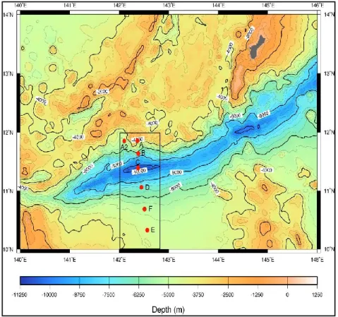
Surface sediment samples (n=3) were collected by using a gravity corer with the Lander system of the ROV ABISMO within the Izu-Ogasawara Trench at 9776 m depth (29°09.00’ N, 142°48.12’ E) and in the adjacent abyssal plain at 5747 m depth (29°16.79’ N, 143°46.04’ E) during the KR11-11 cruise on board of the research vessel Kairei in collaboration of the Japan Agency for Marine-Earth Science & Technology (JAMSTEC) in December 2011 (Figure 1). Sediment samples (n=3) were also collected in the Japan Trench at 8000 m depth (36°04.00’ N, 142°44.00’ E) during the KR12-19 cruise on board of the R/V Kairei (December 2012) and in the abyssal site used as a control (5253 m depth, 39°00.0’ N, 146°00.1’ E). Finally, sediment samples (n=3) were collected within the Challenger Deep of the Mariana Trench at 10901 m depth (11°22.0533’ N, 142°25.4486’ E) and in the surrounding abyssal plain (4700 m depth, 10°17.9803’ N, 142°36.0157’ E), during the KR14-01 cruise on board of the R/V Kairei (January 2014). Sub-aliquots of the sediment surface were used to carry out the analysis of the biochemical composition of the organic matter and microbiological parameters. Immediately after the sediments collection, the samples for analyses of the trophic parameters (in terms of proteins, carbohydrates, lipids and total phytopigments) were frozen and
stored at -20°C. For the analyses of prokaryotic abundances, samples were fixed with buffered 2% formalin and stored at 4°C until processed. Viral production experiment was set up and processed immediately after the collection of the sediments, and samples for analysing viral abundances were stored at -20° without the addition of any preservative. All analyses were carried out on the top 0-2 cm of sediment from each sediment core in 3 sub-replicates.
Fig. 1 Sampling sites
1.2.2 Sedimentary organic matter
Protein content (PRT) was determined according to Hartree (1972) modified by Rice (1982) to compensate for phenol interference. Concentrations were calculated from calibration curves of standard solutions of BSA, normalized to sediment dry weight and expressed as milligram of albumin equivalents per gram of dry sediment. Carbohydrates (CHOs) were analysed according to Gerchacov and Hachter (1972) and expressed as glucose equivalents normalized to sediment dry weight. The method is based on the same principle as the widely used method of Dubois et al. (1956), but it is specifically adapted for carbohydrate determination in sediments. Lipids (LIPs) were extracted by direct elution with chloroform and methanol and analysed according to Marsh and Wenstein (1966). Lipid concentrations were reported as tripalmitine equivalents normalized
to sediment dry weight. For each biochemical analysis, blanks were made with the same sediment samples as previously treated in a muffle furnace (450°C, 2 h). The sum of the carbohydrate, protein and lipid concentrations converted into carbon equivalents (using the conversion factors of 0.40, 0.49 and 0.75 mg C mg-1, respectively) was defined as the biopolymeric organic carbon
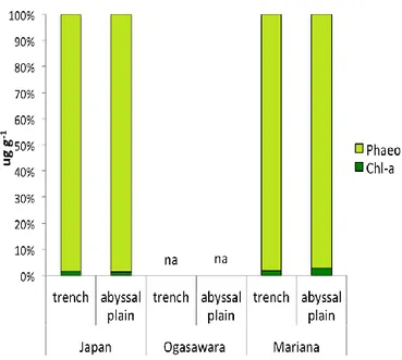
(Pusceddu et al, 2009). With the exception of Ogasawara Trench and it related abyssal site samples, due to the limited amount of available sediment, chloroplastic pigments (chlorophyll-a and phaeopigments) were analysed fluorometrically according to Lorenzen and Jeffrey (1980). Pigments were extracted with 90% acetone (24 h in the dark at 4°C). After centrifugation (800 X g), the supernatant was used to determine the functional chlorophyll-a and acidified with 0.1 N HCl to estimate the amount of phaeopigments. Total phytopigment concentrations (CPEs) were defined as the sum of chlorophyll-a and phaeopigment concentrations (Danovaro et al., 1999a,b).
1.2.3 Prokaryotic abundance and biomass
For prokaryotic counting, ca. 1 cm3 of sediment from the top 0-2 cm of each sediment core were
analysed as previously described by Danovaro et al. (2002). Benthic bacteria were detached from sediment by using pyrophosphate at a final concentration of 5 mM, and were sonicated by ultrasound for three minutes with 30 seconds interval between each minute (Branson 2200 Sonifier, 60 W) to increase the extraction efficiency. For bacterial counting, subsamples were diluted 50-fold with sterilized 0.2 μm-filtered seawater and stained with Acridine Orange (final concentration, 0.01%). After the incubation at the dark, the samples were filtered onto 0.2-μm-pore-size black Nuclepore filters with vacuum pressure of 150 mm of Hg by using sterile glass filter holders for 25 mm diameter filters with 15 ml glass funnel (Millipore). Acridine Orange was used instead of SYBR Green I for bacterial analysis because it provided similar counts but did not result in the overestimation of biovolume. All filters were analysed by epifluorescence microscopy using a Nikon ECLIPSE Ni-U microscope equipped with a micrometric scale to examine the cell dimensions and estimate the biovolume. Ten to 50 fields were viewed at a magnification of X1000, and a
considering the optical field coefficient (filtration area/counting area), the extraction coefficient (1.44), and the dilution factor and it was expressed as viruses per g dry sediment after desiccation at 60°C for 24 h. Bacterial biovolume, expressed as μm3 cell-1, was measured on the base of three
different sizes, small (<0.5 μm), medium (0.5 < x < 1.0 μm), and large ( 1 μm), and considering each cell like a sphere [(4/3) r3×π]. The biovolume was converted to carbon content (bacterial
biomass) by using a conversion factor of 310 fg of C μm-3 (J. C. Fry, 1990).
1.2.4 Extracellular enzymatic activities
Analyses of l-aminopeptidase, β-d-glucosidase, and alkaline-phosphatase activity were carried out on sediment sub-samples (n = 3). Sediment slurries (prepared with pre-filtered sterilized seawater, 1:1 v/v) were incubated for 2 h (enzymatic activity increased linearly with time) in the dark and at in situ temperature with l-leucine-4-methylcoumarinyl-7-amide (MCA), 4- methylumbelliferone β-d-glucopyranoside (β-Glu), and 4-methylumbelliferone phosphate (MUF-P) respectively as substrates (final concentrations of MCA 200 μM, MUF 200 μM, and MUF-P 50 μM determined after kinetic experiments). Sediment sub-samples incubated without fluorogenic substrates were used as blanks and processed according to the procedure followed for fresh sediment samples. After incubation, samples were centrifuged (800X g) and the supernatants were analysed fluorometrically according to Hoppe (1993). Enzymatic activities were expressed as nmol of substrates hydrolysed in one hour and normalized to sediment dry weight. The ratio between enzymatic activities and prokaryotic abundances leaded to estimate the specific degradation activity per cell that were expressed as nmol of substrate hydrolysed in one hour normalized to sediment dry weight per cell.
1.2.5 Viral abundance
Viral counting was carried out on ca. 1 cm3 of sediment from the top 0-2 cm of each sediment core
as described by Danovaro et al. (2001). Sediment sub-samples were diluted in a final volume of 5 ml of virus-free seawater filtered at 0.02 μm pore size using disposable syringe filters (Anotop 25, Whatman), and treated with pyrophosphate at a final concentration of 5mM. Samples were sonicated by ultrasound for three minutes with 30 seconds interval between each minute (Branson 2200 Sonifier, 60 W) to increase the extraction yield. The samples were then diluted 25-fold in virus-free seawater and incubated with the addition of DNase I from bovine pancreas (2-5 Uml-1) in
order to eliminate uncertainties in virus counting due to extracellular DNA interference. After the incubation, samples were filtered onto 0.02 μm pore size filters (Anodisc Al2 O3; 25mm diameter,
Whatman) with vacuum pressure of 150 mm of Hg by using sterile glass filter holders for 25 mm diameter filters with 15 ml glass funnel (Millipore). Then the filters were stained using SYBR Gold (10000X concentrated in anhydrous dimethyl sulphoxide; Molecular Probes-Invitrogen, Grand Island, NY, USA) at a final concentration of 2X. SYBR Gold was used instead of SYBR Green I to detect both double- and single-stranded DNA and RNA viruses, and because its strong brightness and stability as fluorescence stain. Viral counting was performed under epifluorescence microscopy, using a Nikon ECLIPSE Ni-U microscope, by examining at least 20 fields per slide, and counting at least 400 viral particles per filter. The viral abundance was calculated considering the optical field coefficient (filtration area/counting area), the extraction coefficient (1.66), and the dilution factor, and it was expressed as viruses per g dry sediment after desiccation at 60°C for 24 h.
1.2.6 Viral production, virus-induced prokaryotic mortality (VIPM) and turnover
Viral production was analysed as previously described by Dell’Anno et al. (2009). Sub-replicate sediment samples were transferred into sterile tubes and mixed with an equal volume of 0.02 μm pre-filtered virus-free seawater. Aliquots of 0.5 ml were collected at 0, 3, 6 and 12 hours. After the
extraction of viruses from each sediment samples as described above, viral counting was performed under epifluorescence microscopy by examining at least 20 fields per slide, and counting at least 400 viral particles per filter. The increment in virus number was determined calculating the difference between the maximum viral abundance value, considering the abundances of the samples of all the incubation times, and the viral abundance at the t 0h (VA tn -
VA t0) normalized to the number of hours of incubation.
The number of prokaryotes killed by viruses was estimated from the ratio between viral production and burst size, which were calculated on the base the equation: BS = 30.2 + (261.4 cell biovolume μm3; Weinbauer & Hofle, 1998). VIPM was estimated as the ratio between the number of
prokaryotes killed by viruses and the total number of prokaryotes present in the samples and expressed as percentage. Viral turnover was calculated as the ratio between viral production and abundance and expressed as turnover time (h).
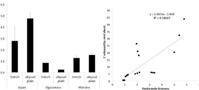
1.2.7 Carbon contribution to microbial C cycle due to viral shunt
The concentration of C released from the infected cells due to viral lysis in each sediment sample (expressed as mg C m-2 d-1)was calculated by multiplying the cell biomass for the number of cells
killed per hour, which was deducted by the ratio between viral production and burst size. The potential contribution of the C released by host cells due to viral lysis to the prokaryotic C degraded was obtained by values of extracellular aminopeptidase and β-glucosidase activities converted in C equivalents degraded per m-2 per day (Hoppe et al., 1988).
1.2.8 Statistics
A one-way univariate analysis of permutational variance (ANOVA) was applied to assess differences between sampling stations (6 levels) and each analysed variable by using the PRIMER 6+ software.
1.3 Results
1.3.1 Biochemical composition of organic matter content and photosynthetic pigments
The results of the biochemical composition of organic matter in the surface sediments of the investigated sites are shown in Figure 2a. Protein concentrations ranged from 0.19±0.08 to 9.51±0.77 mg g-1 of dry sediment, respectively in the abyssal site related with the Mariana Trench
and in the Japan Trench. Carbohydrate concentrations ranged from 1.02±0.22 to 4.60±0.33 mg g-1
(in the abyssal site close to the Mariana Trench and the Japan Trench respectively), while lipid concentrations from 0.11±0.03 to 2.48±0.55 mg g-1 (in the abyssal site related with Ogasawara
Trench and within the Japan Trench, respectively).
The Japan Trench bottom sediments presented significantly higher protein and lipid contents (P<0.05) compared to the Ogasawara and Mariana trenches, exceeding their concentration 7-19 and 6-3 times respectively. Ogasawara Trench presented the highest carbohydrate concentrations (2.63±0.03 mg g-1) among the three trenches. Both protein and lipid contents were significantly
higher within the three trenches if compared with their related abyssal sites, while carbohydrates concentration was double in the abyssal plain respect to the Japan Trench, and between the Mariana Trench and its related abyssal site there was not a significant difference (1.03±0.11 and 1.02±0.22 mg g-1 respectively). Ogasawara Trench, instead, showed significantly higher
carbohydrates concentrations compared to its relative abyssal site.
Biopolymeric carbon contents (Figure 2b) ranged from 0.56±0.14 to 7.47±0.82 mg g-1 with
significantly different values among the trenches (the Japan Trench exceeding 3 and 5 times the concentrations that were present in Ogasawara and Mariana trenches, respectively) but higher when compared to their related abyssal sites. In Figure 2c the contributions of the different biogeochemical components of organic matter to biopolimeric carbon are showed. The total phytopigments were significantly higher in the Japan Trench (269.32±13.81 μg g-1) than both the