UNIVERSITÀ DEGLI STUDI DI NAPOLI
“FEDERICO II”
DOTTORATO IN SCIENZE VETERINARIE
XXXI CICLO
PhD THESIS
“Staphylococcus pseudintermedius-associated canine
skin disorders: isolation, identification, antibiotic
resistance patterns, sequence typing and alternative
therapeutic strategies.”
Coordinatore Candidata Tutor
Prof. G. Cringoli Dr.ssa F. P. Nocera Prof.ssa L. De Martino
Non basta guardare. Occorre guardare con occhi che vogliono vedere,
che credono in quello che vedono. Galileo Galilei
7 List of abbreviations 15 List of figures 19 List of tables 21 Abstract 23 Introduction 27 References 31 Chapter 1 1. Introduction 35 1.1 Staphylococcus pseudintermedius the main opportunistic dog pathogen 41
1.2 Phenotypic features of Staphylococcus pseudintermedius 46
2. Materials and Methods 52
2.1 Collection of samples 52
2.2 Bacterial isolation and phenotypic identification of the isolates 52
2.3 Genotypic identification of the isolates 56
3. Results 59 3.1 S. pseudintermedius isolation and
8
phenotypic identification 59
3.2 S. pseudintermedius genotypic identification 61
4. Discussion and conclusions 62
5. References 66
Chapter 2 1. Introduction 75
1.1 Antimicrobial resistance in Staphylococcus pseudintermedius 78
1.1.1 Methicillin resistance in Staphylococcus pseudintermedius 79
1.1.2 Resistance to non-β-lactam antibiotics in S. pseudintermedius 81
2. Materials and Methods 85
2.1 Antimicrobial susceptibility testing 85
2.2 Genotypic characterization of antibiotic resistance 89
2.2.1 Methicillin resistance determinats – molecular characterization of MRSP 89
2.2.2 Genotypic characterization of tetracycline and erythromycin resistance 90
3. Results 94
3.1 Phenotypic and genotypic characterization of MRSP antibiotic resistance profiles 94
3.2 Phenotypic and genotypic characterization of MSSP antibiotic resistance profiles 97
9
4. Discussion and conclusions 102
5. References 107
Chapter 3 1. Introduction 121
2. Materials and Methods 124
2.1 Multilocus sequence typing of MRSP isolates 124
3. Results 127
3.1 MRSP clones 127
4. Discussion and conclusions 131
5. References 133
Chapter 4 1. Introduction 139
2. Materials and Methods 143
2.1 In vitro sensitivity of clinical canine S. pseudintermedius isolates to DIBI 143
2.1.1 Bacterial cultivation 143
2.1.2 Susceptibility testing 144
2.2 Antimicrobial properties of abietic acid against clinical canine S. pseudintermedius strains 145
2.2.1 S. pseudintermedius cultivation and susceptibility testing 146
2.2.2 Killing rate and checkerboard method 146
10
3.1 Susceptibility of S. pseudintermedius
isolates to DIBI 148 3.2 In vitro abietic acid antimicrobial activity 148 3.3 Synergistic assay of abietic acid with oxacillin
against MRSP 151 4. Discussion and conclusions 152 5. References 155
11
Dedicato ai miei nonni, ai miei genitori, alla mia famiglia, ai miei più cari amici. Dedicato a chi mi ha accolta nella propria vita, a chi ha reso le mie insicurezze ali di farfalla.
12
A conclusione di questo percorso di Dottorato, desidero ringraziare tutte le persone che ho incontrato e conosciuto in questo periodo della mia vita. Persone che, in vario modo, hanno contribuito alla mia crescita professionale ed umana, nonché a rendere migliori questi ultimi tre anni. Siete in tanti ed è difficile ricordarvi tutti in poche righe. Grazie!
Un ringraziamento sentito va alla mia tutor, Prof.ssa Luisa De Martino per essere stata un punto di riferimento ed un esempio di determinazione e perseveranza. È doveroso ringraziarla per la fiducia che ha sempre riposto in me (è stata fondamentale) e per l’umanità con la quale ha saputo incoraggiarmi nei momenti di difficoltà (non sono stati pochi) incontrati in questo triennio.
Ringrazio di cuore la Dr.ssa Filomena Fiorito che, anche se a distanza, mi ha sempre sostenuta ed aiutata con pazienza e spirito critico. I suoi preziosi insegnamenti sono stati fondamentali per la mia crescita intellettuale e professionale.
Un ringraziamento particolare va al Professore Giuseppe Iovane e a tutti i Professori e Ricercatori della Sezione di Malattie Infettive per l’opportunità concessami, la disponibilità e professionalità dimostratemi. Inoltre, ringrazio con tanto affetto i tecnici Salvatore, Gaetano, la signora Lucia, i miei colleghi ed amici di laboratorio sui quali ho potuto contare e che sono stati pronti ad ascoltarmi e consigliarmi nelle giornate di lavoro e studio, che hanno caratterizzato questi anni. Grazie perché mi avete fatto sentire a casa!
13
Un ringraziamento speciale va ai miei genitori e a tutta la mia famiglia. Grazie per tutto l’amore, l’affetto, il sostegno che non sono mai mancati in tutti questi anni, nei momenti belli e meno belli della mia vita. Vi voglio bene!
Ai miei nonni, che mi hanno insegnato a sorridere e mi hanno trasmesso i valori che mi hanno reso la persona che sono. Grazie perché, anche se da lassù, continuerete ad aiutarmi ad affrontare la vita con serenità e dolcezza.
Infine, non per importanza, ringrazio di cuore i miei più cari amici: Cristina, Teresa, Elena, Federica, Maria Laura, Francesco, Rosaria, Giuseppe, Ciro, Anna, Bianca, Veriano, Claudia, Anna C. Grazie per la vera amicizia, ormai rara, e per la continua presenza che mi donate.
Grazie per le risate, le chiacchiere, i bei momenti condivisi, per l’incoraggiamento a non mollare mai e a superare gli ostacoli che spesso si incontrano lungo il cammino della vita. Grazie per l’affetto che non avete mai mancato di dimostrarmi.
A tutte le persone a me care, grazie per la vostra insostituibile presenza.
15
agr accessory gene regulator AIP auto-inducing peptide AMC amoxicillin-clavulanate AMP ampicillin
ATCC® American Type Culture Collection CD clindamycin
CDSs predicted protein-coding sequences CIP ciprofloxacin
CLSI Clinical and Laboratory Standards Institute CN gentamicin
CoNS coagulase-negative Staphylococci CoPS coagulase-positive Staphylococci CRO ceftriaxone
DIBI Hydroxypyridinone-based iron-chelating co-polymer E erythromycin
ENR enrofloxacin
FIC fractional inhibitory concentration FOX cefoxitin IMI imipenem K kanamycin KF cephalothin LNZ linezolid Luk I leucotoxin MALDI-TOF-
16
MS matrix assisted laser desorption ionization time of flight mass spectometry
MBC minimal bactericidal concentration MDR multidrug resistance
MIC minimal inhibitory concentration MLST multilocus sequence typing
MRSA methicillin-resistant Staphylococcus aureus
MRSP methicillin-resistant Staphylococcus pseudintermedius MSA Mannitol Salt agar
MSSP methicillin-susceptible Staphylococcus pseudintermedius NCCLS National Committee for Clinical Laboratory Standards OX oxacillin
P penicillin
PBP2a modified penicillin-binding protein PCR polymerase chain reaction
PVA immunoglobulin-binding protein
RFLP PCR-restriction fragment length polymorphism RPMI Roswell Park Memorial Institute Medium 1640 S streptomycin
SaPI staphylococcal pathogenicity island family sar staphylococcal accessory regulator
SCCmec staphylococcal cassette chromosome mec SIG Staphylococcus intermedius Group STs Sequence types
17
SXT sulfamethoxazole-trimethoprim TE tetracycline
TOB tobramycin VA vancomycin
19 1.1 Canine otitis externa
1.2 Canine superficial pyoderma (Foglia Manzillo et al., 2016) 1.3 Staphylococcus pseudintermedius ecological niche
1.4 Staphylococcus pseudintermedius colonization in dog (Bannoehr and Guardabassi, 2012)
1.5 S. pseudintermedius virulence armamentarium 1.6 S. pseudintermedius: Gram stain
1.7 Typical workflow of new and old methods used in clinical microbiology
1.8 Culture media used for S. pseudintermedius isolation 1.9 Maldi-Tof MS technique
1.10 MicrobankTM: preservation and retrieval procedure
1.11 S. pseudintermedius growth on MSA
1.12 Molecular characterization of S. pseudintermedius: PCR for detection of species-specific nuc gene
1.13 Molecular characterization of S. pseudintermedius: PCR for detection of species-specific hlb gene
2.1 Bacterial antimicrobial resistance mechanisms 2.2 Mobile gene elements (Fang, 2015)
2.3 Antibiotic resistance rates of MRSP and MSSP to the 20 tested antibiotics
3.1 Population genetic structure of S. pseudintermedius lineage (Ruscher et al., 2010)
4.1 DIBI 4.2 Abietic acid
21
1.1 Primers sequences, amplicon sizes, amplification programs 1.2 S. pseudintermedius strains isolated from diseased dogs from
Campania and Lazio Regions
1.1 Mobile genetic elements associated with antibiotic resistance in
S. pseudintermedius ED99
1.2 Antibiotics and antibiotic classes tested for canine S.
pseudintermedius isolates
1.3 Primers sequences, amplicon sizes, amplification programs of
mec genes
1.4 Primers sequences, amplicon sizes, amplification programs of tet
and erm genes
2.5 Antibiotic resistance profiles of canine MRSP
2.6 Antibiotic resistance rates of MRSP and MSSP isolates of canine origin
2.7 Molecular profiles of tetracycline and erythromycin resistance in canine S. pseudintermedius isolates
3.1 Primers for MLST-7 of S. pseudintermedius and amplicon sizes 3.2 Geographical, biological origin and clones of methicillin-resistant S. pseudintermedius
3.3 Origin, MLST analysis, phenotypic and genotypic antimicrobial resistance of MRSP isolates from canine cutaneous swabs 4.1 Antibiotic resistance, methicillin resistance profiles and DIBI
antimicrobial activity in 5 clinical canine otitis
22
4.2 Antibiotic resistance and methicillin resistance profile in 2
clinical canine otitis S. pseudintermedius strains
4.3 MIC90, MBC, MICindex, FICindexvalues of abietic acid on selected
S. pseudintermedius strains
23
Sthaphylococcus pseudintermedius (S. pseudintermedius) is the main
inhabitant of skin and mucosa of dogs, where it represents the major bacterial pathogen causing skin diseases.
Firstly described in 2005, S. pseudintermedius is a member of the
Staphylococcus intermedius Group (SIG), together with S. intermedius
and S. delphini.
The recent emergence and rapid dissemination of multidrug-resistant S.
pseudintermedius strains, particulary methicillin-resistant S. pseudintermedius (MRSP), showing more and more often resistance to
the antibiotic agents licensed for use in small animal practice, represent a relevant threat to small animal and public health. In the last decade, the antibiotic resistance issue has become increasingly relevant, highlighting the need to control and limit it by the means of a continuous surveillance
of both methicillin-resistant and methicillin-susceptible S.
pseudintermedius isolates as well as the development of new and
alternative therapeutic options.
This study aims to describe the phenotypic and genotypic features, the antibiotic resistance patterns, the prevalence of virulence genes of both MRSP and methicillin-susceptible S. pseudintermedius (MSSP) strains, isolated from dogs suffering from otitis externa and pyoderma. MRSP sequence type clones circulating in Southern Italy were also investigated. Moreover, the antimicrobial activity of hydroxypiridone-based iron chelating co-polymer DIBI and abietic acid was analyzed against S.
pseudintermedius isolates, as alternative treatment options.
From a total of 259 staphylococcal cultures, 126 S. pseudintermedius strains were selected during the years 2015-2017.
24
S. pseudintermedius strains (49%) were identified by proteomic
(MALDI-TOF MS) and molecular profiling (nuc and hlb genes). S.
pseudintermedius strains were recovered from dogs suffering from otitis
externa (84%) and pyoderma (16%), originating from Naples, Campania Region (52%) and Latina, Lazio Region (48%).
Amongst the 126 S. pseudintermedius strains, 18% were MRSP, carrying
mecA gene. The remaining 82% were classified as MSSP. None of the
isolates was positive to mecC gene.
Furthermore, all the MRSP showed a complete resistance to amoxicillin-clavulanate and ampicillin (100%), while the highest resistance rates to selected non-β-lactam antibiotics were registered for erythromycin (91%); tetracycline (87%); sulfamethoxazole/trimethoprim (78%); kanamycin (78%); streptomycin (78%); clindamycin (65%) and enrofloxacin (61%). However, there was no resistance to vancomycin and linezolid. It is worth noting that 91% MRSP strains were found to be multidrug-resistant strains. The 82% MSSP isolates, showed interesting antibiotic resistant profiles, but their resistance rates were of about 50% lower than MRSP strains.
The phenotypic tetracycline- and erythromycin-resistant MRSP and MSSP strains harbored tetK and tetM genes, alone or in association, and
ermB gene.
Referring to MRSP isolates, they mainly belonged to multidrug-resistant sequence type ST71 (26%) European clone. In this study, 9 new clones of MRSP were identified and described in Italy and worldwide for the first time. The newly described sequence types (STs) were named from ST1053 up to ST1061.
25
In addition, DIBI and abietic acid resulted to be effective against MRSP and MSSP strains, inhibiting their growth. Thus, they may be considered as valid non-antibiotic alternative treatment approaches for S.
27
The bacterial genus of Staphylococcus consists of many species that are part of the natural microbiota of humans and animals.
Staphylococcus intermedius, Staphylococcus pseudintermedius, and Staphylococcus delphini together compose the Staphylococcus intermedius Group (SIG). In particular, Staphylococcus pseudintermedius is the most
common coagulase-positive staphylococcus isolated from dogs, in which this opportunistic Gram-positive bacterium can be responsible for a great variety of infections including pyoderma, otitis, abscesses, urinary tract infections (UTIs) and wound infections (Weese and van Duijkeren, 2010; Rubin et al., 2011; Youn et al., 2014).
Over the last decade, S. pseudintermedius has become a critically opportunistic small animal pathogen, being frequently the primary aetiological agent of pyoderma and otitis (De Martino et al., 2016; Loeffler and Lloyd, 2018). Even though dog is its natural host, human carriage and infections have been reported, highlighting the potential zoonotic transmission of this species (Somayaji et al., 2016).
Since its first description as a novel coagulase-positive staphylococcal species in 2005, the phenotypic identification of S. pseudintermedius has persisted problematic. Commercial kits, commonly used in routine laboratory diagnostics, are not able to identify and distinguish S.
pseudintermedius from the other members of the SIG and from S. aureus,
because of their similar phenotypic characters. Moreover, as coagulase-negative Staphylococci (CoNS) also S. pseudintermedius generally gives negative results to rapid slide clumping and commercial latex agglutination tests. For these reasons, the real prevalence of S.
28
microbiology laboratories (Weese and van Duijkeren, 2010). So, molecular profiling continues to represent a necessary tool for a proper determination and diagnosis of S. pseudintermedius. However, Decristophoris et al. (2011), reported that MALDI-TOF MS, provided with a reliable database, is an effective technique for a rapid identification of S. pseudintermedius and the other two bacterial species of the SIG. Antibiotic resistance is the most puzzling question of public health of the earlier decade of this 21st century and already since 2006
methicillin-resistant S. pseudintermedius (MRSP) strains have been identified, which have been also proved to be multidrug-resistant displaying resistance to most of the antibiotics approved for use in pet animals (Kadlec and Schwarz, 2012; Moodley et al., 2014).
This high prevalence of multidrug-resistant strains seems to be related to the dissemination of dominant clones, such as MRSP belonging to the clonal lineage ST71. ST71 is a widespread clone, being isolated across the European, American and Asiatic continents. As reported by Perreten at al. (2010) in their multicenter study, a high percentage of MRSP ST71 were resistant to at least six antimicrobial classes. Furthermore, new multidrug MRSP clones are constantly reported worldwide.
Therefore, the increasing spread of multidrug-resistant strains represents an important threat for animal and public health, since dogs, and in general pet animals, represent potential sources of spread of antimicrobial resistance due to the extensive use of antimicrobial agents; this spread may concern also humans, because of their close contact with pets. Thus, it is not only of veterinary significance, but has a zoonotic importance, with pets acting as reservoirs for humans, particularly pet owners and
29
veterinarians. Moreover, the lack of effective antimicrobial therapeutic treatments underlines the need of new alternative approaches to prevent and limit the dissemination of multidrug resistant bacterial isolates.
The purpose of this PhD thesis was to investigate the phenotypic and genotypic antibiotic resistance patterns of clinical methicillin-resistant and
methicillin-susceptible Staphylococcus pseudintermedius strains
associated with the most common clinical cases of otitis externa and pyoderma in dogs, attending the Veterinary Teaching Hospital (OVUD) of the Department of Veterinary Medicine and Animal Production, University of Naples “Federico II” and the Provet Lab srl of Latina. The dominant MRSP clones circulating in Southern Italy were also investigated. Furthermore, the antimicrobial activity against S.
pseudintermedius of the two non-antibiotic agents DIBI (Chelation
Parteners Inc. proprietary) and abietic acid was analyzed as alternative therapeutic options.
The topics of the thesis are organized in four chapter as follows.
Chapter 1 is focused on the:
- isolation and collection of S. pseudintermedius strains obtained from
swabs of dogs suffering from otitis externa and pyoderma;
- comparison of Api Staph conventional biochemical test, matrix- assisted laser desorption ionization time-of-flight mass spectrometry (MALDI-TOF MS) system and polymerase chain reaction (PCR) for the S.
30 Chapter 2 is focused on the:
- discrimination of methicillin-resistant S. pseudintermedius (MRSP) from methicillin-susceptible S. pseudintermedius (MSSP) isolated strains by PCR for mecA and mecC genes;
- evaluation of the antibiotic resistance profile to a panel of 20 antimicrobial agents by the disk diffusion method on Mueller-Hinton agar plates (Liofilchem, Teramo, Italy), according to the guidelines of the Clinical Laboratory Standard Institute (CLSI, 2015);
- determination of the presence of the different tet and erm genes in tetracycline- and erythromycin-resistant strains by PCR.
In Chapter 3, multilocus sequence typing (MLST) was performed to investigate the MRSP sequence type clones circulating in Southern Italy and to correlate their antibiotic resistance patterns to the belonging clones.
The aim of Chapter 4 was to investigate new therapeutic options, in order to prevent the spread of antibiotic resistance, since it is one of the most urgent threat to the public’s health. So, the antimicrobial properties of DIBI, a novel iron chelating polymer developed by Chelation Partners Inc. (Canada), and abietic acid against MRSP and MSSP strains were tested.
31
Clinical and Laboratory Standards Institute (CLSI) 2015. Performance Standard for Antimicrobial Disk and Dilution Susceptibility Tests for Bacteria Isolated from Animals, 3rd Edition CLSI VET01S (Wayne, Pennsylvannia, Clinical Laboratory and Standards Institute), 128, 2015. Decristophoris P, Fasola A, Benagli C, Tonolla M, Petrini O, 2011.
Identification of Staphylococcus intermedius Group by MALDI-TOF MS. Syst Appl Microbiol 34, 45-51.
De Martino L, Nocera FP, Mallardo K, Nizza S, Masturzo E, Fiorito F, Iovane G, Catalanotti P, 2016. An update on microbiological causes of canine otitis externa in Campania Region, Italy. Asian Pac J Trop Biomed 6, 384-389.
Kadlec K, Schwarz S, 2012. Antimicrobial resistance of Staphylococcus
pseudintermedius. Vet Dermatol 23, 276-282.
Loeffer A, Lyod DH, 2018. What has changed in canine pyoderma? A narrative review. Vet J 235, 73-82.
Moodley A, Damborg P, Nielsen SS, 2014. Antimicrobial resistance in methicillin susceptible and methicillin resistant Staphylococcus
pseudintermedius of canine origin: Literature review from 1980 to 2013.
Vet Microbiol 171, 337-341.
Perreten V, Kadlec K, Schwarz S, Grönlund Andersson U, Finn M, Greko C, Moodley A, Kania SA, Frank LA, Bemis DA, Franco A, Iurescia M, Battisti A, Duim B, Wagenaar JA, van Duijkeren E, Weese JS, Fitzgerald JR, Rossano A, Guardabassi L, 2010. Clonal spread of methicillin-resistant Staphylococcus pseudintermedius in Europe and North America: an international multicenter study. J Antimicrob Chemother 65, 1145-1154.
32
Rubin JE, Ball KR, Chirino-Trejo M, 2011. Antimicrobial susceptibility of Staphylococcus aureus and Staphylococcus pseudintermedius isolated from various animals. Can Vet J 52, 153-157.
Somayaji R, Rubin JE, Priyantha MA, Church D, 2016. Exploring
Staphylococcus pseudintermedius: an emerging zoonotic pathogen?
Future Microbiol 11, 1371-1374.
Weese JS, van Duijkeren E, 2010. Methicillin-resistant Staphylococcus
aureus and Staphylococcus pseudintermedius in veterinary medicine.
Vet Microbiol 140, 418-429.
Youn JH, Park YH, Hang’ombe B, Sugimoto C, 2014. Prevalence and characterization of Staphylococcus aureus and Staphylococcus
pseudintermedius isolated from companion
animals and environment in the veterinary teaching hospital in Zambia, Africa. Comp Immunol Microbiol Infect Dis 37, 123-130.
Chapter 1
Bacterial skin disorders in dogs
Staphylococcus pseudintermedius as main bacterial agent of canine otitis
35 1. Introduction
Canine bacterial skin infections represent the mainly reason of presentation in small animal practice and are generally secondary complications of a range of canine skin diseases including endocrine, allergic, seborrheic and follicular disorders (Miller et al., 2013). Indeed, many bacterial species, especially Staphylococci, are part of the natural skin microbiota but particular conditions such as flea infestation, allergic reaction, immune system depletion can lead to an excessive skin bacteria proliferation with the appearance of cutaneous infection symptoms. Infected dogs can show pruritus, areas of redness, rash-like pustules and hair loss.
In dogs, skin and ear infections are very commonly caused by
Staphylococcus pseudintermedius (S. pseudintermedius) since it is a
normal inhabitant of the skin and mucosa of dogs and cats (Van Duijkeren et al., 2011). Thus, Staphylococcus pseudintermedius is an opportunistic pathogen and a leading cause of skin, ear and post-operative wound infections in dogs and marginally in cats (Fitzgerald, 2009; Weese and van Duijkeren, 2010).
Canine otitis externa and pyoderma are featured consistently as the major diseases affecting canine skin system.
• Canine otitis externa
Otitis externa is the most common ear disease seen in dogs, affecting up to 20% of the canine population (De Martino et al., 2016). It has a multifactorial etiology and the otitis causes can be classified as
36
predisposing, primary and perpetuating factors. Moreover, the predisposing factors can be related to the host and to the environment (Fig 1.1).
Among the predisposing factors related to the host, anatomical changes of the ear canal as conformational abnormalities linked to the belonging breed seem to be the main otitis causes. The most frequently affected breeds are: german shepherds for the high moisture levels in their ear canals, cocker spaniels for their pendulous pinnae, shar-peis for their hypoplastic and stenotic ear canals and poodles for a high density of hair in their ear canals (Zur et al., 2011).
Another aspect to consider is represented by the habits of the animals, that is to say dogs living outdoor and hunting dogs have more possibilities to develop otitis externa because foreign fragment of twigs, plants or dirty materials can contaminate their ear canal. Moreover, dogs that are often washed or do swimming activities are predisposed to otitis for the excessive stimulations of the ceruminous glands. Consequently, the humidity in the ear canal compromise the protective function of the epidermidis.
Some studies have shown that changes in temperature and humidity of the surrounding environment are related to temperature changes and dampness of the ear canal (Logas, 1994).
The most common primary causes of otitis externa are allergies such as atopic dermatitis and adverse food reactions. Keratinization disorders, either primary (idiopathic seborrhea) or secondary as in hypothyroidism and sex hormone imbalance, are also common primary causes affecting
37
the secretions of the ceruminous and sebaceous glands lining the ear canal (Rosser, 2004).
The predisposing factors and primary causes create suitable conditions for the growth and the proliferation in the ear canal of microorganisms such as bacteria and yeast, that represent both predisposing and perpetuating factors. Many studies report that the most common bacterial pathogens isolated from canine otitis externa are
Staphylococcus pseudintermedius, Pseudomonas aeruginosa, Proteus mirabilis, Streptococcus spp. and Escherichia coli (Hariharan et al.
2000; Lyskova et al., 2007). Whilst among yeasts, Malassezia
pachydermatis has been reported as the major causative agent of canine
otitis externa (De Martino et al., 2016).
Fig. 1.1 Canine otitis externa
• Canine pyoderma
Pyoderma refers to a bacterial skin disorder and it comes from Greek
words pyo meaning “pus” and derma meaning “skin. Pyoderma is
38
presentations leading to antimicrobial prescription in small animal practice (Hughes et al., 2012). Bacterial pyoderma is usually triggered by an overgrowth/overcolonization of normal resident or transient flora. Since Staphylococcus pseudintermedius is a normal commensal of dog, it is the most commonly pathogen of canine pyoderma, in particular superficial pyoderma. However, normal resident bacteria in canine skin also include coagulase-negative staphylococci, streptococci,
Micrococcus spp, and Acinetobacter spp. Transient bacteria in canine
skin include Bacillus spp., Corynebacterium spp., Escherichia coli,
Proteus mirabilis, and Pseudomonas spp. These organisms may play a
role as secondary pathogens, but often S. pseudintermedius is required for a pathologic process to ensue.
Bacterial pyoderma can occur either as simple infection or complex infection. Simple infections are those occurring in young animals that are triggered by one-time or simple events, such as flea infestation. Complex infections are recurrent and are associated with underlying diseases, such as allergies, endocrinopathies, seborrheic conditions, parasitic diseases (Demodex canis), or anatomic predispositions. Regarding to the anatomic predisposition, it has been suggested that in dog pyoderma is partly a consequence of the thin and compact stratum corneum, of the paucity of intracellular emulsion in epidermidis and of the absence of a sebum plug in the hair follicle (Loeffler and Lloyd, 2018).
Bacterial pyoderma is generally classified as surface, superficial and deep pyoderma on the basis of the depth of infection.
39
Surface pyoderma remains the less known. It comprises acute moist dermatitis (hot spots, pyotraumatic dermatitis), fold pyoderma (intertrigo) and microbial/bacterial overgrowth syndrome, in which erythema is the only clinical sign.
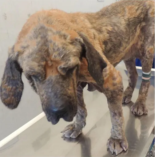
Superficial pyoderma, named also superficial bacterial folliculitis, is the most recurrent type of canine pyoderma (Fig.1.2). It is limited to the epidermis that is invaded by bacteria. The clinical signs are papules, pustules and epidermal collarettes, which are typically confined to the ventral abdomen, medial thighs and to the trunk. Moreover, it is often associated with alopecia and pruritus (Loeffler and Lloyd, 2018). In fact, the most important factor in superficial pyoderma is the bacterial
adherence, or “stickiness,” to the keratinocytes. Warm, moist areas on
the skin, such as lip folds, facial folds, neck folds, dorsal or plantar interdigital areas, vulvar fold, sand tail folds, often have higher bacterial counts than other areas of skin and are at an increased risk of infection. Pressure points, such as elbows and hocks, are prone to infections, possibly because of follicular irritation and rupture due to chronic repeated pressure. Deep pyoderma is less frequent but more serious, since it involves the dermis (deep folliculitis and furunculosis, and cellulitis). Blood vessels may also be involved with a high risk of hematogenous spread and bacteremia.
40
41
1.1 Staphylococcus pseudintermedius: the main opportunistic dog pathogen.
Staphylococcus intermedius was first described in 1976 and it has been
considered as the major strain responsible of canine skin disorders for a long time (Hajek, 1976; Fitzgerald, 2009). However, the development of molecular techniques revealed a genetic diversity among S.
intermedius isolates and in 2005 a novel staphylococcal species, Staphylococcus pseudintermedius, was defined based on the sequential
analysis of 16S rRNA (Devriese et al., 2005).
Therefore, the isolates, that previously could not be distinguished by biochemical and morphological characters, in 2007 were grouped in the
Staphylococcus intermedius Group (SIG) and differentiated thanks to
the sequential analysis of sodA and hsp60. Thus, SIG comprises three
distinct species: Staphylococcus intermedius, Staphylococcus
pseudintermedius and Staphylococcus delphini, each of which has
different ecological niches (Bannoehr et al., 2007; Sasaki et al., 2007).
Importantly, it was discovered that S. pseudintermedius, not S.
intermedius, is a member of the normal skin canine microbiota and an
opportunistic pathogen (Fig. 1.3).
42
Fig. 1.3 Staphylococcus pseudintermedius ecological niche (Bannoehr and Guardabassi, 2012)
43
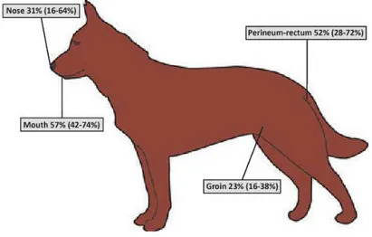
Precisely, Staphylococcus pseudintermedius can be isolated from the nares, oral mucosa, pharynx, forehead, groin and anus of healthy dogs (Garbacz et al., 2013). So, it is the most common bacterial pathogen associated with canine infections, predominantly skin infections (Fig. 1.4).
Fig. 1.4 Staphylococcus pseudintermedius colonization in dog (Bannoehr and Guardabassi, 2012)
Ben Zakour et al. (2011), reported the first whole-genome sequence of a canine clinical isolate of S. pseudintermedius (ED99). Its genome consisted of a single circular chromosome of 2,572,216 bp with an average G+C content of 37,6%, carried five ribosomal operons and 58 tRNA loci and encoded for 2401 predicted proteins coding sequences (CDSs). Moreover, S. pseudintermedius ED99 contained many
44
predicted mobile genetic elements, including insertion elements, transposons mediating resistance to antibiotics, a novel member of the staphylococcal pathogenicity island family (SaPI). The genome of other isolated S. pseudintermedius strains has been also sequenced (Tse et al., 2011; Moodley et al., 2013; Duim et al., 2018). As other staphylococcal species, also S. pseudintermedius has various virulence factors (Fig. 1.5), including some that are closely related to S. aureus (Fitzgerald, 2009; Bannoehr and Guardabassi, 2012). These factors of virulence take part in almost all processes from colonization of the host to bacterial nutrition and dissemination.
S. pseudintermedius has the capability to produce some enzymes such
as coagulase, protease, thermonuclease and toxins. Among toxins,
mainly β-haemolysins (but some strains also α and ᵹ haemolysins),
exfoliative toxins and enterotoxins are produced. Exfoliative toxin seems to be the factor involved in canine pyoderma, because the exfoliative toxin gene has been harbored in many S. pseudintermedius isolated from dogs suffering from skin infections, that is to say chronic otitis and pyoderma (Iyori et al., 2010).
Furthermore, similarly to S. aureus, also S. pseudintermedius produces a leucotoxin and an immunoglobulin-binding protein, which are known as Luk-I and staphylococcal protein A (spa), respectively (Moodley et
al., 2009). Genes encoding virulence factors are usually located on
mobile genetic elements such as pathogenicity islands. The production of virulence factors is regulated by several regulatory loci such as the accessory gene regulator (agr) and the staphylococcal accessory regulator (sar) in response to cell density, energy availability and
45
environmental signals. The accessory gene agr is a staphylococcal quorum-sensing system, which encodes the auto-inducing peptide (AIP). All staphylococcal species encode a unique to each species AIPs (Dufour et al., 2002).
Fig. 1.5. S. pseudintermedius virulence armamentarium (Bannoehr and Guardabassi, 2012)
As other staphylococcal species, S. pseudintermedius strains can be biofilm producers, which represents another important virulence determinant, increasing their ability to resist to antibiotics (Casagrande Proietti et al., 2015; Han et al., 2015).
The biofilm production by S. pseudintermedius, as well as S. aureus, may contribute to its ability to persist in the environment, cause nosocomial infections, and chronic recurrent infections that respond poorly to antimicrobial treatment.
46
However, the knowledge on the pathogenesis of S. pseudintermedius is very limited, since the majority of virulence factors have not been characterized in detail yet.
In the past, the S. pseudintermedius isolates were generally susceptible
to β-lactams, whose major antimicrobial agent is penicillin, and many
other antibiotics. Therefore, methicillin-susceptible S.
pseudintermedius (MSSP) strains originally circulated in canine
population. However, already since 2006 methicillin-resistant S.
pseudintermedius (MRSP) strains have been isolated in Europe,
becoming a relevant problem in veterinary medicine. A veterinary health issue comparable to the public health concern due to methicillin-resistant S. aureus (MRSA) (Deurenberg et al., 2007).
Referring to S. pseudintermedius virulence potential, its zoonotic transmission should not be underestimated, even though it is not often reported. However, S. pseudintermedius, particularly MRSP, has been sometimes isolated from humans, especially in pet owners. It is worth noting that infections caused by S. pseudintermedius in humans are often underreported due to inaccurate identification as S. aureus (Van Hoovels et al., 2006; Stegmann et al., 2010; Somayaji et al., 2016; Robb et al., 2017; Lozano et al., 2017).
1.2 Phenotypic features of Staphylococcus pseudintermedius
Staphylococcus pseudintermedius comes from Greek words pseudes or pseudos meaning false and from intermedius meaning intermediate.
47
Thus, the adjective pseudintermedius indicates a false (Staphylococcus)
intermedius, because of its high phenotypic similarity to S. intermedius.
Indeed, S. pseudintermedius is a Gram-positive coccus arranged in groups as the other staphylococcal species (Fig. 1.6).
During the diagnostic activity, the media used for growth and the isolation of S. pseudintermedius strains are different, but generally Columbia sheep blood agar is routinely used. Here colonies appear non-pigmented and always surrounded by double zone haemolysis. The outer band, which is incompletely haemolytic, becomes completely haemolytic after being put at 4°C (hot–cold haemolysis). This is due to
the activity of the staphylococcal β-haemolysin, that is a
sphingomyelinase (Devriese et al., 2005). Furthermore, also Mannitol Salt Agar (MSA) plates are used for the isolation of this strain. In particular, MSA was developed in 1945 for the selective isolation of pathogenic staphylococci. Since S. pseudintermedius is a mannitol negative strain, differently from S. aureus but similarly to coagulase-negative staphylococci, its colonies on this medium appear light pink for the missing fermentation of mannitol sugar contained in MSA plates. This is one of the reasons for S. pseudintermedius was often misidentified as coagulase- negative staphylococcus.
Together with the other members of the SIG, S. pseudintermedius is a coagulase-positive species (CoPS), producing the coagulase enzyme, which is a relevant virulence factor since it represents the way to evade the host immune system. Coagulase is an extracellular protein that binds the host prothrombin to form a complex called staphylothrombin (McAdow et al., 2012). The complex formation allows the conversion
48
of fibrinogen into fibrin (Władyka and Pustelny, 2008). Moreover, it is catalase positive and DNase positive, but it is normally negative to rapid slide clumping test and to commercial latex agglutination tests that detect the clumping factor.
The identification of S. pseudintermedius is problematic. Phenotypic identification is defective and there are not commercial kits that can identify S. pseudintermedius and distinguish it from the other members of the SIG and from S. aureus, because they share many phenotypic characters. Since phenotypic discrimination of CoPS species is difficult, the real prevalence of S. pseudintermedius might have been underestimated being misidentified with other CoPS, especially S.
intermedius or S. aureus, in routine laboratory diagnostics (Weese and
van Duijkeren, 2010).
Consequently, the molecular identification embodies the only reliable tool for a proper determination and diagnosis of S. pseudintermedius.
49
In fact, to distinguish the belonging species of the SIG, a species-specific multiplex PCR based on the thermonuclease (nuc) gene is routinely performed as described by Sasaki et al. (2010). S.
pseudintermedius constitutively produces β-haemolysin. Kmieciak et al. (2016) have designed, on the basis of the S. pseudintermedius ED99
complete genome deposited in Genbank, a new pair of primers for hlb gene, which enable the analysis of S. pseudintermedius strains. Their data together with species-specific nuc gene and multilocus sequence typing allow to better identify S. pseudintermedius and overcome the many difficulties in the differentiation among the species of the SIG group.
Furthermore, Bannoehr et al. (2009) developed a rapid, simple and inexpensive PCR-restriction fragment length polymorphism (RFLP) which allows the discrimination of S. pseudintermedius from the closely related SIG members through the sequence analysis of the pta gene (Bannoehr et al., 2009).
Multilocus sequence typing (MLST) is a nucleotide sequence-based method, which defines strains from the sequences of internal fragments of 7 housekeeping genes, in order to define a sequence type (ST) (Aanensen and Spratt, 2005). New sequence types are assigned when a new allelic profile is obtained. Even though MLST is a very expensive technique, it is the best method for genetic screening (Deurenberg and Stobberingh, 2008) and for a reliable discrimination of clinical isolates of S. pseudintermedius, which has a considerable genetic diversity within the species (Fitzgerald, 2009). So, from this point of view,
50
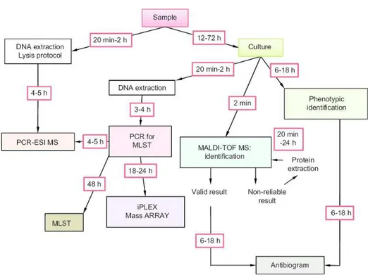
MLST appears to be the most effective method used to classify bacteria into clonal lineages and provide information about their relatedness. On the other hand, the molecular tools can be performed only in few diagnostic and clinical laboratories and require at least 24-36 hours for the results, so in the last years the use of matrix assisted laser desorption ionization time of flight mass spectrometry (MALDI-TOF MS) has notably increased (Fig 1.7). MALDI-TOF MS allows a rapid bacterial characterization and identification with high accuracy by a proteomic approach. Precisely, identification by MALDI-TOF MS is based on the analysis of protein spectrum from bacterial ribosome. Hence, this technique produces a fingerprint spectrum of the peptides and proteins of the analyzed bacterial strains. The extraction protocols are several, either prescribed by MALDI-TOF MS manufacturers or published in literature, and they are specific for certain groups of bacteria and yeasts. Referring to S. pseudintermedius or, better, to the SIG complex, Decristophoris et al. (2011), reported that, MALDI-TOF MS, provided with a reliable database, is a valid and effective technique for a rapid identification of the bacterial species belonging to the S. intermedius Group.
51
Fig. 1.7 Typical workflow of new and old methods used in clinical microbiology (Lavigne et al., 2012)
52 2. Materials and Methods
2.1 Collection of samples
During the years 2015-2017 the specimens, represented by auricular and cutaneous swabs, were collected from dogs suffering from otitis externa and pyoderma at the Bacteriology laboratory of the Veterinary Teaching Hospital of the Department of Veterinary Medicine and Animal Production, University of Naples “Federico II”.
Moreover, further auricular swabs, screened for staphylococci growth, were obtained from Provet Lab srl of Latina, Lazio Region.
From a total of 259 staphylococcal cultures, S. pseudintermedius strains were selected.
2.2 Bacterial isolation and phenotypic identification of the isolates
All samples were cultured and streaked in parallel on Columbia CNA agar (Liofilchem, Teramo, Italy) and on MSA (Liofilchem, Teramo, Italy) and incubated aerobically at 37 °C for 24-48 h (Fig. 1.8). As
positive control S. pseudintermedius ATCC® (American Type Culture
Collection) 49444TM was used. Suspected S. pseudintermedius isolates
were firstly identified by using standard, rapid screening techniques: colony morphology, β-haemolysis on Columbia CNA agar, absence of
mannitol fermentation on MSA, cellular morphology (after Gram’s
staining method), catalase test. Additionally, each mannitol salt negative colony was also subjected to staphylocoagulase (tube coagulase) reaction (Oxoid, Ltd, UK) to confirm their capacity to produce coagulase enzyme.
53
Fig. 1.8 Culture media used for S. pseudintermedius isolation Columbia CNA agar on the left, MSA on the right
For an initial identification, a biochemically-based commercial system, manual API Staph (bioMérieux, Marcy L’Etoile, France) was performed and the presumptive S. pseudintermedius strains, reported as
S. intermedius, if of animal origin, were selected. These strains were
then identified by MALDI-TOF MS analysis (Bruker Daltonics, Germany), which discriminates S. pseudintermedius from S.
intermedius, at the diagnostic service of the Department of
Experimental Medicine, University of Campania “Luigi Vanvitelli”. For MALDI-TOF MS identification, fresh colonies, grown on Columbia CNA agar, were used.
The protocol used was the following: the bacterial colony was first inoculated in the plate for mass spectrometry. Subsequently, 1 µl of the organic matrix, usually cinnamic acid, was added to the sample. Afterwards, the plate was placed in the equipment for MALDI-TOF MS analysis (Fig. 1.9). The identification by MALDI-TOF MS is based on the score value released by equipment’s instructions.
54
Values from 2.3 to 1.9 indicated the best identification of genus and species (Santos et al., 2013).
55
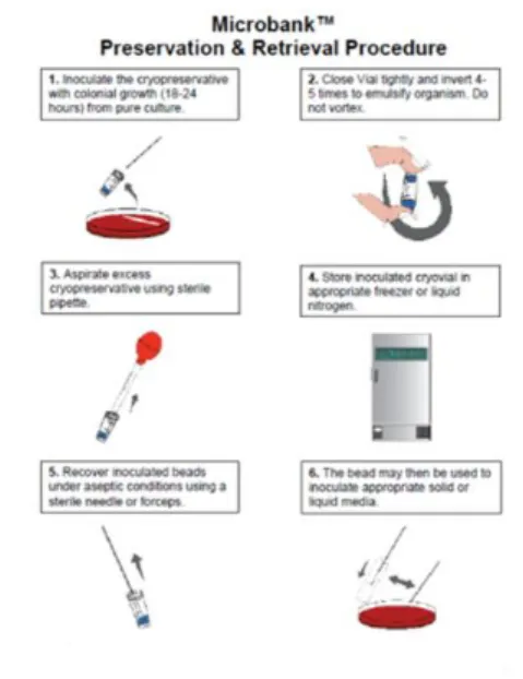
All S. pseudintermedius cultures were stored at -70 °C by using Microbank™ vials (Pro-lab Diagnostics, Canada) for further analysis. Microbank™ is a ready to use system designed for the long term
storage and retrieval of bacterial and fungal isolates. Each Microbank™
vial contains a 25 sterile coloured beads (single colour) and the cryopreservative. The specially treated beads are of a porous nature allowing microorganisms to readily adhere onto the bead surface (Fig. 1.10). After inoculation the Microbank™ vials are kept at -70°C for extended storage. When a fresh culture is required, a single bead is easily removed from the Microbank™ vial and used to directly inoculate a suitable culture medium.
Fig. 1.10 Microbank™: preservation and retrieval procedure (Pro-lab Diagnostics)
56
2.3 Genotypic identification of the isolates
For the molecular characterization of the selected strains, each S.
pseudintermedius isolate was cultured again on MSA with incubation at
37°C overnight. The bacterial DNA extraction of the isolates was carried out using two different protocols:
- by boiling;
- by using a commercial kit.
Referring to the first method, 1-5 colonies were taken from a pure culture of the isolated strain and then homogenized in 50 µL of distilled water and then denatured at 100°C for 10 minutes. Then, the obtained bacterial suspensions were stored at -20°C.
The second technique was performed by using the commercial Isolate II Genomic DNA Kit (Bioline, London, UK) and following the manufacturer’s instructions. Briefly, the isolated bacterial colonies were dissolved in 180 µL of lysis buffer (buffer GL) and in 25 µL of proteinase K solution and then the obtained suspension was vortexed. At that point, the suspension was incubated at 56°C for at least one hour, in order to obtain the complete bacterial cell lysis. Subsequently, 200 µL of buffer G3 were added, followed by another incubation at 70°C for 10-15 minutes. The sample was, then, briefly vortexed and 210 µL of absolute ethanol were added to it and the suspension was vigorously vortexed. The mix of each sample, placed in the isolate II genomic DNA spin column, was transferred into a collection tube and centrifuged at 11.000 x g for 1 minute. Afterwards, the silica membrane was washed twice with two different wash buffers (500 µL of wash buffer G1 and 600 µL of wash buffer G2) and, obviously, centrifugated
57
at each wash at 11.000 x g for 1 minute. This last step ensured that no ethanol residue was transported in the subsequent elution step. Once placed the isolate II genomic DNA spin column in a 1.5 ml sterile microcentrifuge tube, 100 µL of elution buffer G (elution step) were directly pipetted onto the silica membrane. The last centrifugation at 11.000 x g for 1 minute allowed the collection of bacterial DNA in elution liquid in the Eppendorf tubes. Before the molecular characterization of all isolated strains, genomic DNA was tested by
Biophotometer Eppendorf to determine the absorbance ratio (OD
A260/A280). The A260/A280 was considered appreciable when ranged between 1.5-1.9 value. Bacterial DNA was stored at -20°C and used for further studies.
Molecular profiling, using species-specific nuc gene (Sasaki et al., 2010) and species-specific hlb gene (Kmieciak et al., 2016) was performed by single PCR to confirm the identification of S.
pseudintermedius strains. Also for the genotypic characterization of the
isolated strains, S. pseudintermedius ATCC® 49444TM was used as
positive control.
Primers sequences, amplicon sizes and amplification programs are reported in Table 1.1.
The single PCR reaction mixture for each gene (nuc and hlb genes) was prepared by using the Green Hot Start PCR Master Mix Direct Load, 2x (Biotechrabbit, GmbH, Germany) as follows:
- Green Hot Start PCR Master Mix: 12,5 µL - primers (F+R): 1 µL
58
- nuclease free water: 10,5 µL - DNA template: 1 µL
The mixture final volume for one reaction was of 25 µL.
For every PCR reaction there was always a positive and a negative
control. Genomic DNA was amplified using Biorad T100TM Thermo
cycler (BioRad, Hercules, CA).
Tab. 1.1. Primers sequences, amplicon sizes, amplification programs
The amplified products were analyzed by gel electrophoresis. 1.5 g of
agarose (Agarose electrophoresis grade, Gibco BRL) were placed in a flask and dissolved by heating in a microwave oven in 100 ml of TBE
1X for few minutes. Once melted the agarose, in order to stain the gel,
2 μL of Real Safe nucleic acid staining solution (Durviz s.l., Valencia, Spain) were added. At this point, the gel was poured into the appropriate bed equipped with a comb for the formation of wells and left to solidify for about 30 minutes. For each sample, 12 µL of PCR
Gene Primer sequences
(5’-3’ sense and antisense)
Amplicon
size (bp) Amplification program
nuc F: TRGGCAGTAGGATTCGTTAA R: CTTTTGTGCTYCMTTTTGG 926 94°C 5 min; 94°C 30 s, 58°C 60s, 72°C 90s, for 30 cycles; 72°C 5 min. hlb F: GACGAAAATCAAGCGGAA R: TCTAAATACTCTGGCGCAC 734 94°C 2:30 min; 94°C 30s, 56°c 30s, 72°C 1min, for 30 cycles; 72°C 10 min.
59
reaction mix were loaded in each gel well. 5 μL of 100 bp DNA ladder
with 6x loading dye, consisting of 100- 3000 bp, were loaded as molecular marker (Biotechrabbit, GmbH, Germany).
Electrophoresis was performed in a BioRad electrophoresis tank with 1X TBE as running buffer at 80V for 45-50 minutes. Electrophoresed gels were visualized under blue-light and their images taken using the
ChemiDocTM XRS+ with Image LabTM Software (BioRad, Hercules,
CA).
3. Results
3.1 S. pseudintemedius isolation and phenotypic identification
During the years 2015-2017, a total of 259 staphylococcal strains responsible for canine skin disorders were collected. The phenotypic bacterial identification by proteomic profiling defined a relevant number of 126 (49%) S. pseudintermedius strains. Moreover, all the isolated S. pseudintermedius strains were identified with good scores (1.9 or 2.0).
The other most isolated species belonged to S. aureus (8%), S.
schleiferi (7%), S. xylosus (6%), S. sciuri (2%), S. intermedius (2%), S.
delphini (1%). The remaining 25% consisted of unsuspected Staphylococcus spp.
S. pseudintermedius strains were recovered from dogs suffering with otitis externa (84%) and pyoderma (16%), originating from two different Italian geographical areas (Naples, Campania Region [52%] and Latina, Lazio Region [48%]). The Table 1.2 describes the
60
percentage of S. pseudintermedius isolates from the above reported canine skin disorders.
Tab. 1.2 S. pseudintermedius strains isolated from diseased dogs of Campania and Lazio regions
Geographical area of origin
Biological origin % isolated
S.pseudintermedius strains Naples otitis pyoderma 71 29 Latina otitis pyoderma 98 2
All the isolated S. pseudintermedius strains appeared to be
β-heamolytic on Colombia CNA agar and mannitol negative on MSA (Fig 1.11). Furthermore, they were positive at the staphylocoagulase and catalase reactions.
61
3.2 S. pseudintermedius genotypic identification
All 126 S. pseudintermedius strains harboured the species-specific nuc and hlb genes (Fig. 1.12, 1.13) confirming, thus, the proteomic identification by MALDI TOF MS.
Fig. 1.12 Molecular characterization of S. pseudintermedius: PCR for detection of species-specific nuc gene. Data from one of three experiments are shown. Lanes 3,4,6,7,8, positive samples; Lane P+, positive control; lane N-, negative
control; lane M, 100-bp DNA ladder.
62
Fig. 1.13 Molecular characterization of S. pseudintermedius: PCR for detection of species-specific hlb gene. Data from one of three experiments are shown. Lanes 33,36,39,51,54,67,70,75,77,83,84,92, positive samples; lane N-, negative
control; lane M, 100-bp DNA ladder.
4. Discussion and conclusions
Bacterial otitis externa and pyoderma are the most common canine skin diseases and Staphylococcus pseudintermedius is the staphylococcal species most frequently isolated from dogs suffering from these infections. In fact, Staphylococcus pseudintemedius, an opportunistic canine skin pathogen, is the major CoPS that inhabits healthy dogs (Gόmez-Sanz et al., 2013). In particular conditions, such as dog injures or sickness, this species can take advantage of the weakened defenses and cause infection and illness.
According to literature, the results of this study show a high prevalence (49%) of S. pseudintermedius among the other isolated staphylococcal species, approving its role as the main causative agent of canine skin disorders.
63
Due to the high rates of colonization of dogs with S. pseudintermedius and S. aureus, these species make up the majority of Staphylococcus related infections in dogs. According to this finding also the results of this research reported S. aureus as the second most common causative
agent of staphylococcal skin infection.
The literature has always reported a higher interest for coagulase-positive staphylococci such as S. aureus and SIG, which is composed, besides S. pseudintermdius, by S. intermedius and S. delphini which, here, were found responsible agents of canine skin disorders with percentage in the range of 1-8%.
Even though S. intermedius mainly colonizes pigeons (Sasaki et al., 2007), it was also isolated from dogs with otitis externa (Dziva et al., 2015).
Sasaki et al. (2007) found that S. intermedius can be chemically distinguished from S. pseudintermedius by positive arginine dihydrolase and acid production from beta-gentiobiose and D-mannitol. The two species can also be distinguished using molecular and genetic testing, as well as MALDI-TOF mass spectrometry, although this is not commonly done (Wang et al., 2013).
Furthermore, S. intermedius should be included in the differential diagnosis of invasive infection amongst human patients with close contact with dogs.
S. delphini, first isolated from a dolphin (Varaldo et al. 1988), was also
64
camels. These data indicate that S. delphini may be more widespread and clinically important than was previously thought. Surprisingly, in this study, this staphylococcal species was isolated from infected dogs (3/259, 1%), therefore it should be warranted to examine the importance of S. delphini as a veterinary pathogen in pets.
Coagulase-Negative Staphylococci (CoNS), members of normal flora of human and animal skin, have long been considered as nonpathogenic possessing fewer virulence properties than CoPS. Recently they have assumed an important role as pathogens in skin and soft tissue infections, overall, because of their increasing multidrug-resistance profiles.
In this regard, the percentage of CoNS positivity (15%), precisely of S.
schleiferi, S. xylosus and S. sciuri, among the processed samples,
highlights a relevant dog susceptibility to these bacterial species.
In veterinary medicine, S. schleiferi has been repeatedly documented in literature as both an inhabitant and as a pathogen, and May et al. (2012) demonstrated that S. schleiferi could be recovered from the ears and anterior nares of healthy dogs as well as those with otitis and/or pyoderma. In this study, S. schleiferi was isolated from 20/259 samples (7%) followed by S. xylosus (6%) and S. sciuri (2%).
In conclusion, this study underlines that S. pseudintermedius is the most common bacterial isolate from dogs suffering with otitis externa
and pyoderma in Naples – Campania Region, and Latina, Lazio
65
However, its phenotypic identification remains problematic in many clinical microbiology laboratories, especially in the human ones, where it is often misidentified as S. aureus or CoNS. So, genotypic profiling (species-specific nuc and hlb genes) still represents the main molecular technique for the identification of this species when MALDI-TOF MS analysis is not available.
MALDI-TOF MS for the diagnosis of infectious diseases has been
rapidly embraced by laboratories around the globe, and, as seen in this study, represents a valid bacterial identification method also in veterinary medicine. In fact, it represents the most accurate and rapid method for the identification of S. pseudintermedius and the other members of the SIG.
This study has provided a further evidence that MALDI-TOF MS is a useful and reliable technique for S. pseudintermedius identification. Further confirmation of proteomic profiling identification by MALDI-TOF MS was always given by genotypic profiling results, linked to the detection of species-specific S. pseudintermdius genes.
66 5. References
Aanensen DM, Spratt BG, 2005. The multilocus sequence typing
network: mlst.net. Nucleic Acids Res 33, W728–W733.
Bannoehr J, Ben Zakour NL, Waller AS, Guardabassi L, Thoday KL, van den Broek AH, Fitzgerald JR, 2007. Population genetic structure of the Staphylococcus intermedius group: insights into agr diversification and the emergence of methicillin-resistant strains. J Bacteriol 189, 8685-8692.
Bannoehr J, Franco A, Iurescia M, Battisti A, Fitzgerald JR, 2009.
Molecular diagnostic identification of Staphylococcus
pseudintermedius. J Clin Microbiol 47, 469-471.
Bannoehr J, Guardabassi L, 2012. Staphylococcus pseudintermedius in the dog: taxonomy, diagnostics, ecology, epidemiology and pathogenicity. Vet Dermatol 23, 253-266.
Ben Zakour NL, Bannoehr J, van den Broek AH, Fitzgerald JR, 2011. Complete genome sequence of the canine pathogen Staphylococcus pseudintermedius. J Bacteriol 193, 2363-2364.
Casagrande Proietti P, Stefanetti V, Hyatt DR, Marenzoni ML,
Capomaccio S, Coletti M, Bietta A, Franciosini MP, Passamonti F, 2015. Phenotypic and genotypic characterization of canine pyoderma isolates of Staphylococcus pseudintermedius for biofilm formation. J Vet Med Sci 77, 945-951.
Decristophoris P, Fasola A, Benagli C, Tonolla M, Petrini O, 2011. Identification of Staphylococcus intermedius Group by MALDI-TOF MS. Syst Appl Microbiol 34, 45-51.
67
De Martino L, Nocera FP, Mallardo K, Nizza S, Masturzo E, Fiorito F, Iovane G, Catalanotti P, 2016. An update on microbiological causes of
canine otitis externa in Campania Region, Italy. Asian Pac J Trop Biomed 6, 384-389.
Deurenberg RH, Vink C, Kalenic S, Friedrich AW, Bruggeman AC, Stobberingh EE, 2007. The molecular evolution of methicillin-resistant of Staphylococcus aureus. Clin Microbiol Infect 13, 222-235.
Deurenberg RH, Stobberingh EE, 2008. The evolution of
Staphylococcus aureus. Infect Genet Evol 8, 747-763.
Devriese LA, Vancanneyt M, Baele M, Vaneechoutte M, De Graef E, Snauwaert C, Cleenwerck I, Dawyndt P, Swings J, Decostere A, Haesebrouck F, 2005. Staphylococcus pseudintermedius sp. nov., a coagulase-positive species from animals. Int J Syst Evol Microbiol
55, 1569-1573.
Dufour P, Jarraud S, Vandenesch F, Greenland T, Novick RP, Bes M, Etienne J, Lina G, 2002. High genetic variability of the agr locus in
Staphylococcus species. J Bacteriol 184, 1180-1186.
Duim B, Verstappen KMHW, Kalupahana RS, Ranathunga L, Fluit AC, Wagenaar JA, 2018. Methicillin-resistant Staphylococcus
pseudintermedius among dogs in the description of novel SCCmec
variants. Vet Microbiol 213, 136-141.
Dziva F, Wint C, Auguste T, Heeraman C, Dacon C, Yu P, Koma LM, 2015. First identificationof methicillin-resistant Staphylococcus
68
isolated from dogs with otitis externa in Trinidad, West Indies. Infect Ecol Epidemiol 5, 29170.
Fitzgerald JR, 2009. The Staphylococcus intermedius group of bacterial pathogens: species reclassification, pathogenesis and the emergence of methicillin resistance. Vet Dermatol 20, 490-495.
Foglia Manzillo V, Nocera FP, De Martino L, Gizzarelli M, Oliva G, 2016. A successful vancomycin treatment of multidrug-resistant MRSA-associated canine pyoderma. J Dermatol Res Ther 1, 12-18. Garbacz K, Zarnowska S, Piechowcz L, Haras K, 2013. Pathogenicity potential of Staphylococcus pseudintermedius strains isolated from canine carriers and from dogs with infection signs. Virulence 4, 255-259.
Gόmez-Sanz E, Torres C, Lozano C, Zarazaga M, 2013. High diversity
of Staphylococcus aureus and Staphylococcus pseudintermedius
lineages and toxigenic traits in healthy pet-owing household members. Underestimating normal household contact? Comp Immunol Microbiol Infec Dis 36, 83-94.
Han JI, Yang CH, Park HM, 2015. Emergence of biofilm-producing
Staphylococcus pseudintermedius isolated from healthy dogs in
South Korea. Vet Q 36, 207-210.
Hájek V, 1976. Staphylococcus intermedius, a new species isolated from animals. Int J Syst Bacteriol 26, 401-406.
Hariharan H, Coles M, Poole D, Lund L, Page R, 2006. Update on antimicrobial susceptibilities of bacterial isolates from canine and feline otitis externa. Can Vet J 47, 253-255.