TOR VERGATA UNIVERSITY
ROLE OF THE HIV-1 NEF PROTEIN IN THE ACTIVATION
OF INFECTED CD4
+T LYMPHOCYTES
Giorgia Giolo
Supervisor
Dr. Margherita Doria
Ospedale Pediatrico Bambino Gesù Rome
Dottorato di Ricerca in Immunologia, XXI ciclo
Tor Vergata University, Rome, Italy
INDEX
ABSTRACT... 1
1 - INTRODUCTION ... 3
1.1 The life cycle of HIV-1 ... 3
1.2 HIV-1 pathogenesis ... 7
1.3 T cell activation during HIV-1 infection ... 10
1.3.1 T cell activation modulates viral replication ... 10
1.3.2 Bystander and direct T cell activation ... 11
1.3.3 The T cell receptor signaling ... 12
1.4 Role of HIV-1 Nef protein ... 18
1.4.1 Nef increases HIV-1 replication and infectivity ... 19
1.4.2 Nef allows immune evasion by HIV-1 ... 20
1.4.3 Down-regulation of CD4 by Nef ... 21
1.4.4 Effects of Nef on T cell activation ... 22
2 - MATERIALS AND METHODS ... 25
3 - RESULTS ... 33
3.1 Nef expression in HIV-1-infected quiescent CD4+ T lymphocytes ... 33
3.2 Nef increases viral replication and IL-2 production upon activation of HIV-infected T cells... 37
3.3 Protein tyrosine phosphorylation upon activation of quiescent HIV-infected T cells... 39
3.4 Activation of Akt and NF-!B pathways in HIV-1-infected CD4+
T lymphocytes... 42
3.5 CD4/Lck interaction in HIV-1-infected resting T cells... 46
3.6 Activation of Lck upon triggering of CD4 in HIV-1-infected cells... 48
3.7 Phosphorylation of the TCR "-chain upon CD4 stimulation of infected T cells ... 50
3.8 Nef activity in HIV-1-infected activated CD4+ T lymphocytes…... 52
3.9 Protein tyrosine phosphorylation upon secondary TCR stimulation of HIV-infected T cells... 57
4 - DISCUSSION ... 60
5 - REFERENCES... 67
ACKNOWLEDGMENT ... 75
ABSTRACT
The replication of HIV-1 is closely linked to the activation state of the cells: in activated memory CD4+ T lymphocytes, HIV-1 readily undergoes multiple rounds of replication, whereas resting T cells are largely refractory to productive infection due to multiple early post-entry replication blocks. Reported evidences suggest that HIV-1 has evolved functions to contrast these blocks. In particular, the product of an early viral gene, Nef, is able to sensitize HIV-1-infected quiescent CD4+ T lymphocytes to activation thus favoring IL-2 production and viral replication in response to TCR stimulation. On the other hand, in productively infected T cells, Nef was shown to impair early activation events during synapse formation with antigen presenting cells that result in reduced release of IL-2. In addition, former studies have shown that Nef associates with several signaling proteins (e.g. Lck, Vav, PI3-K, PAK2), and affects the expression of the CD4 and CD28 co-receptors, but the functional consequences of these interactions in T cell activation is unclear.
This study is focused on the function of the Nef protein in the activation of primary HIV-1-infected CD4+ T lymphocytes. First, the impact of HIV-1 infection, with or without Nef expression, on the steady-state levels and intracellular localization of Lck, CD4 and CD28 was analyzed in both resting and pre-activated CD4+ T lymphocytes. Next, the capacity of CD4+ T lymphocytes infected with wt or Nef-deficient virus to respond to primary and secondary TCR stimulation was investigated. Moreover, CD4 signaling and Lck activation was analyzed after CD4-cross-linking of quiescent T cells infected with wt or Nef-deficient HIV-1. Results show that, despite the steady-state levels of CD4 and Lck are reduced by Nef, the CD4 signaling pathway is preserved in latently infected resting CD4+ T lymphocytes. Upon activation via CD3/CD28 of HIV-1-infected quiescent T cells, early
tyrosine-phosphorylated effector molecules as well as the down-stream NF-!B pathway are induced to the same extent and with the same kinetics in cells infected with wt virus, Nef-deficient virus, and uninfected cells. Conversely, secondary activation via CD3/CD28 of resting HIV-infected T cells, results in a Nef-dependent delay of PLC-#1 phosphorylation.
Finally, in activated HIV-1-infected T cells the dramatic down-regulation of surface CD4 induced by Nef and late viral proteins (Env, Vpu) as well as the Nef-dependent CD28 down-regulation, may account for the previously described Nef’s capacity to impair the formation of the immune synapse. Apparently, that Nef differentially regulates T cell activation depending on the intracellular environment: Nef enhances activation of quiescent cells to favour initial viral replication whereas it inhibits second rounds of TCR stimulation, possibly to avoid apoptosis that would restrict viral replication.
The future identification of target molecules regulated by Nef is an important step for understaing how HIV-1 interferes with T cell function to facilitate viral spread.
1-INTRODUCTION
1.1 The life cycle of HIV-1
The Human Immunodeficiency Virus type 1 (HIV-1) is a member of the lentivirus family of retroviruses and is the etiologic agent of the acquired immunodeficiency syndrome (AIDS). The genome of HIV-1 is 10 Kb in length and encodes 16 distinct proteins. Those proteins that are derived from the gag (group specific antigen), pol (polimerase), and env (envelope) genes are classical structural and enzymatic factors that are required by all retroviruses. In addition, HIV-1 contains open reading frame for six additional regulatory and accessory proteins. The Trans Activator of Transcription (Tat), the Regulator Expression of Virion genes (Rev), the ill-named “Negative Factor” (Nef), the Viral Infectivity Factor (Vif) and the Viral Proteins r (Vpr) and u (Vpu) [1].
HIV-1 enters the body through the exchange of bodily fluids and its major target of infection are cells expressing the CD4 surface antigen, such as T helper (Th) cells, and cells of the monocyte/macrophage lineage including monocytes, macrophages, dendritic cells (DC) and brain microglial cells. Virus-CD4 receptor binding is mediated by a high affinity interaction between the viral outer envelope glycoprotein, gp120, and the CD4 molecule. Subsequentially, the gp120 interacts with one of the additional coreceptors that are strain and target specific and determines the tropism of the virus. The R5 strains of HIV-1 use CC chemokine receptor 5 (CCR5) as their coreceptors and can, therefore, enter macrophages, DCs and T cells, whereas X4 strains use CXCR4 as a coreceptor and can infect T cells only [2] . Early in infection, only R5 viruses are usually detected in infected individuals. At this stage, the virus might need to transit through DCs and
macrophages which in turn could pass the virus to CD4+ T cells possibly during the process of antigen presentation. DCs are likely to have an important role in transporting the virus from its portal of entry to lymphoid organs. These cells can be productively infected by HIV-1 or they can capture the virus trough DC-specific ICAM3-grabbing non integrin receptor (DC-SIGN) and store it in an infectious form before presenting it to T cells that simultaneously become primed for infection. With time, X4 viruses come to predominate which hastens the demise of Th cells, the hallmark of AIDS [3].
Once internalized, HIV-1 is uncoated and its RNA genome is reverse transcribed to a double-stranded DNA that is integrated into the genome of the target cell, yielding the long terminal repeat (LTR)-flaked provirus (Fig. 1). Unlike other retroviruses, HIV-1 does not require disintegration of the nuclear membrane given that nuclear localization signals on integrase (IN), matrix and Vpr proteins ensure that the viral genome passes through the nuclear pores. The linear double stranded cDNA integrates in the genome with a preference for active genes, although other regions, for example heterochromatin-rich centromeric regions, are targeted also [4]. The activity of the integrated viral genome or provirus is greatly influenced by the metabolic and activation state of the host cell and the longevity of the provirus is dictated by the life span of the cell that contains it [5].
In an activated T cell, viral replication is rapid and efficient. The viral long terminal repeat contains binding sites for cellular factors that positively regulate transcription of HIV-1 and that are abundant in activated T cells. However, these host factors are not enough, and the viral Tat protein also markedly augments viral gene expression. The situation is very different in resting lymphocytes. These cells are refractory to viral replication in vitro because there are several barriers that preclude
the completion of early steps, such as, inefficient reverse transcription and energy levels too low for effective nuclear import of double-stranded viral genomes [5]. In infected quiescent cells, the provirus might exist in a silent or latent state and it can be rescued upon cell activation. Once they have been activated even partially, T cells become fully permissive for HIV infection. The nature of the induced factors that are responsible for the acquisition of HIV permissiveness is not known yet. Under conditions where integration was undetectable, some genes are already transcribed. All HIV transcripts are derived from a common full-lenght precursor, which also serves as the mRNA for the gag-pol genes and the viral genomic RNA. By alternative splicing, this full-lenght precursor generates about 30 different mRNAs, including singly spliced transcripts coding for Env, Vpu, Vpr and Vif, and multiply spliced transcripts coding for Nef-, Tat- and Rev-specific transcripts [6]. Among the multiply spliced transcripts, Nef-encoding mRNA was prominent in quiescent cells, along with lesser levels of Tat mRNA. By comparison, activated cells generated abundant levels of Nef-, Tat- and Rev-specific transcripts. When viral DNA is integrated in the host genome, the provirus behaves like any human gene, with transcription being initiated at the 5’ end and terminating at the 3’ end. Genomic RNA is exported to the cytoplasm where it is used as a template for translation of Gag full-lenght polypeptide that it is cleaved into the matrix (MA), capsid (CA), nucleocapsid (NC) and p6 proteins. This production is followed by assembly, budding and maturation, in which the new HIV-1 particles are packaged up and sent out to infect new cells [4].
1.2 HIV-1 pathogenesis
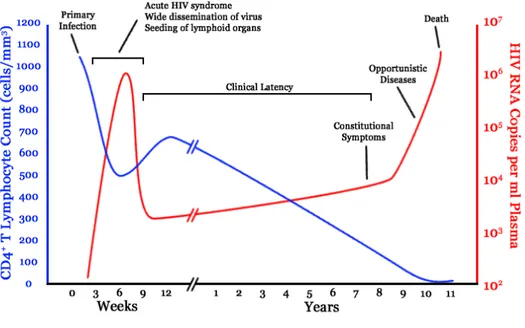
Primary infection of humans with HIV-1 is associated with an acute mononucleosis-like clinical syndrome which appears approximately 3-6 weeks following infection (Fig. 2. ). The severity and persistence of the symptoms vary considerably. Significant declines in the levels of CD4+ T lymphocytes in the peripheral blood occur in the first 2-8 weeks following HIV-1 infection. These levels may rebound toward normal as the patients enters the clinically latent stage of disease, although they rarely if ever return to pre-infection levels [6, 7]. The acute syndrome associated with primary HIV-1 infection is accompanied by a burst of viral replication that can be detected in the blood approximately 3 weeks following infection. During this period, infectious virus and viral proteins can be detected in the cell-free plasma, and the number of virions can reach 106 to 107 per milliliter.
Approximately 3-6 weeks after the infection with HIV-1, specific antiviral immune responses can be detected in association with a decline in the viral burden and a temporary stabilization of the number of CD4+ T cells. Multiple factors probably contribute to the decline in viral titer, including innate and adaptive immune responses, the secretion of suppressing cytokines, and the depletion of CD4+ target cells [7] .
Following the induction of an immune response to HIV-1, there is a relatively long period of latency that is characterized by few, if any, clinical manifestations. The length of the latency is regulated by a complex interplay between the host genetic and immunologic factors and the pathogenic potential of the virus. Throughout the latent period there is usually a continuous, yet variable, decline in the numbers of CD4+ lymphocytes as well as continuous viral replication. Once the CD4+ T cell count falls
below 200 cell/µl, the patient is susceptible to AIDS-defining opportunistic infections and neoplastic diseases. In addition, HIV-1 infection leads to the dysregulation of several leukocyte subpopulations and generalized immune activation with the subsequent development of other infections and malignancies [7]. By progressive destruction of the helper T lymphocyte pool, HIV-1 precludes both the efficient production of antibodies by B cells and the proper function of cytotoxic T lymphocytes (CTLs). In parallel, the virus exploits a great diversity of strategies to alter the cellular arm of the immune response. Indeed, through mechanisms that are not clear as yet, HIV infection leads to B-cell hyperplasia, hypergammaglobulinemia, elevated autoantibody titers, a poor response to neoantigens and mitogens [8]. These abnormalities may contribute to disease progression and development of opportunistic infections.
Several evidence indicate that the HIV-1 induced perturbation of innate immune responses contributes to permissive immunosuppression. For instance, natural killer (NK) cells, display an aberrant phenotype and reduced citotoxic activity [8, 9].
The macrophage lineage play an important role in initial infection with HIV-1 and contribute to pathogenesis. The infected macrophages are resistant to cytopathic effects and persist throughout the course of infection as long-term stable reservoirs for HIV-1 capable of disseminating the virus to tissues [10].
In general, elevated levels of activation are observed in the immune systems of HIV-1-infected individuals and activation marker expression correlates with disease progression [7].
Fig. 2. Viral load and CD4+ T cell count over the course of HIV-1 infection in an untreated patient (from Nat Rev Immunol, 2003, 3:97).
1.3 T cell activation during HIV-1 infection
1.3.1 T cell activation modulates viral replication
As mentioned earlier, the cellular activation state is critical for productive HIV-1 infection. In vitro studies with peripheral blood mononuclear cells (PBMC) have indicated that specific cellular characteristics govern the susceptibility of T cells to productive HIV-1 infection, including activation, maturation, and proliferation [5, 11]. In particular, some studies indicate that higher HIV-1 LTR activation levels were consistently observed in memory cells if compared to naïve cells [12]. The authors observed that memory CD45RO+ display a more pronounced NFAT (nuclear factor of activated T cells) activation, which may explain higher replication levels [13].
Studies performed with cells derived from HIV-1 patients, showed that memory CD4+ T cells produce fourfold more replication competent virus and contain six fold more provirus than naïve CD4+ T cells [14]. These observations suggest that HIV-1 can enter naïve CD4+ T cells in vivo and initiate reverse transcription but it is unable to integrate into genome as efficiently as in memory CD4+ T cells. In vitro studies have also demonstrated that naïve CD4+ T cells can be acutely infected with HIV-1 but are less efficient in replicating virus upon physiologic stimulation [15]. The mechanisms responsible for these differences in viral replicative capacity have not been clearly defined and possible explanations include lower levels of deoxynycleoside triphosphate substrates and a deficiency of other cellular factors in naïve cells that are necessary for completion of viral replicative cycle [16].
1.3.2 Bystander and direct T cell activation
HIV infection is known to be associated with changes in the cytokine milieu that can lead to the induction of bystander T cell activation. Bystander activation is defined as activation of a T cell to produce phenotypic or functional changes through a mechanism which is independent of specific T cell receptor stimulation [17]. Numerous studies in mice have shown that cytokines alone can elicit T-cell responses. Cytokines such as IL-2, IL-4, IL-6, IL-7 and IL-15 can render resting T cells susceptible to productive HIV infection, and this might further enhance disease progression.
Previous work has demonstrated that the exogenous viral protein Nef is able to mediate bystander activation of T cells, to increase PBMC proliferation and to enhance HIV-1 replication through IL-15 synthesis induction by the monocyte/macrophage population [18].
Association of the viral envelope gp120 with surface CD4 has been demonstrated to activate pathways in both primary T cells and T cell lines. The gp120 has innate biological activity as a result of a specific interaction with CD4, inducing increases in intracellular levels of inositol triphosphate and of calcium, and in interleukin-2 receptor expression and cell motility [7].
Direct effects of HIV infection on the activation state of human T cells have been described. For instance, it was shown that infection of by HIV-1 renders T cells hypersensitive to T cell receptor and CD28 stimulation [19]. In particular, Nef protein associates with membrane microdomains critically involved in the initiation and propagation of T cell signaling and it primes T cells for activation [20]. Nef promotes the activation of transcription factors such as NFAT and NF-!B in response to CD3/CD28 stimulation that induce enhancement of IL-2 secretion and HIV-1
replication in a stimulus-dependent manner [19, 20]. Another HIV regulatory protein capable to enhance cytokine gene transcription, Tat, is expressed early during infection. Both Nef or Tat enhances IL-2 secretion in Jurkat and primary T cells upon T cell activation [21].
1.3.3 The T cell receptor signaling
The engagement of the T cell receptor (TCR) initiates the earliest events leading to T cell activation. The TCR is a complex consisting of the variable $% chains noncovalently associated with the nonpolymorphic CD3 proteins. CD3 exist as a series of dimers including #&, '&, and "" associated with a single $% heterodimer. The "-chain of the CD3 display an extra-cellular region of 9 amino acids, a trans-membrane domain, and a intra-cytoplasm domain of 113 amminoacids with three ITAM (Immunoreceptor Tyrosine-based Activation Motif). The CD3 and "-chain drive the signal transduction induced by antigen recognition and biochemicals events leading to T cell activation.
CD4 and CD8 are two important co-receptors that participate to the triggering and signal transduction of the TCR. The interaction between CD4 and the non variable regions of MHC II molecules on the surface of the antigen presenting cell (APC) enhances the T cell response to antigenic stimulation. CD4 is a 55 kDa glycoprotein comprised of four Ig-like extracellular domains, a single transmembrane domain, and a short cytoplasmic tail of 38 residues. CD4 is associated noncovalently with a src-related tyrosine kinase, p56Lck, through two cysteine residues at positions 420 and 422 of the cytoplasmic domain [22]. The CD4/Lck association is required to
bring Lck in the vicinity of the TCR signaling complex and initiate T cell activation upon antigen recognition [24, 25].
In addition, the outcome of TCR stimulation is regulated by the simultaneous engagement of the costimulatory molecule CD28. Indeed, T cells costimulated through CD28 respond more rapidly to lower levels of TCR occupancy enhancing distinct signaling pathways that lead to gene transcription [26].
The earliest step in intracellular signaling following TCR ligation is the tyrosine phosphorylation of ITAM sequences in the intra-cytoplasm tail of "-chain and CD3. The phospho-tyrosine becomes a binding site for SH2 domain of src kinase (Lck, Fyn) and ZAP-70. ZAP-70 binds the transmembrane adapter protein linker for the activation of T cells (LAT) and the cytosolic adapter domain-containing leukocyte phosphoprotein of 76 kDa (SLP-76) [27]. These two adapters form the backbone of the complex that organizes effector molecules in the correct spatiotemporal manner to allow for the activation of multiple signaling pathways. LAT contains nine tyrosines that are phosphorylated upon TCR engagement, which bind the C-terminal SH2 domain of PLC-#1, the p85 subunit of phosphoinositide 3-kinase (PI3K), and the adapters growth factor receptor-bound protein 2 (GRB2) and the adapter downstream of Shc (Gads). PLC-#1 also directly binds to SLP-76, LAT and Vav1. These interactions collectively are required to stabilize PLC-#1 in the correct conformation within the complex to allow for its optimal activity. The proximal signaling complex results in the activation of PLC-#1-dependent pathways including Ca2+-and DAG-induced responses, cytoskeletal rearrangements, and integrin activation pathways. Activated PLC-#1 then hydrolizes the membrane lipid PI(4,5)P2, producing the
second messengers IP3 and DAG. The production of DAG results in the activation of two major pathways involving Ras and PKC. Ras is a guanine nucleotide-binding protein and is required for the activation of the Raf-1 kinase, which initiates a mitogen-assiciated protein kinase MAPK phosphorilation, which in turn phosphorylate Erk. Erk kinase activity results in the activation of Elk, which contributes to the activation of AP-1, Jun and Fos transcription complex. One critical pathway that PKC regulates involves the NF-!B transcription factor. In resting cells, NF-!B is found in the cytosol associated with inhibitor of NF-!B (I!B) family members that keep NF-!B from moving into the nucleus. Upon T cell activation, IÍ!B is phosphorylated by the I!B kinase (IKK) complex, ubiquitinylated, and degraded, allowing NF-I!B to translocate into the nucleus, where it activates genes involved in the function, survival and homeostasis of T cells [28].
TCR recruitment and Vav activation induce Rac/cdc42/MKK and p38 activation. Rac-GTP activates Fos and JNK (c-Jun N-terminal kinase) which phosphorylates c-Jun. TCR-induced increases in intracellular Ca2+ levels result in the activation of Ca2+ and calmodulin-dependent transcription factors, including the phosphatase calcineurin and the Ca2+-calmodulin-dependent kinase that in turn activate a variety of transcription programs. Activated calcineurin dephosphorylates members of the NFAT family, leading to their translocation to the nucleus. In the nucleus, NFAT isoforms can form cooperative complexes with a variety of other transcription factors, thereby integrating signaling pathways resulting in differential gene expression patterns and functional outcomes. The NFAT/AP-1 interaction integrates Ca2+ and Ras signals and results in the expression of genes important for T
cell activation including IL-2. In contrast, NFAT activity in the absence of AP-1 activation induces a pattern of gene expression that results in T cell anergy and a characteristic lack of IL-2 production.
Numerous studies have shown that CD28 promotes T cell proliferation, cytokine production, cell survival and cellular metabolism. Following binding of CD28 to its ligands CD80 or CD86 on APCs, the p85 regulatory subunit of PI3K associates with a cytoplasmic tail of CD28. This regulatory subunit recruits the p110 catalytic subunit of PI3K, which converts PIP2 to phosphatidylinositol (3,4,5) trisphosphate (PIP3) at the membrane [29]. Localized PIP3 generation serves as a docking site for the PDK1 and its target Akt. Akt phosphorylates multiple proteins, and thus affects numerous cellular responses. Activation of Akt enhances the nuclear translocation of NF-kB, which has positive effects on the expression of prosurvival genes. The ability of Akt to promote prosurvival gene expression, coupled with the ability of Akt to inhibit trancription factors that promote cell cycle arrest, results in Akt-driven cell survival and proliferation. Akt also affects optimal trascription of NFAT-regulated genes, such as IL-2 [24].
During HIV-1 infection different viral proteins are involved in alteration of the TCR signaling. In particular the function of Tat is to dysregulate cytokine production resulting in perturbation of the host immune response and enhancement of the retrovirus survival. Previous work demonstrate that Tat impairs the IFN#-receptor signaling pathway. In addition the Nef protein of HIV-1 has a important role in the activation pathway of T cells. In T cell lines, Nef induces transcription of an array of genes almost identical to that triggered upon exogenous stimulation of T cell receptor
[30]. However, the extent of T cell activation imprinted by expression of Nef is a matter of controversy. Although in some studies Nef facilitates the cell surface exposure of the early activation marker CD69 as well as IL-2 production [19], other studies demonstrated that Nef has no effect on T cell activation markers or even reported an inhibitory effect of Nef on T cell activation, such as disruption of immunological synapse and the accumulation and degradation of Lck [19, 20].
Fig. 3. TCR receptor signaling
T Cell Receptor (TCR) activation promotes a number of signaling cascades that ultimately determine cell fate through regulating cytokine production, cell survival, proliferation, and differentiation. (from Immunol. Rev. 2007)
1.4 Role of the HIV-1 Nef protein
The nef gene product is a small protein of about 27 kDa that is syntesized at the earliest stage of viral gene expression. It was originally thought to exert a negative influence on viral replication, hence the acronym Nef for Negative factor. These early results were not confirmed and Nef was instead convincingly shown to stimulate viral growth both in cell culture and in vivo. Nef is post-translationally modified by myristoylation of its amino terminus, and this modification targets the protein to the inner surface of the plasma membrane and to the trans-golgi network. Nef is present also in the cytosol, in the nucleus and is incorporated in the virion. To date, no enzymatic activity has been directly attributed to the Nef protein. Nevertheless, several major distinct activities of Nef have been demonstrated in vitro: down-regulation of cell surface molecules such as CD4 and MHC-I, alteration of cellular signaling pathways and stimulation of HIV-1 infectivity and replication [31, 32].
The Nef protein contains several domains that are critical for its function [33]. The N-myristoylation signal of Nef is required for its association with cellular membranes and is critical for virtually all of its biological activities. The conserved proline-rich motif (PxxP) of Nef was accredited as a Src homology domain 3 (SH3)-binding site, with its central prolines, P72 and P75, contacting two hydrophobic pockets in the SH3 domain, whereas P69 and P78 assist the PxxP positioning. Through PxxP, Nef binds SH3-containing signaling molecules such as Hck and Vav. To date, several regions of Nef have been involved in the internalization and trafficking of molecules, events that mediate the degradation of CD4 and MHC-I [34]. Two Nef’s motifs located in the C-terminal flexible loop are involved specifically in CD4
internalization: LL165 and DD175. The highly conserved endocytic LL165 signal is essential for the sorting of Nef into clathrin-coated pits, a process mediated by the interaction with clathrin adaptor complexes AP-1 and AP-2. This motif likely binds adaptor protein complexes, although its recognition domain has not yet been mapped precisely. In the case of the two aspartic acids (DD175) motif, mutation to alanine completely abolishes the ability of Nef to down-regulate CD4. The catalytic subunit H of the vacuolar ATPase (V1H) has been shown to interact with this region, and to promote CD4 internalization by bringing it to the endocytic machinery. The interaction between Nef and PACS-1, which is supposed to mediate retention of MHC I by Nef in the trans-Golgi, was mapped to an acidic cluster of four successive glutamic acids in Nef [EEEE(62)].
1.4.1 Nef increases HIV-1 replication and infectivity
Lack of Nef protein synthesis compromises the virus infection process by markedly decreasing the ability of HIV-1 to replicate in vitro systems [32, 35]. In vivo studies confirmed that Nef confers an advantage for virus replication. The positive influence of Nef on viral growth is due, at least in part, to its capacity to enhance the infectivity of virions. Indeed, end-point titrations of isogenic wild-type and nef-deleted viruses have determined that virus containing an intact nef gene have a greater infectivity per picogram of capsid antigen (p24) than nef-deleted virus. The mechanism behind Nef enhancement of infectivity is not clear.
In addition, Nef may increase viral release and infectivity by down-regulating CD4, thus counteracting the negative effect of the receptor on Env incorporation into viral particles and on the output of progeny virions. However, Nef is also able to enhance the viral infectivity of HIV-I particles produced in CD4 negative cells [35,
36, 37]. Several properties of Nef may explain its CD4-independent capacity to improve the function of the viral envelope. Recent evidences suggest that Nef increases the cell-surface levels of Env products and cholesterol which, as a consequence, accumulate into virions increasing their infectivity. In addition, it was suggested recently that Nef’s ability to associate with cell membrane lipid rafts may favor viral egress from this sites, a phenomenon that seems to increase HIV-1 infectivity.
1.4.2 Nef allows immune evasion by HIV-1
HIV-1 has evolved several mechanisms to evade the immune defence of the host and establish a chronic infection. The Nef protein reduces the surface expression of MHC-I molecules thus protecting infected cells from CTL recognition and killing . However, MHC-I downregulation can alert NK cells that preferentially lyse target cells with reducted MHC-I expression. Of note, Nef selectively decreases HLA-A and -B, leaving the levels of HLA-C and -E unchanged. This selective MHC-I downregulation has been shown to protect HIV-infected cells from lysis mediated by NK cells expressing inhibitory receptors that are specific for HLA-C and -E [9].
Several evidences suggest that the ability of Nef to disrupt MHC-I-mediate antigen presentation is very important for viral disease pathogenesis in vivo. For instance, our group found that Nef proteins derived from rapid progressor patients down-regulate MHC-I more efficiently than proteins derived from slow or non-progressor patients [38].
1.4.3 Downregulation of CD4 by Nef
The best characterized among Nef’s activities is down-regulation of cell surface CD4 molecules. Nef acts as a CD4-specific sorting adapter by accelerating receptor endocytosis and misdirecting internalized molecules to lysosomes where they are degraded instead of entering recycling pathways [39]. This process requires a membrane-proximal dileucine motif (LL414) of CD4 that functions as an internalization signal, as well as various residues in Nef that mediate its association with plasma membranes (the myristoylation site G2), CD4 (WLE59), adaptor protein (AP) complexes (LL165), and the catalytic subunit of the vacuolar ATPase (DD175) . In our laboratory we demonstrated that to accumulate and degrade CD4 Nef uses mechanisms distinct from those used to down-regulate cell-surface CD4 [40].
It is currently unclear how Nef-mediated CD4 down-regulation may increase in vivo viral loads and influence pathogenesis. The first effect attributed to this Nef’s activity was protection of infected cells against viral superinfection, a phenomenon that can lead to reduced viral replication and premature cell death. On the other hand, it was recently shown that Nef variants defective for CD4 down-regulation were still able to inhibit to some extent HIV-1 superinfection [43]. An important effect accredited to Nef-mediated removal of CD4 consists in avoiding Env sequestration into a CD4-Env complex thus favoring the release and infectivity of HIV-1 particles [44, 45, 46].
Since the disruption of the CD4/Lck complex precedes internalization of CD4 [47], a probable indirect consequence of Nef-induced down-regulation of CD4 is the accumulation of Lck in recycling endosomes. Lck is usually translocated from recycling endosomes to the immunological synapse to sustain signal transduction, but this process is inhibited by Nef, which induce intracellular accumulation of Lck [48].
It was observed that disruption of CD4/Lck complex by Nef correlates tighly with CD4 down-modulation suggesting that both processes are coupled [49, 50], although discordant data have been reported. For instance, it was shown that CD4 mutants that cannot dissociate Lck are efficiently down-regulated by Nef, suggesting that CD4/Lck association is not involved in downregulation by Nef. In addition, by in vitro binding of purified proteins, it was observed that Nef associates directly with Lck and inhibits its in vitro catalytic activities [53, 54].
1.4.4. Effects of Nef on T cell activation
Despite early reports described a positive effect of Nef on TCR signaling, opposing effects have been demonstrated by a large number of recent papers.
A positive effect of Nef in T cell activation has been attributed to the protein's capacity to lower the T cell activation threshold, that is associated with increased IL-2 production. By examining human T cell line (Jurkat) and primary T lymphocytes after transduction with retroviral vector expressing only the Nef protein of HIV and after CD3/CD28 stimulation, some groups observed that Nef increase of IL-2 production is due to the enhancement of cells secreting IL-2, but not to a higher amount of IL-2 secreted per cell [19, 20]. A positive role of Nef was observed also in quiescent primary CD4+ T cells infected with NL4-3 strain of HIV-1, and 5 days post infection stimulated with anti-CD3/CD28 coated beads. Wu and colleagues showed a stimulus-dependent increase in IL-2 generation and an increase in viral replication when a functional nef gene was present in resting cells exposed to HIV [21]. Moreover, some reports reveled that the Nef primes human T lymphocytes for signaling through the CD3/CD28 stimulation promoting the activation of transcription factors such as NFAT and NF-!B [20, 55]. In another study, the effects
of Nef on PI3K-dependent signaling pathways were analyzed. The Jurkat cell line was transduced with Nef-expressing vaccinia virus and the phosphotyrosine-associated fraction of PI3K was precipitated from cells stimulated with PMA and anti-CD28. As shown in this work, the level of PI3K activity was two fold higher in cells expressing Nef [56].
In addition, the TCR signaling events were analyzed in thymocytes transduced with HIV and stimulated through their CD3. This study showed that Nef expressing thymocytes were in a state of activation and hyperresponsiveness with respect to the tyrosine phosphorylation of several substrates of TCR signaling including LAT and MAP kinase [57].
Different results were obtained in a more recent study in which quiescent CD4+ T cells were transfected with expression plasmids encoding a GFP-tagged Nef protein of the SF2 strain of HIV-1. In this experimental model, Nef-GFP did not trigger the production of IL-2 in resting CD4+ T lymphocytes but modestly enhanced IL-2 production in the context of a potent T cell stimulation [58]. Analogue results were obtained in Jurkat cells transfected with a Nef-encoding plasmid: little or no effect by Nef was observed on T cell activation after CD3/CD28 activation [59].
A negative effect of Nef in T cell activation was observed in other studies showing that the viral protein dysregulates the function of the immunological synapse (SI) [60]. One study proposed that HIV-1 Nef perturbs T cell receptor clustering and internalization and reduces T cell activation following receptor ligation [48]. In this experimental system, primary T lymphocytes and Jurkat T cell line were infected with HIV-1 and then incubated with super-antigen pulsed APC. In addition, Nef reduced the recruitment of Lck and tyrosine-phosphorylated proteins at the IS by both
inducing endosomal retention of Lck and reducing the accumulation of tyrosine-phosphorylation protein at contact site between T cell and APC [48, 61].
Finally, Nef has been proposed to functionally interact with the actin cytoskeleton that drives segregation of surface receptors and receptor-proximal signaling machinaries favouring the stable IS formation. In particular, it was shown that Jurkat T cells expressing a GFP-Nef fusion protein had a markedly impaired formation of actin rings upon contact with anti-CD3 antibody coated surfaces mimicking the IS [62].
2-MATERIALS AND METHODS
Cells and antibodies
293T cells (kindly provided by G. Nolan, Stanford, CA) were maintained in Dulbecco’s modified Eagle’s medium supplemented with 10% fetal bovine serum, 2 mM L-glutamine, 100 units/ml penicillin-streptomycin. For isolation of primary human CD4+
T lymphocytes. Peripheral blood mononuclear cells (PBMCs) were derived from healthy donors by Ficoll-Hypaque (Amersham Pharmacia Biotech) density gradient sedimentation of buffy coats. CD4+
T cells were purified from PBMC with anti-CD4 mAb-coated magnetic beads (MACS Miltenyi Biotec) according to manufacturer’s instructions and cultivated in complete RPMI 1640 medium supplemented with 10% fetal bovine serum, 2 mM L-glutamine, 100 units/ml penicillin-streptomycin. The purity of CD4+
T cells was >95% as determined by flow cytometry.
For the analysis of the surface expression of CD4 and CD28 molecules were used, respectively; (PE)-conjugated CD4 (MT310, Dako), and FITC-conjugated anti-CD28 antibodies (BD).
For cells stimulation; we used monoclonal antibody (mAb) anti-CD4 (clone RPA-T4, BD Biosciences), anti-CD3, anti-CD28 (BD Biosciences Pharmingen) cross-linked with goat anti-mouse IgG (Sigma).
For immunofluorescence microscopy, the following antibodies were used; mAb Nef MATG (kindly provided by Schwartz O.), mAb CD4 and anti-CD28 (BD Biosciences Pharmingen), polyclonal rabbit anti-CD4 and anti-Lck (Santa
Cruz Biotechnology), Alexa 488- or Alexa 555-conjugated goat anti-mouse IgG, texas red-conjugated-goat-anti rabbit, IgG.
Western blotting and immunoprecipitation analyses were performed with the following antibodies; polyclonal rabbit serum anti-CD4, mAb anti-p-Tyr, polyclonal rabbit serum Lck and mAb CD3-"(chain, polyclonal rabbit serum anti-PLC#1, polyclonal rabbit serum anti-Vav (all from Santa Cruz Biotechnology), polyclonal sheep anti-Nef (clone 444, kind gift of M. Harris), polyclonal rabbit serum anti-ZAP-70 (Upstate), mAb anti-Akt, monoclonal rabbit IgG anti-IKK$/%, mAb anti-NF-!B, mAb anti-I!B (all from Cell Signaling). As secondary antibodies, HRP-conjugated goat anti-mouse, or anti-rabbit IgG or, alternatively, G-protein-coupled HRP were used.
DNA constructs
The following proviral HIV-1 constructs, were used: NL4-3 (wt; NHI Reagent Program), PDS ()Nef; NHI Reagent Program), HIV-GFP wt and ) Nef (kindly provided by Kirchhoff F.), the mutated DDAA virus was generated in the genetic background of the NL4-3 strain cloned in the pNLblue vector [63]. The DDAA mutation was introduced by standard PCR-based mutagenesis in the NL4-3 nef gene, that was amplified by PCR with specific primers introducing NcoI and NotI sites at the 5’ and 3’ ends, respectively, and cloned in the NcoI/NotI sites of pNLblue. All DNA constructs have been confirmed by sequencing of both strands.
HIV-1 production and detection
Stocks of infectious NL4-3 viruses were prepared by transfecting 20 µg of proviral plasmids into 293T cells with the standard calcium-phosphate method. 48 h post-transfection, cell culture supernatants were collected, clarified by low-speed centrifugation, and stored in aliquots at – 80°C.
Viral stocks were titrated by anti-p24 ELISA (Immunogenetics) according to manufacturer’s instructions.
HIV-1 infection and stimulation of T cells.
Freshly isolated quiescent CD4+
T cells were infected by incubation with occasional agitation for 4 h at 37°C with 250 ng of p24 x 106
cells of VSV-G-pseudotyped wt or Nef-deleted virus. Cells were then washed twice, re-suspended at 5 x 10⁶/ml in complete RPMI medium. 5 days post-infection, the cells were either
analyzed immediatelly or stimulated. To stimulate the CD4 receptor, infected CD4+
T cells were incubated on ice with 10 µg/ml of anti-CD4 mAb for 30 minutes. Alternativelly, infected CD4+
T cells were incubated with 10 µg/ml of anti-CD3 and anti-CD28 mAb to stimulate the TCR signalling. Then, cells were wash, re-suspended with 20 µg/ml of anti-mouse goat IgG and incubated for variable time at +37°C. Finally, the cells were collected, washed twice with PBS and lysed for western blotting analysis. In order to analyze viral replication and IL-2 production, quiescent T cells have been infected with low viral doses (25 ng p24/ 106
cells), stimulated 5 days p.i. via CD3/CD28 described above, then cultivated in the absence of IL-2 for 48 hrs. At various time after stimulation (16, 24, 48 hours), an aliquot of cell culture
supernatant was collected for p24 and IL-2 quantification by specific ELISA (Innotest and Endogen Test respectively) according to manufacturer’s instructions.
To infect T cells with the HIV-GFP viruses, the procedure described in ref. 64 was followed. Freshly isolated CD4+
T cells were activated with 1µg/ml of PHA and maintained in culture in complete RPMI medium with IL-2 for three days. Then, the cells were infected as described above and re-suspended at 2 x 10⁶
/ml in complete RPMI medium. Three days post-infection the cells were stimulated a second time with PHA, maintained in culture with IL-2 for 3 before analysis. To analyze surface CD4 expression, CD4+
T cells were infected with 50 ng p24/106
cells, stimulated with PHA and IL-2, then harvested after 3, 5, 7 and, 10 days for FACS analysis.
To analyze secondary activation of infected T cells, freshly isolated CD4+
T cells were first stimulated with anti-CD3 and anti-CD28, mAb as described above, washed and maintained in culture in complete RPMI medium with IL-2 for six days. Then, cells have been infected as described above, re-suspended at 5 x 10⁶
/ml in complete RPMI medium with IL-2 and, after three days, stimulated again via CD3/CD28 for various time prior analysis.
Flow cytometry
The following procedures were performed at 4°C in phosphate-buffered saline (PBS) containing 0.5% BSA and 0.1% sodium azide. CD4+
T cells were incubated with saturating amounts of PE-conjugated anti-CD4 and FITC-conjugated anti-CD28, for 30 min on ice. After staining, cells were washed three times and re-suspended in 1% para-formaldehyde for 30 minutes at RT, then fluorescence intensities for CD4 and CD28 were analyzed by flow cytometry on a FACS Calibur with CellQuest
software (Becton Dickinson). Parameters were set in order to acquire 104
living cells/sample. The amounts of cell surface CD4 molecules were determined as the geometric mean of the specific fluorescence in cells gated (R1 region).
For the simultaneous detection of surface CD4 and intracellular p24, T cells were incubated with anti-CD4 mAb or mouse IgG1 as isotype control. After 3 washes, cells
were incubated with Cy5-conjugated goat anti-mouse IgG. Cells were then washed, fixed and permeabilized with reagents from BD Biosciences and incubated with the PE-conjugated anti-HIV p24 mAb. Finally, cells were washed, resuspended in 1% paraformaldehyde and analyzed by two-colour flow cytometry
Western blotting analysis
CD4+
T lymphocytes uninfected and infected cells were washed two times in PBS 1X and lysed for 20 min on ice in JS buffer (50 mM Tris-HCl, pH 8.0, 1% Triton X-100, 1.5 mM MgCl2, 150 mM NaCl, 5 mM EGTA, 10% glycerol, 0.2 mM
phenylmethylsulfonyl fluoride, 1µg of pepstatin/ml, 5 ug of aprotinin/ml, 1µg of leupeptin/ml and 1X phosphatase inhibitor cocktail (SIGMA). After centrifugation at 12000 x g for 15 min to 4°C, equal amounts of total cellular extracts (20-30µg) were separated by 10% SDS-PAGE, transferred on nitrocellulose and analyzed by immunoblotting. In order to evaluate the level of molecule phosphorylation, the membranes was blocked with TBS-0.05% Tween, 5% milk for 1h and ibridizated with anti-p-Y antibody diluted 1:1000 and secondary anti-mouse IgG antibody diluted 1:10.000. Next the membrane was stripped with stripping buffer (2% SDS, 62.5% TRIS HCl pH 6,7, 100 mM !-Mercaptoetanol) for 20 min 50°C with gentle agitation to remove the antibodies of first ibridization. After three wash the membrane was re-probed with specific antibodies anti-PLC-"1 1:200, anti-Vav 1:500,
anti-ZAP-70 1:1000, anti-Lck 1:200, anti-"-chain 1:200 in TBS 1X, 0,1% Tween, 5% milk and anti-Akt 1:1000, anti-IKK$/% 1:1000, anti-NF-!B p56 1:1000, anti-I!B 1:1000 in TBS 1X 0,1% Tween 5% BSA and secondary anti-mouse IgG-HRP 1:10000, anti-rabbit IgG-HRP 1:10000. The signals were detected with the ECL normal and ECL Advance system (Amersham Pharmacia Biotech) and proteins’ specific signals were quantified by densitometric analysis.
Immunoprecipitation assay
Total cellular extracts of quiescent CD4+
T cells infected with wt and )Nef virus were subjected to immunoprecipitation with anti-CD4. Equal amounts of total extracts (50-80µg) were incubated with 10 µg/ml of monoclonal mouse anti-CD4 antibody (clone RPA-T4, BD Biosciences) 1h on ice. To immunoprecipitate CD4/Lck complex was added 10% glutathione-sepharose beads according to the manufacturer's instructions (Amersham Pharmacia Biotech) and incubated with gentle rotation for 1 h at 4°C. The beads were then collected by centrifugation at 400 x g for 5 min at 4°C followed by supernatant removal, and washed three times at room temperature by addition of 800 µl of JS with inhibitors, inversion three times, and centrifugation. Moreover the supernatant was collected and subjected to the second immunoprecipitation with anti-CD4 antibody. The CD4/Lck complex were eluted by boiling in reducing sample loading buffer at 95°C for 5 min, divided in equal partin two gels, separated by 10% SDS-PAGE and analyzed by immunoblotting with anti-CD4 and anti-Lck specific antibodies.
Analogously, total extracts of quiescent T cells infected with wt and )Nef virus were subjected to immunoprecipitation by anti-p-Tyr and anti-"-chain. Equal amounts of lysetes were incubated with 10 µg/ml anti-p-Tyr or anti-" -chain
antibodies 1 h on ice. Then it was added 10% glutathione-sepharose beads according to the manufacturer's instructions (Amersham Pharmacia Biotech) and incubated with gentle rotation for 1 h at 4°C. The beads were then collected by centrifugation at 400 x g for 5 min at 4°C followed by supernatant removal, and washed three times at room temperature by addition of 800 µl of JS with inhibitors, inversion three times, and centrifugation. The samples were boiled in reducing sample loading buffer at 95°C for 5 min, divided in equal partin two gels and separated by 10% SDS-PAGE and analyzed by immunoblotting with anti- "-chain and anti- p-Tyr respectively.
Immunofluorescence microscopy
Uninfected and infected CD4+
T lymphocytes were counted and equal number of cells 105
cells were let to adhere on polilysine-treated glass covers-lips. The covers-lips were rinsed twice with PBS and fixed with 4% para-formaldehyde 30 min RT. The cells were washed twice with PBS and permeabilized/blocked with PBS, 0.05% Saponine, 0.5% BSA for 10 min at RT. Then the cells were incubated for 1 h at RT with monoclonal mouse anti-Nef (1:10000), alone or together with polyclonal rabbit anti-CD4 (1:10), polyclonal rabbit anti-Lck (1:100), the covers-lips were washed twice with PBS and incubated with goat anti-mouse IgG antibody conjugated with Alexa 488 or Alexa 555 (1:100), and Texas Red-conjugated goat anti-rabbit antibody 1:100 1 h at RT. To analyze the CD28 distribution in infected cells we used monoclonal mouse anti-Nef with secondary goat anti-mouse IgG antibody conjugated with Alexa 555, and FITC-conjugated anti-CD28 (1:5) antibodies. After washing twice with PBS the nucleus was permeabilized with PBS/0,15% Triton-X-100 5 minutes RT, then the cover-lips were incubated 10 minutes with DRAQ5 1:5000.
Finally, the covers-lips were washed with 5 µg/ml of DAPI in PBS and mounted on glass slides.
Analogously, we performed the immunfluorescence of HIV-GFP infected cells and uninfected cells. The cells were let to adhere on polilysine treated glass covers-lips, fixed with para-formaldehyde 4%, and permeabilized/blocked with PBS, 0.05% Saponine, 0.5% BSA for 10 min at RT. Then the cells were incubated for 1 h at RT monoclonal mouse anti-CD4 1:400, polyclonal rabbit anti-Lck 1:100 and monoclonal mouse anti-CD28 1:100. Finally, the covers-lips were washed twice with PBS and incubated with Alexa 555-conjugated goat anti-mouse IgG antibody 1:100, goat anti rabbit Texas Red 1:100 secondary antibodies. After washing twice with PBS the nucleus was permeabilized with PBS/0,15% Triton-X-100 5 minutes RT, then the cover-lips were incubated 10 minutes with DRAQ5 1:5000. Finally, the covers-slips were washed with 5 µg/ml of DAPI in PBS and mounted on glass slides.
Images were acquired by Confocal Laser Microscope with immersion objective 60X and Olympus FV1000 software (Pediatric Hospital Bambino Gesù, Rome, Italy).
3-RESULTS
3.1 Nef expression in HIV-1 infected quiescent CD4
+T
lymphocytes
The capacity of the HIV-1 Nef protein to affect the activation state of quiescent CD4+ T lymphocytes was investigated. To this aim, a wt and Nef-deficient HIV-1 ()*ef) viruses were produced by transfecting 293T cells with the NL4-3 molecular clone and its mutated variant containing two premature termination codons in the nef coding region [63]. Since )*ef is less infectious than wt virus due to the absence of Nef expression, the vesicular stomatitis virus glycoprotein (VSV-G) was co-expressed to allow its incorporation into the viral envelope and induce, consequently, viral entry by endocytosis [65]. Primary quiescent CD4+ T lymphocytes derived from healthy donors were infected with equal amounts of VSV-G-pseudotiped wt or )Nef viruses and cultivated in the absence of mitogen for 5 days during which no measurable viral replication occurred. After this period, the cells were lysed and equal amounts of total cell extracts were analyzed by western blotting with anti-Nef antibody, showing that in cells infected with wt virus Nef expression was readily detected despite the absence of viral replication (Fig. 3A). This observation is in agreement with a previous study showing expression of Nef soon after infection in the absence of viral DNA integration [21]. Next, we investigated the levels of CD4 and Lck that are known targets of Nef-induced degradation. By western blotting, it was evident that CD4 levels were reduced by 30% in cells expressing wt virus if compared to cells infected with )Nef or not infected, indicating that early during infection Nef was already able to degrade CD4. On the other hand, 20% reduced Lck levels were
observed in both wt- or )*ef-infected cells, suggesting the existance of a Nef-independent mechanism through which the virus degrades Lck (Fig. 3A).
Next, we analyzed by immunofluorescence the intracellular localization of CD4, Lck and CD28, another molecules down-regulated by Nef. Uninfected and wt-infected T cells were doubly-stained with Nef antibody to identify infected cells and, anti-CD4, anti-CD28 or anti-Lck antibodies. )Nef-infected cells could not be identified due to the lack of an HIV-1-specific marker different from Nef expressed in infected quiescent cells. Nef+ cells represented about 3,5% of total cells (average of three experiment), although this measurement probably underestimate the real percentage of infected cells. Indeed, upon stimulation infected cells become 10-20% p24+ after 48h as a consequence of the first round of viral replication. It is likely that, before activation only those that express high levels of Nef could be detected with anti-Nef antibody by immunofluorescence microscopy thus underestimating the efficiency of infected cells. As shown in Fig. 3B, if compared to uninfected cells, in Nef-expressing cells both CD4 and Lck accumulate intracellularly in vesicle-like structures. As opposed to western blotting analysis, an overall reduction of CD4 and Lck proteins could not be appreciated by immunofluorescence microscopy because this latter tecnique is likely inadequate for quantitative analysis. Next, we analyzed CD28 localization in uninfected and wt-infected cells and, as shown in Fig. 3B, this molecules was similarly distributed in the two cell types. Then, the cell surface expression of CD4 and CD28 was analyzed by flow cytometry in uninfected, wt- and )*ef-infected cells. As shown in Fig. 3C, there was a 10% reduction of the surface CD4 expression in wt- and )Nef-infected cells if compared to uninfected cells. This weak CD4 reduction was Nef-independent and probably due to receptor down-regulation during the process of viral entry. Therefore, in quiescent HIV-infected
cells Nef is not able to down-regulate surface CD4 expression. This observation also suggests that Nef-induced overall reduction of CD4 resulted from Nef’s capacity to retain intracellularly and degrade newly synthesized CD4 molecules. By analysing the cell surface expression of CD28 we observed that there were no differences between uninfected, wt- or ) Nef-infected cells, in agreement with the immunofluorescence analysis shown above.
Fig. 3. Analysis of CD4, Lck and CD28 expression in quiescent CD4+ T lymphocytes infected with HIV-1
(A-C) Freshly isolated CD4+
T lymphocytes uninfected (n.i) and infected with HIV-1 NL4-3 virus either wt or Nef-deficient (!Nef) were cultivated for 5 days in the absence of mitogen. (A) The cells were lysed and equal amounts of total cell extracts were analyzed by western blotting with anti-Nef, anti-CD4 and anti-Lck specific antibodies. (B) 5 days post infection (p.i.) cells were counted and equal number of cells were let to adhere on polilysine-treated glass covers-lips. Next, cells were permeabilized and blocked with saponine/BSA to perform the intracellular double-staining with anti-Nef and anti-CD4, anti-Lck, anti-CD28 antibodies. Finally the nucleus was marked with DRAQ5. Images were acquired using confocal mycroscope. (C) 5 days p.i., n.i., wt- and !Nef-infected cells were analyzed by flow cytometry for the cell surface expression of CD4 and CD28 with specific antibodies.
3.2 Nef increases viral replication and IL-2 production upon
activation of HIV-infected T cells
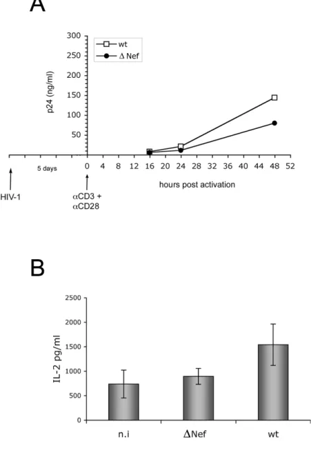
We then investigated the contribution of the HIV-1 Nef protein in both viral replication and T cell activation. Freshly isolated CD4+ T lymphocytes have been infected with VSV-G-pseudotiped wt or )Nef viruses and five days post infection (p.i.) the cells were stimulated through their T cell receptor with CD3 and anti-CD28 cross-linked antibodies. 16, 24, 48 hrs post stimulation, the cells were monitored for viral release by measuring soluble p24 in the culture supernatant by ELISA. As expected, a positive effect of Nef was consistently observed with a two-fold increase in viral production by cells infected with wt virus if compared to those infected with )Nef (Fig. 4A).
As described earlier, Nef may favor viral replication by lowering the threshold of T cell activation. Since the increase of IL-2 production represent a hallmark of T cell activation [19, 20, 21], we analyzed whether in our experimental conditions Nef expression altered the release of IL-2 from infected cells. As described above, CD4+ T lymphocytes were infected and, after 5 days, stimulated with CD3 and anti-CD28 cross-linked antibodies. 16, 24, 48 hrs post-stimulation, an aliquot of the culture supernatant was tested for the presence of IL-2 by ELISA. As shown in Fig. 4B, 48 hrs post activation we observed a two-fold increase of IL-2 production from cells infected with wt virus if compared to those infected with )Nef virus or uninfected. This result is in agreement with other studies [56] and confirms the positive role of the HIV-1 Nef protein in T cell activation.
Fig. 4. Nef enhances viral replication and IL-2 production in response to
CD3/CD28 stimulation in quiescent CD4+
T lymphocytes infected with HIV-1 (A, B) Freshly isolated CD4+ T lymphocytes uninfected (n.i) and infected with HIV-1 NL4-3 virus either wt or Nef-deficient ()Nef) were cultivated for five days in the absence of mitogen. Next, the cells were activated with anti-CD3 and anti-CD28 cross-linked antibodies. (A) 16, 24, 48 hrs post stimulation, the supernatant was collected for p24 quantification by ELISA one out of three independent experiments is shown. (B) 48 hrs post activation an aliquot of the culture supernatant was tested for the presence of IL-2 by ELISA. Results shown represent the average value of three independent experiments.
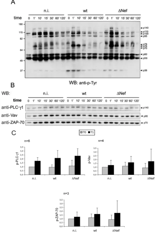
3.3 Protein tyrosine phosphorylation upon activation of
quiescent HIV-1-infected T cells
As shown in previous work, Nef profoundly manipulates HIV target cells by altering a variety of signal transduction pathways and protein sorting processes [58]. With respect to CD4+ T lymphocytes, Nef is known to affect the TCR signal transduction. Successful TCR signal initiation induces a characteristic cascade of tyrosine phosphorylation events, Ca2+ release and the activation of transcription factors [19]. Thus we investigated the role of Nef in the TCR signaling of primary HIV-1 infected CD4+ T lymphocytes. Freshly purified CD4+ T cells were infected with equal amounts of VSV-G-pseudotyped wt or )Nef virus or not infected and cultivated in the absence of mitogen for 5 days. The cells were then stimulated at 37°C for variable time (0, 1, 10, 15, 30, 60, 120 minutes) with anti-CD3 and anti-CD28 cross-linked antibodies. Finally, cells were lysed and equal amounts of total cell extracts were analyzed by western blotting. By probing the membrane with anti-p-Tyr antibody, the typical pattern of protein phosphorylation of activated T cells was observed: p140, p95, p70, p56, p36 corresponding to p-PLC-#1, p-Vav, p-ZAP-70, p-Lck and p-"-chain of TCR complex respectively (Fig. 5A). Next, the membrane was stripped with reducing buffer and re-probed with specific antibodies anti-PLCg-#1, antia-Vav, antia-ZAP-70 (Fig. 5B). The relative phosphorylation was calculated by measuring bounds by densitometry, normalizing phospho-bands for their loading controls, and setting to 1 the signal of uninfected cells at time 0. By comparing uninfected, wt- and )Nef-infected cells, it was evident that upon TCR stimulation the extent and the
kinetic (with a peak after 1' of stimulation) of tyrosine phosphorylation of PLC-#1, Vav, and ZAP-70 did not differ significantly (Fig. 5C). By this analysis, the extent of Lck phosphorylation was extremely variable between different experiments and thus it could not be evaluated. This was possibly due to the high basal level of Lck phosphorylation and/or to the presence of other phosphorylated protein co-migrating with Lck.
Fig. 5. Activation of HIV-infected T cells via CD3/CD28 Five days post infection, quiescent CD4+
T lymphocytes were stimulated for variable time with cross-linked anti-CD3 and anti-CD28 antibodies (A). Equal amounts of cell lysates were analyzed by western blotting with anti-p-Tyr antibody. (B) The membrane shown in panel (A) was stripped and re-probed with specific antibodies anti-PLC-#1, anti-Vav, anti-ZAP-70. (C) The average phosphorylation of PLC-#1, Vav, ZAP-70 measured after 1 minute of stimulation in 6, 4 and 3 independent experiments is shown.
3.4 Activation of Akt and NF-!B pathways in
HIV-1-infected CD4
+T lymphocytes
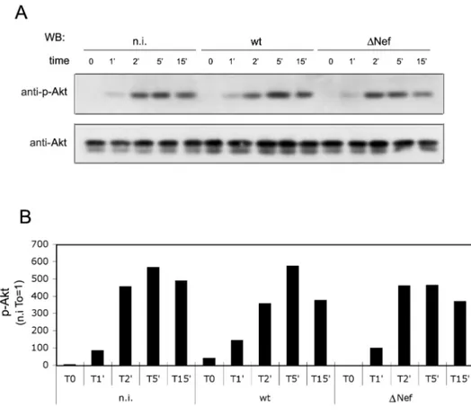
Since Akt has an important role in the T cell activation pathway [29], we investigated its phosphorylation/activation in the context of wt- and )Nef-infected CD4+ T lymphocytes. Primary CD4+ T lymphocytes uninfected or infected for 5 days were stimulated through CD3/CD28 as described above and, at various times after stimulation, the cells were lysed. Equal amounts of total cell extracts were analyzed by western blotting with anti-Akt antibody and, after stripping, the membrane was re-probed with anti-p-Akt specific antibodies (Fig. 6A). The rate of Akt phosphorylation was obtained by densitometric analysis and by normalizing Akt phospho-bands for their loading control. The ratio between p-Akt/Akt in uninfected cells before the stimulation (T0) was set to 1. We observed that in each cell sample (n.i., wt, )Nef), the Akt protein became phosphorylated to a similar extent upon stimulation and that the peak of phosphorylation was reached after five minutes (Fig. 6B). Thus, Akt phosphorylation did not differ significantly in uninfected or HIV-1-infected cells, with or without Nef expression.
To examine whether Nef could affect the NF-!B pathway, we analyzed the level of phosphorylation of IKK, I!B and NF-!B in primary CD4+ T lymphocytes uninfected or infected with wt and )Nef virus for 5 days. The cells were either treated or not with anti-CD3 and anti-CD28 antibodies for variable time (15, 60, 360 minutes) then collected, lysed and equal amounts of cell extracts were analyzed by western blotting with anti-IKK antibodies specific for $ and % isoforms, anti-I!B and anti-NF-!B (p50) antibodies. Next the membrane was stripped and re-probe con IKK,
anti-I!B and anti-NF-!B specific antibodies (Fig. 7A). The relative phosphorylation was calculated by densitometric analysis normalyzing phospho-bands obtained by anti-p-Tyr antibody for their loading control with specific antibody and setting to 1 the signal of uninfected cells at time 0. As shown in the Fig. 7B, the phosphorylation of IKK, I!B and NF-!B was induced and reached a peak after 15, 60 minutes and 6 hrs respectively. By comparing uninfected with wt- and )Nef-infected cells, we observed that there were no differences in the phosphorylation level of IKK, I!B and NF-!B.
Fig. 6. Effect of Nef on Akt activation in HIV-1-infected cells. (A) Quiescent CD4+
T cells were infected and five days post infection were stimulated for variable time (0, 1, 2, 5, 15 minutes) with cross-linked anti-CD3 and anti-CD28 antibodies. Then equal amounts of cell extracts were analyzed by western blotting. The membrane was immunoblotted with anti-p-Akt antibody and, after stripping, re-probed with anti-Akt specific antibody. (B) Relative Akt phosphorylation was obtained by densitometric analysis normalizing phospho-bands for their loading controls and setting to 1 the signal of n.i. cells at T0. Results shown are representative of three independent experiments.
Fig. 7. Activation of the NF-!B pathway in HIV-1-infected cells.
(A) CD4+ T cells were uninfected, infected with wt or )Nef virus. Five days post infection the cells were stimulated for 0, 15, 60, 360 min with cross-linked anti-CD3 and anti-CD28 antibodies. Equal amounts of cell lysates were analyzed by western blotting with anti-p-IKK$/% , anti-p-I!B and anti-p-NFk-!B antibodies. After stripping the membrane was re-probed with anti-IKK, anti-I!B and anti-NFk-!Ba/b, antibodies. (B) The phosphorylation level of the proteins in one representative experiment out of three is shown. The signal of n.i. cells at time 0 was set to 1.
3.5 CD4/Lck interaction in HIV-1-infected resting T cells
Next, we investigated the association of Lck with CD4 in HIV-1-infected T cells, with and without Nef expression, by a co-immunoprecipitation analysis. Primary quiescent CD4+ T lymphocytes uninfected, infected with wt or )Nef HIV-1, were harvested five days post infection to prepare total cell extracts. As shown by western blotting (Fig. 3A), HIV-1 expression results in decreased levels of CD4 and Lck in a Nef-independent manner, while the intracellular re-distribution of both CD4 and Lck occur only if the viral Nef protein is expressed. Equal amounts of total cell extracts were subjected to two rounds of immunoprecipitation with anti-CD4 antibody. Western blotting analysis of cell lysates before and after anti-CD4 immunoprecipitation confirmed that the overall amount of cell-associated CD4 was bound to antibodies (Fig. 8A). In addition, Lck levels were almost completely removed from the cell lysate upon anti-CD4 immunoprecipitation indicating that the vast majority of Lck is associated to CD4 within primary T cells. The immunoprecipitated proteins were divided in two aliquots that were analyzed by western blotting with anti-CD4 and anti-Lck antibody (Fig. 8B). The levels of both CD4 and Lck in the immunoprecipitated samples were measured by densitometry and the relative amount of Lck was divided by the corresponding CD4 amount. This results shows that CD4 and Lck associates efficiently in HIV-1-infected T cells, despite the levels of both proteins are reduced and, due to Nef's activity, relocalized within intracellular compartments.Fig. 8. CD4/Lck association in HIV-1 infected quiescent CD4+
T lymphocytes 5 days post infection, total cell extracts of uninfected, wt- and )Nef-infected cells were quantified and subjected to immunoprecipitation with anti-CD4 antibody. (A) The lysates before and after two rounds of anti-CD4 immunoprecipitation were analyzed by western blotting with anti-CD4 and anti-Lck specific antibodies. (B) Immunocomplexes were analyzed as described in panel (A) and the relative amount of Lck was divided by the amount of immunoprecipitated CD4 in each sample. The Lck/CD4 ratio was calculated by setting to 1 that found in the n.i. sample. As control, lane ctr corresponds to an immunoprecipitation reaction performed without cellular lysate.
3.6 Activation of Lck upon triggering of CD4 in
HIV-1-infected cells
We investigated whether the HIV-1 Nef protein is involved in the regulation of Lck activation upon triggering of the surface CD4 receptor. Uninfected, wt- and )Nef-infected CD4+ T lymphocytes were stimulated five days post infection with cross-linked anti-CD4 antibody for variable time (0, 1, 5, 10 min). Next the cells were collected and lysed. The total cell extracts were quantified and equal amounts were analyzed by western blotting with anti-p-Tyr antibody followed by re-probing with anti-Lck specific antibody (Fig. 9A). Levels of total and Tyr-phosphorylated Lck (p-Lck) were calculated by densitometric analysis and expressed by setting to 100 and 1, respectively, the signal of n.i. cells at time 0. As shown in Fig. 9B, upon CD4 stimulation the phosphorylation of Lck rapidly increased while the protein levels decreased, possibly due to the rapid consumption of activated Lck. Despite the basal levels of Lck were lower in wt- and )Nef-infected cells, after 1' of CD4 stimulation about 50% of the Lck protein disappeared in both infected and uninfected cells. In addition, the phosphorylation of Lck was maximally induced 5 minutes after the CD4 stimulation. Importantly, the kinetic as well as the extent of Lck phosphorylation was similar in uninfected and HIV-1 infected cells. Thus, despite Lck is reduced and re-localized, the function of this kinase in maintained, in HIV-1-infected cells consistently with the full capacity of these cells to respond to TCR activation.
Fig. 9. Lck phosphorylation upon CD4 stimulation of HIV-1-infected CD4+ T cells.
(A) CD4+
T lymphocytes uninfected, infected with wt or )Nef virus, were stimulated with cross-linked anti-CD4 antibody for variable time (0, 1, 5, 10 min). To investigate the level of Lck phosphorylation, equal amounts of cell extracts were analyzed by western blotting with anti-p-Tyr (Top). After stripping the membrane was re-probed with anti-Lck (Bottom). (B) Lck and p-Lck in n.i., wt- and )Nef-infected cells was calculated by densitometric analysis and expressed setting to 100 and 1 the value obtained for n.i. cells at time T0, respectively. Results of one representative experiment out of three are shown.