The anti-inflammatory effects of
dimethyl fumarate in astrocytes involve
glutathione and haem oxygenase-1
Shao Xia Lin*, Lucia Lisi{, Cinzia Dello Russo{, Paul E Polak*, Anthony Sharp*, Guy Weinberg*,{, Sergey Kalinin* and Douglas L Feinstein*,{1
*Department of Anesthesiology, University of Illinois, Chicago, IL, U.S.A.
{Institute of Pharmacology, Catholic Medical School, Rome, Italy {Jesse Brown VA Hospital, Chicago, IL, U.S.A.
Cite this article as: Lin S, Lisi L, Russo CD, Polak PE, Sharp A, Weinberg G, Kalinin S and Feinstein DL (2011) The anti-inflammatory effects of dimethyl fumarate in astrocytes involve glutathione and haem oxygenase-1. ASN NEURO 3(2):art:e00055.doi:10.1042/AN20100033
ABSTRACT
DMF (dimethyl fumarate) exerts anti-inflammatory and pro-metabolic effects in a variety of cell types, and a formulation (BG-12) is being evaluated for monotherapy in multiple sclerosis patients. DMF modifies glutathione (GSH) levels that can induce expression of the anti-inflammatory protein HO-1 (haem oxygenase-1). In primary astrocytes and C6 glioma cells, BG-12 dose-dependently suppressed nitrite production induced by either LI [LPS (lipopolysaccharide) at 1mg/ml plus IFNc (interferon c) at 20 units/ml] or a mixture of pro-inflammatory cytokines, with greater efficacy in C6 cells. BG-12 reduced NOS2 (nitric oxide synthase 2) mRNA levels and activation of a NOS2 promoter, reduced nuclear levels of NF-kB (nuclear factor kB) p65 subunit and attenuated loss of IkBa (inhibitory kBa) in both cell types, although with greater effects in astrocytes. In astrocytes, LI decreased mRNA levels for GSHr (GSH reductase) and GCL (c-glutamylcysteine synthetase), and slightly suppressed GSHs (GSH synthetase) mRNAs. Co-treatment with BG-12 prevented those decreased and increased levels above control values. In contrast, LI reduced GSHp (GSH peroxidase) and GCL in C6 cells, and BG-12 had no effect on those levels. BG-BG-12 increased nuclear levels of Nrf2 (nuclear factor-erythroid 2 p45 subunit-related factor 2), an inducer of GSH-related enzymes, in astrocytes but not C6 cells. In astrocytes, GSH was decreased by BG-12 at 2 h and increased at 24 h. Prior depletion of GSH using buthionine-sulfoximine increased the ability of BG-12 to reduce nitrites. In astrocytes, BG-12 increased HO-1 mRNA levels and effects on nitrite levels were blocked by an HO-1 inhibitor. These results demonstrate that BG-12 suppresses inflammatory activation in astrocytes and C6 glioma cells, but
with distinct mechanisms, different dependence on GSH and different effects on transcription factor activation.
Key words: nitric oxide synthase 2 (NOS2), inflammation, glial cells, glutathione, multiple sclerosis.
INTRODUCTION
BG-12 is an FAE (fumaric acid ester), an oral formulation of DMF (dimethyl fumarate) with known anti-inflammatory and neuro-protective effects. FAEs were first considered for use as treatment for psoriasis, a Th1-mediated disease, due to anti-proliferative effects on lymphocytes (Stoof et al., 2001; Mrowietz and Asadullah, 2005). One FAE (Fumaderm) has been approved for psoriasis in Europe for over 15 years. Subsequent studies showed that DMF reduces inflammatory gene expression, including that of pro-inflammatory cytokines and chemokines, and increases anti-inflammatory expression (Stoof et al., 2001; Loewe et al., 2002; Seidel et al., 2009) – effects likely to contribute to its anti-psoriasis efficacy. These findings have led to increased interest for using DMF in other auto-immune or inflammatory diseases, including MS (multiple sclerosis) (Kappos et al., 2008; Moharregh-Khiabani et al., 2009). In animal studies, BG-12 reduced glial inflammation during MOG-peptide-induced EAE (experimental autoimmune encephalomyelitis) and increased plasma levels of IL-10 (interleukin-10; Schilling et al., 2006). A Phase 2B trial of BG-12 in RRMS (relapsing remitting MS) patients showed significant decreases in new gadolinium enhancing lesions, T1 and T2 lesions, and a non-significant decrease in the annualized relapse rate (Kappos et al., 2008).
1To whom correspondence should be addressed (email [email protected]).
Abbreviations: BSO, buthionine-sulfoximine; DMEM, Dulbecco’s modified Eagle’s medium; DMF, dimethyl fumarate; EAE, experimental autoimmune encephalomyelitis; EMSA, electrophoretic mobility-shift assay; FAE, fumaric acid ester; FCS, fetal calf serum; GSHp, GSH peroxidase; GSHr, GSH reductase; GSHs, GSH synthetase; HO-1, haem oxygenase-1; HSP32, heat-shock protein 32; IFNc, interferon c; IkBa, inhibitory kBa; IL, interleukin; LDH, lactate dehydrogenase; LPS, lipopolysaccharide; LI, LPS at 1 mg/ml plus IFNc at 20 units/ml; MS, multiple sclerosisi; NF-kB, nuclear factor kB; Nrf2, nuclear factor-erythroid 2 p45 subunit-related factor 2; NOS2, nitric oxide synthase 2; qPCR, quantitative PCR; TNFa, tumour necrosis factor a; ZnPP, zinc protoporphyrin IX.
The mechanisms of the action of BG-12 are not fully known. BG-12 can suppress NF-kB (nuclear factor kB)-dependent transcription (Stoof et al., 2001; Gerdes et al., 2007), thus accounting for some of its anti-inflammatory effects. BG-12 can also activate the Nrf2 (nuclear factor-erythroid 2 p45 subunit-related factor 2) pathway (Lukashev et al., 2007; Kappos et al., 2008), which induces the transcription of various genes, including anti-oxidative ones, reduces oxidative neuronal death and helps maintain myelin integrity. BG-12 induces detoxifica-tion enzymes in astrocytes and microglial cells (Wierinckx et al., 2005). As a consequence, BG-12 can modulate GSH levels in cells leading to cytotoxic or protective effects (Dethlefsen et al., 1988; Spencer et al., 1990), including in primary astrocytes (Schmidt and Dringen, 2010). The anti-inflammatory effects of DMF have been shown, in some cases, to involve induction of HO-1 (haem oxygenase 1) also termed HSP32 (heat-shock protein 32) (Lehmann et al., 2007), which occurs following GSH depletion. HO-1 can suppress a variety of inflammatory responses (Horikawa et al., 2002), including those in astrocytes (Calabrese et al., 2005; Guo and Bhat, 2006), as well as confer protection against oxidative stress (Min et al., 2006).
A role for astrocyte activation during the pathogenesis of MS and EAE has been implicated in several studies (Smith and Lassmann, 2002). Inflammatory activation of astrocytes can lead to the expression of pro-inflammatory cytokines, as well as of the inducible form of NOS2 (nitric oxide synthase 2) whose overproduction of NO contributes to oligodendrocyte and neuronal damage. In vitro, astrocytes express NOS2 in response to a variety of stimuli, and in MS the predominant glial localization of NOS2 has been reported to be in astrocytes (Brosnan et al., 1994; Broholm et al., 2004). An astrocytic localization of NOS2 has also been reported in EAE (Brenner et al., 1997; Tran et al., 1997) as well as in Theiler’s virus model of MS (Oleszak et al., 1997). The effects of BG-12 in astrocytes have not been well characterized. A recent study shows anti-inflammatory actions in microglial cells, but weaker effects in primary astrocytes (Wilms et al., 2010). In the present study, we have re-examined the ability of BG-12 to reduce NOS2 expression in primary astrocytes and compared those findings with results using rat C6 glioma cells, often used to model astrocyte biology (Feinstein et al., 1994). A comparison of the anti-inflammatory actions of BG-12 in astrocytes versus C6 cells, combined with measurements of effects on intracellular GSH levels, has allowed us to establish a linkage between these two phenomena, and suggests that changes in GSH modulate the suppressive efficacy of BG-12.
MATERIALS AND METHODS
Reagents
Cell culture reagents [DMEM (Dulbecco’s modified Eagle’s medium) and antibiotics] were from Cellgro Mediatech
(Manassas, VA, U.S.A.). FCS (fetal calf serum) (,10 units endotoxin/ml) was from Invitrogen (Carlsbad, CA, U.S.A.). Bacterial endotoxin LPS (Salmonella Typhimurium), BSO (buthionine-sulfoximine), ZnPP (zinc protoporphyrin IX), gentamicin (G418), antibiotics, and other common reagents were from Sigma–Aldrich (St Louis, MO, U.S.A.). Recombinant rat IFNc (interferon c), human IL-1b, and humanTNFa (tumour necrosis factor a) were from Gibco (Gaithersburg, MD, U.S.A.). BG-12 was provided by Biogen Idec (Cambridge, MA, U.S.A.). Antibodies against NF-kB p65 subunit and IkBa (inhibitory kBa) were from Santa Cruz Laboratories (Santa Cruz, CA, U.S.A.), and those against b-actin were from Sigma.
Cells and treatments
Primary enriched astrocyte cultures were obtained from cerebral cortices of postnatal day 1–2 Harlan Sprague– Dawley rats as described previously (Galea et al., 1992). All experiments using animals were carried out according to institutional guidelines and were approved by the local Institutional Animal Use and Care Committee. Media were changed every 3 days. After 2 weeks of growth in DMEM containing 10% FCS and antibiotics (100 i.u./ml penicillin and 100 mg/ml streptomycin) the cultures consisted of 95–98% astrocytes and 2–3% microglia. C6 rat glioma cells were grown in DMEM containing 10% FCS and antibiotics. C6 cells stably transfected with a 2.2 kb fragment of the rat NOS2 promoter (Gavrilyuk et al., 2001) driving expression of luciferase were passaged in complete medium containing 200 mg/ml G418. Cell cultures when 80–90% confluent were transferred to DMEM with 1% FCS and antibiotics and treated with LI (LPS at 1 mg/ml plus IFNc at 20 units/ml) or TII (10 ng/ml IL-1b, 10 ng/ml TNFa and 5 ng/ml IFNc) for robust induction of NOS2. NOS2 activity was assessed indirectly by nitrite production in the cell culture media (Green et al. 1982). Briefly, an aliquot of cell culture media (80 ml) was mixed with 40 ml of Griess reagent and the absorbance was measured at 550 nm. Cell viability was assessed by the release of LDH (lactate dehydrogenase) into the culture media using the Cell-tox 96 kit (Promega, Madison, WI, U.S.A.).
mRNA analysis
Total cytoplasmic RNA was prepared from cells using TRIzolH reagent (Invitrogen); aliquots were converted into cDNA using random hexamer primers. Quantitative changes in mRNA levels were estimated by real-time PCR [qPCR (quantitative PCR)] using the following cycling conditions: 35 cycles of denaturation at 95
˚
C for 10 s, annealing at 58–61˚
C for 15 s depending on the specific set of primers and extension at 72˚
C for 20 s. Reactions were carried out in the presence of SYBR Green (1:10000 dilution of stock solution from Molecular Probes, Eugene, OR, U.S.A.), carried out in a 20-ml reaction volume in a Corbett Rotor-Gene apparatus (Corbett Research, Mortlake, NSW, Australia). The primers used for NOS2 were forward 59-GGAGAAGGGGACG-AACTCAGT-39, and reverse: 59-GCATTGGAAGTGAAGCGTTTC-39;for the catalytic subunit of GCL (c-glutamylcysteine ligase) were forward AAAGGCCTCTAAGCCAGACCACAT-39, and reverse 59-GCGATGCAGCACTCAAAGCCATAA-39; for GSHp (GSH peroxidase) were forward 59-ACCCGGGACTACACCGAAATGAAT-39, and reverse 59-TGCCATTCTCCTGATGTCCGAACT- 39; for GSHs (GHS synthetase) were forward 59-AGCCTTCCATCCAAGGACCAGAAA-39 and reverse AGGAGCTTCCATTCCCACTCCAAA-59-AGCCTTCCATCCAAGGACCAGAAA-39; for GSHr (GSH reductase) were forward 59-AAAGAAGACCTCATCGGGCTTGGA-39 and reverse 59-AGAGCAGGCAATCGACATCTGGAA-39; for HO-1 were forward CTTTCAGAAGGGTCAGGTGTC-39 and reverse TGCTTGTTTCGCTCTATCTCC-39; and for b-tubulin were forward 59-CCCTCGCCATGGTAAATACAT-39, and reverse 59-ACTGGATGG-TACGCTTGGTCT-39. Relative mRNA levels were estimated by the comparative analysis of Ct values, and normalized to values measured for b-tubulin in the same samples.
Luciferase activity assay measurements
Cells were lysed by Chaps buffer (10 mM Chaps and 10 mM Tris, pH 7.4), the plate frozen at 280
˚
C, thawed and shaken on a rotary shaker for 10–15 min at room temperature (22˚
C). Aliquots of cell lysates (10–20 ml) containing approximately equal amounts of protein (10–20 mg) were placed into wells of an opaque, white 96-well microplate. An equal volume of luciferase substrate (Steady Glo reagent, Promega) was added to all samples, and the luminescence measured in a microplate luminometer (Rosys-Anthos, Hombrechtikon, Switzerland).Glutathione assay
Total GSH levels (GSH+GSSG) were measured in whole cell lysates using a commercial glutathione assay kit (Sigma CS0260). In brief, following treatments, cells were collected, pelleted, lysed, deproteinized, and aliquots of the resulting supernatant mixed with 5,59-dithiobis(2-nitrobenzoic acid) and the formation of coloured 59-thio-2-nitrobenzoic acid mea-sured at 412 nm over 20 min.
Immunoblot analysis
After treatments, cells were lysed in RIPA buffer (1 mM EDTA, 150 mM NaCl, 1% Igepal, 0.1% SDS, 0.5% sodium deoxycholate and 50 mM Tris/HCl, pH 8.0) containing protease inhibitors. Protein content was determined by Bradford reagent (Bio-Rad, Hercules, CA, U.S.A.) with BSA as standard. A 10 mg aliquot of protein was mixed with Laemmli buffer (Bio-Rad), boiled for 5 min and separated through 10% polyacrylamide SDS gels. Separated proteins were transferred to PVDF membranes by semi-dry electrophoretic transfer. The membranes were blocked with 10% (w/v) non-fat dried skimmed milk powder in TBST (10 mM Tris, 150 mM NaCl and 0.1% Tween-20, pH 7.6) for 1 h at room temperature, then incubated with rabbit polyclonal anti-IkBa (Santa Cruz Biotechnology, Santa Cruz, CA, U.S.A.) or mouse monoclonal anti-b-actin antibody (Sigma) both at 1:1000 dilutions overnight with gentle shaking at 4
˚
C. Primary antibodies were removed, membranes washed three times inTBST, then incubated 1 h at room temperature with specific secondary antibodies (Vector Laboratories, Burlingame, CA, U.S.A.). Following three washes in TBST, bands were visualized by incubation in ECLH (enhanced chemiluminescence) reagents and exposure to Hyperfilm ECL (GE Healthcare, New York, NY, U.S.A.). The same membranes were washed three times in TBST, blocked with 10% (w/v) non-fat dried skimmed milk powder in TBST for 1 h at room temperature and used for b-actin immunoblot. Band intensities were determined using ImageJ software (National Institutes of Health) from autoradiographs obtained from the minimum exposure time that allowed band detection, and background intensities (determined from an equal-sized area of the film immediately above the band of interest) were subtracted.
Immunocytochemical staining
Astrocytes and C6 cells were plated directly on to poly-lysine-coated glass coverslips and grown until near confluency. The cells were then treated with LI and 20 mM BG-12 or vehicle for the indicated lengths of time. After treatment, medium was removed, and cells were fixed in 4% formaldehyde for 10 min at room temperature. The cells were then washed three times with PBS, blocked in 5% normal donkey serum in PBS containing 0.1% Triton X-100 for 30 min; primary antibody was added (goat anti-NF-kB p65 subunit, 1:100 dilution) and the mixture incubated overnight at 4
˚
C. The primary antibody was removed, cells washed three times with PBS, then appropriate fluor-escent-conjugated secondary antibodies (donkey anti-goat 1:200; Jackson ImmunoResearch Laboratories, West Grove, PA, U.S.A.) were added; they were incubated for 45 min at 37˚
C, washed, and then Vectashield mounting solution was added (Vector Laboratories, Burlingame, CA, U.S.A.). The coverslips were mounted on to glass slides and imaged.Transcription factor DNA-binding activity assay
Nuclear levels of active DNA-binding NF-kB p65 subunit and Nrf2 were measured using ELISA-based TransAM kits (Active Motif, Carslbad, CA, U.S.A.). In brief, the cells were incubated with LI and 20 mM BG-12 or vehicle, and, at indicated times, nuclear extracts were prepared, aliquots (2–10 mg) added to wells containing consensus NF-kB or Nrf2-binding-site element oligonucleotides, incubated 2 h at room temperature, washed and then HRP (horseradish peroxidase)-conjugated antibodies against p65 or Nrf2 added for 1 h, washed, substrate added and colour intensity (for Nrf2) or chemiluminescence signal (for p65) measured. Binding activity values were normalized for the amount of protein per sample, and presented relative to activity measured in non-treated (zero time) cells.Data analysis
All experiments were performed at least in triplicate, and repeated at least twice. Comparisons were made using one-way ANOVA for dose–response curves, followed by Bonferroni
post-hoc analysis, by two-way ANOVA for transAM assays, or by unpaired t-test for group comparisons. Two-tailed analyses were used, and statistical significance was taken at P,0.05.
RESULTS
BG-12 reduces NOS2 activity and expression in
astrocytes and C6 cells
We first tested the effects of BG-12 on the expression and activity of NOS2 in glial cells. In both primary astrocytes and rat C6 glioma cells (Figure 1), BG-12 dose-dependently reduced the LPS and IFNc (LI)-dependent production of nitrites. In astrocytes, a slight (,20%) but significant inhibition was observed at 3 mM, with maximal inhibition of near 50% at 20 mM the highest dose tested. In contrast, in C6 cells, 3 mM BG-12 reduced nitrite production approx. 50%, with over 90% inhibition observed at a dose of 20 mM. In all cases, incubation for 24 h with up to 20 mM BG-12 did not increase LDH release from either astrocytes or C6 cells. BG-12 was also more effective at blocking nitrite production in C6 cells than in astrocytes when activation was carried out with a combination of pro-inflammatory cytokines (Figure 2).
In both astrocytes and C6 cells, BG-12 significantly reduced NOS2 mRNA levels (Figures 3A and 3B), suggesting an effect at transcriptional or post-transcriptional levels. Consistent with this, treatment with BG-12 potently suppressed the LI-dependent activation of a 2.2 kb NOS2 promoter stably expressed in C6 cells (Figure 3C), with significant inhibition observed at 3 mM, and complete suppression at 20 mM, suggesting a reduction in NOS2 gene transcription.
Figure 1 BG-12 reduces nitrite production
(A) Primary rat astrocytes were treated with LI to induce NOS2 expression, in the presence of the indicated concentration of BG-12 or vehicle (20 mM DMSO). After 24 h, nitrite levels in the media were measured by Griess assay. Results are means¡S.D. of n56 per group and show % nitrite release as compared with LPS/IFNc alone (none). The experiment was repeated twice with similar results. *P,0.001 versus vehicle, 1-way ANOVA, Bonferroni post hoc. (B) Measurements of LDH release after 24 h did not reveal any significant increase due to incubation with BG-12 up to 20 mM. (C) Rat C6 cells were treated with LI to induce NOS2 expression, in the presence of the indicated concentration of BG-12 or vehicle (20 mM DMSO). After 24 h nitrite levels in the media were measured by Griess assay. Results are means¡S.D. of n56 per group and show % nitrite release as compared with LPS/IFNc alone (none), which was 8.1 nmol of nitrite per mg per 24 h. The experiment was repeated twice with similar results. *P,0.001 versus vehicle, one-way ANOVA, Bonferroni post hoc. (D) Measurement of LDH release after 24 h did not reveal any significant increase due to incubation with BG-12 up to 20 mM.
Figure 2 Effects of BG-12 on cytokine-induced NOS2 expression
(A) Astrocytes and (B) C6 cells were treated with TII (a mixture of TNFa, IL-1b and IFNc) to induce NOS2 expression, in the presence of the indicated concentration of BG-12 or vehicle (20 mM DMSO). After 24 h, nitrite levels in the media were measured by Griess assay. Results are means¡S.D. of n54 per group and show % nitrite release as compared with TII alone (0 BG-12), which was 16 nmol (for astrocytes) and 46 nmol (for C6 cells) nitrite per mg per 24 h. The experiment was repeated twice with similar results. *P,0.001 versus vehicle, one-way ANOVA, Bonferroni post hoc.
BG-12 reduces NF-kB activation in glial cells
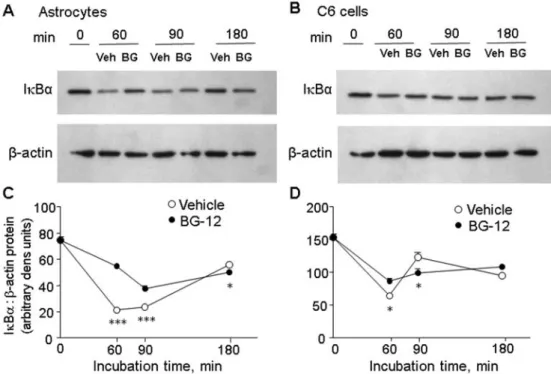
Since NOS2 gene transcription is dependent on activation of the transcription factor NF-kB, we tested the effects of BG-12 on activation of this factor using an EMSA (electrophoretic mobility-shift assay)-based kit (Figure 4A). In astrocytes, LI induced the appearance of nuclear p65 DNA-binding activity by 30 min, which returned to baseline by 4 h; in C6 cells nuclear activity was still high at 2 h, the longest time point measured. At all time points nuclear p65 activity was significantly reduced by BG-12. Immunostaining (Figure 4B) showed that, after 1 h incubation with LI, BG-12 almost completely reduced nuclear levels of the NF-kB p65 subunit in astrocytes. In contrast, in C6 cells nuclear p65 could still be detected in many cells, suggesting that the nuclear p65 was unable to bind DNA. Western-blot analysis of whole cell lysates showed that, in astrocytes, BG-12 significantly reduced the rapid LI-dependent loss of IkBa (Figures 5A and 5C). In contrast, in C6 cells the loss of IkBa measured after 30 min was only slightly reduced by BG-12, while after 60 min levels were lower in the BG-12 treated cells. Together, these results suggest that the effects of BG-12 on NOS2 are due, at least in part, to the inhibition of NOS2 gene expression, and that suppression of IkBa degradation and NF-kB activation contributes to that reduction, although differences exist between its actions in astrocytes and C6 cells.BG-12 differentially modulates GSH levels in
astrocytes versus C6 cells
DMF can increase GSH levels which in turn can influence inflammatory responses; we therefore tested if Figure 3 BG-12 reduces NOS2 expression
(A) Primary astrocytes and (B) C6 cells were treated with LPS/IFNc for 6 h alone, or in the presence of 10 mM BG-12 or equivalent amount of DMSO (Veh), then RNA samples were measured by qPCR for NOS2 mRNA levels. Results show the relative NOS2 mRNA level after normalization tob-tubulin mRNA values measured in the same samples. Results are means¡S.D. for three samples from each group. LPS/IFNc increased NOS2 mRNA levels approx. 2600-fold in astrocytes, and 1800-fold in C6 cells over baseline levels. *P,0.05 versus vehicle. (C) C6 cells stably transfected with rat 2.2 kb NOS2 promoter driving luciferase activity were treated with LPS/IFNc in the presence of the indicated concentrations of BG-12 or vehicle (20 mM DMSO). After 5 h, luciferase activity was measured. Results are means¡S.D. for three to six samples per group and show promoter activation relative to no BG-12. Treatment with LPS/IFNc increased the NOS2 promoter 65-fold over control levels. Experiments were repeated twice with similar results. *P,0.05 versus vehicle, one-way ANOVA, Bonferroni post hoc.
Figure 4 BG-12 reduces NF-kB activation
(A) Astrocytes and C6 cells were treated with LPS/IFNc in the presence of 20 mM BG-12 (open circles) or vehicle (filled circles) and after the indicated times nuclear lysates prepared and levels of active nuclear p65 binding to DNA measured with the TransAM (p65) assay kit. The amount of active p65 was normalized to levels measured in the non-treated (zero time) samples. Results are means¡S.E., n53 or 4 per group. BG-12 significantly reduced p65-binding activity in both cells types ( F [3,1]5 6.7 in astrocytes, 7.1 in C6 cells; P,0.005, two-way ANOVA; *P,0.05; ***P,0.001, Bonferroni post hoc). (B) Astrocytes and C6 cells grown on glass coverslips were treated as described above then fixed and stained for the presence of NF-kB p65. After 1 h incubation, nuclear staining for p65 was observed in most of the vehicle-treated cells, was absent from the BG-12-vehicle-treated astrocytes and was still present in many of the BG-12-treated C6 cells (arrowheads).
BG-12 modified glial cell mRNA levels of enzymes involved in GSH metabolism (Figure 6). Quantitative PCR showed that, in astrocytes, after 6 h incubation, levels of GSHr and GCL mRNAs were significantly reduced by LI, while the levels of GSHs were reduced but that decrease did not reach statistical significance. Treatment with BG-12, but not vehicle, prevented the decrease of both GCL and GSHr mRNAs, and significantly increased levels of GSHs compared with incubation with LI alone. The effect of BG-12 on the GSHr was significantly different compared with vehicle, and almost (P50.063) significant for GSHr. In contrast with astrocytes, while treatment of C6 cells with LI significantly reduced levels of GSHp and GCL, BG-12 did not modify expression of any of the mRNAs tested compared with LI alone or to vehicle.
Since expression of some GSH related mRNAs can be induced by transcription factor Nrf2, we tested effects of BG-12 on the activation of this factor (Figure 7). In astrocytes, but not in C6 cells, BG-12 significantly increased nuclear Nrf2 DNA-binding activity compared with vehicle-treated cells (Figure 7A), with significant pairwise differences present at 1 and 4 h, the longest time tested.
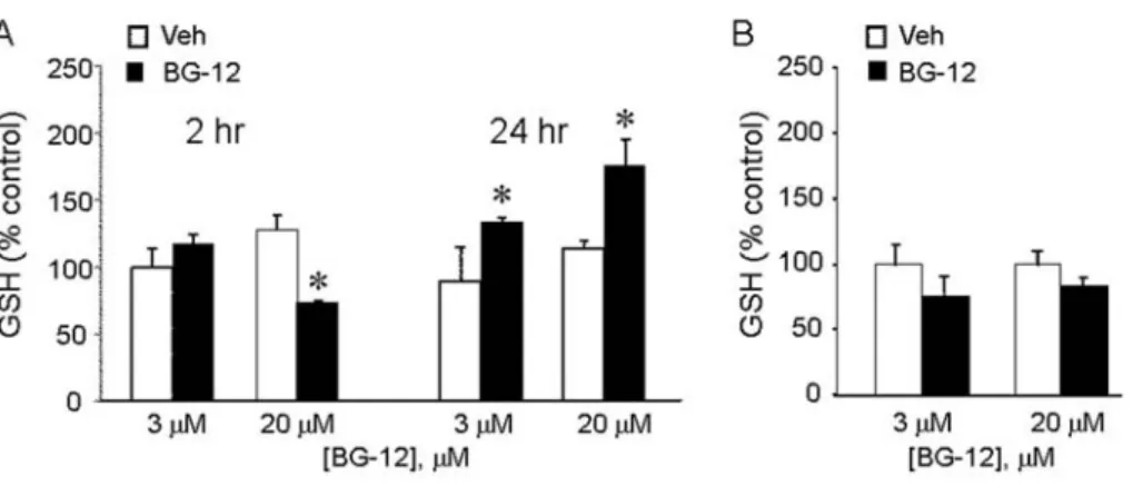
Consistent with increased GSHr and GCL mRNA levels, measurements of total intracellular GSH (GSH plus GSSG) content showed that BG-12 significantly increased GSH levels in primary astrocytes (Figure 8A) but not C6 cells (Figure 8B). In astrocytes, the increase was both time- and dose-dependent, since, at the lower dose of 3 mM, an increase was
not observed at 2 h, but only after 24 h. In contrast, at the higher dose of 20 mM, we observed a significant reduction in GSH after 2 h incubation, but a significant increase after 24 h.
Reduction of GSH levels increases NOS2
suppression by BG-12
To determine if changes in GSH levels played a role in NOS2 suppression, we examined the effects of BG-12 on NOS2 ex-pression in cells pretreated with BSO to deplete GSH levels (Figure 9). In both astrocytes and C6 cells, pretreatment with BSO had a small but non-significant effect on the ability of 3 mM BG-12 to reduce nitrite production. However, the suppression by 10 mM BG-12 was significantly increased by BSO in both C6 cells and primary astrocytes. At 20 mM BG-12, the highest dose tested, there was potent inhibition (close to 90%) of C6 cell nitrites that was not significantly increased by BSO; in contrast, the suppression observed in the astrocytes (approx. 70% inhibition compared with DMSO control) was significantly increased by BSO to near the levels achieved in C6 cells. These results suggest that GSH normally restricts the ability of BG-12 to reduce NOS2 expression in glial cells.
BG-12 effects in astrocytes involve induction of
HO-1
The anti-inflammatory effects of GSH depletion have been shown to involve induction of HO-1 [also called HSP32
Figure 5 Effects of BG-12 on IkBa levels
(A) Astrocytes and (B) C6 cells were treated with LPS/IFNc in the presence of 20 mM BG-12 (‘BG’) or vehicle (‘Veh’), and after indicated times whole cell lysates prepared and used for Western-blot analysis of IkBa protein. The membranes were stripped and re-probed for levels ofb-actin. (C) Band intensities were determined using ImageJ. Results are means¡S.E. ratio IkBa to b-actin, n53–6 samples per group. BG-12 significantly reduced IkBa loss in both cell types (P,0.005, two-way ANOVA; *P,0.05, ***P,0.0001 versus BG-12, Bonferroni post hoc).
(heat-shock protein 32)] in some cells. In primary astrocytes, mRNA levels of HO-1 were low, and slightly decreased (,50%) by incubation with LI, whereas co-incubation with 10 mM BG-12 increased steady state mRNA levels over 15-fold (Figure 10A). HO-1 mRNA levels were approximately three times higher in untreated C6 cells compared with
untreated astrocytes; however, those levels were not altered either by incubation with LI or by BG-12 (Figure 10B). Consistent with this, co-incubation with ZnPP, a selective inhibitor of HO-1, significantly attenuated the suppressive effects of BG-12 in astrocytes, but not in C6 cells (Figure 11). These results suggest that, despite having similar effects on GSH levels, the anti-inflammatory actions of BG-12 are mediated by different mechanisms in primary astrocytes compared with transformed C6 cells.
DISCUSSION
In the present study, we examined the effects of BG-12, currently being tested in clinical trials for treatment of MS, on a robust inflammatory activation of glial cells. During disease progression in MS, activation of parenchymal glial cells, both microglia as well as astroglia, results in sustained production of pro-inflammatory substances which contribute to neuronal and oligodendrocyte damage. Although BG-12 was introduced as a potential therapy for MS by virtue of its ability to reduce activation of lymphocytes (Gerdes et al., 2007; Lehmann et al., 2007; Milenkovic et al., 2008), its possible effects on brain glial cell activation have not been well characterized. Our results confirm and expand on a recent study that 10 mM DMF reduced cytokine (IL-1b, IL-6 and TNFa) mRNA levels in primary rat astrocytes, although levels of NOS2 mRNA were not significantly reduced (Wilms et al., 2010). Interestingly, those authors found that DMF more potently (e.g. greater reductions) suppressed microglial compared with astroglial cytokine expression. Although GSH levels were not measured in that study, our present findings suggest that the effects of DMF on GSH in microglia may be similar to what is observed in C6 cells.
Figure 6 Effects of BG-12 on GSHs, GSHp, GSHr and GCL mRNA levels Primary astrocytes (A) or C6 glioma cells (B) were treated with nothing (control), LPS/IFNc or LPS/IFNc in the presence of 10 mM BG-12 or equivalent amount of DMSO vehicle. After 6 h, the relative levels of the indicated mRNAs were determined by qPCR. Results show expression levels after normalization tob-tubulin mRNA values measured in the same samples, and relative to levels measured in the control samples. Results are means¡S.D. for three samples per group. Results were analysed by one-way ANOVA and Bonferroni post hoc; *P,0.05 versus LI alone; #P,0.05 versus vehicle; 1P,0.05 versus control.
Figure 7 BG-12 induces Nrf2 activation in astrocytes
(A) Astrocytes and (B) C6 cells were treated with LPS/IFNc in the presence of 20 mM BG-12 (open circles) or vehicle (filled circles), and after the indicated times the nuclear lysates were prepared and levels of active nuclear Nrf2 binding to DNA measured with the TransAM Nrf2 assay kit. The amount of active Nrf2 was normalized to levels measured in the non-treated (zero time) samples. Results are means¡S.E., n53 or 4. BG-12 significantly increased Nrf2-binding activity in astrocytes (F [3,1]53.6, P50.031; two-way ANOVA; *P,0.05 versus BG-12, Bonferroni post hoc) but had no effect in C6 cells.
Our results demonstrate that the induction of NOS2 in primary astrocytes is suppressed by BG-12. Nitrite accumula-tion after 24 h was modestly reduced by lower doses (3 and 10 mM) of BG-12, and significantly reduced in the presence of 20 mM drug. BG-12 more potently suppressed NOS2 activity in C6 cells, since incubation with 3 mM BG-12 reduced nitrite production approx. 50%, and 20 mM BG-12 almost completely abolished nitrite. Despite these differences, BG-12 had a similar effect on steady-state NOS2 mRNA levels in both cell types when measured after 5 h. This suggests that in both these cells, BG-12 is effective at reducing NOS2 transcription at early times, but that compensatory changes may occur at later times in astrocytes, but not C6 cells that allow for further or increased NOS2 expression or activity.
We observed that BG-12 potently prevented activation of the NOS2 promoter, suggesting an effect occurring at the level of NOS2 gene transcription. Since optimal NOS2
expression depends upon NF-kB activation, we examined the effects of BG-12 on this transcription factor. In both astrocytes and C6 cells, BG-12 potently reduced the nuclear levels of NF-kB p65 able to bind to DNA, determined using an EMSA-based kit. In astrocytes, immunostaining showed a large reduction in the presence of nuclear p65, while immunoblot analysis showed that BG-12 prevented the loss of the IkBa inhibitory subunit. Together these findings are consistent with the idea that, in astrocytes, BG-12 inhibits IkBa degradation, reduces NF-kB nuclear transport, and thereby reduces NOS2 gene expression. On the other hand, in C6 cells similar immunostaining revealed nuclear p65 in some cells and only modest effects on the loss of IkBa, despite the fact that EMSAs did not detect any NF-kB p65 DNA-binding activity at that time point. These findings suggest that whereas BG-12 does not prevent IkBa loss or NF-kB p65 nuclear translocation, the nuclear p65 is less able to bind to cognate DNA. DMF can covalently modify proteins owing to the presence of an a/b unsaturated carbonyl group (Frycak et Figure 8 Effects of BG-12 on GSH levels
(A) Primary astrocytes were treated with LPS/IFNc together with BG-12 or vehicle at the indicated doses and for either 2 or 24 h, after which intracellular total GSH levels were measured. (B) C6 cells were incubated with LPS/IFNc in the presence of the indicated concentration of BG-12 or vehicle. After 24 h, levels of free GSH were measured in whole cell lysates. Results show the GSH level relative to control value measured after 2 h in 3 mM vehicle, and are means¡S.D. for three samples per group. *P,0.05 versus vehicle.
Figure 9 GSH depletion increases inhibition by BG-12
Primary astrocytes and C6 cells were pretreated with 500 mM BSO overnight to deplete GSH levels. The next day the cells were treated with LPS/IFNc together with the indicated doses of BG-12 or vehicle, and nitrite levels were measured 24 h later. Results are means¡S.D. for three samples per group, and shows the % nitrite production measured in the presence of BG-12 versus that measured in the equivalent amount of vehicle. *P,0.05 versus no BSO pretreatment.
Figure 10 BG-12 induces HO-1 expression in astrocytes
Primary astrocytes (A) or C6 glioma cells (B) were treated with nothing (C, open bars) or with LPS/IFNc (filled bars) alone, with 10 mM BG-12 or with an equivalent amount of vehicle. After 6 h, relative levels of HO-1 mRNA were determined by qPCR. Resuls are means¡S.D. of three samples per group of mRNA levels after normalization tob-tubulin mRNA values measured in the same samples, and is fold-increase relative to levels in astrocyte control samples. *P,0.05 versus vehicle.
al., 2005; Schmidt et al., 2007), and it has been suggested that this modification reduces the ability of p65 to bind to DNA (Seidel et al., 2009). In C6 cells, the low levels of NF-kB p65 activity could therefore be due to modification of this subunit by BG-12. BG-12 could also have important effects on nuclear levels or DNA-binding activity of other NF-kB subunits such as c-Rel, p50 or p52, which are differentially regulated by DMF in other cell types (Vandermeeren et al., 2001; Loewe et al., 2002; Meili-Butz et al., 2008) and whose relative levels could influence overall NOS2 gene expression. Measurements of mRNA levels for enzymes involved in GSH synthesis show that BG-12 induced several of these (GSHs, GSHr and GCL) in astrocytes, but was without effect in C6 cells. BG-12 also increased GSH levels in astrocytes, but not in C6 cells, when measured after 24 h. These results point to differences in the ability of BG-12 to induce an anti-oxidant response in these cells, and measurement of nuclear levels of transcription factor Nrf2, a potent inducer of anti-oxidant genes, confirmed that BG-12 only activated this factor in astrocytes. Nrf2 is kept in the cytoplasm by association with the Keap1 protein, which normally promotes Nrf2 ubiquitination and degradation. In response to various stresses (Giudice et al., 2010), Keap1 thiol groups are modified which prevents Nrf2 degradation, and allows Nrf2 to translocate into the nucleus where it can activate transcrip-tion of several anti-oxidant genes, including that encoding GCL. It was reported that DMF treatment led to the activation of Nrf2 in the CNS (central nervous system; Lukashev et al., 2007; Kappos et al., 2008), which likely contributes to the beneficial effects of BG-12 seen in MS. Our findings suggest that the induction of Nrf2 in astrocytes increases their production of GSH which can have cytoprotective effects for glial cells as well as for neighbouring neurons. The reason for lack of effect of BG-12 on Nrf2 activation in C6 cells is not clear. It was suggested that, similar to modification of NF-kB p65, a direct reaction between DMF and Keap1 thiol groups could occur, leading to Nrf2 stabilization (Lukashev et al., 2007). If so, than interactions of BG-12 with other targets in C6 cells could limit its ability to interact with Keap1.
Our findings indicate that BG-12 more potently inhibits NOS2 in C6 cells than in astrocytes, the cells in which GSH levels are also increased. Several studies have reported that GSH forms a stable complex with DMF thus reducing both their concentrations (Nelson et al., 1999; Schmidt et al., 2007); if so, then higher GSH levels might be expected to reduce the ability of BG-12 to suppress NOS2 expression. To test this possibility, we made use of BSO to selectively deplete GSH levels. We used a dose (500 mM) reported to lead to over 80% depletion at 24 h (Schmidt et al., 2007). Under these conditions, we found that BG-12 more potently reduced nitrite production in both cell types, and that 20 mM now reduced astrocyte nitrite pro-duction to an extent comparable with that in C6 cells.
In the present study a comparison of the effects of BG-12 in astrocytes to its effects in C6 glioma cells has identified interactions between the anti-inflammatory and anti-oxidant effects of this drug. While NF-kB activation was significantly reduced in both cell types, the basis for that inactivation was different, since p65 nuclear levels were strongly attenuated in astrocytes, but less so in C6 cells. Findings that BG-12 activated Nrf2 in astrocytes, but not in C6 cells, help explain results that GSH enzymes are induced and GSH levels only increased in astrocytes. Although the basis for differences between astrocytes and C6 cells is not clear, the increased metabolic demands of transformed cells likely plays a role. Finally, since increased GSH levels can reduce the effective concentrations of BG-12 able to react with its protein targets (e.g. p65 and Keap1), our findings suggest a complex relationship in which compensatory responses to BG-12, designed to maintain an anti-oxidative state, may limit its anti-inflammatory properties.
ACKNOWLEDGEMENTS
We thank Stefan Lanker for helpful discussions.
FUNDING
This work was supported, in part, by the National Institutes of Health [grant number NS55337], and a grant from Biogen ldec. Figure 11 Effects of BG-12 in astrocytes involve HO-1 activity
Primary astrocytes (A) or C6 glioma cells (B) were treated with LPS/IFNc in the presence of indicated concentration of BG-12, and either 0 (open bars) or 1 mM (filled bars) ZnPP to block HO-1 activity. Nitrite levels were measured after 24 h. Results are means¡S.D. for three samples per group and are nitrite production relative to that measured in the absence of BG-12 or ZnPP. *P,0.05 versus no ZnPP.
REFERENCES
Brenner T, Brocke S, Szafer F, Sobel RA, Parkinson JF, Perez DH, Steinman L (1997) Inhibition of nitric oxide synthase for treatment of experimental autoimmune encephalomyelitis. J Immunol 158:2940–2946.
Broholm H, Andersen B, Wanscher B, Frederiksen JL, Rubin I, Pakkenberg B, Larsson HB, Lauritzen M (2004) Nitric oxide synthase expression and enzymatic activity in multiple sclerosis. Acta Neurol Scand 109:261– 269.
Brosnan CF, Battistini L, Raine CS, Dickson DW, Casadevall A, Lee SC (1994) Reactive nitrogen intermediates in human neuropathology: an overview. Dev Neurosci 16:152–161.
Calabrese V, Ravagna A, Colombrita C, Scapagnini G, Guagliano E, Calvani M, Butterfield DA, Giuffrida Stella AM (2005) Acetylcarnitine induces heme oxygenase in rat astrocytes and protects against oxidative stress: involvement of the transcription factor Nrf2. J Neurosci Res 79:509– 521.
Dethlefsen LA, Lehman CM, Biaglow JE, Peck VM (1988) Toxic effects of acute glutathione depletion by buthionine sulfoximine and dimethyl-fumarate on murine mammary carcinoma cells. Radiat Res 114:215– 224.
Feinstein DL, Galea E, Roberts S, Berquist H, Wang H, Reis DJ (1994) Induction of nitric oxide synthase in rat C6 glioma cells. J Neurochem 62:315–321. Frycak P, Zdrahal Z, Ulrichova J, Wiegrebe W, Lemr K (2005) Evidence of covalent interaction of fumaric acid esters with sulfhydryl groups in peptides. J Mass Spectrom 40:1309–1318.
Gavrilyuk V, Horvath P, Weinberg G, Feinstein DL (2001) A 27-bp region of the inducible nitric oxide synthase promoter regulates expression in glial cells. J Neurochem 78:129–140.
Gerdes S, Shakery K, Mrowietz U (2007) Dimethylfumarate inhibits nuclear binding of nuclear factor kappaB but not of nuclear factor of activated T cells and CCAAT/enhancer binding protein beta in activated human T cells. Br J Dermatol 156:838–842.
Giudice A, Arra C, Turco MC (2010) Review of molecular mechanisms involved in the activation of the Nrf2-ARE signaling pathway by chemopreventive agents. Methods Mol Biol 647:37–74.
Green LC, Wagner DA, Glogowski J, Skipper PL, Wishnok JS, Tannenbaum SR (1982) Analysis of nitrate, nitrite, and [15N]nitrate in biological fluids.
Anal Biochem 126:131–138.
Guo G, Bhat NR (2006) Hypoxia/reoxygenation differentially modulates NF-kappaB activation and iNOS expression in astrocytes and microglia. Antioxid Redox Signal 8:911–918.
Horikawa S, Yoneya R, Nagashima Y, Hagiwara K, Ozasa H (2002) Prior induction of heme oxygenase-1 with glutathione depletor ameliorates the renal ischemia and reperfusion injury in the rat. FEBS Lett 510:221– 224.
Kappos L, Gold R, Miller DH, Macmanus DG, Havrdova E, Limmroth V, Polman CH, Schmierer K, Yousry TA, Yang M, Eraksoy M, Meluzinova E, Rektor I, Dawson KT, Sandrock AW, O’Neill GN (2008) Efficacy and safety of oral fumarate in patients with relapsing-remitting multiple sclerosis: a multicentre, randomised, double-blind, placebo-controlled phase IIb study. Lancet 372:1463–1472.
Lehmann JC, Listopad JJ, Rentzsch CU, Igney FH, von BA, Hennekes HH, Asadullah K, Docke WD (2007) Dimethylfumarate induces immuno-suppression via glutathione depletion and subsequent induction of heme oxygenase 1. J Invest Dermatol 127:835–845.
Loewe R, Holnthoner W, Groger M, Pillinger M, Gruber F, Mechtcheriakova D, Hofer E, Wolff K, Petzelbauer P (2002) Dimethylfumarate inhibits TNF-induced nuclear entry of NF-kappa B/p65 in human endothelial cells. J Immunol 168:4781–4787.
Lukashev M, Zeng M, Goelz S, Lee D, Linker R, Drukach B, VanDam A (2007) Activation of Nrf2 and modulation of disease progression in EAE models by BG-12 (dimethyl fumarate) suggests a novel mechanism of action combining anti-inflammatory and neuroprotective modalities. Multiple Sclerosis 13:149.
Meili-Butz S, Niermann T, Fasler-Kan E, Barbosa V, Butz N, John D, Brink M, Buser PT, Zaugg CE (2008) Dimethyl fumarate, a small molecule drug for psoriasis, inhibits nuclear factor-kappaB and reduces myocardial infarct size in rats. Eur J Pharmacol 586:251–258.
Milenkovic M, rsenovic-Ranin N, Vucicevic D, Bufan B, Jancic I, Stojic-Vukanic Z (2008) Beneficial effects of dimethyl fumarate on experimental autoimmune myocarditis. Arch Med Res 39:639–646.
Min KJ, Yang MS, Kim SU, Jou I, Joe EH (2006) Astrocytes induce hemeoxygenase-1 expression in microglia: a feasible mechanism for preventing excessive brain inflammation. J Neurosci 26:1880–1887. Moharregh-Khiabani D, Linker RA, Gold R, Stangel M (2009) Fumaric Acid and
its esters: an emerging treatment for multiple sclerosis. Curr Neuropharmacol 7:60–64.
Mrowietz U, Asadullah K (2005) Dimethylfumarate for psoriasis: more than a dietary curiosity. Trends Mol Med 11:43–48.
Nelson KC, Carlson JL, Newman ML, Sternberg Jr, P, Jones DP, Kavanagh TJ, Diaz D, Cai J, Wu M (1999) Effect of dietary inducer dimethylfumarate on glutathione in cultured human retinal pigment epithelial cells. Invest Ophthalmol Vis Sci 40:1927–1935.
Oleszak EL, Katsetos CD, Kuzmak J, Varadhachary A (1997) Inducible nitric oxide synthase in Theiler’s murine encephalomyelitis virus infection. J Virol 71:3228–3235.
Schilling S, Goelz S, Linker R, Luehder F, Gold R (2006) Fumaric acid esters are effective in chronic experimental autoimmune encephalomyelitis and suppress macrophage infiltration. Clin Exp Immunol 145:101–107. Schmidt MM, Dringen R (2010) Fumaric acid diesters deprive cultured
primary astrocytes rapidly of glutathione. Neurochem Int 475:56–60. Schmidt TJ, Ak M, Mrowietz U (2007) Reactivity of dimethyl fumarate and
methylhydrogen fumarate towards glutathione and N-acetyl-L-cysteine – preparation of S-substituted thiosuccinic acid esters. Bioorg Med Chem 15:333–342.
Seidel P, Merfort I, Hughes JM, Oliver BG, Tamm M, Roth M (2009) Dimethylfumarate inhibits NF-{kappa}B function at multiple levels to limit airway smooth muscle cell cytokine secretion. Am J Physiol Lung Cell Mol Physiol 297:L326–L339.
Smith KJ, Lassmann H (2002) The role of nitric oxide in multiple sclerosis. Lancet Neurol 1:232–241.
Spencer SR, Wilczak CA, Talalay P (1990) Induction of glutathione transferases and NAD(P)H:quinone reductase by fumaric acid derivatives in rodent cells and tissues. Cancer Res 50:7871–7875.
Stoof TJ, Flier J, Sampat S, Nieboer C, Tensen CP, Boorsma DM (2001) The antipsoriatic drug dimethylfumarate strongly suppresses chemokine production in human keratinocytes and peripheral blood mononuclear cells. Br J Dermatol 144:1114–1120.
Tran EH, Hardin-Pouzet H, Verge G, Owens T (1997) Astrocytes and microglia express inducible nitric oxide synthase in mice with experimental allergic encephalomyelitis. J Neuroimmunol 74:121–129.
Vandermeeren M, Janssens S, Wouters H, Borghmans I, Borgers M, Beyaert R, Geysen J (2001) Dimethylfumarate is an inhibitor of cytokine-induced nuclear translocation of NF-kappa B1, but not RelA in normal human dermal fibroblast cells. J Invest Dermatol 116:124–130.
Wierinckx A, Breve J, Mercier D, Schultzberg M, Drukach B, Van Dam AM (2005) Detoxication enzyme inducers modify cytokine production in rat mixed glial cells. J Neuroimmunol 166:132–143.
Wilms H, Sievers J, Rickert U, Rostami-Yazdi M, Mrowietz U, Lucius R (2010) Dimethylfumarate inhibits microglial and astrocytic inflammation by suppressing the synthesis of nitric oxide, IL-1beta, TNF-alpha and IL-6 in an in-vitro model of brain inflammation. J Neuroinflammation 7:30.
Received 29 November 2010/28 February 2011; accepted 7 March 2011 Published as Immediate Publication 7 March 2011, doi 10.1042/AN20100033