Alma Mater Studiorum – Università di Bologna DOTTORATO DI RICERCA IN
Scienze farmacologiche e tossicologiche, dello sviluppo e del movimento umano
Ciclo 29°
Settore Concorsuale di afferenza: 05/E1 Settore Scientifico disciplinare: BIO/10
TITOLO TESI
Longevity, aging and DNA damage:
investigation of the role of 53BP1 in the maintenance of genomic stability
Presentata da: Dott.ssa Eleonora Croco
Coordinatore Dottorato Relatore Prof. Claudio Stefanelli Prof.ssa Patrizia Hrelia Co-relatore Dr. Antonello Lorenzini
4
CONTENTS
Graphical abstract ……….7
1. Introduction……….………..8
1.1 Common features of cellular aging……….………..9
1.1.1 Telomere attrition ………9
1.1.2 Epigenetic alteration. ……….10
1.1.3 Genomic instability………11
1.2 Theories of aging……….12
1.2.1 Free radical theory of aging………13
1.2.2 DNA damage theory of aging……….14
1.3 DNA damage………15
1.3.1 DSB, NHEJ PATHWAY and DNA END BINDING ACTIVITY……….16
1.3.2 Role of 53BP1 in DNA damage recognition……….18
1.3.3 DNA damage foci………21
1.3.4 Micronuclei……….22
2. Aim of the thesis……….24
2.1 Project description……….25
2.1.1 Part I- DNA damage and species longevity: possible role of 53BP1…………25
2.1.2 Part II- Genetic instability and aging under the scrutiny of comparative biology………..………….27
Genomic stability and body mass………28
Advantages of the comparative approach………29
Analysis of micronucleated erythrocyte frequency across mammals………29
2.1.3 Part III- Role of 53BP1 in chronological age……….30
3. Material and methods………..32
3.1 Cell culture………..32
3.2 Freezing and thawing cells………34
3.3 Genotoxic treatment……….……..34
3.4 Preparation of whole lysates………..35
5
3.6 SDS-PAGE and western blotting………36
3.7 Immunofluorescence: foci determination……….36
3.8 Micronuclei assay………37
3.9 DNA synthesis assessment………38
3.10 Comet assay………39
3.11 Cellular viability assay………..39
3.12 Cell cycle analysis……….39
3.13 Β-galactosidase assay………40
3.14 Annexin V assay………40
3.15 CRISPR-CAS9 technology to break down 53BP1 expression……….41
3.16 Preparative restriction digestion………..42
3.17 Ligation, electrophoresis and gel recovery of DNA from agarose gels…………..42
3.18 Transformation of competent E. Coli………43
3.19 Mini prep………..………..…….43
3.20 Maxi prep………..……..43
3.21 Sequencing of plasmids………..43
3.22 Keratinocytes transfection for stable transformation………44
3.23 Clonogenic assay formation……….44
3.24 Survival assay……….44
3.25 Microscopy………..45
3.26 Data analysis and database………..45
3.27 Statistical analysis………45
4. Results………..………..48
4.1 Part I- DNA damage and species longevity: possible role of 53BP1……….48
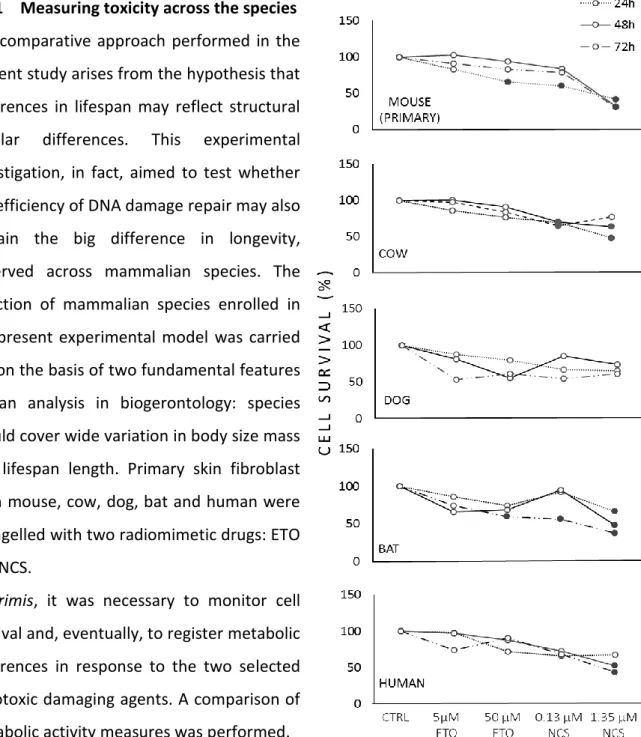
4.1.1 Measuring toxicity across the species……….48
4.1.2 Dose response experiments in 53BP1 foci formation………..49
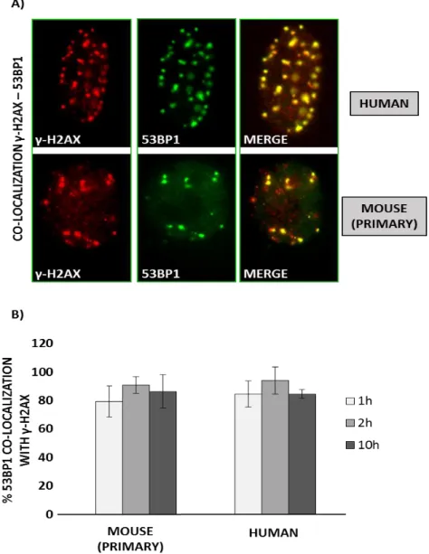
4.1.3 53BP1 co-localize with γH2AX after 5μM ETO treatment……….51
4.1.4 Long lived species exhibit the highest percentage of 53BP1 foci and protein level………52
4.1.5 Genotoxic treatment sinduce 53BP1-PMLnb co-localization and increase of senescence in longer lived species……….……….56
6
4.1.6 Genotoxic stress induce cell cycle arrest in all the species…...…………..58
4.1.7 Long-lived species induce higher level of apoptosis………..61
4.1.8 Human DNA generally appears more or equally resistant to fragmentation………..61
4.1.9 Measuring unresolved damage………64
4.1.10 53BP1 foci are inversely correlated with micronuclei abundance…………..66
4.2 Part I- 53BP1 down regulation in keratinocytes cell lines………..……...67
4.2.1 53BP1 knock down clone selection………67
4.2.2 Stable 53BP1 knock down clone reduces cellular growth.………69
4.2.3 Down regulation of 53BP1 reduces survival rate in response to damage………..69
4.2.4 53BP1 down regulation is associate to increased genomic instability……….70
4.3 Part II- Genetic instability and aging under the scrutiny of comparative biology: a meta-analysis of spontaneous micronuclei frequency……….………72
4.3.1 Body mass and MNEF are inversely correlated……….72
4.3.2 Residuals and phylogenetically independent contrasts analyses…………..…74
4.4 Part II- Role of 53BP1 in chronological age………75
4.4.1 Small increase of spontaneous 53BP1 foci in middle and old donors………..75
4.4.2 Absence of micronuclei increase with chronological age……….76
4.4.3 Higher rate of DNA-SCARS in old donors………..77
5. Conclusion……….79
6. List of references…..………..87
7
Graphical abstract
In brief: a comparative biology approach offers a powerful observational point, through
which we aimed to explore the possible link between DNA damage and longevity, by investigating the role of 53BP1 protein as a determinant for a long lifespan.
Highlights:
1. A comparative analysis in five mammalian species of 53BP1 repair foci and micronuclei expression shows an inverse correlation of these two DNA damage markers: higher level of 53BP1 foci are associated to lower incidence of micronuclei formation. 2. By CRISPR-CAS9 methodology, a 53BP1 knock down model, comparable to the
difference observed among species, was reproduced: stable downregulation of 53BP1 is associated to lower survival and increase of micronuclei frequency in response to DNA damage.
3. Spontaneous micronuclei (MN) frequency is inversely proportional to adult body mass. 4. 53BP1 as DNA damage marker for the aging process: increased spontaneous 53BP1
foci level and small decrease of repair efficiency are associated to the increase of chronological age, in human donors.
8
1. INTRODUCTION
Aging is a universal and inevitable evolution of life, characterized by global loss of physiological integrity and homeostatic imbalance [1] associated to a progressive decline of tissues and organ functions, to an impaired ability to respond to stress and to an increasing incidence of diseases. All these changes are the results of a side effect of normal metabolism, which is in turn exacerbated by environmental influence and unhealthy lifestyle [2]. The general worsening of the physiological state, which rise up during aging, is considered the primary risk factor for several diseases including neurodegenerative pathologies, diabetes, cardiovascular disorders, and cancer [3]. Therefore, death is a consequence of advancing of age. In the last decades, the improvements in hygiene and social health led significant increase in life expectancy and to a growing number of elderly people worldwide.
So, now more than ever, modern society has to deal with a whole range of social and health problems related to the global population’s aging. The need to solve the aging-related problems and the utopian dream of counteracting the effect of time, have prompted the scientific community to
investigate all the
phenomena related to aging and, more in general, to longevity. The “systemic” aging can be considered as the resulting effect of all the changes taking place inside our body. In fact, the physiological basis of these phenomena lay in the progressive lifelong accumulation of deleterious changes that impair the structure of our body at the tissue, cellular and, primarily, molecular level [2]. This is the reason why scientists attempted to find some common features of the aging process and tried to elaborate valuable theories to explain aging; some of them will be explained in the present work.
9
1.1 Common features of cellular aging
Aging research has developed deeply in the last decades and, over this time, a growing knowledge of the molecular and cellular basis of life and disease has been produced. In 2013 a group of scientists, driven by the need to conceptualize the basic and common features of aging, draw up a “list” of nine hallmarks of aging [3]; these hallmarks are: 1) altered intercellular communication; 2) stem cell exhaustion; 3) cellular senescence; 4) mitochondrial dysfunction; 5) deregulated nutrient sensing; 6) loss of proteostasis; 7) telomere attrition; 8) epigenetic alteration; 9) genomic instability. Only the last three will be presented in this thesis.
1.1.1 Telomere attrition
Telomeres are specialized structures of terminal repeated sequences of DNA, which lay at the extremity of each chromosome. During every cell division, a small part of the telomeric DNA is inevitably lost, due to the inability of DNA polymerase (which in human is active in the germline and stem cell lines, but not in somatic cells) to replicate the DNA ends. the consequence of this physiological process is the gradual shortening of telomeres, the alteration of the telomeric structure and, consequently, replicative senescence [4][5].
10
Telomeres can be regenerated by the activation of the telomerase enzyme; for example ectopic expression of telomerase can promote immortalization of primary cell culture, without inducing oncogenic transformation [6]. Telomeres’ shortening is a phenomenon occurring both in human and in mice, as a feature of normal aging [7] . In fact, it has been showed that telomerase deficiency in human is associated with premature development of diseases, such as pulmonary fibrosis, dyskeratosis congenita, and aplastic anemia, diseases connected to the loss of the regenerative capacity of different tissues [8]. Experimental evidence support that the same hypothesis is valuable in mice, where telomere length is associated with aging and control of lifespan: mice with shortened or lengthened telomeres exhibit respectively decreased or increased lifespans length [9][10][11].
Since it has become clear that in most cases what it is limiting replicative capacity in vitro is telomere shortening [6], telomere biology has been deeply investigated also across species in cell culture systems [12][13][14]. Stuart et al. [15] clearly showed that telomerase activity is low or absent and telomeres are short in long lived and large species and the opposite is true in short and small species. Than it is reasonable to suggest that convergent evolution is at play using short telomere and absence of telomerase activity as a protective mechanisms to limit tumor growth. Larger species, in fact, having more cells, have more potential targets to transformation; additionally, long lived species have more time to accumulate tumor predisposing mutations. This contradiction is clearly summarized into the “Peto’s paradox” which states that the risk to develop a tumor per gram of tissue is 3 trillion times smaller in a human than in a mouse [16]. Moreover it has been widely shown that telomerase activity is linked to tumorigenesis, as well: a big percentage of tumors, which generally originate from the transformation of somatic cells, exhibits absence of telomeres shortening and restoration of telomerase activity, with the consequence of unlimited growing capacity and inhibition of replicative senescence.
All these evidence led to the idea that telomere attrition, in mammals, can be considered a common feature of physiological aging.
1.1.2 Epigenetic alteration
Conrad Waddington (1905–1975) defined epigenetics as “the branch of biology which studies the causal interactions between genes and their products, which bring the phenotype into being” [17]. More in general epigenetic can be defined as a set of changes which alter the final
11
outcome of the expression of a locus or a chromosome, without changes in the correspondent DNA sequence. In this context, the idea that many of the epigenetic changes which arise during organisms’ development and growth may be involved in the aging processstarted to spread. For example chromatin remodelling, posttranslational modification of histones, as increased histone H4K16 acetylation, as well as modification of the DNA methylation patterns (H4K20 or H3K4 trimethylation, as well as decreased H3K9 methylation) have been showed to be related to age-associated epigenetic marks [18][19]. In fact some epigenetic markers that are remodelled during reprogramming (like the above mentioned DNA methylations, post-translational modifications of histones and chromatin remodelling) are impaired during aging [20][21][22]. Moreover, cellular differentiation may be also considered an epigenetic phenomenon, largely governed by epigenetic changes, accordingly to Waddington, rather than alterations in genetic inheritance [23]. Thus, epigenetic dysregulation has emerged as one of the key hallmark of the aging process [1]. Studies on the SIRT6, a stress responsive protein deacetylase and mono-ADP ribosyl-transferase enzyme involved in epigenetic changes, showed that its loss of function reduces longevity; on the contrary SIRT6 gain of function was associated to an extended longevity in mice [24][25]. Furthermore, the discovery of the Yamanaka factors, also known as “reprogramming factors” (the transcription factors Oct4 (Pou5f1), Sox2, cMyc, and Klf4), supports the idea that epigenetic changes have a strong influence on lifespan length and opens a new era on the epigenetic studies. Several groups have observed an amelioration of age-associated cellular phenotypes during in vitro cellular reprogramming [26][27][28][29]. It has been also shown that cellular reprogramming obtained by transient expression of Yamanaka factors ameliorated age-associated symptoms, prolonged lifespan in progeroid mice, and improved tissue homeostasis in older mice [30]. Taken all together, these works suggest that understanding epigenetic mechanisms is a fundamental step towards an improvement for the cure of age-related pathologies and for extension healthy lifespan.
1.1.3 Genomic instability
The DNA integrity and stability are constantly challenged by exogenous physical, chemical, and biological agents; but endogenous activity, such as DNA replication, spontaneous hydrolytic reactions and reactive oxygen production, can also represent a source of damage which may impair DNA integrity. Several scientific evidences strengthen the idea of a tight link
12
between genomic stability and aging. The most important experimental evidence comes from the observation that several progeroid syndromes, such as Werner syndrome, Bloom syndrome, xeroderma pigmentosum, trichothiodystrophy, Cockayne syndrome or Seckel syndrome are characterised by defects on DNA damage repair pathways [31][32][33]. Somatic mutations accumulate within cells from aged humans and model organisms [34]. Other forms of DNA damage can accumulate during aging: Faggioli et al. showed an increase of chromosomal aneuploidies in the brain of aged mice [35] and Forsberg et al. identified in human an association between aging and the copy-number variations, in vivo [36].
Genomic instability can also induce structural and numerical chromosome aberrations in somatic cells which, if not properly repaired, can lead to dangerous outcomes for cell’s life. An investigation on the efficiency of the spindle assembly checkpoint, which is a critical element in regulating chromosome segregation by preventing chromosome mis-segregation and aneuploidy, showed that its efficiency is higher in large species; thus species’ longevity does not appears as a key factor [37]. Although this analysis suggests that an increased efficiency of the spindle assembly check point could be more important for evolving large body size than longevity, this does not exclude the possibility that aneuploidy may negatively affect life span. Baker et al., in fact, report that mice with low levels of the spindle assembly checkpoint protein BubR1, display progeroid features, dwarfism, lordokyphosis, and short lifespan [38]. Moreover they showed that transgenic mice overexpressing BubR1 exhibit an increased protection against aneuploidy and cancer, and extended healthy lifespan [39]. All these data together suggest that maintenance of genomic stability is an important feature in order to achieve long lifespan length.
1.2 The theories of aging
Multiple theories have been proposed to explain the aging process with significant experimental support, and it is easy to understand that the various theories of aging can overlap at different levels of organization. In the 90’s, Kowald and Kirkwood elaborated a theory according to which a “single-factor” process strived for explain the complex mechanism of aging, assuming that a single cause could be responsible for this phenomenon [40]. But in biogerontology there is a growing appreciation that the process of aging is a multifactorial process and that it may not operate in similar fashion in all species [41]. This is probably because different species have evolved different strategies to counteract spontaneous
13
molecular damage and random malfunctioning of biological machineries. The different efficiency of these strategies could explain the different rate of aging observed across species [42] and why aging is not a universal process [43]. So based on evidence that several mechanisms may interact simultaneously operating at different levels of functional organization, the "single-factor" theory of aging was completely replaced by the vision that aging is the result of a complex multifactorial process, characterized by physiological, genetic and molecular changes that occur over time, as determined by intrinsic, extrinsic (environmental) and stochastic (spontaneous changes within molecules) causes that involve all living species. Therefore, it is possible to speculate that several “single-factor” theories alone can explain just a part of the phenomena, but cannot clarify the aging process in its entirety. Although there are many theories regarding aging, in the present thesis we will deep analyse just two of these theories, which are mostly accredited by the scientific community and useful for the present discussion: 1) free-radicals theory of aging (FRTA); 2) DNA damage theory of aging.
1.2.1 Free-radical theory aging
The free-radical theory of aging is based on the hypothesis that aging is the result of the cellular accumulation of oxidative damage, as a consequence of reactive oxygen species (ROS) released by the mitochondria. We are all living in an environment which leads to ROS production and it has been extensively demonstrated that the concentration of free radicals growss with the increase of metabolic activity [44]. Moreover it is supposed that the individual reactivity or resistance to free radicals stress is improved over time, indeed it is not a pre-determined factor. DNA molecules, proteins and lipids are all possible targets of oxidative damage that occur at cellular level [45]. The FRTA in fact suggests that 1) the production of mitochondrial ROS determines the rate of aging; 2) over time, free radicals are produced faster; 3) with aging, damage caused by ROS production is no longer efficiently repaired. Differences in how different organism handle oxidative damage were also found among the mammalian species. Some authors suggest that long-lived species produce lower levels of ROS when compared to short-lived species; while others demonstrated that rodents under caloric restriction live longer and produce less ROS when compared to controls [46].
Rossinni used and tested the FRTA to explain the exceptional longevity of M. lucifugus (maximum lifespan= 34 years). In a comparative study, the O2 consumption and the
14
production of mitochondrial H₂O₂ in several organs (heart, brain and kidneys) was measured in M. lucifugus (little brown bat, long-lived bat) and Blarina brevicauda (short-tailed shrew , short-lived). As supposed by the FRTA, M. lucifugus produced about half of the amount of H₂O₂ produced by the short-tailed shrew. Rossinni aimed also to observe oxidative damage, due to the high metabolic rate associated with the flight in M. lucifugus. Thus, in order to evaluate potential differences in oxidative damage, he measured and compared the O2 consumption and the production of H₂O₂ in both adults and young individuals (fully developed and able to fly). Surprisingly and against his hypothesis, young individuals had significantly higher levels of H₂O₂ production when compared to adults. The supposition of Rossinni was that lower production of free radicals in adults could be the result of a selection, in the individual, of efficient mitochondria, caused by the selective pressure due to the high energy level required to fly [47].
In invertebrates oxidative damage, in particular the level of superoxide, does not appear to be critical to determine species longevity; instead, in mammals, the effect of oxidative stress on lifespan’s length is poorly understood yet, but several authors claimed the potential role of antioxidant treatments as a protection against age-related organ dysfunction and cognitive decline [48]. Despite numerous scientific evidences proving that antioxidants introduced by the diet may reduce the ROS accumulation in rodents without extending lifespan, another study showed that mice with mutations in the superoxide dismutase (SOD) are more prone to develop pathologies and die prematurely. Anyhow, hyper-ubiquitous expression of SOD does not extend lifespan of these animals; therefore, this enzyme does not regulate longevity [49]. Numerous studies showed a link between the dysfunction of the respiratory chain, oxidative damage induced by ROS activity, and aging in the majority of the model organism[50]. All these evidences taken together suggest that oxidative stress is a determinant factor involved in aging.
1.2.2 DNA damage theory of aging
For a long time, accumulation of molecular damage has been considered key factor for the aging process [51]. The theory of DNA damage dates back to the late 50’s when Failla [52] and Szilard [34] independently hypothesized that accumulation of DNA lesions maybe be the driving force of the aging process. It is now clear that DNA mutations and chromosomal abnormalities increases with age in mice [53] and humans [54], and that longevity is correlated
15
with the ability to repair the DNA [55]. It is interesting to note that the more serious progeroid syndromes (Werner's syndrome, Hutchinson-Gilford, and Cockayne) originate from mutations which impair genes involved in DNA repair and metabolism [56].
The emerging hypothesis suggest that only certain types of DNA lesions are crucial for aging and this would explain why mutations falling in certain DNA repair genes affect aging, while others do not. A recent study, through an analysis of the genes involved in the DNA repair, showed that genes related to non-homologous repair mechanism are closely associated to aging [57]. In addition, damage to DNA that interfere with transcription appear to contribute to senescence exerting effects on signalling and cellular aging [31].
Figure 3: Schematic representation of cellular aging. Figure adapted from [58].
Understanding which aspects of DNA biology play a role in aging is still a challenge. Although it is well known that DNA changes over time could play a very important role in aging, the mechanisms which lag behind these changes have to be completely clarified.
1.3 DNA damage
DNA is constantly exposed to exogenous and endogenous damaging sources which may threaten DNA molecules, generating several types of DNA lesions. In order to preserve genome integrity, cells have evolved complex mechanisms which allow to point out and detect the presence of the DNA. Connecting to the DNA damage theory of aging, it is possible to hypothesize that the capacity to detect molecular damage is a potential cellular determinant of longevity. Thus, the detection of the presence of damage is a necessary step in order to proceed with repair or, if the damage is irreparable, with other means of damage control, as the induction of cellular senescence or apoptosis. Collectively these mechanisms have been termed as the DNA damage response (DDR). There is a growing evidence that the DDR efficiency can be also modulated by reorganization of the chromatin in answer to DNA damage
16
[59]. In fact the DDR machinery needs to exploit its function on a molecule, DNA, which is tightly wrapped around histone proteins in the form of chromatin [60]. Albeit chromatin acts as a physical barrier to DNA recognition and repair, it can be modified by some structural changes which take place consequently to DNA damage, such as hystone methylation [61] or histone post-translational modifications [62], which in turn can facilitate DDR’s function. In the context of the DDR activation, the most documented chromatin modification is the phosphorylation of the histone variant H2AX (γ-H2AX) [63]. Thus, the DNA damage response is a complex scenario where repair proteins are recruited at the lesion’s site and chromatin-associated proteins are mobilized to and from DNA breaks [64].
1.3.1 DSB, NHEJ pathway and DNA end binding activity
Among the different types of DNA damage, the most harmful and dangerous lesions are double-strand breaks (DSBs): one unrepaired DSB is sufficient to trigger permanent growth arrest and cell death [65][66][67]. DSB can be induced by endogenous and exogenous causes: physiological causes which include V(D)J recombination and class switch breaks (AID / UNG / EPA); or external factors as ionizing radiations, ROS production, the action of enzymes on the nuclear DNA (including topoisomerase II) and mechanical stress [68]. To repair DSBs, mammals have evolved two major repair pathways: non-homologous end-joining (NHEJ) and homologous recombination (HR). The majority of DSBs in mammals are repaired by NHEJ [69]: in the adult body, the majority of cells, are in G0 phase and, whereas HR is typically active only during the S and G2 phases, NHEJ is active throughout the cell cycle [70]. Additionally, NHEJ is significantly faster than homologous recombination [71].
NHEJ can be categorized in two different alternative pathways: 1) the canonical NHEJ (c-NHEJ), which involves core proteins such as Ku70, Ku80, DNA-PKcs, Artemis and the Ligase IV complex (Lig IV, XRCC4, and XLF) [72]; and 2) the alternative NHEJ (alt-NHEJ), also called microhomology-mediated end joining (MMEJ) a DNA-PK-independent repair mechanism, which uses a distinct set of proteins, including but not limited to PARP-1, CtIP, XRCC1, and Ligase III [73][74][75][76][77][78][79]. NHEJ is also regulated by SIRT6 [80] and Werner syndrome proteins [81]. In vertebrates, the Ku heterodimer (composed by Ku70 and Ku80 proteins) is the first proteic complex to be recruited at the DNA damage site; its recruitment to the double strands lesion is a fundamental step in order to activate the NHEJ pathway. Due to its ability to recognize and bind DNA free ends, Ku forms a ring structure at the level of the
17
damaged DNA free ends [82]; once loaded on the DNA damaged ends, it functions as a scaffold for the assembly of all other proteins involved in the NHEJ pathway.
Figure 4: Model for repair of a DSB by NHEJ. Figure from [83].
This complex secondarily interacts with the DNA dependent protein kinase (DNA-PKcs) (catalytic subunit), forming the DNA-PKcs complex, necessary for the recruitment of ATM (Ataxia telangiectasia mutated), which in its turn promotes phosphorylation of the H2A histone variant, H2AX, at the site of Ser 139 (called γ-H2AX)[84] and the recruitment of p53 binding protein 1 (53BP1) at cleavage site [85]. DNA-PKcs also induces the phosphorylation of other factors such as Artemis, an enzyme whose 5 '> 3' single-stranded exonuclease activity is switched to 5 'end and 3' endonuclease activity once bound to DNA [86]. The final step of the repair process consists in the ligation of the DNA broken ends through the intervention of LIG4, XRCC4 and XLF (Figure 4) [87].
Thus, efficient detection of the presence of DNA lesion appears to be a fundamental event for a proper cellular answer to DNA damage. Several experimental hypothesis suggest that DNA end-binding activity appears to be more closely related to longevity, than other mechanisms
18
of genomic surveillance. Stamato and colleagues showed a striking positive relationship between DNA-end binding and species longevity. This is a property which is strictly ascribable to the Ku70/Ku80 protein heterodimer [88]. What made this relationship interesting is the lack of a correlation between DNA-end binding activity with species adult body mass. The underlying hypothesis is that the different abundance of the proteins involved in the early phase of NHEJ (the Ku heterodimer and DNA-PKcs) might likely explain the different binding activities observed across the species [13].
Indeed, other mechanisms of genomic surveillance such as poly(ADP-ribose) polymerase (PARP) activity [89] and the repair of UV induced DNA lesions [90][91], positively correlates both with longevity and body mass; see dedicated reanalysis in [92][15]. For example, mice with loss-of-function mutations in Ku70, Ku80, and SIRT6 each as well as Werner syndrome patients exhibit symptoms of premature aging [93][94][95][96][25][97]. So far, the appearance of accelerated aging in human patients and in mouse models with mutations in genes involved in NHEJ is the strongest evidence supporting the relation which link together impairment of the NHEJ and aging. These mutant studies, however, do not tell us whether NHEJ is affected during normal aging [98].
1.3.2 Role of 53BP1 and its role in DNA damage recognition
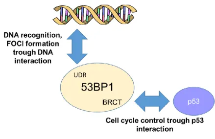
In the presence of DSBs, 53BP1 is recruited at the level of DNA breaks. 53BP1 is a large DNA damage sensor and oligomeric chromatin reader of 1972 amino acids. It is characterised by the absence of an apparent enzymatic activity and by the presence of several structural domains which can directly interact with DSB-responsive proteins.
19
Among these important structural elements, 53BP1 includes the UDR domain, the BRCA1 carboxy-terminal (BRCT) repeats, the tandem Tudor domains and 28 amino-terminal Ser/Thr‑Gln (S/T‑Q) sites, which are phosphorylated, at least in part, by the ataxia telangiectasia mutated (ATM) kinase5 [65]. The UDR domain is used for DNA recognition of the damaged site, in order to form nuclear foci (protein agglomerates) in response to a double strand breaks, where it can co-localize with other DNA damage response proteins such as γ-H2AX, Rad50/Mre11/NBS1, BRCA1, and Rad51. The BRCT domain, indeed, is involved in the interaction with p53. Notably, 53BP1 owes its name to the ability to bind p53 and precisely through this interaction it was originally identified [100]. But, while the role of 53BP1 in the DDR pathways has been deeply studied and its contribution completely clarified, 53BP1 involvement to p53-dependent cellular activities remain still undefined. Cuella-Martin et al. carefully investigated the influence of 53BP1 activity upon p53-depedent signalling after DNA damage; they demonstrated that 53BP1 stimulates genome-wide p53-dependent gene transactivation and repression events in response to ionizing radiation (IR) and synthetic p53 activation. Overall, these experimental reports suggest that these two functions are structurally separated and independent [101]. In figure 6 53BP1’s interactive targets are described in detail.
Figure 6: 53BP1 has two structural and functional domains. 1) The UDR domain leads 53BP1
to be rapidly relocated at DNA damage site after cellular exposure to ionizing radiation (IR); 2) the tandem BRCT (BRCA1 C terminus) motif, through which 53BP1 directly binds p53 enhancing p53-mediated transcription of reporter genes. These two functions can be fully separated: mutations in the 53BP1 protein sequence can selectively impair its enrichment at nuclear foci or impede p53 binding. When UDR domain is impaired, cells are more sensible to IR displaying a decrease survival. If the BRCT domain is impaired, cells show improved survival after irradiation [85][101].
20
53BP1-dependent NHEJ is also crucial in the immune system, where it is necessary for immunoglobulin class switch recombination (CSR) and T cell receptor rearrangements [102][103]. It was observed that 53BP1 knockout mice (53BP1 -/-) have a severe reduction of CSR, thus, results in an over immunodeficiency [104][105]. In order to foster DNA recognition and to promote NHEJ, 53BP1 binds a combination of methylated (H4 Lys20me1/2) and ubiquitinated histone’s (H2A ubi-Lys15) epitopes within DSB-associated chromatin [106]. Through the Tudor domain, 53BP1 is recruited to H2AK15 and H4K20 hystones, which are respectively ubiquitylated by RNF8- and mono-dimethylated by RNF168. But the first step towards its recruitment is the formation of the MRE11–RAD50–NBS1 (MRN) complex, which is a sensor of DNA lesion.
Figure 7: The signal transduction of 53BP1 pathway. Figure adapted from [65].
Once MRN complex is aggregated at the level of DSB lesion, it induces auto-phosphorylation of active ataxia-telangiectasia mutated (ATM), which in turn phosphorylates the histone 2A (H2A) variant H2AX at Ser139, in order to generate γ-H2AX. γ-H2AX is in turn recognized by mediator of DNA damage checkpoint protein 1 (MDC1). MDC1 activates a positive feedback loop by recruiting more MRN complexes and activated ATM to the damaged chromatin. This event leads to the recruitment of the E3 ubiquitin ligase RING finger 8 (RNF8) (phosphorylation-dependent). MDC1 linked to RNF8 is recognized by a second E3 ubiquitin ligase, RNF168.[65][85]. This last complex, as previously mentioned, recruits 53BP1 to the DNA damage surrounding chromatin (Figure 5). Moreover, it was found that 53BP1 foci formation is reduced in response to a reduction of the H4K20 methylation [107].
21
Additionally, 53BP1 possesses tumor suppressor functions. Germline 53BP1 mutations predispose mice to T cell lymphoma, in manner exacerbated by p53 loss [108][109]. Ward et
al. showed that 53BP1 mice knock out display a higher sensitivity to IR and an increase rate of
tumorigenesis [110]. On the whole, these informations track the “portrait” of a key protein, fundamental for the cellular destiny after a DNA damage.
1.3.3 DNA damage nuclear foci
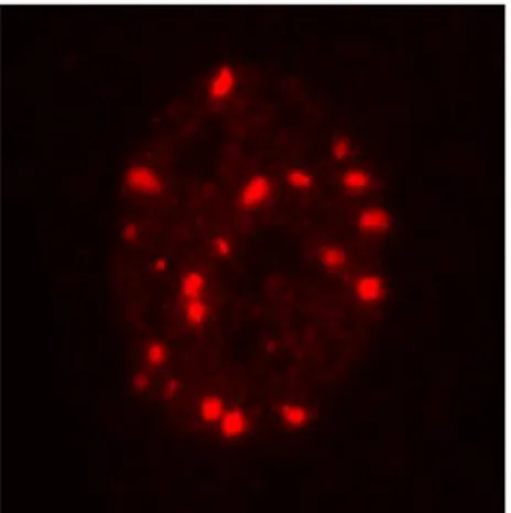
As previously mentioned, 53BP1 agglomerates in clusters at double break sites and these clusters are generally called nuclear foci. Nuclear foci are agglomerates of DNA damage repair proteins, each with a different but necessary function, which form into the nucleus of stressed cells within minutes after damage. 53BP1 foci, as well as all the different nuclear foci, are easily identifiable trough immunofluorescence assays and are universally used as a DNA damage marker [111]. Nowadays very much is known about the role and the kinetics of the DNA damage repair complex
assembly. For example the phosphorylation of the histone variant H2AX (γ-H2AX) and the accumulation of the 53BP1 protein in distinct nuclear foci [112] are widely used as DSBs markers; γ-H2AX phosphorylation promotes the formation of a protein scaffold necessary for the repair of DBSs [113]. Depending on the extent of the damage, γ-H2AX and 53BP1 may co-localize [114] and, since each focus represents a single DSB, their amount is proportional to the intensity of damage [115]. Therefore, nuclear foci represent not only a marker of the presence of DNA damage but also a measure of the damage’s intensity.
Kinetic of nuclear foci, such as 53BP1 foci, can be divided in two temporal phases: 1) early foci which rapidly form after damage and are a sign of an active detection DNA damage system; 2) long lasting foci which, if DNA damage is not properly repaired, can persist for longer time into the nucleus. Moreover, the nuclear location of these persistent foci define two different classes of long lasting foci: 1) if foci localized at the telomeres are called Telomere Dysfunction Induced Foci (TIF) [116]; 2) if foci are localized all over the nucleus, will be called DNA-SCARS (segments of DNA with chromatin alterations) and they can persist for several days after
Figure 8: Representative
image of 53BP1 foci in a human primary fibroblast.
22
damage [45]. DNA SCARS, which may include both γ-H2AX and 53BP1, often completely or partially co-localize with PMLnbs (promyelocytic leukemia protein nuclear body) [117][118]. PMLnbs are small dynamic nuclear bodies found in most cell-lines and many tissues. They belong to the nuclear matrix in a structure which has been proposed to regulate many nuclear functions as DNA replication, transcription, or epigenetic silencing [119]. They may also participate to cellular responses to genotoxic stress [120]. Although their biological role is not completely clarified, they are often associated to stress induced senescence [121]. In fact persistent foci are usually abundant in tissues undergoing to genotoxic stress [122] and in senescent tissues of mice and primates [116].
1.4.4. Micronuclei
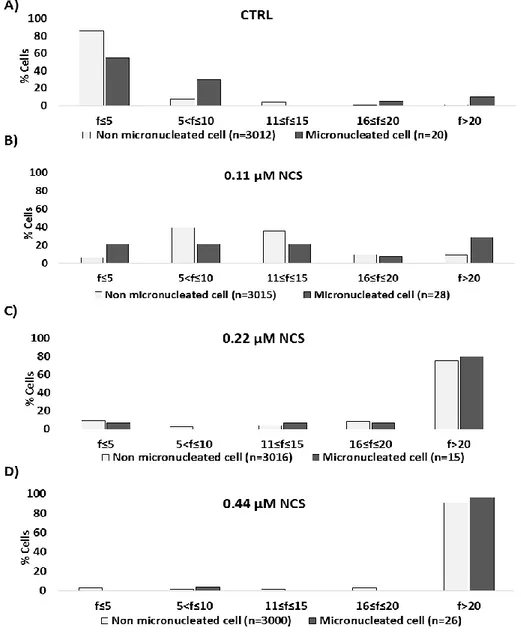
Another well-known DNA damage marker are micronuclei (MN). MN formation is widely considered as a cytogenetic direct marker of unresolved damage and the analysis of their appearance is used as a measure of the residual genomic instability in a cellular population, after a genotoxic stress. MN are defined as small, round, DNA containing cytoplasmic bodies derived by loss of nuclear material, not incorporated into the nucleus of a daughter cell after an aberrant mitosis [123] [124] (fig 8). Thus, MN can be constituted by different nuclear portions: acentric chromosomes, chromatid fragments or whole chromosomes. MN generation is strictly connected to the action of chemical or physical agents which induce, in vivo and in vitro, genotoxic stress. Mutagenic agents can be classified into (1) clastogenic agents, able to give rise to or induce disruption or breakages of chromosomes, leading to chromosomal deletion, insertion or rearrangements or (2) aneugens agents, that damage the mitotic spindle or the centromere of a whole chromosomes [125]. Both mutagenic mechanisms lead to a separation of portion of chromatin from the main nucleus, in favour of the formation of a new structure similar to the main nucleus, but independent and smaller when compared to it [126]. Fenech
et al. draw a list of characteristics which need to be applied in order to correctly identify MN.
Thus MN, must 1) be round or oval shaped 2) to lay close to the main nucleus 3) have a Figure 9: Representative image of a
23
diameter between 1/3 and 1/16 of the diameter of the main nucleus 4) have the same staining intensity and the same appearance of the latter [127] (see Figure 9). Because cell division is necessary for the generation of MN, it is recommended that MN are scored by the cytokinesis block micronucleus (CBMN) assay developed by Fenech and Morley [128]. Therefore, the presence of micronuclei in the cellular cytoplasm is considered an indicators of genetic damage.
24
2.
AIM OF THE THESIS
In mammals, species lifespan can vary by more than 100 fold (shrew 2 years, bowhead whale 211 years). It would be plausible to think that differences in lifespan may reflect structural differences between species at the cellular level by influencing fundamental processes. The DNA damage theory of aging, mentioned above, postulates that accumulation of mutations over time is one driving force of the aging process; additionally one may ask whether accumulation of DNA damage could also have determined the average lifespan of a species, during evolution. Accordingly, less efficient repair systems of a given species could leave more frequently unsolved damages in the cellular population, thus leading to increased genomic instability, which in turn would decrease the overall longevity of the species. This hypothesis is supported by the observation that a positive correlation exists between longevity and the ability to repair damage in model organism [55]. In addition, it has been shown that the abundance of proteins like Ku80 and DNA-PKcs, which are directly involved in DNA end-binding during DSB repair, correlates with the longevity of certain species [13].
This thesis aims at further verification of the hypothesis that the efficiency of DNA damage repair, which likely contributes to the individual aging process, is also suited to explain the big difference in longevity observed in different mammalian species. Specifically, I intended to test whether long-lived species have more efficient cellular DNA repair machineries and investigated potential differences among five mammalian species, with respect to DNA damage-repair and the maintenance of genomic stability in response to low and high genotoxic damage.
53BP1 is widely known as a key regulator and orchestrator of the cellular response to double strand break damage: on one hand it is directly recruited on the DNA damage site and on the other hand it regulates cellular responses through its direct interaction with p53. Thus, 53BP1 should be investigated to assess the efficiency of the DNA damage response in the comparative experimental model. In addition, formation of micronuclei should be monitored to evaluate long-term effects of genotoxic stress in these different species. And finally, similar experiments should be applied to cultures of primary skin fibroblasts from human donors of
25
different age to explore a potential association between DNA damage, senescence markers and the chronological age.
2.1 Project description
The project has been structured in three parts, each investigating the same theme from different observational point of view: interspecies longevity, in vitro studies (PART I); interspecies longevity, in vivo studies (PART II) and intraspecies longevity ex vivo and in vitro studies (PART III).
2.1.1 PART I- DNA damage detection by 53BP1: relationship to species longevity
Croco E., Marchionni S., Bocchini M., Angeloni C., Stamato T., Stefanelli C., Hrelia S., Sell C., Lorenzini A.
Abstract
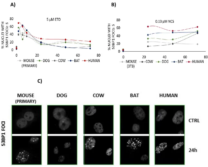
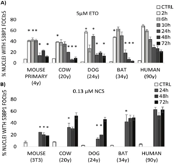
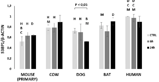
In order to examine potential differences in genomic stability, we have challenged fibroblasts derived from five different mammalian species of variable longevity with the genotoxic agents, etoposide and neocarzinostatin. We report that cells from longer-lived species exhibit more tumor protein p53 binding protein 1 (53BP1) foci for a given degree of DNA damage relative to shorter-lived species. The presence of a greater number of 53BP1 foci was associated with decreased DNA fragmentation and a lower percentage of cells exhibiting micronuclei. These data suggest that cells from longer-lived species have an enhanced DNA damage response. We propose that the number of 53BP1 foci that form in response to damage reflects the intrinsic capacity of cells to detect and respond to DNA harms.
INTRODUCTION
Double-strand breaks (DSBs) are the most harmful DNA lesions a cell can encounter. In mammals, non-homologous end-joining (NHEJ) is the cellular mechanism responsible for repairing the majority of DSBs [69]. This is because NHEJ is much faster than homologous recombination (HR) [71] and it is active throughout the cell cycle, whereas HR is typically active only during the S and G2 phases [70]. Moreover, the majority of cells in the adult body are in G0. At the sites of DSBs, nuclear foci of phosphorylated histone H2AX (γ-H2AX) and tumor protein p53 binding protein 1 (53BP1) can be detected by immunofluorescence and are widely used as DNA damage markers since their abundance has been reported to correlate very closely with the degree of genotoxic insult [reviewed in [129]. 53BP1 accumulates in DNA
26
damage foci after the occurrence of DSBs in order to facilitate NHEJ repair [130]. Previous works had shown that 53BP1 is involved in activation of the DNA damage response during the G1, S, and G2 phases of the cell cycle [131][132][133].
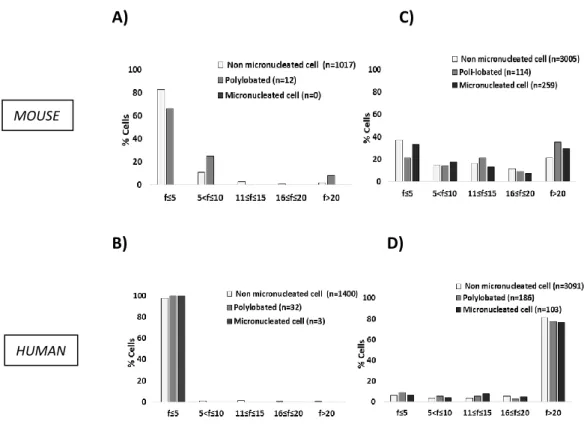
We have previously observed an unexpected difference in the formation of DNA damage foci between human and mouse cells following exposure to the same concentration of neocarzinostatin (NCS). Despite similar exposure to the DNA-damaging agent, human cells exhibited a significantly greater number of 53BP1 foci compared with mouse cells. The greater abundance of foci in human cells correlated inversely with the appearance of micronuclei, which represent the presence of severe genomic damage [134]. We have also reported an impressive correlation between mammalian longevity and the capacity of nuclear proteins to bind DNA ends, which appears to be largely dependent on increased expression of the evolutionarily highly conserved heterodimer Ku80/Ku70 [13]. The DNA end-binding assay represents the accumulation of proteins required for the initial DNA damage recognition, a necessary step before a cell can proceed with repair, cell cycle arrest or apoptosis. These data persuaded us to conduct a deeper investigation into the relationship between the appearance of markers of DNA damage and mammalian longevity.
To this end, we have compared fibroblast cells from mouse (maximum life span, 4 y; weight, 20.5 g), cow (maximum life span, 20 y; weight, 750 Kg), dog (maximum life span, 24 y; weight, 40.0 Kg), little brown bat (maximum life span, 34 y; weight, 10.0 g), and human (maximum life span, 122 y; weight, 62.0 Kg) in their response to the genotoxic insult caused by etoposide (ETO) and neocarcinostatin (NCS). ETO is a poison that binds reversibly to topoisomerase IIα/β to block re-ligation of cleaved DNA strands, causing DSBs [135] while NCS is a macromolecular chromoprotein antibiotic that causes single- and double-strand breaks [136]. Our experimental approach was based on the assumption that the biological mechanisms that facilitate longevity are cell autonomous and thus will be reflected in fundamental cellular processes. Consequently, these mechanisms can be studied in cell culture away from the complexity of the whole organism. For example, using a cell culture approach, Dostàl and colleagues have demonstrated that long-lived rodent species are more efficient in excluding Cadmium that is known for disturbing DNA base excision and mismatch repair [137].
With the present work, we propose that quantitative, species-specific differences in 53BP1 foci formation observed following exposure to genotoxic agents correlate with differences in
27
lifespan and adult body mass, suggesting a new interpretation of nuclear foci as markers of a species’ ability to detect and mitigate DNA damage to ensure genomic stability.
2.1.2 PART II- Genetic instability and aging under the scrutiny of comparative biology: a meta-analysis of spontaneous micronuclei frequency
Eleonora Croco, Silvia Marchionni, Antonello Lorenzini.
The idea that accumulation of DNA damage may be responsible for the aging process dates back to the late 1950s [52][34]. The key aspects of this theory are twofold: the idea that damage accumulates with time and the fact that damage to the DNA is of fundamental importance. The first concept is easily explainable if we look at many of the lifeless objects of our daily life that, after a period of service, accumulate some sort of damage (cars, buildings, etc.). These objects are designed to last for a specified time and they maintain themselves well for that period. However, damage eventually appears and accumulates due to intrinsic failure of their parts, harmful extrinsic factors, and lack of proper maintenance and repair. The result is a period of good function followed by a gradual decline. Biological systems are similar: there is a period of low mortality up to a point, followed by a gradual decline in fitness and increased mortality. Linking this concept to longevity, each species possesses an intrinsic design that ensures functionality for the life span of that species. Repair of DNA damage is critical to that design.
The second concept, that damage to the DNA is of fundamental importance, is also readily understandable thanks to a recent scientific breakthrough. Gibson and colleagues, having created the first synthetic life form through the synthesis of a 1.08-megabase-pair mycoplasma genome, have de facto demonstrated that DNA has a master role in the hierarchy of organic molecules [138]. DNA damage will, in fact, carry consequences at any level of biological organization or function (RNA, protein, metabolism, etc.).
Szilard, in his theory paper “On the nature of the aging process”, discussed “hits” to chromosomes that render genes inactive[34]. However, genetic instability may also be obtained in the absence of actual damage to the DNA molecule. Aneugens, for example, are substances that cause genetic instability by simply interfering with chromosome segregation. Genetic, genomic, or chromosome instability are thus broader terminologies that are being used progressively more in gerontology [139].
28
Many observations and theoretical considerations support a role for genetic instability in aging. In rats, for example, inducing DNA damage with a single dose of ionizing radiation at a young age (6 weeks) shortens average life span by 10% for a dose of 1 Gy and 25% for a 3-Gy dose [140]. The very existence of more than 150 genes involved in DNA damage repair attests to the importance of DNA integrity in sustaining life [141]. Following this reasoning, the perturbation of one such repair gene should, to some degree, negatively affect life span. In support of this, the Ku heterodimer, comprising the Ku70 and Ku80 polypeptides, is critical for double-stand break (DSB) repair. Double mutants, as well as mutants of either Ku component, display an approximate 66% reduction in life span, with signs of early aging [96]. Despite a dramatic effect of the mutant Ku on life span, not all data are supportive of the theory. Kaya and colleagues, for example, have monitored a mutant of Saccharomyces cerevisiae that accumulates high levels of mutations. Whereas old wild-type cells with few mutations were dying, young mutant cells with many more mutations continued budding [142]. In conclusion, although the DNA damage accumulation theory is among the “oldest” of the aging theories, discussion of its true relevance is still very active among gerontologists.
Genomic stability and body mass
The somatic mutation theory of aging predicts that long-lived species will have superior genomic stability. In fact, if alterations at the level of DNA are supposed to be responsible for the aging process, these alterations must accumulate at a lower rate in longer-lived species. With similar reasoning, we can hypothesize that genomic stability will also be positively related to adult body mass. In fact, two species with similar longevity but different adult body mass will differ in their total number of cells in adulthood. Consequently, the number of total cellular divisions that the genome must sustain, from the creation of the zygote onward, can be very different. Additionally, body mass is positively correlated with longevity among mammals, birds, amphibians, and reptiles [143]. In the multitude of cellular mechanisms involved in preserving genomic stability, some will be more essential in order to guarantee longevity, while others will be more essential in order to guarantee the development of greater body mass. For example, we have found that the capacity to bind DNA ends (a capacity of the Ku heterodimer) correlates with longevity, but not with body mass in 12 mammalian species [13]. In contrast, we have found that the efficiency of the spindle assembly checkpoint correlates strictly with body mass and more loosely with longevity in six species of mammals
29
[144]. The efficiency of excision repair after UV damage and the activity of Parp, an enzyme involved in single-strand break (SSB) repair, are other examples where a correlation is found both with longevity and body mass [89][92][91][15].
Advantages of the comparative approach
Life span studies may be extremely long lasting, being clearly linked to the life span of the tested species. Rats and mice, which require at least three to four years of study, are the most studied mammals in the laboratory. Nonetheless, these two rodents are not the best representation of the aging process in mammals since their longevities lay at the lowest extreme of the vast range of life spans exhibited by this class. Maximum longevity in mammals spans 100-fold, from 2.1 years for the forest shrew to 211 years for the bowhead whale (data from the AnAge database; [145]). Comparing characteristics of species known to differ significantly in longevity represents a unique and powerful approach to aging studies [146]. Comparative cellular biology also gives the potential to manipulate the environment and challenge cells with different stressors to monitor their differential reactivity (reviewed in [15]). In a recent and comprehensive review, Moskalev and colleagues analyzed evidence for and against the DNA damage theory cataloguing studies based on Koch-like criteria [34]. The authors concluded, in part, that comparative biology data are thus far scarce in the literature (both for and against the theory). In the present article, we will demonstrate to the gerontological community that comparative biology data are readily available and may need only to be extracted from the literature. To this end, we conducted a meta-analysis on spontaneous in vivo micronuclei (MN) frequency in mammalian erythrocytes of species with different longevity and body mass. Our aim was to search for a possible relationship between these two parameters (longevity and body mass) and the propensity to this type of genomic instability.
Analysis of micronucleated erythrocyte frequency across mammals
MN are small cellular bodies containing genomic material enclosed in nuclear membranes. These small bodies are produced during mitosis when a whole chromosome or chromosome fragment is left behind by the mitotic spindle and fails to be incorporated into either one of the daughter cell nuclei. The spontaneous or induced formation of MN is used as a cytogenetic test using different cellular substrates: epithelial cells from mouth swab, lymphocytes from blood, and erythrocytes from blood, bone marrow, or spleen [147][148]. Erythrocytes are
30
particularly useful since they naturally expel the nucleus during maturation, but do not expel the MN [149]. With staining such as Giemsa or acridine orange, micronucleated erythrocytes (MNE) are scored to assess the possibility of genotoxicity in vivo [150]. Observing MN in bone marrow erythrocytes may testify to recent genotoxic damage, while the presence of MN in circulating erythrocytes is an indication of chronic exposure [151]. If the aging process is seen as an accumulation of damage, then it may be likened to a chronic exposure. Consequently, we have screened the literature for reports on the spontaneous frequency of MN in circulating erythrocytes in different species of mammals.
2.1.3 PART III- 53BP1, DNA DAMAGE AND CHRONOLOGICAL AGE
The aging process is accompanied by an accumulation of cellular damage, which compromises the viability and function of cells and tissues. Dermal fibroblasts are mostly quiescent cells that are regularly exposed to environmental stimuli, thus these cells provide a paradigmatic model of long-term cellular adaptation to exogenous stress [152]. One of the biggest goal in the aging field is to find markers which can be applied for early identification of aging process. Human diploid fibroblasts have been used as the election model for in vitro-studies of cellular aging, but so far, only a small amount of information about the aging process of these cells in their physiological tissue environment, i.e. the dermis , is known [153]. Since DNA damage markers can be easily identified and studied, the major part of these studies attempted to find an association between their expression and aging. In a recent study, numbers of 53BP1 foci in dermal fibroblasts were also positively associated with the age of the donor [154]. Fibroblasts in the dermis of aged baboons exhibit accumulation of nuclear foci positive for 53BP1. Many of these foci are co-localised with telomere markers suggesting they indicate telomere dysfunction [155]. Micronuclei numbers were shown to be higher at older age in lymphocytes (reviewed in [156]) and buccal cells [147].However, an inverse relation between the number of 53BP1 foci and micronuclei has also been described between mouse and human cells [134]. In our work [157] a wide cohort of individuals with different chronological age was tested, in vitro, for the presence of two DNA damage markers: MN and 53BP1 foci formation. The expression of these two markers was investigated in untreated and rotenone treated primary fibroblasts from 100 donors. No association between micronuclei and chronological age was found; DNA damage foci of cultured fibroblasts are significantly associated with chronological age, but not with biological age, of donors.
31
32
3.
MATERIAL AND METHODS
3.1 Cell culture
All fibroblast strains and lines used are described in Table 1. Fibroblast strains from mouse, little brown bat, dog, and cow were established as previously described [158]. Adult human fibroblasts were obtained from Claudio Franceschi (DIMES, University of Bologna).
Fibroblasts from mammals species were maintained in Minimum Essential Medium (MEM) with Earle’s Salts and L-glutamine containing 10% fetal bovine serum (FBS), MEM vitamins and amino acids, and penicillin-streptomycin (all from Sigma Aldrich, St. Louis, Mo). Fibroblasts were passaged weekly, washing them twice with PBS 1X and using Trypsin-no EDTA 1X for detaching the cell layers. The number of cells were counted using the Burker chamber and they were seeded according to the standard cell density (10.000/cm²). Cumulative population doubling (PD) was determined as previously described [159].
Skin fibroblast from 15 human female donors were used in the present study. They were aged 20, 23, 25, 26, 26, 40, 41, 42, 43, 49, 60, 62, 63, 64 and 67 years, thus covering a wide age spectrum 20 – 67 years. They were divided into three different chronological age groups, each with five biological replicates: “young” (20–30 years), “middle aged” (40–50 years) and “old” (60–70 years). All these strains were provided from Prof. Boege (Uniklinik, University of Düsseldorf).
HaSK-pw keratinocytes (KT) (characterised by Petra Boucamp, German Cancer Research Center, Heidelberg, Germany), spontaneously immortalized keratinocytes, described in [160], were kindly provided from Prof. Boege (Uniklinik, University of Düsseldorf). They were maintained in FAD II medium, a 3:1 mixture of DMEM and Ham's F12 medium supplemented with 10% FBS, 24 ng/ml Adenin, 1 ng/ml hEGF, 0.4 μg/ml hydrocortisone, and 5 μg/ml insuline. The KT cell line was authenticated by short tandem repeat (STR) profiling confirming its uniqueness. KT were passaged 1:5 every three days, in order to avoid high confluence level.
33
All cell strains were maintained at 37°C in a humidified atmosphere containing 5% CO2. With the exception of the immortalized 3T3 mouse line and keratinocytes, little brown bat strains that had undergone spontaneous immortalization, lines displayed cellular senescence and were used for all experiments at approximately the first half way through their replicative lifespan.
TAB.1 Cell culture strains and lines
STRAIN
NAME SPECIES
COMMON NAME
(strain) ORIGIN (Age) EXPERIMENTS
MEF Mus musculus Mouse Embryo, F FOCI, MN,
M3M Mus musculus Mouse Adult (3 months), F FOCI ETO, MN, WB,
MTT
M2W Mus musculus Mouse Young (2 weeks), F
FOCI ETO, MN, WB, MTT
3T3 Mus musculus Mouse Embryo
(Immortalized)
FOCI NCS, MN, COMET
YA35CF Homo sapiens Human Adult (25 years), F FOCI, MN, WB, MTT
IMR90 Homo sapiens Human Embryo, F FOCI, MN, COMET,
WB
WI38 Homo sapiens Human Embryo, F FOCI, MN
B#1 Bos taurus Cow Young (about 6
months), F
FOCI, MN, COMET, MTT
D#1 Canis familiaris Dog (Beagle) Young adult, F FOCI, MN, COMET,
MTT
D#BAS Canis familiaris Dog (Basset hound) Young adult, F WB, MTT
LBB#4B Myotis lucifugus Little Brown Bat Young adult,
(immortalized), F
FOCI, MN, COMET, MTT
KT Homo sapiens Human Immortallized, KT
GENOME EDITING, MN, SURVIVAL
ASSAY, CFA
34
3.2 Freezing and Thawing of cells
In order to freeze cells, they were collected, counted and cellular suspension was centrifuged at 300 x g for 5 minutes. The cellular pellet was suspended, according to the standard cellular density, in freezing medium (FBS 70%; complete MEM 20%; DMSO 10%), previously prepared and stored at 2°-8°C until its use. The suspension was transferred into crio-tubes. After an overnight passage at -80°C, tubes were transferred into liquid nitrogen for long term storage. For thawing cells, tubes from liquid nitrogen were warmed in the water bath at 37°C for 1 minute. Cells were mixed and suspended in 5-10mL of fresh MEM and seeded in T25 or T75 flask. The day after, fresh new medium was added.
3.3 Genotoxic treatment
Two different type of genotoxic treatment were performed 1) radiomimetic treatment, ETO and NCS treatment; 2) ionizing radiation (IR) treatment.
1) For the chemical treatment, cells were treated with ETO and NCS (both from Sigma Aldrich, St. Louis, Mo). The majority of the experiments were performed on S phase synchronized cells and when unsynchronized cells were used, this is specified. S phase was chosen as the most appropriate time for exposure to genotoxic stress, accordingly to a previous study where differences in susceptibly to genotoxic damage were determined between human and mouse cells, showing that susceptibility is most apparent in S-G2 [134]. Additionally, topoisomerase II inhibitors are most effective during DNA replication in S phase [161]. Cultures were initially arrested in G0 by serum starvation for 48 hours (h) in serum-free medium. Cells were then stimulated to enter the cell cycle by the addition of fresh complete medium with 10% FBS. For each species, the time following FBS stimulation required for entry into S phase was defined by cell cycle analysis (data not shown). Mouse and bat cells were stimulated in complete medium with 10% FBS for 16 h; dog, cow, and human for 18 h. Once synchronized, cells were treated in complete serum-free medium to avoid the potential for interference of serum on drug bioavailability. Unsynchronized cells were used only on initial dose response experiments (Figure 14 and 15). Dose ranging from 5 µM to 100 µM of ETO and ranging from 0.13 µM to 1.35 µM of NCS were used.
35
2) IR treatment was performed by the use of the Gulmay’s machine, available at a separate facility of the University of Dusseldorf. All the IR experiments were performed on unsynchronized cells, under the same condition of voltage (150kV) and amperage (15mA). Time exposure is described in Tab. 2.
3.4 Preparation of whole cells lysates
Cell whole lysates from fibroblasts derived from mouse, little brown bat, dog, cow and human were prepared by adding ice-cold RIPA buffer (50mM Tris, 150mM NaCl, 1% NP-40, 0.1% SDS, 0.5% sodium deoxycholate, 1mM PMSF, 1 µg/mL leupeptin, 1 µg/mL pepstatin). Lysates were collected and protein amount was dosed by using DC protein assay (5000111, Bio-Rad kit), based on a modification of the Lowry’s method. A standard curve was prepared by obtaining 3-5 standards of protein (Bovine Serum Albumine, BSA), containing from 0.2 mg/ml to about 1.5 mg/ml protein. For the dosage, samples were prepared according to manufacturers’ instructions. Then, to determine protein concentration absorbance was read at 750 nm. To collect whole lysates from primary fibroblasts from young, middle, old donors and for KT (wild type and knock down) pellets were resuspended in 2X lysis buffer (250 mM Tris-HCl, pH 6.8, 2% glycerol, 4% SDS, 20 mM DTT, 1.4 M urea, 20 mM EDTA, 2 mM PMSF, 5 mM pefa block, 0.04% bromphenol blue) in a volume sufficient to get a final density of 1x10⁵cells/ 20 µl. Thus, lysates were homogenized by ultrasound (10 s at 20% power).
All samples where boiled (100°C, 5 min). 50 micrograms of cell extracts or 30 µl of warm aliquots were loaded onto SDS-polyacrylamide gels.
TAB.2 Ionizing radiation settings
Gy TIME
1 1’23”
36
3.5 Polyacrylamide gel electrophoresis
Electrophoresis was performed in 1x TGS buffer running buffer at voltage of 90-160V.
3.6 DS-PAGE and Western blotting
After separation, proteins were electrophoretically transferred from the gel to a nitrocellulose membrane by the tank-blot method. Therefore the nitrocellulose membrane was activated in MeOH for 1min, washed in H2O and equilibrated in transfer buffer (48 mM Tris, 39 mM glycin, 0-20 % methanol, pH ~9.2), while the gel was equilibrated for at least 20 min in transfer buffer. After transfer, the nitrocellulose membranes were blocked for 1 h in 5% bovine serum albumin (BSA) or Milk in Tris-buffered saline containing 0.1% Tween-20 (TBST). Membranes were incubated with 5% BSA or Milk TBST primary antibody shaking overnight and then were incubated for 1 h with goat anti-mouse-HRP or goat anti-rabbit-HRP (Cell Signalling, Billerica, Ma) in 5% BSA or Milk TBS-T. Blots were washed in TBS-T and incubated with ClarityTM Western ECL substrate (Bio Rad, Hercules, Ca) for 5 minutes before devoloping. Densitometry measurements were captured using the ChemiDoc station and software (Bio Rad, Hercules, Ca). All densitometric measurements were normalized to loading controls.
3.7 Immunofluorescence: foci determination
Fibroblasts were seeded onto glass coverslips, and treated with ETO or NCS at varying concentrations for 6 or 2 h respectively. Cells were fixed at the indicated times after the beginning of damage in 3,7 % paraformaldehyde for 10 minutes and permeabilized in 0.5% Triton X-100 in PBS for 15 minutes. Cells were washed three times in PBS and blocked for 1 hour in 5% goat serum 1% BSA in PBS containing Tween-20 (PBST) (blocking solution), after which they were incubated with primary antibody in 0,1% blocking solution buffer for 1 h at room temperature in a humidified chamber. All the antibodies used are listed in Tab. 3. 53BP1 antibody (NB 100-304) binds, in tested species, epitopes with homologies that range between 90% and 92% compared to the human epitope. Anti-γH2AX antibody, used for human and mouse, binds an epitope with a homology of 90% between these two species. Slides were washed three times in blocking solution and incubated with secondary antibody in 0,1% blocking solution for 1 h. Cells were washed three times, stained with DAPI, and mounted with Vectashield mounting medium (Vector Laboratories, Burlingame, Ca) before
37
visualizing. Images were captured using an Olympus BX61 fluorescence microscope (Tokyo, Japan) equipped with a Hamamatsu ORCA-ER camera (Hamamatsu City, Japan) or an Olympus IX50 equipped with a Diagnostic Instruments 9.0 Monochrome - 6 camera (Sterling Heights, Mi). Clearly identifiable 53BP1 or γH2AX foci inside the nucleus were counted as positive foci. Nuclei were scored as containing: 53BP1 foci (F) < 5; 5 ≤ F < 20; F ≥ 20. Nuclei with fewer than 5 foci were considered foci-negative because this number of foci was common in untreated cells. Overlapping and/or contiguous 53BP1 and PML foci were scored in co-localization experiments.
3.8 Micronuclei assay
Unsynchronized cells or cells synchronized in S phase, as previously described, were treated with ETO or NCS at varying concentrations for 6 or 2 h, respectively; or with IR at different grade of irradation. At the times indicated in each figures, cells were fixed in 4% paraformaldehyde for 10 minutes and permeabilized in 0.2% Triton X-100 in PBS for 1 minute. Fixed cells were stained with DAPI and washed with PBS. Cells were mounted with Vectashield and scored using fluorescence microscopy as above. Micronuclei, defined as DAPI-positive bodies that were morphologically identical but smaller than the nucleus, were scored according to Thomas and Fenech [127]. Cells were considered micronucleus-positive if they contained, at least, one micronucleus.