DOTTORATO DI RICERCA
Metodologie di Ricerca nelle Malattie Vascolari e Toraciche
XX Ciclo
Settori scientifici disciplinari di afferenza: MED22 - MED05
RESIDENT ANGIOGENIC
MESENCHYMAL STEM CELLS
FROM MULTIORGAN DONOR THORACIC AORTAS
Tesi di Dottorato
Coordinatore Dottorato : Presentata da:
Chiar.mo Prof. Andrea Stella Dott.ssa Laura Foroni
Relatore :
Chiar.mo Prof. Gianandrea Pasquinelli
I
INDRODUCTION
1. STEM CELLS
1.1
Definition and classification of stem cells……….11.2
Embryonic stem cells………31.3
Adult stem cell………..51.4
Mesenchymal stem cells ……….112. STEM CELLS NICHE
2.1
The hematopoietic stem cell niche in the bone marrow …...192.2
The epithelial stem cell niche in skin………...212.3
The intestinal stem cell niche………...232.4
The neuronal stem cell niche………252.5
The heart stem cell niche………..273. THE BLOOD VESSELS SYSTEM
3.1
Histology and classification………293.2
The vascular wall as a source of stem cells………323.3
Angiogenesis………...373.3.1
Vascular endothelial growth factor………..383.3.2
MSC and Angiogenic differentiation………393.4
The cardiovascular tissue banking………...40EXPERIMENTAL DESIGN
4. AIM OF THE WORK ………..42
5. MATERIALS AND METHODS………44
5.1
Human arterial sample……….44II
5.4
Fresh and cryopreserved arterial tissue: histology analisys…..455.5
Fresh and cryopreserved arterial tissue : tem analysis…………465.6
In situ cell presergvation: immunohistochemistry protocol……475.7
In situ cell death: tunel assay………...485.8
Isolation cells from cryopreserved allografts………495.9
Flow cytometry analysis………..495.10
Organ culture: lm and tem………...505.11
Antigenicity preservation:immunohistochemical staining for HC-10 e NAMB-1………..515.12
Identification in situ of a “vasculogenic zone”: immunohystochemical studies………525.13
Isolation and espansion of adult stem cells from fresh human thoracic aortas………..535.14
Immunophenotyping: flow cytometry analysis………..545.15
Immunofluorescence analysis……….555.16
Ultrastructural analysis of cell : tem analysis ………565.17
Stem cells mrnas expression by rt- pcr………565.18
Endothelial differentiation of adult stem cells from human thoracic aortas……….585.18.1
In Vitro Matrigel assay ………595.18.2
TEM analysis………...595.18.3
Flow Cytomery Assay………..595.18.4
In situ Immunofluorescence assay………...605.18.5
CD133 and KDR mRNAs expression by RT-PCR……….605.18.6
Quantification of eNOS mRNA expression by Real Time PCR………616. RESULTS………..62
6.1
Fresh and cryopreserved arterial tissue: histology analisys….62III
6.3
In situ cell preservation : immunohistochemistry results……..656.4
In situ cell death: tunel assay………..676.5
Isolation cells from cryopreserved allografts: flow citometry study………696.6
Organ culture: lm e tem………..706.7
Antigenicity preservation: immunohistochemical staining for HC-10 e NAMB-1………...736.8
Identification in situ of a “vasculogenic zone”: immunohistochemical studies………776.9
Isolation and espansion of adult stem cells from fresh human thoracic aortas………..836.10
Immunophenotyping: flow cytometry analysis………..866.11
Immunofluorescence analysis……….886.12
Ultrastructural analysis of cell : tem analysis………896.13
Stem cells genes expression by rt- pcr………916.14
Endothelial differentiation of adult stem cells from human thoracic aortas……….936.14.1
In Vitro Matrigel assay……….936.14.2
TEM analysis………...946.14.3
Flow Cytomery Assay………..956.14.4
In situ Immunofluorescence assay………...976.14.5
CD133 and KDR mRNAs expression by RT- PCR……….986.14.6
Quantification of eNOS mRNAs expression by Real Time PCR………987. DISCUSSION………100
1
1. STEM CELLS
1.1 DEFINITION and CLASSIFICATION OF STEM CELLS
In the last years stem cells have became a new fascinating topic of research in biology due to their extensive regeneration potential and their functional differentiation capacity. Stem cells rapidly became attractive for many applications like tissue engineering, cellular therapies and drug screening. The biology of stem cells is very important to understand how an organism develops from a single cell and how healthy cells replace damaged cells in adult organisms. Stem cells are particular types of cells that differ from other kinds of cells in the body. All stem cells- regardless of their source- must fulfill three criteria (Fig.1) (National Institute of Health,
2002;Ulloa-Montoya et al.,2005)
1. They must be capable of self-renewal, i.e. dividing and renewing themselves for long periods: when cells replicate themselves many time over it is called proliferation. If the cell descendants continue to be unspecialized, like the stem cells parents, the cells are said to be capable of long term self renewal.
2. They are unspecialized cells : they don‟t have any tissue-specific structures that allow it to performs specialized functions, but unspecialized cells can give rise to specialized cells.
3. They can give rise to specialized cell types: when unspecialized cells give rise to specialized cells, the process is called differentiation that is trigger by internal signs (cells genes) and external signs (cytokines secreted by other cells, physical contacts with neighboring cell and different molecules present in the microenviroment).
2
Fig. 1: Criteria for definition of “stem cells”
Stem cells are classified according to their differentiation capability. The
Zygote is defined the totipotent stem, whereas the Embryonic Stem Cells
are considered Pluripotent Stem Cells for their capacity to differentiate into the three somatic germ layers: mesoderm, ectoderm and endoderm. The
Adult Stem Cells or Somatic Stem Cells can be divided into multipotent and
paucipotent stem cells. The multipotent stem cells maintained the undifferentiated state and their multipotentiality through asymmetric divisions. The paucipotent stem cells or committed progenitors can differentiate only into cells of one tissue or germ layer in which they reside like bone marrow, brain, skin, liver, skeletal muscle, peripheral blood and blood vessels.(Fig.2)
3
Fig.2: Classification and destiny of stem cells
1.2 THE EMBRYONIC STEM CELLS
The Embryonic Stem Cells (ESCs) are derived from the inner cell mass of the blastocyst from the 3 to 5 day old embryos. These cells can give origin to hundreds of highly specialized cells needed to make an adult organism. The ESCs have unlimited self renewal and differentiation potential. They are capable of giving rise to cells of the three somatic germ layers that constitute an organism: mesoderm (muscle, bone), ectoderm (neurons, skin, etc.) and endoderm (hepatocytes, pancreatic beta cells,)(Fig.3); for this property the ESCs are defined pluripotent stem cells.
4
Fig.3: Characteristics and differentiation potential of ESCs
The ESCs were first isolated from mice in the 1981 (Martin, 1981,Evans et
al., 1981), then from non human primates and in the 1998 from humans
(Thomson et al., 1995,1998). At the beginning, ESCs were created for infertility purposes through in vitro fertilization procedures and when they were not longer needed for that purpose, they were donated for research with the informed consent of the donor. Mouse ESCs are able to give rise embryogenesis when injected into a pre-implantation embryo, producing functional differentiated progeny in all tissue and organs even after numerous propagation and manipulation in vitro (Smith, 2001). In theory, also the human ESCs should be able to give the same results, but for ethical reasons this cannot demonstrated. The pluripotency of ESCs is tested by the colony-forming ability in culture, the expression of numerous surface markers like SSEA-1 in the mouse and SSEA3, SSEA-4 and TRA-1 in humans (Henderson et al., 2002, Carpenter e al., 2003). Further it is crucial the expression of transcription factor correlated with the stem ability and
5
the undifferentiated state like OCT4, NANOG and SOX2 (Niwa et al.,
2000, Chambers al., 2003, Avilion et al 2003). Besides, the pluripotent
differentiation of ESCs has been show when they were transplanted into post natal animals: they generated tumors called teratomas consisting of different type of tissues belong to the three germ layers (Wobus et al, 1984,
Reubinoff et al., 2000). However, this feature represents a challenge on
ESCs application for clinical therapies because of the need to control undifferentiated cells to avoid teratoma formation after transplantation.
Another aspect about the property of ESCs that must be considerate for the clinical application is their immunogenicity. In particular the ESCs expression of Human Leukocyte Antigen Class II (HLA-II) can trigger immune rejection in the case of heterologus transplantation. Considering this problem and for the ethical issue connected with the ESCs, the researchers have addressed their study on the Adult Stem Cells (Watt et al.,
2000).
1.3 ADULT STEM CELLS
Also the Adult Stem Cells or Somatic Stem Cells answered to the criteria that defined Stem Cells. They had a self-renewal degree and differentiation potential lower than ESCs. The adult stem cells are undifferentiated cells able to renew itself and to differentiate into specialized cell types of the tissue or organ in which they reside (multipotent potential). Thus the fundamental role of Adult Stem Cells in living organism is to maintain the homeostasis and to repair the damaged tissues. Scientists have identified adult stem cells in different tissues and this finding had led to speculate if these cells are suitable for transplantation. The possibility to control their differentiation potential could propose these cells as the best candidate for cellular therapy in common diseases (National Institute of Health, 2002).
6
The researches on adult stem cells starts in the 1960 when were found in the bone marrow two kind of cells: the HEMATOPOIETIC STEM CELLS (Islam, 1985) and the MESENCHYMAL STEM CELLS (Friedenstein et
al., 1974) (Fig.4).
Fig.4: The ADULT STEM CELLS in the bone marrow
The most extensively studied adult stem cells are the HEMATOPOIETIC
STEM CELLS (HSCs): a type of the adult stem that is the source of all
blood cell lineages. The HSCs were first isolated from bone marrow in the mouse (Spangrude et al., 1988) and now they can be obtained in humans from bone marrow, peripheral blood and umbilical cord blood
(Ulloa-Montoya, et al., 2005); the human HSCs have been characterized for the
7
HSCs is to continuously differentiate into multiple lineages of different blood cell types (Fig. 5) , simultaneously replicating themselves through self-renewal to prevent depletion of the stem cells pool in the bone marrow (Huang et al, 2007).
Fig.5: HSCs and their differentiation lineages
HSCs are used in the clinical practice (bone marrow transplantation) for reconstitution of patient‟s hematopoietic system after undergoing chemotherapy or radiotherapy to treat cancer and diseases such as aplastic anemia, thalassemia, etc. (Ulloa-Montoya et al.,2005). Bone Marrow-HSCs (BM-HSCs) are normally quiescent, the fate choice to either self-renewal or
8
differentiate is controlled by complex interplay mechanisms between intrinsic and extrinsic signals, e.g the microenvironment of the niche in which they reside. Furthermore, it is well demonstrated that the HSCs undergo asymmetrical divisions, in which individual HSCs give rise to non identical cell descendants, one keeping the features of HSCs and the other becoming a differentiated progenitor cell. External environmental signals must integrate with intrinsic molecular mechanisms to control the fate decisions of HSCs. In particular several transcription factors, playing as intrinsic molecular mechanism, are implicated in the regulation of self renewal like: Transcription factor translocation Ets leukemia (tel)(Hock et
al., 2004), Hox4 (Sauvageau et al., 2004), Stat5 (Kato et al., 2005), Stat3
(Chung et al., 2006). In addition several proteins involved in the modulation of gene expression have been found to regulate HSCs self-renewal like BMI-1,which, together with proteins, leading to the repression of transcriptional activity through the maintaining of epigenetic memory (Iwama et al., 2004). On the other hand, the environment signals implicated in the regulation of HSCs self renewal are numerous like the transduction pathways of Notch, WNT, BMP and Sonic hedgehog (Shh). Functional genomics and newly developing technologies will continue to extend the understandings of HSCs characteristics in order to obtain protocols for HSCs in vitro generation, expansion and of differentiation control (Huang
et al., 2007).
Different kind of adult stem cells have been studied although they are not well characterized as HSCs. Neuronal stem cells give rise to neurons, astrocytes and oligodendrocytes (Gage et al.,2000), where as Mesenchymal stem cells can differentiate into fibroblast, osteoblastas, chondroblasts, adipocytes and skeletal muscle (Friedestein, 1982, Prockopt 1997,
Pittenger et al., 1999). In the last years, other stem cells have been
identified like gastrointestinal stem cells (Potten, 1998), epidermal stem cells (Watt, 1998) and hepatic stem cells (Alison, 1998). Recently a new
9
population of pluripotent stem cells defined Multipotent adult progenitors cells (MAPC) have been isolated from bone marrow of post natal human and rodents, that can differentiate in vitro in cells of the three germ lineages (Reyes et al., 2001).
The hypothesis that Somatic Stem Cells can differentiate only in the cell types of the tissue in which they reside has been revaluated. In the last years, several experiments showed the possibility that adult stem cells from one tissue may be able to give rise to cell types of a completely different tissue . This Property is called Plasticity (Fig.6). For example, bone marrow derived cells differentiate or trans–differentiate into myoblasts (Ferrari et
al,1998), endothelium (Rafii et al., 1994, Asahara et al., 1997, Lin et al., 200, Orlic et al., 2001), liver, biliary duct (Peterson et al., 1999, Theise et al 2000), lung, gut (Lagasse et al., 2000) or neural tissue (Mezey et al., 2000); MAPC were shown to differentiate into neurons and hepatocytes
(Zhao et al 2002, Schwartz et al., 2002); neuronal stem cells differentiate into hematopoietic stem cells (Bjornson et al.,1999), bone marrow cells regenerate infarcted myocardium (Orlic et al, 2001) and Mesenchymal Stem Cells (MSCs) from fetal membrane enhanced cardiac repair in infarcted rat hearts when induced in culture with a mixed ester of hyaluronan with butyric and retinoic acid (HBR) (Ventura et al., 2007).
10
Fig.6: Plasticity of Adult stem cells
Even if cumulative experimental in vitro and in vivo evidences suggested the plasticity of the adult stem cells lines, their clinical use can be consider safety in replacing cells and human tissues. Finally, the ability of adult stem cells to differentiate into specific cell types offer the possibility of a renewable sources of replacement cells and tissues to treat diseases like Parkinson, Alzheimer, stroke, heart disease, diabetes and rheumatoid arthritis. However the principal problem for the therapeutically use of adult stem cells is the lack of a sufficient number of stem cells available. Unlike embryonic stem cells, adult stem cells like MSCs lacked telomerase activity (Zimmermann et al., 2003) and showed limited ex vivo proliferation capability, reaching senescence and losing multilineage differentiation potential after 34-50 population doublings in culture.
11
1.4 MESENCHYMAL STEM CELLS
For many years the researchers hypothesized the presence of cells with the property of stem cells among the non hematopoietic stem cells. The pioneering work of Friedestein et al, who first demonstrated that bone marrow derived cells were capable of osteogenesis (Friedenstein et al.,
1961), described the existence of a subpopulations of stromal cells that
support normal adult hematopoiesis (Dexter et al., 1977, Calvi et al, 2003)
.
The Term Mesenchymal Stem Cells (MSCs) was popularized by Caplan (Gao et al 2001) describing a cell with a fibroblast like morphology isolated by Percoll density centrifugation that grew adherent when cultured in a plastic support and that expressed SH2 and SH3 antigen. Today the general protocol for isolating MSCs from bone marrow involves the isolation of mononuclear cells using a gradient centrifugation and seeding these cells on tissue culture plates in medium with fetal bovine serum. After attachment of the adherent fraction, the medium is removed to eliminate non–adherent cells and remaining cells are expanded in vitro for a limited number of passages (Ulloa-Montoya et al., 2005). The adjective Mesenchymal can give some ambiguity. In fact, the mesenchyme is correlated with tissue of mesodermal origin, the middle embryological germ layer, giving rise to blood, vascular, musculosketal, urinogenital system and to connective tissue including dermis. So, the term mesenchymal should include both blood and connective tissue ; in fact, now there are some evidences of the existence of a common precursors for the HSCs and endothelial cells (EC), called haemangioblast (Ema et al., 2003, Jaffredo et al., 2005), a cell derive from embryonal mesoderm. Today, the more reliable hypothesis is that hemangioblast derived from mesenchymal stem cells. Accordingly, the nomenclature for this type of cells is not consistent, so the cells with non hematopoietic multipotency included “ colony-forming units-fibrobalsts”, “stromal cells stem”, “mesodermal progenitors cells”, “skeletal stem cells”, “mesodermal progenitors cells”, “ non hematopoietic stem cells”, and so on
12
(Sethe et al., 2006). In 2005, the International Society for cellular Therapy have defined the nomenclature for MSCs: In this position paper, the Society proposed that the plastic-adherent cells currently described as mesenchymal stem cells could be termed Multipotent mesenchymal stromal cells, while the term mesenchymal stem cells should be reserved for a subset of these (or other) cells that demonstrate stem cells ability by clearly stated criteria. For both the population, the acronym MSCs may be used (Horwitz et al.,
2005).
Now is clearly described the presence of a niche within the bone marrow that support the survival and the growth of the HSCs. This niche is formed by stromal cells (endothelial cells, adipocytes, macrophages, reticular cells, fibroblasts, osteoprogenitors cells) and extracellular microenvironment where MSCs are presumed to exist (Baksh et al., 2004). Since the early work of Castro-Malaspina (Castro-Malaspina et al., 1980), many researchers have developed different way to isolate and purified a populations of mesenchymal stem cells. In the 1999 MSCs were isolated from human post natal bone marrow (Pittenger et al 1999) and even if bone marrow is considered a well-accepted source of MSCs, in the last years, MSCs were isolated from many other human sources like umbilical cord blood (Erices et al., 2000), adipose tissue, connective tissue , peripheral blood, skeletal muscle (Baksh et al., 2004) and more recently from deciduous tooth (Miura et al., 2003), umbilical cord mesenchime (Romanov
et al., 2003) and fetal membrane (Zhang et al, 2004, Alviano et al , 2007).
Term Amniotic membrane is a very attractive source of MSCs due to the fact that is a fetal tissue usually discarded without ethical conflicts leading to a high efficiency in MSCs recovery with no intrusive procedures(Alviano
et al., 2007); the umbilical cord blood is rich in MSCs because during
embryonic development the embryonic hematopoiesis change site: from the yolk sac as an initial site to liver and then to bone marrow with a consequent migration of HSCs and MSCs
13
In the last years, considerable progress has been made towards characterizing the cells surface antigenic profile of Human bone marrow derived MSCs populations using FACS (Fluorescence activated cell
sorting) and magnetic bead sorting techniques. To date, a single marker to
definitely delineates this cells has yet to be identified, so now the principal criteria for the definition of MSCs are (Prockopt et al, 1997): the ability to adhere to tissue culture plastic, the typical fibroblast-like morphology and the formation of colonies (termed colony forming unit-fibroblasts [CFU-f]). However the primary important feature of MSCs is their ability to differentiate in vitro, in different culture conditions, into several cell types (Fig.7).
Fig.7: The Mesengenic Process of Mesenchymal Stems Cells
Now is well accepted that MSCs are capable of multipotent differentiation into connective tissue like bone, cartilage, tendon , muscle , adipose tissue, hematopoietic supporting stroma (Baksh et al., 2004) and also
non-14
mesoderm-type cells, for example, neuronal-like and endoderm-like cells (D’Ippolito et al., 2004, Zhao et al, 2002,Sanchez-Ramos et al, 2000).
The MSCs are a heterogeneous populations in terms of their multilineage differentiation potential. For instance, Pittenger et al demonstrated that only one-third of the initial adherent bone-marrow derived MSCs clones are pluripotent ( osteo/adipo/chondro)(Pittenger et al 1999). Several studies both in vivo and in vitro confirmed this observations. (Baksh et al, 2004). This characteristic of MSCs, e.g. their heterogeneity, could be explained by the fact that the MSCs in the bone marrow are a pool of cells that comprises Mesenchymal stem cells and different subpopulations at different state of differentiation. Baksh et al in 2004 suggested a model (Fig.8) in which the MSCs in the bone marrow constituted a “primitive stem cells”, like HSCs, with multipotent differentiation potential and self-renewal capacity, that with asymmetric divisions, give rise to MSCs with limited self renewal and different multilineage potential (Baksh et al, 2004).
Fig.8: Models MSC differentiation (Baksh et al., 2004)
The multilineage differentiation potential of MSCs remain today an important feature to define their stemness by in vitro test. To date a specific universal antigen to define the immunophenotype of MSCs is still lacking.
15
For this reason MSCs are described negative for hematopoietic surface marker : CD34 ( a transmembrane protein that defined ˜ 1% of normal bone marrow mononuclear cells including hematopoietic precursors/stem cells and normal endothelial cells), CD45( recognizes a family of proteins known as the leukocyte common antigen exclusively expressed on the surface of almost all haematolymphoid cells and their progenitors), CD14 (LPS-receptor) , CD31( glycoprotein also designed platelet endothelial cell adhesion molecule-1- PECAM-1-that is normally expressed on endothelial cells, circulating and tissue phase hematopoietic cells including platelets, monocytes/macrophages, granulocytes and B-cells) and positive for the coexpression of several antigen like (D’Ippolito et al, 2004, Short et al,
2003, Reyes et al 2001, Pittenger et al, 1999):
CD105: this antibody SH2 identifies an epitope of endoglin (CD105),
the TGFβ receptor III present on endothelial cells, erythroblasts, monocytes, and connective tissue stromal cells and facilitates enrichment of stromal progenitors from bone marrow (Short et al., 2003)
CD166 or ALCAM (activated leucocyte adhesion molecule) : is
involved in the osteogenic differentiation and also is expressed on lymphocyte B, T and macrophages.
CD54 or ICAM-1: Intercellular adhesion molecule -1 is an inducible
cell surface glycoprotein expressed at a low level on a subpopulations of hematopoietic cells, vascular endothelium, fibroblasts and certain epithelial cells.
CD44 : is a receptor for hyaluronic acid and it is involved in adhesion
between hyaluronate and other proteoglicans in the extracellular matrix including collagen and fibronectin.
16
CD90 or Thy1: a 25–37 kDa heavily N-glycosylated, glycophosphatidylinositol (GPI) anchored conserved cell surface protein, originally discovered as a thymocyte antigen. Thy-1 can be used as a marker for a variety of stem cells and for the axonal processes of mature neurons. Structural study of Thy-1 lead to the foundation of the Immunoglobulin superfamily.
CD73: a glycoprotein , identified by monoclonal antibody SH3 and SH4,
involved in B-cell activation (Short et al., 2003) It is expressed by lymphocytes and endothelial cells
CD29: is a beta 1 hyntegrin involved in the cell-cell interactions and in
adhesion with extracellular marker.
In the last years , the research have tried to improved isolation and ex vivo expansion of MSCs by optimization of initial plating density, and by a immunoselection of a homogeneous MSCs population based on cell surface markers, cell size or expression of telomerase (Ulloya-Montoya et
al, 2005). STRO-1, yet uncharacterized cell marker, is one of the first
antibody used to enrich approximately 10 - 20 fold for CFU-F in fresh aspirates of human BM respect their incidence in unsepareted BM. Further, the STRO1 enriched subset of marrow cells is capable of differentiating into hematopoiesis-supportive stromal cells with a vascular smooth-like phenotype, adipocytes, osteoblasts and chondrocytes(Short et al., 2003). The use of antibody anti-STRO1 combined with anti-vascular cell adhesion molecule-1 (V-CAM-1/CD106) has allowed to obtained a population of cells that could be expanded for over of 40 population doublings and that showed a 50% cloning efficiency to form CFU-F (Short et al., 2003,
Ulloya-Montoya et al., 2005).
For the Immunological profile, Mesenchymal Stem Cells express intermediate levels of human leukocyte antigen (HLA) major histocompatibility complex (MHC) class I molecules and negligibly low
17
levels of HLA class II and Fas ligand; they do not express the costimulatory molecules B7-1, B7-2, CD40, or CD40L. MSCs in vitro were able to suppress T-cell proliferation, and intravenous infusion of MSCs didn‟t elicited toxicity. The immunosuppressive nature of MSCs is of clinical relevance in allogeneic transplantation since it could reduce the incidence and severity of graft-vs-host disease (GVD) (Le Blanc , 2003 a,b). For the properties above described ( multipotential potential, extensive self renewal and immunological property), adult mesenchymal stem cells shown a great promise in cell therapy, gene therapy and in the new emerging area of tissue engineering. In several animal transplantation studies , MSCs expanded ex vivo were able to differentiate in the cells of the tissue in which they were implanted, to repair the tissue and, in some cases, partially to restore its normal function without any immunoreactivity in the host. (Baksh et al,
2004). Recently, the use, with local implantation, of MSCs or subset of the cells with vascular endothelial phenotype or a mixture of hematopoietic stem cells have been tried in a small number of patients with vascular ischemia (Tateishi-Yuyama et al., 2002), coronary artery disease (Assmus et
al., 2002), but this results must to be confirmed in a randomized clinical
trials. In particular the researchers have studied the possibility to use MSCs as a carrier to deliver genes into the tissue for gene therapy application to either promote repair and regeneration of diseased or damaged tissue or rescue defective genes. For this purpose, the researchers have used different methods for transfecting MSCs (Kassem et al.,2004). Another important area where the use of MSCs seems to be very useful, is the tissue engineering. Ex vivo engineering procedures, by the means of three-dimensional bio-scaffoldf seeded with mature cell or stem cells and cultivated in bioreactors, could lead to the formation of tissues or organs (Stock, 2001) . Now several kind of scaffolds are available (collagen type 1, fibronectin, alginate, polylactide, plyglicolide and different combination) and different pre-clinical animal model showed the success of this
18
approach, particularly for treatment of large bone defects (Kon et al., 2000). Now there are many researches based on the study of tissue engineering with applications in different field of Regenerative Medicine (Fig.9). The hope is to achieve an established human transplantation of these engineered tissues in the near future.
Fig.9: Potential use of adult stem cells in regenerative medicine and recustructive surgery. (Conrad et al., 2004)
19
2. STEM CELL NICHE
After birth adult stem cells are located in a special microenviromental termed “Niche” , that varies in relationship with the tissue in which they reside. The principal role of adult stem cells is to maintained the tissue homeostasis , replacing cells lost for natural cell death (apoptosis) or after injury. For this particular function, a delicate balance between self-renewal and differentiation must be maintained. In the 1978, Schofield, for first, hypothesized the presence of the “Niche”, a special microenviromental that support the stem cells‟s maintaining (Schofield., 1978). This particular structure has the important function to sequester stem cells from stimuli of differentiation, apoptosis and others that would challenge stem cells reserves. Further, the niche prevents the excessive stem cells production that could lead to a cancer development. The stem cell niche is composed of cellular components and molecular signals produced by the support cells. The studies to identified the niche started in different genetic models like Drosophila and C. Elegans. In the last years, different type of Niche in the mammals systems have been identified and described (Li et al., 2005).
2.1 THE HEMATOPOIETIC STEM CELLS IN THE BONE MARROW
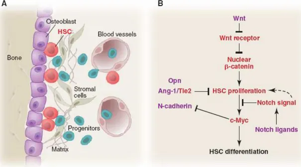
The HSCs represent the best characterized stem cell population, but little is known about the in situ information that define the anatomical and structural relationships of stem cells with their progeny and the microenviromental cells (Moore et al., 2006). The anatomical sites in which this cells reside are located in the bone marrow proximal to the endosteal surface of trabecular bone. (Fig .10 A)
20
Fig 10: Stem cells and their niche in the bone marrow . A) HSCs and their niche cellular component ; B) possible molecular signals that HSCs send and receive for regulating their proliferation and differentiation (Moore et
al., 2006)
Recently two studies indicated that: 1) osteoblastic cells are required for the physically attach of the HSCs in the bone marrow , 2) N-Cadherin/ β Catenin complex between HSCs and Osteoblast has been identified 3) Jagged1,generated from osteoblasts, influences HSCs by signaling through the Notch receptor, 4) the number of osteoblasts controls the number of HSC (Calvi et al., 2003,Zhang et al., 2003)
In vitro studies of HSCs coculture with osteoblasts demonstrated the expansion of HSCs (Taiehman et al.,1998), while a depletion of osteoblasts leads to the loss of hematopoietic tissue (Visnjic et al., 2004). The Osteoblast presence is relevant and play a fundamental role in the BM-HSCs niche even if others type of cells may supply the same function. The contribution of stromal cells or perivascular cells is yet to be defined.
21
N- Cadherin is fundamental for anchored stem cells to the niche and other type of adhesion molecules, like Integrins, are involved (Simmons et al.,
1997) Further, several studies provide direct evidence for the involvement
of matrix components in HSCs regulation (Moore et al., 2006).
About the analysis of the molecular signals, different gene expression studies of HSCs have revealed some signals involved in the regulation of self renewal / differentiation balance (Fig.10B). These components include: WNT/β catenin (Reya et al., 2003), important for the maintenance of self renewal , Notch that maintained HSCs in undifferentiated state (Calvi et al.,
2003, Ducan et al., 2005), Transforming growth factor βeta –BMP (TGF/β-
BMP), for the control in the number of HSCs (Zhang et al., 2003). In addition, a comprehensive genomic analysis of an HSC-supportive microenviromental cells has been performed (Hackney et al., 2002).
2.2. THE EPHITELIAL STEM CELL NICHE IN SKIN
The hair follicle present in the skin is composed by a basal layer or permanent portion that normally give rise to stratified skins layers and a specially zone called the Bulge, in which reside the hair follicle stem cells that are responsible for the regeneration of hair and sebaceous gland (Fig.11A)(Alonso et al., 2003). So, the bulge area is a sort of Niche where epithelial stem cells are located and maintained (Niemann et al.,2002,
Cotsarelis et al., 1990, Sun et al., 1991). This stem cells give rise to
daughter cells that either migrate upward to serve as epidermal progenitors for generating epidermal cells during wound repair or migrate downward to convert to hair-matrix progenitors which give rise to the hair shaft (Niemmann et al., 2002, Oshima et al., 2001, Taylor et al., 2000). In the last years different studies confirmed that the hair follicle stem cells (HFSCs) are quiescent. Multipotentiality of single hair follicle stem cells has been
22
shown by using cells expanded in vitro; it is possible that in situ single cells in the bulge are destined to produce distinct lineages. Freshly isolated cells from bulge have been used as a populations and not as a single cell in transplantation (Morris et al., 2004).Such techniques, or an ability to track the progeny of single HFSCs in situ, will be required to accurately assess the multipotential activity of these cells in normal homeostasis (Moore et
al., 2006). The molecular analysis performed on the hair follicle stem cells
reveled the following features (Li et al., 2005):
1 The expression of adhesion molecules is essential for the interactions between stem cell and niche
2 The presence of growth inhibition factors such as TGFβ/BMP molecules and cell cycle
3 The components of Wnt signaling pathways, including receptors and inhibitors
The expression of several signaling pathways like WNT and BMP have important role in the regulation of HFSCs development and regeneration. The WNT/β catenin systems is important for controls the stem cells activation, fate determination and differentiation while BMP signaling , as opposed to WNT, restricts the activation of stem cells and favors epidermal cell fate (Fig.11 B) (Moore et al., 2006).
23
Fig 11: Stem cells within their niche in the hair follicle. A) cells present in the hair follicle and B) molecular mechanisms that mediate the hair follicle stem cells proliferation ( Moore et al., 2006)
2.3. THE INTESTINAL STEM CELL NICHE
The presence of a stem cell niche, in the intestine reside in the architecture of the epithelial villus-crypt structure based on surrounding perycryptal fibroblasts and mesenchymal cells. (Fig.12 A).The intestinal regeneration begins with intestinal stem cells that give rise to four different type of cells: columnar enterocytes, mucin producing goblet cells, paneth cells and eteroendocrine cells (Bjerknes et al., 1999, Hermiston et al., 1995, Winton
et al, 2000). In every crypt, there are four or six intestinal stem cells that are
located in a ring from the bottom crypt; the activated intestinal stem cells migrate upward to became progenitor or transit amplifying cells and at the top of the crypt, they stop proliferating and differentiate (Moore et al.,
2006). The intestinal stem cells (ISCs) are normally quiescent and their
24
stem cells surround the crypt and during postnatal intestine regeneration, play a important role in directing epithelial cell proliferation, differentiation and apoptosis; for example BMP4 expressed by mesenchymal stem cells is one of the molecular mechanisms purposed for the niche signals (He et al.,
2004). The endothelial cells that composed the vascular vessels provided
the intestinal stem cells survival with different signals like FGF (Paris et
al., 2001), while myofibroblasts, that surrounding epithelial cells, are
proposed to be the candidate “niche” supporting ISCs and influencing other epithelial cells (Millis et al., 2001). Also in the intestinal stem cells niche, some molecular signals have been proposed for regulate the balance of intestinal stem cells between self-renewal and differentiation. (Fig. 12 B).This molecule and gene expression included WNT, BMP, FGF, Notch, Myc, PI3K/Akt (Li et al., 2005). In this complex mechanisms, WNT play a positive role in promoting intestinal stem cells activation/self renewal and the crypt cell fate (Van de Wetering et al.,2002), while BMP restricts this process. (Haramis et al., 2004, He et al., 2004)
25
Fig 12: The intestinal stem cells niche. A) Principal type of cells and their spatial disposition in the crypt niche. B) Molecular signals pathways that mediate proliferation of Intestinal Stem Cells (Moore et al., 2006).
2.4. THE NEURONAL STEM CELL NICHE
Neuronal stem cells have been identified in the 1990 (Alvarez-Buylla et al.,
1990).The well characterized germinal regions in which the neuronal stem
cells reside and support the neurogenesis in the adult brain are two important regions in the hippocampus: the SUBVENTRICULAR ZONE (SVZ) and the SUBGRANULAR ZONE (SGV)(Doetsch et al., 1999, Lois
et al., 1993,Palmer et al.,1997, Temple et el., 2001). In the SVZ there are
different kinds of cells. Ependymal cell layers the lateral ventricle; SVZ astocytes are located adjacent to the ependymal cells. The immature cells derived from SVZ astrocytes are precursors for neuroblasts; this cells have stem cell features: self-renewal and give rise to transient amplifying precursors C cells that give rise to neuroblasts, which than differentiate into neurons. Also the astocytes in the SVZ can generate oligodendrocytes
26
(Doetsch et al., 2003, Mirescu et al., 2003, Temple et al., 2001). In this zone a specialized basal lamina extending from the blood vessels wrapped all types of cells (Fig. 13).
Fig.13: Neuronal stem cells niche in the SVZ zone: B are astocytes, E are ependymal cells, C are amplyfing cells and A are neuroblast. All types of cell are wraped by the basal lamina of the blood vessel (Li et al., 2005).
The SUBGRANULAR ZONE (SGZ) is a zone in the hippocampus between the limbus and the dentate gyrus; in the SGZ the neurogenesis occurs directly with the contribution of blood vessels. The astrocytes act as stem cells able to produce granule neurons (Fig.14) (Doetsch et al., 2003,
Temple et al., 2001).
Fig.14 : The SGZ neuronal stem cell niche: B are astrocytes directly attached to the blood vessels, D are proliferenting cells and G are differentiated cells into granule neurons.(Li et al., 2005)
27
In both zone (SVZ and SGZ), the blood vessel (endothelial cells and basal lamina) are principal components of the niche; these endothelial cells secreted signals and factors essential for control self-renewal and lineage commitment like BMP and their antagonists, WNT and β catenin, Noggin, FGF, IGF, VEGF, TGF α and BDNF (Shen et al., 2004, Doetsch et al.,
2003, Temple et al., 2001).
2.5 THE HEART STEM CELL NICHE
Recently, a new concept of niche was postulated for the heart. Urbanek et al in 2006 demonstrated the presence of a niche in the adult heart of the mice. The author demonstrated that this niche is present particularly in the atria and in the apex of the heart. The Cardiac Niche is constituted by Cardiac stem cells (CSC) and early lineage committed cells that are supported by similar cells found in the bone marrow and in the brain. Homeostasis of cardiac niche is mediated by asymmetric and symmetric division of CSCs. Different internal and external signals like Integrin, Numb, histone H3 , Connexins and Cadherin seem to be involved in the regulation of the niche in the heart (Urbanek et al., 2006) The presence of a stem cells niche in the human heart must be demonstrated yet.
Taken together, the concept of stem cells niche presents in different adult tissue, could give important insights to identification of the stem cells niche in other systems, like in the vascular system, where isn‟t yet identified a complete understood niche. Further, in the concept of the niche, the blood vessels plays a fundamental role, therefore in the adult vessels would be reside stem cells and the other components that created a niche, in which the stem cells could be maintaining in a undifferentiated state and could be involved in the vessels homeostasis and turnover.
28
3. THE BLOOD VESSELS SYSTEM
Blood vessels constitute the first organ in the embryo and form the largest network in the adult. There are three types of vessels - arteries, veins, and capillaries; they are not anatomically the same and they are not just tubes through which the blood flows.Arteries have to expand to accept the blood being forced into them from the heart, and then squeeze this blood on to the veins when the heart relaxes. Arteries have the property of elasticity, meaning that they can expand to accept a volume of blood, then contract and squeeze back to their original size after the pressure is released. It is the elasticity of the arteries that maintains the pressure on the blood when the heart relaxes, and keeps it flowing forward. If the arteries did not have this property, the blood pressure would be more like 120/0, instead of the 120/80 that is more normal. Arteries branch into arterioles as they get smaller. Arterioles eventually become capillaries, which are very thin and branching. It is in the capillaries that the exchange between the blood and the cells of the body takes place. Here the blood releases its oxygen and takes on carbon dioxide, except in the lungs, where the blood picks up oxygen and releases carbon dioxide. As the capillaries begin to thicken and merge, they become venules. Venules eventually become veins and head back to the heart. Veins do not have as many elastic fibers as arteries. Veins do have valves, which keep the blood from pooling and flowing back to the legs under the influence of gravity. When these valves break down, as often happens in older or inactive people, the blood flows back and pool in the legs. The result is varicose veins, which often appear as large purplish tubes in the lower legs. Capillaries are the smallest diameter vessels and the site of exchange of metabolites between blood and tissues. Capillaries consist of a single layer of endothelial cells and their basement membrane. The endothelial cells are joined together by tight junctions. At intervals, these tight junctions are interrupted, leaving small spaces allowing the passage of fluid (Pasqualino,1996).
29
3.1. HYSTOLOGY OF THE BLOOD VESSELS
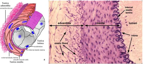
The blood vessels are made of three layers, called from the luminal side outward, the tunica intima, the tunica media and the tunica adventitia and the thickness of these three layers varies greatly depending upon the size and type of vessel (large, medium & small arteries and veins; capillaries).(Fig. 15)(Gallegher, 1992).
Fig. 15: The layers in the artery (intima, media, adventitial layer). a) Schematic representation; b) Hystology ( H&E).
The tunica Intima:
The intima is the inner layer of a vessel. It consists of endothelium (present in all vessels) and subendothelial connective tissue may be present. The subendothelial layer is highly variable, depending on the type of vessel and also grows with age or disease conditions like arteriosclerosis. The endothelium of vessels entering or leaving the heart is continuous with that of the heart. Both connective tissue and smooth muscle are present in the intima. The border of the intima, in the arteries, is delineated by the internal elastic lamina (elastic tissue) that forms the boundary between the intima and the media. Veins do not contain the elastic membrane lining that is found in arteries. In some veins the tunica intima layer also contains valves.
30
The tunica intima increases in thickness with age, and may also become expanded by lipid deposits, so the integrity is critical since damage can lead to atherosclerosis or clotting.
The tunica Media
The tunica media is the middle layer of arteries and veins wall. It is composed of smooth muscle and elastic fibers. This layer is thicker in arteries than in veins. The tunica media is the layer of concentrically-arranged smooth muscle, the autonomic control of which can alter the diameter of the vessel and affect the blood pressure. Smooth muscle cells (in contrast to cardiac and skeletal) have secretory capabilities, and (depending on the vessel), the tunica media contains varying amounts of collagen fibres, elastic fibres, elastic lamellae, and proteoglycans secreted by the smooth muscle cells. The elastic tissue that forms the boundary between the media and the adventitial is called external elastic membrane.
The tunica Adventitia
The tunica adventitia is the strong outer covering of arteries and veins. It is composed of connective tissue as well as collagen and elastic fibres. These fibres allow the arteries and veins to stretch to prevent overexpansion due to the pressure that is exerted on the walls by blood flow. This adventitial connective tissue is usually more or less continuous with the stromal connective tissue of the organ in which the vessel is found. The presence of adventitial connective tissue tightly adhering to vessels facilitates the surgical isolation and repair of vessels. It tends to be much larger in veins than arteries. Adventitia may also contain numerous elastic fibres. In the arteries with a diameter > 1 mm, blood vessels supplying the adventitia and outer media are also present, these are called vasa vasorum ("vessels of the vessels").
31
Arteries carry blood away from the heart. They are classified into three types according to their size: large or elastic arteries; medium (or muscular or distributive) arteries; and small arteries or arterioles, which are less than 0.5 mm in diameter. The aorta is the largest artery in the body and principal artery of the body that carries oxygenated blood to most other arteries in the body. The aorta in humans rises from the left ventricle (lower chamber) of the heart, arches back and downward through the thorax, passes through the diaphragm into the abdomen, and divides into the right and left iliac arteries at about the level of the fourth lumbar vertebra. The aorta gives rise to the coronary arteries, which supply the heart muscle with blood, and to the innominate, subclavian, and carotid arteries, which supply the head and arms. The descending part of the aorta gives rise, in the thorax, to the intercostal arteries that branch in the body wall. In the abdomen it gives off the coeliac artery, which divides into the gastric, hepatic, and splenic arteries, which supply the stomach, liver, and spleen, respectively; the mesenteric arteries to the intestines; the renal arteries to the kidneys; and small branches to the body wall and to reproductive organs. The aorta is subject to a condition known as atherosclerosis, in which fat deposits attach to the aortic walls. If left untreated, this condition may lead to hypertension or to an aneurysm (a swelling of the vessel wall), which can be fatal (Pasqualino, 1996)(Fig.16).
32
3.2. THE VASCULAR WALL AS A SOURCE OF STEM CELLS
During the embryogenesis, the cardiovascular system is the first functioning developed apparatus, which requires coordinated development of the heart, the vessels and the blood and its function is critical for early embryo survival (Copp et al., 1995). First in 1920 Sambin proposed, in the vascular wall of the chick dorsal aorta, the existence of a common progenitors cells , for endothelial cells and hematopoietic cells, called Hemangioblast (Sabin
et al., 1920). When the blood islands are developed in the yolk sac,
haematopoietic and endothelial cells (ECs) are intimately related, so this association has prompted embryologists to assume the existence of a putative common ancestral progenitor. ECs and HSCs shared several markers and several genes like flk1, flt1, tie1, tie2, tal1 and Runx1 (Ema et
al., 2003) .The clearest evidence for this common progenitors came from
Choi and colleagues who found that differentiating embryonic stem cells contain blast-colony forming cells, which are able to generate both primitive hematopoietic and endothelial cells in appropriate conditions in vitro (Choi et al., 1998). In the intraembryonic aorta-gonad-mesonephros region (AGM), hematopoietic precursors can be seen as clumps of cells apparently budding off from the endothelial cells of the ventral wall of the dorsal aorta and the umbilical and vitelline arteries (Ema et al., 2003). Thus, during definitive as well as primitive yolk sac haematopoiesis, there is a close association between endothelial and hematopoietic cells. In the postnatal life, the adult haematopoiesis take place in the bone marrow, where Hematopoietic stem cells reside in a special microenviromental or niche that promotes stem cells renewal and differentiation. In has been showed that CD34+/KDR+ bone marrow cells, in mice and human, are enriched for endothelial precursors (Asahara et al., 1997, Peichev et al.,
2000) and single CD34+/KDR+ cells from adult bone marrow can generate
both endothelial and hematopoietic cells in vitro. This studies raised the possibility of an adult hemangioblasts that might play a key role in normal
33
haematopoiesis and vessels turnover. In the 2002, Minasi et al identified in the quail embryonic dorsal aortas the existence of a self-renewal, multipotent stem cell that expressed emo-angioblasts markers (CD34, C-kit, flk1) and that was able to differentiate in most mesodermal tissue (chondrogenic, osteogenic, adipogenic, skeletal muscle). The authors identified these vessels associated stem cells with the name of the meso-angioblasts, on the assumption that it represents common progenitors for endothelial and other mesodermal cells, that participate in postembryonic development of the mesoderm (Minasi et al., 2002). Yet, the existence of mesangioblasts in adult tissue has not been reported. The process for the blood vessels formation during the early embryogenesis is called Vasculogenesis, i.e. represents the primary differentiation of endothelial cells from undifferentiated precursors cells called angioblasts.New vessels in the adult arise mainly through angiogenesis, a process defined as outgrowth of new vessels from preexisting blood vessels (Carmeliet, 2003). Recent studies indicate that postnatal vasculogenesis may occur indicating a role for circulating (C-EPCs) and/or bone marrow-derived endothelial precursor cells (BM-EPCs), cells with property of embryonal angioblasts, involved in the new blood vessel formation in response to various stimuli (Asahara et al., 1999a, Kalka et al., 2000, Pelosi et al.,2002). Once mobilized in the blood, EPCs are supposed to participate in physiological and pathological arterial wall remodeling during their lifetime (Carmeliet,
2000, 2003), even if the homing and the contribute of EPCs for the
formation of new vessels at the sites of ischemia have been demonstrated (Hristov et al.,2003), up to now the exact role of this cells in this site has been matter of debate (Asahara et al., 1999b, Bagley, et al.,2003,
Carmeliet et al., 2001, Rajantie et al., 2004).
Immature mesenchymal cells with vascular progenitor features have to been shown to reside in pheripheral tissue such as skeletal muscle, where they participate in angiogenesis after injury (Majka et al., 2003). In the last years
34
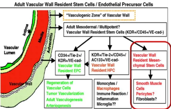
, some experimental evidences suggest that also the vascular wall is much more dynamic than ever before believed. In fact, apart from the unquestionable link with the hematopoietic system, some progenitors seem to be located within the adult vessel wall. Otherwise, the presence of ectopic tissue like cartilage , fat, muscle, bone and vascular tissue in the wall of diseases vessels is a common finding during routine histopathological observations. Several studies also indicated that putative smooth muscle progenitor cells are present within the adult vasculature, and more precisely in the vascular adventitia (Liu et al 2004). In the last years, some experimental artherosclerosis studies demonstrated that the vascular adventitia could be implicated in neointimal proliferation. In 2004, Hu et al identified the presence of stem cell population in the vessel wall. In this study, the author isolated from the adventitia of mouse vessel wall, especially in the aortic root, a population of cells positive for Sca1+, cKit+, CD34+,FLK1+ and negative for the stage-specific embryonic antigen 1 (SSEA-1). In primary culture this cells were heterogeneous, including fibroblast-like, rounded-shaped and adipocyte-like cells. In order to characterize only the adventitial progenitor cells, the cells from primary culture, were sorted for Sca1.The Sca1 sorted cells resulted also C-kit+ and Lin-; they were able to differentiate into smooth muscle cells (SMCs) in vivo and ECs in vitro; they contributed to experimental atherosclerosis in vivo and also they did not originate from the bone marrow. This study also suggested the abundant presence of resident progenitor cells in the aortic root respect others vascular areas demonstrating the embryological role of the aorta.(Hu et al., 2004). In according with this experimental evidence, afterwards it has been demonstrated that postnatal aorta contained an immature subpopulation of vascular progenitor cells, CD34+/ASMA-. These cells expressed markers of early pericyte lineage, had an undifferentiated ultrastructural morphology and differentiated into mural cells in vitro but not into ECs (Howson et al., 2005). Ingram et al in 2005
35
showed that a complete hierarchy of EPCs can be identified also in mature endothelial cells such as human umbilical vein endothelial cells (HUVEC) and human aortic endothelial cells (HAEC) (Ingram et al,2005a). Otherwise, the human fetal aortas contained immature vascular progenitor cells (CD133+,CD34+, KDR+) coexpressing endothelial and myogenic markers in undifferentiated state, where as they can differentiate into ECs and muscular cells in vitro and secrete a complex combination of angiogenic factor that ameliorate ischemic tissue outcome in vivo (Invernici
et al, 2007) This Vascular Progenitor Cells (VPCs) did not express CD45,
indicating that may not arise from hematopoietic system but may derive from resident MSCs located in the niche of the fetal paraortic membrane or at the periphery of aorta wall parenchyma (Alessandri et al., 2001). In 2006, Zengin et al. suggested the existence of a vasculogenic zone in the adult human vascular wall located between the smooth muscle and the adventitial layer that contains vascular wall resident stem cells that could be serve as a reservoir of cells for postnatal vasculogenesis. This vasculogenic zone hypothetically contains cells of different subpopulations: vascular wall resident EPCs, that represents the first evidence of EPCs outside the bone marrow (Zengin et al., 2006), even if Ingram showed that vessel wall derived endothelial cells proliferate because they contain a complete hierarchy of EPCs (Ingram et al., 2005a) This zone may contain multipotent stem cells that are capable of differentiating into macrophage but probably also into hematopoietic progenitor cells (HPCs); theses cells could be a reservoir for inflammatory cells important for local immune response. In this study the author also hyphotized in the vasculogenic zone the presence of multipotent mesodermal stem cells that may serve as a precursors of SMCs, fibroblasts and perycyte (Zengin et al., 2006). According with this, several experimental observations shown that postnatal artery walls contain MSCs with multilineage differentiation. In 2003, Tintut et al described a subpopulation of vascular cells, isolated from bovine aortic
36
medial cells, called “Calcifying vascular cells”(CVC) that are able to differentiate in vitro along different mesenchymal lineages, e.g osteogenic, chondrogenic, leiomyogenic and stromogenic lineages, where as the adipogenic potential of this cells was limited. CVCs, at flow cytometry analysis, were positive for surface markers commonly expressed by marrow-derived mesenchymal stem cells (positive for CD44 and CD29 , and negative for hematopoietic markers like CD14 and CD45). Otherwise, CVCs replicated for 20-25 passages with retention of multipotentiality. Since these cells did not differentiate toward adipogenyc lineage, they could represent a stage of commitment generation below the mesenchymal stem cells in the mesengenic hierarchy (Tintut et al., 2003). In another elegant study, Sainz et al in 2006 defined the possibility to isolate a “Side Population” (SP) of constitutively resident progenitor cells from healthy arteries of adult mice. In this study, living SP was found only in the tunica media layer of thoracic and abdominal aortas and their immunophenotype was Sca1+/Ckit -/low Lin- with low CD34 and flk1 expression. Arterial SP cells did not express SMCs or ECs markers at the basal state but, in the presence of vascular endothelial growth factor (VEGF), differentiated into endothelial cells phenotype and when cultured on Matrigel showed vasculogenic potential, while, in the presence of TGF beta 1 or PDGF-BB, became more similar to SMCs phenotype. Arterial SP cells were unable to generate hematopoietic colonies on Methylcellulose, indicating no capacity of this cells to differentiate toward neither the myeloid lineage not the lymphoid lineage. This cells could participate in arterial homeostasis and remodeling (Sainz et al., 2006). Furthermore MSCs isolated from the human saphena vein are able to differentiate in vitro into osteoblasts, chondrocytes and adipocytes (Covas et al., 2005). Recent observations demonstrated that human vascular adventitia ( pulmonary arteries) contains mesenchymal stem/ progenitors cells ( Vimentina+, Cd29+, Cd44+, Cd105+ ,CD34-,Cd133-,CD14-,CD45-) with the capacity to differentiate in
37
vitro into adypocyes, osteoblsts, and myofibroblasts. (Hoshino et al., 2008). Taken together these experimental evidences suggest the presence of resident stem cells ( multipotent or a mixture of committed progenitors cells) within the postnatal arterial wall, which are involved in the postnatal vasculogenis and in the formation of ectopic tissue. Adult stem cells in the vessels seem to be located in the vasculogenic zone between the media and the adventitial layer. Even so, the definition of a functional microenvironment (niche) in the human postnatal vessels, where stem cell related to mouse and chicken mesoangioblast can be maintained in a quiescent state, must be determinate.
3.3 ANGIOGENESIS
Vessels formation occurs mainly through two mechanisms: Vasculogenesis and Angiogenesis. Vasculogenesis represents de novo formation of blood vessels during embryonic development. Mesoderm-derived stem cells (hemangioblasts) form aggregates (blood islands), and they develop into primitive hematopoietic and endothelial cells (angioblasts). The angioblasts, undifferentiated precursor cells, proliferated and differentiated in situ into endothelial cells to form a vascular labyrinth (Carmeliet, 2000). In postnatal life adult hemangioblasts seemed to be present even if their location and characterization must be determinate. On the other hand, the formation of new vessels from preexisting vessels is called angiogenesis. New vessels in the adult arise mainly through angiogenesis, although vasculogenesis also may occur. Angiogenesis is a tightly regulated process required for a number of physiological process like wound healing, ovulation and menstruation as well as embryonic development. Excessive angiogenesis is seen in a wide range of diseases including tumors, inflammatory diseases and diabetic retinopathy (Kiumura et al., 2003). Angiogenesis is a very complex mechanism that include different steps. Angiogenesis initiates with vasodilation, a process involving nitric oxide. This increase in permeability
38
lead to a preexisting vessel destabilization and loss of the endothelial cell interactions. Different angiogenic growth factors are released. These molecules lead to matrix metallo proteinase activation, crucial for extracellular matrix degradation. After this, endothelial cells proliferate and migrate to assemble in a solid cords that subsequently acquire a lumen. Subsequently, pericytes and SMCs migrate in order to reorganize the cellular interactions and surround the new formed vessels. Angiogenic sprouting is controlled by a balance of activators and inhibitors. Important activator factors are VEGF (vascular endothelial growth factor), FGF (fibroblast growth factor), TGF-β (transforming growth factor β), IL-8 (interleukin 8) and Angiopoietin-I. Inhibitors factors are Interferon α, β, γ, IL-12 (interleukin 12), Angiostatin and Endostatin (Carmeliet, 2000).
3.3.1 VASCULAR ENDOTHELIAL GROWTH FACTOR
The family of VEGF includes VEGF-A, B, C, D and placenta growth factor. VEGF (denoted as VEGF-A) was initially named vascular permeability factor for its ability to induce vascular permeability. Later this vascular endothelial specific mitogen was named VEGF for its ability to promote proliferation of endothelial cells. VEGF play a key role during angiogenesis process as activator. VEGF induces endothelial cells proliferation, migration differentiation and survival. Knock-out experiments have demonstrated that heterozygous VEGF-A knout-out mice were embryologically lethal and that blood vessels formation was dramatically impaired (Ferrara, 1996). The main receptors which seem to initiate signal transduction cascades in response to VEGF binding consist of three kinds of tyrosine kinases : VEGF-R1 or Flt1 (fms-like tyrosine-kinase 1), VEGF-R2 or KDR (kinase-insert domain receptor) and VEGF-R3 or Flt3(fms-like tyrosine-kinase 3). Among them, KDR may mediate the major action on cell growth and permeability. (Gale et al., 1999). Different factors have