Short-Term Treatment With Atorvastatin Reduces Platelet
CD40 Ligand and Thrombin Generation in
Hypercholesterolemic Patients
Valerio Sanguigni, MD; Pasquale Pignatelli, MD; Luisa Lenti, MD; Domenico Ferro, MD; Alfonso Bellia, MD;
Roberto Carnevale, PhD; Manfredi Tesauro, MD; Roberto Sorge, PhD; Renato Lauro, MD; Francesco Violi, MD
Background—Soluble CD40L (sCD40L), a substance that maximally reflects in vivo platelet activation, is increased inpatients with hypercholesterolemia. We investigated the relation between sCD40L and platelet CD4OL in hypercho-lesterolemic patients before and after a short-term treatment with atorvastatin.
Methods and Results—Collagen-induced platelet CD40L and plasma levels of sCD40L and prothrombin fragment F1⫹2, a marker
of thrombin generation, were investigated in 30 hypercholesterolemic patients and 20 healthy subjects. Hypercholesterolemic patients were then randomized to either diet (n⫽15; group A) or atorvastatin 10 mg/d (group B); the aforementioned variables were measured at baseline and after 3 days of treatment. Compared with referents, hypercholesterolemic patients showed higher values of platelet CD40L (P⬍0.005), sCD40L (P⬍0.005), and F1⫹2 (P⬍0.003). Platelet CD40L was significantly correlated with sCD40L (P⬍0.001), and the latter was significantly correlated with F1⫹2 (P⬍0.001). The intervention trial showed no changes in group A but a significant decrease in platelet CD40L (P⬍0.01), sCD40L (P⬍0.002), and F1⫹2 (P⬍0.03) in group B. In vitro studies demonstrated that cholesterol enhanced platelet CD40L and CD40L-mediated clotting activation by human monocytes; also, atorvastatin dose-dependently inhibited platelet CD40L expression and clotting activation by CD40L-stimulated monocytes.
Conclusions—This study shows that, in hypercholesterolemia, platelet overexpression of CD40L may account for enhanced plasma
levels of sCD40L and F1⫹2. Atorvastatin exerts a direct antithrombotic effect via inhibition of platelet CD40L and CD40L-mediated thrombin generation, independently of its cholesterol-lowering effect. (Circulation. 2005;111:412-419.)
Key Words: platelet-derived factors 䡲 platelets 䡲 thrombin 䡲 hypercholesterolemia 䡲 statins
C
D40 ligand (CD40L), a member of the tumor necrosis factor family, is a transmembrane protein found on cells of the immune system as well as on endothelial cells, smooth muscle cells, macrophages, and platelets.1On interaction with its receptor CD40,CD40L exerts proinflammatory and prothrombotic activity, includ-ing increased expression of matrix metalloproteinases, chemokines, cytokines, and tissue factor (TF).1 The role of CD40L in the
pathogenesis of atherosclerosis has been confirmed by the fact that administration to hyperlipidemic mice of an antibody against CD40L reduced atherosclerotic lesions.2CD40L is expressed by
platelets on stimulation with common agonists3; it is then cleaved
from platelets over a period of minutes to hours, thus generating a soluble form (sCD40L).4 It has been calculated that ⬎95% of
circulating sCD40L is of platelet origin.5Elevated plasma levels of
sCD40L have been found in patients with acute coronary syndrome and in those at risk for cardiovascular events.6 – 8 There are also
converging lines of evidence that patients with hypercholesterol-emia show enhanced levels of sCD40L, suggesting that this protein may represent an important promoter of atherosclerotic complica-tions occurring in this setting.9Because sCD40L stems essentially
from platelet CD40L, patients with hypercholesterolemia could be expected to show an upregulation of platelet CD40L; however, there are no clear data in this respect, because both upregulation and downregulation of platelet CD40L have been reported.9 Patients
with hypercholesterolemia are in an ongoing prothrombotic state, as documented by enhanced TF expression and in vivo thrombin generation.10Because CD40L exerts a prothrombotic effect through
overexpression of TF,11it would be reasonable to speculate that in
hypercholesterolemia, clotting activation occurs as a consequence of CD40L overexpression. A significant correlation between sCD40L and prothrombin fragment F1⫹212 could support this
hypothesis, although sCD40L has not been demonstrated to exert any prothrombotic action to date.13
Recent studies have also sought to determine whether statins could influence CD40L expression in patients with hypercholester-olemia: although there are converging data on the efficacy of statins in reducing sCD40L,9,14,15no change in platelet CD40L expression
was reported.9,12Also, a previous study suggested that statins could
reduce sCD40L plasma levels, independently of their cholesterol-lowering effect.15
Received June 18, 2004; revision received October 16, 2004; accepted November 18, 2004.
From the Department of Experimental Medicine and Pathology (P.P., L.L., D.F., R.C., F.V.), University of Rome “La Sapienza,” and the Department of Internal Medicine (V.S., A.B., M.T., R.S., R.L.), University of Rome “Tor Vergata,” Rome, Italy.
Correspondence to Francesco Violi, MD, Dipartimento di Medicina Sperimentale e Patologia, Università di Roma La Sapienza, Policlinico Umberto I, 00185, Rome, Italy. E-mail [email protected]
© 2005 American Heart Association, Inc.
Circulation is available at http://www.circulationaha.org DOI: 10.1161/01.CIR.0000153810.81187.7D 412
In the present study, which was conducted in patients with polygenic hypercholesterolemia, we investigated the following ele-ments: (1) CD40L expression in stimulated platelets; (2) the relation between sCD40L and clotting activation; and (3) the effect of a 3-day treatment with atorvastatin on the aforementioned variables compared with diet alone. Finally, we performed in vitro studies to evaluate (1) the effect of native LDL cholesterol on platelet CD40L expression; (2) the effect of native LDL cholesterol on CD40L-mediated clotting activation by monocytes; and (3) whether atorva-statin was able to influence the aforementioned parameters.
Methods
Clinical Study
The clinical study was divided into 2 parts. In the first part of the study, we compared 30 patients with polygenic hypercholesterolemia (16 males, 14 females; mean age, 53.4 years) and 20 sex- and age-matched normocholesterolemic subjects (10 males and 10 females; mean age, 52.6 years). Both patients and controls were recruited from the same geographic area and followed a typical Mediterranean diet. None of the patients had clinical evidence of cardiovascular disease (as shown by clinical history, physical examination, or ECG), diabetes mellitus, or hypertension. Patients with hypercholesterolemia had not taken any lipid-lowering agents or antiplatelet drugs in the previous 30 days. Lipid profile, CD40 ligand expression by platelets, sCD40L, and F1⫹2 were measured.
In the second part of the study, we tested the hypothesis that atorvastatin could influence platelet CD40L expression and CD40L-mediated clotting activation by a mechanism independent of its cholesterol-lowering effect; thus, only hypercholesterolemic patients were randomized to either a diet (American Heart Association step I diet; n⫽15, 8 males, 7 females) or atorvastatin 10 mg/d (n⫽15, 8 males, 7 females) for 3 days. Lipid profile, CD40 ligand platelet expression, sCD40L, and F1⫹2 were measured at baseline and after 3 days of treatment.
Blood samples mixed with 0.13 mol/L sodium citrate (ratio 9:1, vol/vol) were obtained between 8 and 9AMfrom patients and healthy volunteers who had fasted for 12 hours and had provided their informed consent to participate in the study; an aliquot of serum was used to measure lipid profile. The ethics committee of our university approved the study protocol.
Lipid Profile
Serum levels of total cholesterol and triglycerides were determined with an enzyme-based method. HDL cholesterol was measured after phosphotungstic acid/MgCl2 precipitation of fresh plasma. LDL
cholesterol was calculated according to the Friedewald formula.
Analysis of sCD40L and F1ⴙ2
Blood samples were immediately centrifuged at 2000 rpm for 20 minutes at 4°C, and the supernatant was collected and stored at ⫺80°C until measurement. Plasma levels of sCD40 ligand were measured with a commercial immunoassay (Quantikine CD40 li-gand, R&D Systems). Intra-assay and interassay coefficients of variation were 7% and 9%, respectively. Plasma levels of human prothrombin fragment F1⫹2 were assayed by an enzyme immuno-assay based on the sandwich principle (Enzygnost F1⫹2, Behring-werke). Intra-assay and interassay coefficients of variation for this method were 5% and 7%, respectively.
Platelet Isolation From Whole Blood
Blood samples were drawn between 8 and 9AMwithout stasis from an antecubital vein with a 21-gauge needle from patients after a 12-hour fast and mixed with 0.13 mol/L sodium citrate (ratio 9:1, vol/vol). Washed platelets were prepared as previously described.16
Flow Cytometric Analysis of CD40L Expression
CD40L expression on platelet membranes was analyzed with spe-cific fluorescein isothiocyanate–labeled monoclonal antibodies (mAbs; anti-CD40L antibody by Beckman Coulter). In all assays, an irrelevant isotype-matched antibody was used as a negative control.
Twenty microliters of mAb was added to 200L of platelet suspension (2⫻108/mL) previously fixed with (2%) paraformaldehyde (0.1% bovine
serum albumin) and incubated for 60 minutes at 4°C. The unbound mAb was removed by addition of 0.1% bovine serum albumin–phosphate-buffered saline (PBS) and centrifugation at 5000g for 3 minutes (twice). Fluorescence intensity was analyzed on an Epics XL-MCL cytometer (Coulter Electronics) equipped with an argon laser at 488 nm. For every histogram, 50 000 platelets were counted to determine the proportion of positive platelets. Antibody reactivity is reported in arbitrary units obtained by multiplying the number of positive events resulting from platelet stimulation by the mean value of the fluorescence observed when the specific mAb was used and then correcting the values obtained in unstimu-lated samples treated with the same antibody.
In Vitro Study
Platelet CD40L Expression
Platelets from healthy subjects (n⫽4, 2 males, 2 females; mean age, 50.3 years) and hypercholesterolemic patients (n⫽4, 2 males, 2 females; mean age, 52.2 years) were washed and suspended in fatty acid–free Tyrode’s buffer (2⫻108
/mL). Platelet suspensions were incubated with collagen (4g/mL) for 15 minutes at 37°C with and without native LDL cholesterol (100g/mL) or scalar concentra-tions (0.1 to 10mol/L) of atorvastatin and treated as described earlier to detect CD40L expression on platelet surfaces. Atorvastatin alone did not affect platelet viability, as assessed by the ipotonic shock response test17(not shown).
Monocyte TF Expression and Monocyte-Mediated Thrombin Generation
Peripheral blood mononuclear cells (PBMCs) were isolated from heparinized, venous blood samples of healthy subjects (n⫽4, 2 males, 2 females; mean age, 50.3 years) and hypercholesterolemic patients (n⫽4, 2 males, 2 females; mean age, 52.2 years) by aseptic technique. Platelets were separated by centrifugation, once at 140g and twice at 100g, in PBS at room temperature for 10 minutes. PBMCs were isolated by centrifugation on Lymphoprep (Nyeegard Oslo) at 1200g for 20 minutes at 20°C. Monocytes, identified by May-Grunwald–Giemsa staining, were between 16% and 22% (mean, 19%).
Monocytes (adherent cells) were obtained by incubation of PBMCs for 90 minutes at 37°C in a humidified atmosphere of 5% CO2in air in Petri
dishes containing RPMI 1640, supplemented with 2 mmol/L glutamine; lymphocytes (nonadherent cells) were removed by aspiration with a Pasteur pipette and washing of the dishes with warm medium. The purified monocyte preparation contained 85% to 95% monocytes.
After isolation, cells were washed twice in PBS and preincubated for 1 hour at 2⫻105cells/mL in RPMI 1640 at 37°C 5% CO
2with atorvastatin
(0.1,1 and 10mol/L) or medium (as control) and then incubated with 50 ng/mL CD40L (Recombinant human CD40L/TNFSF5, R&D Systems) for 6 hours. In another set of experiments, monocytes were preincubated for 12 hours at 2⫻105
cells/mL in RPMI 1640 at 37°C under 5% CO2with native
LDL (100g/mL)18or medium (as control) and then incubated with 50
ng/mL CD40L for 6 hours. At the end of the incubation period, the cells and media were separated by centrifugation (2000g⫻15 minutes). The cells were washed with Tris-NaCl buffer (0.1 mmol/L NaCl, 0.1% bovine serum albumin, pH 7.4) and then lysed in the same buffer by adding 15 mmol/L
n-octyl--D-glycopyranoside at 37 C° for 30 minutes. Cell count and trypan blue exclusion were performed on cell suspensions after washing to evaluate cell viability. Cell viability was 90% at the maximum concentra-tion of atorvastatin used (10mol/L). The ELISA for measuring TF antigen in cell lysate was performed with a commercial kit (Imubind tissue factor ELISA kit, American Diagnostica). The lower detection limit is⬇10 pg/mL. The assay recognizes TF-apo, TF, and TF–factor VII complexes and is designed in such a manner as to prevent any interference from other coagulation factors or inhibitors of procoagulant activity.
The rate of thrombin generation by CD40L-stimulated monocytes was evaluated in vitro as previously described.19For this purpose, a
low-molecular-weight heparin–anticoagulated blood sample (ratio 1:10, vol/vol) was taken from healthy volunteers (n⫽4) and hypercholesterolemic patients (n⫽4). The blood was anticoagulated with low-molecular-weight heparin
(820 U/mL) because this agent effectively inhibits the formation of thrombin in solution but has only a small effect on thrombin generated at and bound to surfaces. After isolation, monocytes were washed twice in PBS and preincubated for 1 hour at 2⫻105cells/mL in RPMI 1640 at 37°C
under 5% CO2with atorvastatin (0.1, 1, and 10mol/L) or medium (as
control) and then incubated with CD40L (50 ng/mL) for 6 hours. In another set of experiments, monocytes were preincubated for 12 hours at 2⫻105
cells/mL in RPMI 1640 at 37°C under 5% CO2with native LDL (100
g/mL) or medium (as control) and then incubated with 50 ng/mL CD40L for 6 hours. The medium was removed, and a 1000-mL overlay of heparinized standard plasma was added to each well and incubated at 37°C for 24 hours. After incubation, samples were harvested, centrifuged at 4000g, and assayed for F1⫹2 generation, calculated as the increase in F1⫹2 levels compared with the value observed in control samples, which consisted of heparinized plasma added with CD40L. All samples were assayed in duplicate.
Statistical Analysis
Assuming that a short-term (3-day) treatment with atorvastatin would reduce platelet CD40L expression and monocyte tissue factor by 25%, we postulated that the study sample should consist of at least 12 patients for each group (␣⫽0.05 and 1⫺⫽0.90).
Comparisons between groups were carried out by 1-way and repeated-measures ANOVA20and were replicated as appropriate with nonparametric
tests, like the Wilcoxon and Kolmogorov-Smirnov (z) test in case of nonhomogeneous variances as verified by Levene’s test. MANOVA with a Bonferroni test for multiple comparisons was applied in in vitro experi-ments. The correlation analysis was carried out by Pearson’s test. Data are presented as mean⫾SD. Statistical significance was defined at P⬍0.05. Statistical analysis was performed with SPSS 10.0 software for Windows.
Results
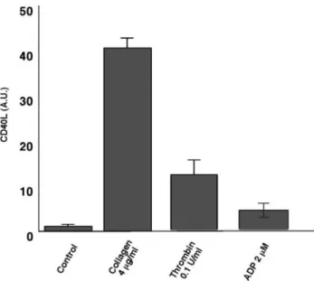
Platelet CD40L, Expression, and Plasma F1ⴙ2 in Healthy Subjects and Hypercholesterolemic Patients To choose the platelet agonist that maximally expressed platelet CD40L, we evaluated the effect of 3 different agonists, namely, ADP, collagen, and thrombin. Because the highest expression was obtained with collagen (Figure 1), all of the experiments in hyper-cholesterolemic patients were performed with collagen as the platelet agonist.
Table 1 shows the characteristics of patients and referents. Compared with referents (n⫽20), patients with hypercholesterol-emia (n⫽30) showed higher expression of platelet CD40L (43.9⫾13.4; 95% confidence interval [CI], 38.8 to 48.9 versus 30.1⫾5.6; 95% CI, 27.6 to 32.5 AU; P⬍0.005; Figure 2A); 11 hypercholesterolemic patients had values⬎41.3 AU (mean⫹2SD) versus 1 referent. Compared with referents, patients with hypercho-lesterolemia showed higher plasma levels of sCD40L (4.3⫾1.9; 95% CI, 3.5 to 5 versus 2.2⫾0.7; 95% CI, 1.8 to 2.5 ng/mL;
P⬍0.005; Figure 2B); 19 hypercholesterolemic patients had values
⬎3.6 ng/mL (mean⫹2SD) versus 1 referent. Compared with referents, patients with hypercholesterolemia showed higher plasma levels of F1⫹2 (2⫾0.9; 95% CI, 1.6 to 2.3 versus 1.12⫾0.49; 95% Figure 1. Agonist-dependent platelet CD40L expression in
healthy subjects (n⫽4, 2 males, 2 females; mean age, 50 years). Results are mean⫾SD. Abbreviations are as defined in text.
TABLE 1. Characteristics of Study Participants
Hypercholesterolemic Patients Healthy Subjects Age, y 53⫾2 52⫾3 Sex 16 males, 14 females 10 males, 10 females
Smokers, n 3 2
LDL cholesterol, mg/dL 186⫾13 112⫾13 Triglycerides, mg/dL 88⫾18 84⫾16
Figure 2. Platelet CD40L expression (a), SCD40L (b), and
pro-thrombin F1⫹2 (c) plasma levels in hypercholesterolemic patients (HC, n⫽30) and healthy subjects (HS, n⫽20). Repeated-measures ANOVA: P⬍0.001, HC vs HS; **P⬍0.005 HC vs HS. Results are mean⫾SD. Box plots depict median and 95% CIs; whiskers represent minimum and maximum values. Abbreviations are as defined in text.
CI, 0.8 to 1.3 nmol/L; P⬍0.003; Figure 2C); 13 hypercholesterol-emic patients had values of⬎2.1 nmol/L (mean⫹2SD) versus 0 referents. Platelet CD40L expression was significantly correlated with sCD40L (r⫽0.87, P⬍0.001) and F1⫹2 (r⫽0.75, P⬍0.001), and sCD40L was significantly correlated with F1⫹2 (r⫽0.86,
P⬍0.001). Platelet CD40L was also significantly correlated with
LDL cholesterol (r⫽0.58, P⬍0.05). In healthy subjects, platelet CD40L expression was significantly correlated with sCD40L (r⫽0.79, P⬍0.001) and F1⫹2 (r⫽0.58, P⬍0.01), and sCD40L was significantly correlated with F1⫹2 (r⫽0.67, P⬍0.001). Platelet CD40L was also significantly correlated with LDL cholesterol (r⫽0.73, P⬍0.001). In hypercholesterolemic patients, platelet CD40L expression was significantly correlated with sCD40L (r⫽0.82, P⬍0.001) and F1⫹2 (r⫽0.68, P⬍0.001), and sCD40L was significantly correlated with F1⫹2 (r⫽0.85, P⬍0.001). Platelet CD40L was also significantly correlated with LDL cholesterol (r⫽0.82, P⬍0.001).
Effect of Atorvastatin on Platelet CD40L and F1ⴙ2 in Hypercholesterolemic Patients
At baseline, patients randomized to diet alone (group A) and those randomized to diet plus atorvastatin (10 mg/d, group B) had similar values of platelet CD40L, sCD40L, and F1⫹2. Both groups did not show any changes in lipid profile after 3 days of treatment (Table 2). In group A (n⫽15), no changes in platelet CD40L (44.5⫾12.3; 95% CI, 37.7 to 51.4 versus 43.3⫾14.8; 95% CI, 35 to 51.4 AU) sCD40L (4.2⫾2; 95% CI, 3.1 to 5.3 versus 4.3⫾2; 95% CI, 3.2 to 5.4 ng/mL), and F1⫹2 (2⫾1; 95% CI, 1.4 to 2.6 versus 1.9⫾0.7, 95% CI, 1.5 to 2.4 nmol/L) were observed (Figure 3). In group B (n⫽15), a significant decrease in platelet CD40L (46.3⫾14.6; 95% CI, 38.2 to 54.5 versus 32.2⫾6.4; 95% CI, 28.6 to 35.8 AU;
P⬍0.01), sCD40L (4.1⫾1.9; 95% CI, 3 to 5.2 versus 3⫾1; 95% CI,
3 to 5.2 ng/mL; P⬍0.002), and F1⫹2 (2⫾1.1; 95% CI, 1.4 to 2.7 versus 1.4⫾0.4; 95% CI, 1.2 to 1.6 nmol/L; P⬍0.03) was observed within and between groups (A and B; Figure 3) after 3 days of atorvastatin. In group B, before-after treatment changes in platelet CD40L and sCD40L (r⫽0.66, P⬍0.01), sCD40L and F1⫹2 (r⫽0.87, P⬍0.001), and CD40L and F1⫹2 (r⫽0.55, P⬍0.05) were significantly correlated.
In Vitro Study
Effect of Cholesterol on Platelet CD40L
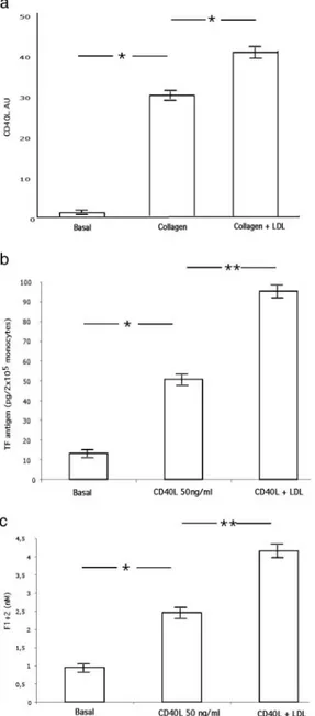
Compared with unstimulated platelets, collagen induced a signifi-cant increase in CD40L expression; a further increase was observed when collagen-stimulated platelets were added with native LDL cholesterol (100g/mL; MANOVA, P⬍0.001; Figure 4A). No
change in CD40L was observed in platelets treated with native LDL cholesterol alone (not shown).
Effect of Cholesterol on CD40L-Induced Clotting Activation Compared with unstimulated monocytes, CD40L-stimulated mono-cytes showed a significant increase in TF and F1⫹2; further increases in monocyte TF (MANOVA, P⬍0.005; Figure 4B) and F1⫹2 (MANOVA, P⬍0.005; Figure 4C) were observed when monocytes were added with native LDL cholesterol (100g/mL). No change in clotting activation was observed in monocytes added with native LDL cholesterol alone (not shown).
TABLE 2. Lipid Profile Before and After 3 Days of Treatment
Diet Only Atorvastatin LDL cholesterol, mg/dL Before 185⫾14 187⫾12 After 185⫾16 182⫾13 Triglycerides, mg/dL Before 88⫾22 87⫾14 After 88⫾18 86⫾17
Figure 3. Platelet CD40L expression, sCD40L, and prothrombin
F1⫹2 plasma levels in group A (diet only, n⫽15) and group B (atorvastatin 10 mg/d, n⫽15) patients before (A) and after (B) 3 days of either treatment. ●Repeated-measures ANOVA: CD40L, * P⬍0.01: group B, before vs after 3 days; group B vs group A after 3 days; sCD40L and F1⫹2, ** P⬍0.05: group B, before vs after 3 days; group B vs group A after 3 days. ●Results are given as mean⫾SD. ●Box plots depict median and 95% CIs; whiskers represent minimum and maximum values. Abbrevia-tions are as defined in text.
Effect of Atorvastatin on Platelet CD40L
Because these data suggested that atorvastatin reduced CD40L by a mechanism that was independent of its cholesterol-lowering effect, we investigated whether it had a direct influence on platelet CD40L expression. Incubation of platelets from healthy subjects or hyper-cholesterolemic patients with atorvastatin showed a significant decrease in platelet CD40L expression (MANOVA, P⬍0.005; Figure 5A and 5B). This effect was dependent on the concentration of atorvastatin in both patiens and controls, as demonstrated by comparison of dose-response curves for hypercholesterolemic pa-tients (r⫽0.82, P⬍0.001) and healthy control subjects (r⫽0.92,
P⬎0.001; Figure 5C).
Effect of Atorvastatin on CD40L-Induced Clotting Activation Finally, we investigated in vitro whether atorvastatin was able to influence the prothrombotic effect of CD40L in a
thrombin-generating system. This study showed that incubation of human monocytes with CD40L induced a significant increase in F1⫹2 compared with unstimulated monocytes in both healthy subjects and hypercholesterolemic patients (MANOVA, P⬍0.005); this effect, however, was more marked in patients than in controls, as documented by the higher rate of thrombin generation in hypercho-lesterolemic patients (Figure 6A). Similar findings were obtained when TF expression by unstimulated and CD40L-stimulated mono-cytes was investigated (MANOVA, P⬍0.005; Figure 6B). Incuba-tion of monocytes with atorvastatin dose-dependently inhibited the rate of thrombin generation (Figure 7A and 7B) and TF expression Figure 4. Effect of native LDL cholesterol (100g/mL) on
CD40L platelet expression in collagen-stimulated platelets (a) and on TF (b) and F1⫹2 (c) in CD40L-stimulated monocytes (n⫽4 healthy subjects, 2 males, 2 females; mean age, 50.3 years). MANOVA, *P⬍0.001, **P⬍0.005. Results are mean⫾SD.
Figure 5. CD40L expression by collagen-stimulated platelets (A)
and collagen-stimulated platelets together with 0.1mol/L (B), 1 mol/L (C), or 10 mol/L (D) atorvastatin in healthy subjects (HS; n⫽4, 2 males, 2 females; mean age, 50 years) (a) and hypercholesterolemic patients (HC; n⫽4, 2 males, 2 females; mean age, 53 years) (b). c, Dose-response curves for HC vs HS subjects. MANOVA with Bonferroni test: *P⬍0.001, B vs A; **P⬍0.005, C vs A and D vs A. Results are mean⫾SD. Abbrevi-ations are as defined in text.
(Figure 7D and 7E) by monocytes (MANOVA, P⬍0.05). This effect was demonstrated in both patients and controls, as demon-strated by comparison of dose-response curves of hypercholester-olemic and healthy subjects (Figure 7C and 7F; for TF and F1⫹2,
r⫽0.97, P⬍0.001, and r⫽0.87, P⬍0.001, respectively).
Discussion
This study provides the first evidence that in hypercholesterolemia, platelet CD40L is overexpressed and is significantly correlated with sCD40L, thus suggesting that enhanced levels of sCD40L are likely to reflect platelet CD40L upregulation.
Cholesterol, CD40L, and Clotting Activation The significant correlation between serum cholesterol and platelet CD40L suggests a role for cholesterol in enhancing platelet CD40L overexpression. We also sought to investigate whether CD40L has an influence on clotting system activation. The experiments
con-ducted in monocytes from healthy subjects and hypercholesterol-emic patients demonstrated that CD40L, at a concentration close to that observed in the peripheral circulation, was able to increase TF and prothrombin fragment F1⫹2, with more marked effect in patients with hypercholesterolemia.
Because these findings suggested a role for cholesterol in en-hancing platelet CD40L expression and clotting activation, we performed further experiments to support this hypothesis. Our study provides the first evidence that cholesterol is implicated in the overexpression of platelet CD40L elicited by agonist; also, we demonstrated that CD40L-induced clotting activation is amplified in monocytes coincubated with cholesterol. Both findings provide a novel insight into the atherothrombotic role of cholesterol and warrant further studies to identify the mechanism accounting for such effects.
Atorvastatin and Platelet CD40L
Because CD40L has a key role in the initiation and progression of atherosclerosis, we investigated whether statins, that are potent inhibitors of atherosclerotic progression.21–23could influence
plate-let CD40L expression. Although our data suggest that cholesterol plays a major role in enhancing platelet CD40L expression, it is also possible that statins influence CD40L expression independently of their principal mechanism of action. In one study, Semb et al15
randomized 100 patients with familiar hypercholesterolemia to either atorvastatin or simvastatin and demonstrated a significant decrease in sCD40L that was independent of the cholesterol-lowering effect of these agents. To further investigate this issue, we studied platelet CD40L and sCD40L before and immediately after 3 days of atorvastatin treatment; using the same approach, we had previously found no changes in the lipid profile of hypercholester-olemic patients treated with a statin.24Compared with diet-assigned
patients, atorvastatin-treated patients demonstrated a significant and parallel decrease of platelet CD40L and sCD40L, suggesting that atorvastatin inhibits sCD40L by downregulating platelet CD40L through a mechanism that is independent of its cholesterol-lowering effect. This hypothesis was supported by an in vitro study, which demonstrated that atorvastatin dose-dependently inhibited collagen-induced CD40L expression; it should be noted that inhibition of platelet CD40L expression was observed at atorvastatin concentra-tions as low as 0.1mol/L, which may be achieved in the peripheral circulation of subjects treated with 5 to 20 mg/d atorvastatin.25
Atorvastatin and Clotting Activation
Another novel finding of the present study was an early reduction in the levels of prothrombin fragment F1⫹2, suggesting that atorva-statin has an inhibitory effect on activation of the coagulation cascade. At least 2 mechanisms may account for this finding. One mechanism may be related to atorvastatin-induced CD40L down-regulation, which could be responsible for reduced TF expression and perhaps a lower thrombin generation rate. Another mechanism may involve direct interference of the drug with the intracellular signaling responsible for TF expression. This hypothesis is in accordance with previous studies showing that statins inhibit nu-clear factor NF-B, which has a key role in TF expression by monocytes26 –28; however, this hypothesis should be wisely
consid-ered because inhibition of clotting activation was achieved with relatively high concentrations (1mol/L) of atorvastatin. Figure 6. Prothrombin F1⫹2 (a) and TF expression (b) by
unstimulated monocytes (A and C) and monocytes stimulated with CD40L (50 ng/mL; B and D) in healthy subjects (HS; n⫽4, 2 males, 2 females; mean age, 50 years) and hypercholesterol-emic patients (HC; n⫽4, 2 males, 2 females; mean age, 53 years). MANOVA: *P⬍0.003, B vs A; **P⬍0.001, D vs C; ***P⬍0.005, D vs B. Results are mean⫾SD. Abbreviations are as defined in text.
Atorvastatin and Coronary Artery Disease
The effect observed on platelet CD40L and F1⫹2 as early as after 3 days of treatment suggests that atorvastatin has an immediate antiinflammatory and antithrombotic action that may prove useful in cases of plaque instability. In fact, inflammation with the ensuing activation of platelets and the coagulation cascade is a typical feature of vulnerable plaque and triggers coronary thrombus growth. So far, statins have been mainly used in patients with stable coronary syndromes or in those at risk of cardiovascular disease to retard atherosclerotic progression. Atorvastatin’s early antiinflam-matory and antithrombotic effect could suggest the use of statins even in patients with acute coronary syndromes; in these patients, timely halting of plaque instability could retard thrombus growth and perhaps have a favorable effect on clinical outcome. This concept is in agreement with the results of a recent trial in patients with acute coronary syndromes, in whom atorvastatin was shown to reduce cardiovascular events as early as 30 days after starting the
treatment.29Our findings may also explain the significant reduction
of myocardial necrosis observed after 7 days of atorvastatin treat-ment in patients undergoing percutaneous coronary angioplasty.30
In conclusion, we provide evidence that in hypercholesterolemia, platelet CD40L is upregulated and is likely responsible for en-hanced sCD40L. Overexpression of CD40L has important impli-cations for activation of the clotting system and may represent a novel mechanism accounting for the thrombotic events occurring in hypercholesterolemia. The inhibition of platelet CD40L expression and CD40L-mediated thrombin generation provides new insight into the antiatherothrombotic action of atorvastatin and may open new paths to the use of statins in the prevention of cardiovascular disease.
References
1. Schoenbeck U, Libby P. The CD40/CD154 receptor/ligand dyad. Cell
Mol Life Sci. 2001;58:4 – 43.
Figure 7. Prothrombin F1⫹2 (a and b) and TF (d and e) generation by monocytes stimulated with CD40L (50 ng/mL, A) and
CD40L-stimulated monocytes added with 0.1mol/L (B), 1 mol/L (C), or 10 mol/L (D) atorvastatin in healthy subjects (HS; n⫽4, 2 males, 2 females; mean age, 50 years) and hypercholesterolemic patients (HC; n⫽4, 2 males, 2 females; mean age, 53 years). Dose-response curves for HC versus HS subjects (c and f). MANOVA with Bonferroni test: *P⬍0.01, C vs A; **P⬍0.02, C vs A; ***P⬍0.005, D vs A. Results are mean⫾SD.
2. Mach F, Schonbeck U, Sukhova GK, Atkinson E, Libby P. Reduction of ath-erosclerosis in mice by inhibition of CD40 signalling. Nature. 1998;394: 200–203.
3. Freedman JE. CD40-CD40L and platelet function: beyond hemostasis. Circ Res. 2003;92:944–946.
4. Henn V, Slupsky JR, Grafe M. Anagnostopoulos I, Forster R, Muller-Berghaus G, Kroczek RA. CD40 ligand on activated platelets triggers an inflammatory reaction of endothelial cells. Nature. 1998;391:591–594.
5. André P, Nannizzi-Alaimo L, Prasad SK, Phillips DR. Platelet-derived CD40L: the switch-hitting player of cardiovascular disease. Circulation. 2002;106: 896–899.
6. Schonbeck U, Varo N, Libby P, Buring J, Ridker PM. Soluble CD40L and cardiovascular risk in women. Circulation. 2001;104:2266–2268.
7. Heeschen C, Dimmeler S, Hamm CW, van den Brand MJ, Boersma E, Zeiher AM, Simoons ML; CAPTURE Study Investigators. Soluble CD40 ligand in acute coronary syndromes. N Engl J Med. 2003;348:1163–1165.
8. Varo N, De Lemos JA, Libby P, Morrow DA, Murphy SA, Nuzzo R, Gibson CM, Cannon CP, Braunwald E, Schonbeck U. Soluble CD40L: risk prediction after acute coronary syndromes. Circulation. 2003;108:1049–1052.
9. Garlichs CD, John S, Schmeisser A, Eskafi S, Stumpf C, Karl M, Goppelt-Struebe M, Schmieder R, Daniel WG. Upregulation of CD40 and CD40 ligand (CD154) in patients with moderate hypercholesterolemia. Circulation;. 2001;104: 2395–2400.
10. Ferro D, Basili S, Alessandri C, Mantovani B, Cordova C, Violi F. Simvastatin reduces monocyte-tissue-factor expression type IIa hypercholesterolaemia.
Lancet. 1997;350:1222.
11. Bavendiek U, Libby P, Kilbride M, Reynolds R, Mackman N, Schonbeck U. Induction of tissue factor expression in human endothelial cells by CD40 ligand is mediated via activator protein 1, nuclear factor-B, and Egr-1. J Biol Chem. 2002;277:25032–25039.
12. Cipollone F, Mezzetti A, Porreca E, Di Febbo C, Nutini M, Fazia M, Falco A, Cuccurullo F, Davi G. Association between enhanced soluble CD40L and pro-thrombotic state in hypercholesterolemia: effects of statin therapy. Circulation. 2002;106:399–402.
13. Henn V, Steinbach S, Buchner K, Presek P, Kroczek RA. The inflammatory action of CD40 ligand (CD154) expressed on activated human platelets is tem-porally limited by coexpressed CD40. Blood. 2001;98:1047–1054.
14. Schönbeck U, Gerdes N, Varo N, Reynolds RS, Horton DB, Bavendiek U, Robbie L, Ganz P, Kinlay S, Libby P. Oxidized low-density lipoprotein augments and 3-hydroxy-3-methylglutaryl coenzyme A reductase inhibitors limit CD40 and CD40L expression in human vascular cells. Circulation. 2002;106: 2888–2893.
15. Semb AG, Van Wissen S, Ueland T, Smilde T, Waehre T, Tripp MD, Froland SS, Kastelein JJ, Gullestad L, Pedersen TR, Aukrust P, Stalenhoef AF. Raised serum levels of soluble CD40 ligand in patients with familial hypercholesterol-emia: downregulatory effect of statin therapy. J Am Coll Cardiol. 2003;41: 275–279.
16. Pignatelli P, Pulcinelli FM, Lenti L, Gazzaniga PP, Violi F. Hydrogen peroxide is involved in collagen-induced platelet activation. Blood. 1998;91:484–490. 17. VandenBroeke T, Dumont LJ, Hunter, Nixon J, Murphy S, Roger J,
Herschel L, AuBuchon JP, Gulliksson H, Dengler T, Hornsey V, Prowse
C, Biomedical Excellence for Safer Transfusion Working Party of the International Society of Blood Transfusion. Platelet storage solution affects on the accuracy of laboratory tests for platelet function: a multi-laboratory study. Vox Sang. 2004;86:183–188.
18. Llorente-Cortes V, Otero-Vinas M, Camino-Lopez S, Llampayas O, Badimon L. Aggregated low-density lipoprotein uptake induces membrane tissue factor pro-coagulant activity and microparticle release in human vascular smooth muscle cells. Circulation. 2004;110:452–459.
19. Ferro D, Basili S, Alessandri C, Cara D, Violi F. Inhibition of tissue-factor-mediated thrombin generation by simvastatin. Atherosclerosis. 2000;149: 111–116.
20. Davis C. Statistical Methods for the Analysis of Repeated Measurements. New York, NY: Springer; 2002:125–136.
21. Scandinavian Simvastatin Survival Study Group. Randomized trial of cholesterol lowering in 4,444 patients with coronary heart disease: the Scandinavian Simva-statin Survival Study (4S). Lancet. 1994;344:1383–1389.
22. Sacks FM, Pfeffer MA, Moye LA, Rouleau JL, Rutherford JD, Cole TG, Brown L, Warnica JW, Arnold JM, Wun CC, Davis BR, Braunwald E. The effects of pravastatin on coronary events after myocardial infarction in patients with average cholesterol level. N Engl J Med. 1996;335:1001–1009.
23. Sheperd J, Cobbe SM, Ford I, Isles CG, Lorimer AR, MacFarlane PW, McKillop JH, Packard CJ. The West of Scotland coronary prevention group: prevention of coronary heart disease with pravastatin in men with hypercholesterolemia. N Engl
J Med. 1995;333:1301–1307.
24. Sanguigni V, Pignatelli P, Caccese D, Pulcinelli FM, Lenti L, Magnaterra R, Martini F, Lauro R, Violi F. Increased superoxide anion production by platelets in hypercholesterolemic patients. Thromb Haemost. 2002;87:796–801. 25. Stern RH, Yang B-B, Hounslow NJ, MacMahon M, Abel RB, Olson SC.
Pharmacodynamics and pharmacokinetic-pharmacodynamic relationship of ator-vastatin, an HMG-CoA reductase inhibitor. J Clin Pharmacol. 2000;40: 616–623.
26. Inoue I, Itoh F, Aoyagi S, Tazawa S, Kusama H, Akahane M, Mastunaga T, Hayashi K, Awata T, Komoda T, Katayama S. Fibrate and statin synergistically increase the transcriptional activities of PPAR␣/RXR␣ and decrease the transac-tivation of NFB. Biochem Biophys Res Commun. 2002;290:131–139. 27. Zelvyte I, Dominaitiene R, Crisby M, Janciauskiene S. Modulation of
inflam-matory mediators and PPAR␥ and NFB expression by pravastatin in response to lipoproteins in human monocytes in vitro. Pharmacol Res. 2002;45:147–154. 28. Hilgendorff A, Muth H, Parviz B, Janciauskiene S. Statins differ in their ability to block NF-B activation in human blood monocytes. Int J Clin Pharmacol Ther. 2003;41:397–401.
29. Cannon CP, Braunwald E, McCabe CH, Rader DJ, Rouleau JL, Belder R, Joyal SV, Hill KA, Pfeffer MA, Skene AM. Pravastatin or Atorvastatin Evaluation and Infection Therapy-Thrombolysis in Myocardial Infarction 22 Investigators. Intensive versus moderate lipid lowering with statins after acute coronary syn-dromes. N Engl J Med. 2004;350:1495–1504.
30. Pasceri V, Patti G, Nusca A, Pristipino C, Richichi G, Di Sciascio G, ARMYDA Investigators. Randomized trial of atorvastatin for reduction of myocardial damage during coronary intervention: results from the ARMYDA (Atorvastatin for Reduction of MYocardial Damage during Angioplasty) study. Circulation. 2004;110:674–678.