INDEX
Abbreviation…………...….……….………1
Abstract……….……….………4
CHAPTER 1: STAT proteins family and signaling pathway
1.1 Signaling circuits and regulatory networks………...201.2 The STAT proteins family………....………21
1.3 STAT3 signal transduction: the canonical and the non-canonical pathways………24
1.4 STAT3-regulated transcription in normal and cancer cells………...27
1.5 STAT3 as a novel molecular target for cancer prevention and treatment………….30
CHAPTER 2: STAT3 inhibitors for cancer prevention and therapy
2.1 Inhibition of STAT3 signaling pathway in cancer cells………322.2 Oligonucleotides and peptides………...34
2.3 Small molecule inhibitors…………...………...34
2.3.1 Natural inhibitors of STAT3 signaling pathway……….35
2.3.2 Synthetic agents inhibiting STAT3 signal…………..……….35
2.3.3 Other small molecule inhibitors of STAT3 signaling pathway………...37
2.3.4 Inhibitors of JAK and Src kinases………..39
2.3.5 Small molecule inhibitors of STAT3 under clinical trial studies………40
2.4 Prospect of STAT3 inhibitors………41
2.5 Identification of new scaffolds for the development of new small molecules targeting STAT3 signaling pathway for cancer therapy……….42
CHAPTER 3: Design, synthesis, and biological evaluation of
[1,2,4]triazolo[1,5-a]pyrimidinium salts
3.1 Design and synthesis of TZC derivatives………..483.2 Chemistry………...48
3.3 Biological evaluation……….51
3.3.1 Antiproliferative activity of TZC derivatives………..51
3.3.2 Evaluation of the direct inhibition of STAT3………..55
3.3.3 Western blot analysis of compound TZC10………...56
3.3.4 Induction of ROS generation and combination study……….56
3.4 Conclusion……….58
3.5 Experimental section……….58
Chemistry 3.5.1 General experimental methods………..58
3.5.2 Synthesis………59 Biology
3.5.3 Cell culture………67
3.5.4 Growth inhibition assay……….………67
3.5.5 Colony formation assay……….68
3.5.6 Wound healing (scratch) assay……….…….68
3.5.7 Fluorescence polarization assay………...68
3.5.8 Western blot………...69
3.5.9 ROS detection assay………...69
3.5.10 Combination assay………...70
CHAPTER 4: Design, synthesis, and experimental validation of
4-methyl substituted 2,6-di-tert-butylphenol derivatives
4.1 Design and synthesis of DTP derivatives………..724.2 Chemistry………...72
4.3 Biological evaluation……….73
4.3.1 Antiproliferative activity of DTP derivatives……….73
4.3.2 Direct inhibition of STAT3 signal………..75
4.3.3 Inhibition of the reductase activity of PDI………..76
4.4 Conclusion……….78
4.5 Experimental section………..79
Chemistry 4.5.1 General experimental methods………...79
4.5.2 Synthesis……….80
Biology 4.5.3 Insulin turbidimetry assay………..82
CHAPTER 5: Design and synthesis of novel
2,3-dihydroquinazolin-4(1H)-one derivatives with remarkable antiproliferative activity
5.1 Design and synthesis of DHP, QZ and DHQ derivatives………..855.2 Chemistry………...85
5.3 Cytotoxic profile of the new DHQ derivatives………..88
5.3.1 The benzene ring of DHQs is necessary for their cytotoxicity………...88
5.3.2 The oxidized form of DHQ has a lower cytotoxicity against cancer cell lines..89
5.3.3 The cytotoxicity of 2-aryl DHQs depends on the feature and the position of different substitutions of the phenyl ring……….……90
5.3.4 2-Indole substituted DHQs are selectively cytotoxic against pancreatic cancer cells but not against normal ones……….91
5.3.5 Direct inhibition of STAT3………..93
5.3.6 Western blot analysis of compounds DHQ27-28 and DHQ30-31………..94
5.4 Conclusion……….94
5.5 Experimental section……….95
Chemistry 5.5.1 General experimental methods………95
5.5.2 Synthesis of 2-substituted 2,3-dihydropyridopyrimidin-4-(1H)-one derivatives
(DHP1-11)………...96
5.5.3 Synthesis of 2-substituted 2,3-dihydropyridopyrimidin-4-(1H)-one derivatives
(DHP12-15)……….98
5.5.4 Synthesis of 2-substituted quinazolin-4(3H)-one derivatives (QZ1-15)………100 5.5.5 Synthesis of 2-aryl substituted 2,3-dihydroquinazolin-4(1H)-one derivatives
(DHQ1-14)………103
5.5.6 Synthesis of 2-indole substituted 2,3-dihydroquinazolin-4(1H)-one derivatives
(DHQ15-35)………..106
References……….111
Page | 1
Abbreviations
5-FU: 5-fluorouracil CBP: Creb-binding protein CDK5: cyclin-dependent kinase 5 CGHC: Cys-Gly-His-Cys motif DFO: deferoxamine DHP: 2,3-dihydropyridopyrimidin-4(1H)-one DHQ: 2,3-dihydroquinazolin-4(1H)-one DNMT1: DNA methyltransferase 1DPBS: Dulbecco's phosphate-buffered saline DTP: 4-methyl substituted 2,6-di-tert-butylphenol DTT: dithiothreitol
E2:estrogen
FDA: Food and Drug Administration FP: fluorescence polarization
GC-MS: gas chromatography coupled mass spectrometry gp130: glycoprotein 130
H2O2:hydrogen peroxide
IC50:median inhibition concentration
IFN-α: interferon alpha IFN-γ: interferon gamma IL-6: interleukin-6
IL6R: IL-6 receptor subunit JAK: Janus Kinase
KO: knock-out
MAPK: mitogen-activated protein kinase MLSD: multiple ligand simultaneous docking mRNA: messenger RNA
MTD: maximum tolerated dose
MTT: 3-(4,5-dimethylthiazol-2-yl)-2,5-diphenyltetrazolium bromide NCI: National Cancer Institute
Necrostatin-1: 5-(1H-Indol-3-ylmethyl)-3-methyl-2-sulfanylideneimidazolidin-4-one NLS: nuclear localization signal
NMR: nuclear magnetic resonance
PACMA: propionic acid carbamoyl methyl amide PDI: protein disulfide isomerase
PIAS3: protein inhibitor of activated STAT3 PKC: protein kinase C
QZ: quinazolin-4(1H)-one ROS: reactive oxygen species
RPMI-1640: Roswell Park Memorial Institute medium
SAR: structure-activity relationship SH2: Src homology 2
siRNA: small interfering RNA
SOCS3: suppressor of cytokine signaling 3
STATs: Signal transducers and activators of transcription TAD: transcriptional activation domain
TZC: [1,2,4]triazolo[1,5-a]pyrimidine nucleus WT: wild-type
Page | 3 z-VAD: methyl (3S)-5-fluoro-3-[[(2S)-2-[[(2S)-3-methyl-2-(phenylmethoxycarbonyl amino)butanoyl]amino]propanoyl]amino]-4-oxopentanoate
Abstract
Nowadays, cancer is one of the primary cause of death in the world. The term “cancer” does not mean a single and precise disease, but it represents a collection of various related diseases, in which abnormal cells grow without control and can invade different tissues and organs. Generally, the initiation of cancer has no symptoms, so it’s difficult to detect it in an early stage and often there are no proper treatments when diagnosed. There are many types of cancer, developed by various signals which involve many molecules and molecular mechanisms. So, specific therapeutic strategies are required to fight cancers and combination of drugs may be needed.
According to the cancer statistics from National Cancer Institute (NCI), the United States is the country with the largest number of diagnosed cancer. In particular, pancreatic cancer is the third leading cause of cancer death in the United States. It is more commonly diagnosed in men than in women, and in increasing age (people aged 65-74). Pancreatic cancer is often associated to high mortality rate and the average survival time for this type of cancer is low and only 8% of patients survive for 5 years. Treatments for this deadly cancer include surgery, radiation therapy and chemotherapy, but they depend on the type and the stage of cancer and most of time they are not completely successful. The Food and Drug Administration (FDA) organization approved several drugs for the treatment of pancreatic cancer, like 5-fluorouracil (5-FU) and gemcitabine, but chemoresistance make it completely non-effective. Drug combinations, especially with cisplatin and oxaliplatin, are widely used to combat this disease. (http://seer.cancer.gov/) Taken together, these statistics show how urgent is the discovery of new drugs to treat pancreatic cancer. Recently, the rates of recovery and 5-years relative survival after diagnosis increased for both genders. This is merit of the continuous research by Translational Medicine, in particular Oncology, which today represents the most modern and important approach for
Page | 5 the treatment of cancer. It allows to move from the discoveries of new molecular and cancer mechanisms in the laboratory directly to the clinical management of the patient, relatively quickly, suggesting innovative therapies for the treatment of this disease. Signal transducers and activators of transcription (STATs) comprise a family of cytoplasmic transcription factors that mediate intracellular signaling usually generated at cell surface receptors and thereby transmit it to the nucleus. Among them, STAT3 was initially recognized as a transcription factor, which mediates the nuclear action of many different cytokines and growth factors. In addition to its role in normal cell function, STAT3 is over-expressed in brain and central nervous system, head and neck, and gastric cancers. In particular, the constitutive activation of STAT3 in a wide range of cancers, especially pancreatic, colorectal, and ovarian ones, has attracted the attention of researchers and has led to consider STAT3 as a drug target for the development of potential anticancer agents. Increasing evidences suggest that aberrant STAT3 signaling promotes the development and progression of human cancers by either inhibiting apoptosis or inducing cell proliferation and survival, angiogenesis, and metastasis. Inhibition of STAT3 signaling pathway suppresses cancer growth and induces apoptosis of cancer cells. Although many natural products and synthetic small molecules have been found to directly or indirectly inhibit STAT3 activation, they present some limits and currently there are no FDA-approved inhibitors of STAT3 for the cancer treatment. Therefore, the identification and the development of novel agents that can effectively target STAT3 remains an important scientific and clinical challenge.
The present work was conducted in collaboration with the Department of Medicinal Chemistry, College of Pharmacy of the University of Michigan (USA), which has long been actively involved in a "Translational Oncology Program". Here, a group of researchers led by Professor Nouri Neamati, initially identified two promising scaffolds
for the development of STAT3 inhibitors, the [1,2,4]triazolo[1,5-a]pyrimidine nucleus (TZC) and the 4-methyl substituted phenyl scaffold (DTP) (Figure I).
N N N N R R H2N TZC DTP N 1 4 HN R1 R2 R
Figure I. Chemical representation of the new identified scaffolds.
They performed a medium-throughput screening of a highly diverse library of 20 000 molecules, representing over one million compounds, in the pancreatic cancer cell line Mia PaCa-2. The most potent compounds, showing median inhibition concentration (IC50) less than 10 μM, were then tested for STAT3 inhibition in a fluorescence
polarization (FP) assay. The two scaffolds demonstrated a significant inhibition of STAT3 (IC50 < 50 μM). They also performed the rational design of new derivatives of
both scaffolds, which may show better activity of the identified lead compounds (Data not shown).
During my first year of the Doctoral School in Translational Medicine at the Department of Pharmacy, Health and Nutritional Sciences under the supervision of Dr. Francesca Aiello and Professor Antonio Garofalo, I synthesized a small library of derivatives for both scaffolds, fully characterized by 1H-NMR, 13C-NMR and GC-MS, and started a structure-activity relationship (SAR) study.
In particular, the modifications related to the TZC nucleus included the introduction of electron-donating groups on the hanging phenyl rings; the substitution of the free amino group on C2 with alkyl and acyl groups; and the functionalization of the heterocyclic N1 with variously decorated bromo-acetophenone, leading to the formation of TZC salts
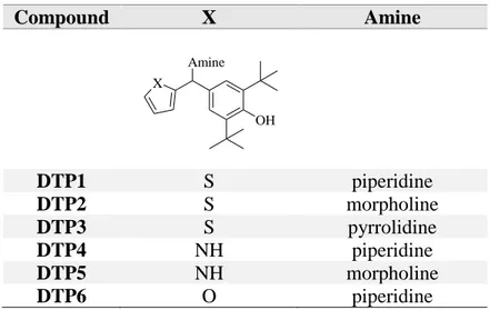
Page | 7 (Table I). The DTP scaffold was decorated with heterocycles such as thiophene, pyrrole and furan, and various aliphatic cyclic amine, like piperidine, morpholine and pyrrolidine (Table II).
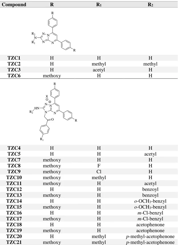
Table I. Small library of TZC derivatives.
Compound R R1 R2 N N N N N R R R1 R2 TZC1 H H H TZC2 H methyl methyl TZC3 H acetyl H TZC6 methoxy H H N N N N O X HN R R R1 R2 TZC4 H H H TZC5 H H acetyl TZC7 methoxy H H TZC8 methoxy F H TZC9 methoxy Cl H TZC10 methoxy methyl H TZC11 methoxy H acetyl TZC12 H H benzoyl TZC13 methoxy H benzoyl TZC14 H H o-OCH3-benzyl
TZC15 methoxy H o-OCH3-benzyl
TZC16 H H m-Cl-benzyl
TZC17 methoxy H m-Cl-benzyl
TZC18 H H acetophenone
TZC19 methoxy H acetophenone
TZC20 H methyl p-methyl-acetophenone
Table II. Small library of DTP derivatives. Compound X Amine Amine OH X DTP1 S piperidine DTP2 S morpholine DTP3 S pyrrolidine DTP4 NH piperidine DTP5 NH morpholine DTP6 O piperidine
Then, I joined the “Translational Oncology Program” at the University of Michigan, for 13 months, under the supervision of Professor Nouri Neamati. In the USA, I had the opportunity to conduct research in both chemistry and biology fields. Firstly, I worked on the characterization of the biological profile and the mechanism of action of the new TZC and DTP derivatives. The antiproliferative activity of all the synthesized compounds was determined in a panel of human cancer cell lines, and the IC50 values of the most active
compounds were calculated. Some TZC salts (TZC7 and TZC10) and DTP derivatives (DTP1-3) significantly inhibited the growth of cancer cells, in particular the pancreatic cancer cell line Mia PaCa-2 (IC50 TZC7 = 2.1 μM; IC50 TZC10 = 1.2 μM; IC50 DTP1 = 5.0 μM).
The ability of the active compounds to inhibit the colony formation and migration of pancreatic cancer cells was also evaluated. The inhibition of STAT3 was determined through FP assay using the protein previously purified in the biology laboratory; then, the levels of phospho-STAT3 in the cells were evaluated by Western blot analysis. While TZC10 showed to inhibit STAT3 similarly to the control Stattic, well-known STAT3 inhibitor, and to reduce phospo-STAT3 levels in a dose-dependent manner, DTP derivatives moderately inhibited the protein. Successively, the possible mechanism of
Page | 9 action of both classes of compounds was investigated using a set of biological assays. TZC salts may stimulate production of reactive oxygen species (ROS) and induce apoptosis in Mia Paca-2 cancer cell line. Because of the structural similarity of DTP derivatives with known modulators of the protein disulfide isomerase (PDI), such as estradiol (E2) and propionic acid carbamoyl methyl amide-31 (PACMA-31) (Figure II),
the inhibition of the reductase activity of PDI by DTP derivatives was also assessed by an insulin turbidimetry assay.
O H H OH H H N S H N O O O O O O E2 PACMA-31
Figure II. Chemical representation of know PDI modulators.
DTP1-3 derivatives significantly inhibited PDI with IC50 values in the low micromolar
range (IC50 DTP1 = 1.51 μM; IC50 DTP2 = 1.69 μM; IC50 DTP3 = 4.17 μM). These results make
them potential multi-target anticancer agents.
In the last months of my permanence in the USA and during my third years of PhD I was working on a third scaffold, the 2,3-dihydroquinazolin-4(1H)-one (DHQ) (Figure III), identified as scaffold for the development of potential anticancer agents, similarly to TZC and DTP scaffolds, after medium-throughput screening. Series of 2,3-dihydropyridopyrimidin-4(1H)-ones (DHPs), quinazolin-4(1H)-ones (QZs) and DHQs were synthesized and lead optimization campaigns were undertaken to improve upon their potency and start a SAR study (Table III). Then, the biological profile as well as the mechanism of action of the new DHQ derivatives was intensely investigated.
DHQ NH H N R O *
Figure III. Molecular structure of the DHQ framework.
Table III. Small library of DHP derivatives.
Compound R N NH H N R O DHP1 Phenyl DHP2 4-(piperidin-1-yl)phenyl DHP3 2-fluoro-5-(trifluoromethyl)phenyl DHP4 4-(4-methylpiperazin-1-yl)phenyl DHP5 1-methyl-1H-indol-3-yl DHP6 2-hydroxy-5-methylphenyl DHP7 4-morpholinophenyl DHP8 1,7-dimethyl-1H-indol-3-yl DHP9 1-ethyl-1H-indol-3-yl DHP10 4-(diethylamino)phenyl DHP11 4-(dimethylamino)phenyl N NH H N R O DHP12 4-(trifluoromethyl)phenyl DHP13 4-(piperidin-1-yl)phenyl N NH H N R O DHP14 4-(diethylamino)phenyl N NH H N R O DHP15 4-(piperidin-1-yl)phenyl
Page | 11 Table III. Small library of QZ and DHQ derivatives.
Compound R X Y Y NH H N R O X QZ1 4-(4-methylpiperazin-1-yl)phenyl H N QZ2 2-hydroxy-5-methylphenyl H N QZ3 4-(diethylamino)phenyl H N QZ4 1-methyl-1H-indol-3-yl H H QZ5 4-(diethylamino)phenyl H H QZ6 6-fluoro-1-propyl-1H-indol-3-yl H H QZ7 1-isopropyl-7-methyl-1H-indol-3-yl F H QZ8 1-cyclopropyl-7-methyl-1H-indol-3-yl F H QZ9 6-fluoro-1-cyclopropyl-1H-indol-3-yl F H QZ10 1-cyclopropyl-7-chloro-1H-indol-3-yl F H QZ11 1-ethyl-6-fluoro-1H-indol-3-yl F H QZ12 1-methyl-1H-indol-3-yl F H QZ13 1-ethyl-1H-indol-3-yl F H QZ14 1-cyclopropyl-6-fluoro-1H-indol-3-yl F H QZ15 7-chloro-1-cyclopropyl-1H-indol-3-yl F H NH H N R O X DHQ1 4-(diethylamino)phenyl H - DHQ2 phenyl F - DHQ3 4-(piperidin-1-yl)phenyl F - DHQ4 4-(4-methylpiperazin-1-yl)phenyl F - DHQ5 4-morpholinophenyl F - DHQ6 4-(diethylamino)phenyl F - DHQ7 4-(dimethylamino)phenyl F - DHQ8 4-(1H-pyrrol-1-yl)phenyl F - DHQ9 4-(pyrimidin-5-yl)phenyl F - DHQ10 3-fluorophenyl F - DHQ11 2-fluoro-5-(trifluoromethyl)phenyl F - DHQ12 2-hydroxy-5-methylphenyl F - DHQ13 benzo[d][1,3]dioxol-5-yl F - DHQ14 4-(imidazo[1,2-a]pyridin-3-yl)phenyl F -
During my three years PhD program, I worked hard to define my profile of young medicinal chemist. The support of my supervisors was indispensable and helpful. I am grateful and honored of their teachings, which allowed me to greatly improve my competence in the field of Medicinal Chemistry. Therefore, I had the opportunity to participate to many National and International Congresses, showing my progresses in research through both poster and oral presentations. I attended the 19° “Course of Mass Spectrometry”, held at Certosa di Pontignano (SI) in 2015. I was also winner of a fellowship offered by the Pharmaceutical Chemistry Division of the Italian Chemical
Compound R X Y NH H N O X N R Y DHQ15 methyl H H DHQ16 methyl H 7-methyl DHQ17 ethyl H H DHQ18 benzyl H 7-methyl DHQ19 propyl H 7-methyl DHQ20 propyl H 7-chloro DHQ21 ethyl H 7-chloro DHQ22 ethyl H 7-methyl DHQ23 H H 7-methyl DHQ24 benzyl H 6-fluoro DHQ25 methyl H H DHQ26 methyl F 7-methyl DHQ27 ethyl F H DHQ28 propyl F 7-methyl DHQ29 propyl F 7-chloro DHQ30 ethyl F 7-chloro DHQ31 ethyl F 7-methyl DHQ32 H F 7-methyl DHQ33 benzyl F 6-fluoro DHQ34 propyl F 6-fluoro DHQ35 ethyl F 6-fluoro
Page | 13 Society to attend the European School of Medicinal Chemistry 2017 in Urbino. Furthermore, this PhD experience allowed me to advance my English understanding. I got a B1 Preliminary Cambridge English Qualification (PET) in 2014 and a C1 qualification through the Test of English as a Foreign Language (TOEFL) in 2015. I also contributed to the scientific field through the publication of my progresses in research. Moreover, during my third year of PhD program, I was tutor of the laboratory teaching activities for the course “Analysis of Medicines” SSD CHIM/08. Finally, I am a member of the team of the project “RYGoldZip”, led by Dr. Francesca Aiello, and we won the “Clinic Center Life Sciences” award and the “Start Cap Calabria 2017” for the discovery and formulation of a topical product with wound healing properties for diabetic ulcers.
Attendance to Congresses:
Merck Young Chemists Symposium – Milano Marittima (November, 13th-15th, 2017)
Badolato Mariateresa – Speaker
“Glycyrrhiza glabra L. leaves: from agrochemical waste to new potential therapeutic agents”
Badolato M., Tundis R., Cappello A., Carullo G., Polerà N., Aiello F.
Glycyrrhiza glabra L. leaves: from agrochemical waste to new potential therapeutic agents
Department of Pharmacy and Health Sciences and Nutrition, University of Calabria, Ed. Polifunzionale, 87036 - Arcavacata Rende (Cs), Italy.
Carullo G.,a Restuccia D.,a Spizzirri U. G.,a Giorgi G.,b Badolato M.,a Aiello F.a
After wine: typical white Calabrian grape pomaces as useful source of functional food
a Department of Pharmacy and Health Sciences and Nutrition, University of Calabria, Ed. Polifunzionale,
87036 – Arcavacata di Rende (Cs), Italy. b Department of Biotechnologies, Chemistry and Pharmacy, University of Siena, Polo Scientifico S. Miniato, Via A. Moro 2, 53100 Siena, Italy.
XXVI Congresso Nazionale della Società Chimica Italiana – Paestum (September, 10th-14th,
2017)
Badolato M.,a,b Carullo G.,a Polerà N.,a Garofalo A.,a Neamati N.,b Aiello F.a
Design, synthesis and biological evaluation of triazolopyrimidinium salts as novel antiproliferative agents
a Department of Pharmacy and Health Sciences and Nutrition, University of Calabria, Ed. Polifunzionale,
87036 – Arcavacata di Rende (Cs), Italy. b Department of Medicinal Chemistry, College of Pharmacy and
Translational Oncology Program, University of Michigan, North Campus Research Complex, 2800 Plymouth Road, Building 520, Ann Arbor, MI 48109, USA.
Carullo G., Trotta F., Sirianni R., Polerà N., Badolato M., Aiello F.
A multi-component one-pot synthesis of 3-amino alkylated indoles, new interesting antiproliferative agents against breast cancer cells
Department of Pharmacy and Health Sciences and Nutrition, University of Calabria, Ed. Polifunzionale, 87036 - Arcavacata Rende (Cs), Italy.
European School of Medicinal Chemistry – Urbino (July, 2nd-6th, 2017)
Fellowship winner
Badolato M., a,b Aiello F., a Garofalo A., a Neamati N., b
Discovery of novel DHQ derivatives with remarkable cytotoxicity profile.
a Department of Pharmacy and Health Sciences and Nutrition, University of Calabria, Ed. Polifunzionale,
87036 – Arcavacata di Rende (Cs), Italy. b Department of Medicinal Chemistry, College of Pharmacy and
Translational Oncology Program, University of Michigan, North Campus Research Complex, 2800 Plymouth Road, Building 520, Ann Arbor, MI 48109, USA.
XXIV National Meeting in Medicinal Chemistry & 10th Young Medicinal Chemists
Symposium – Perugia (September, 11th-14th, 2016)
Carullo G.,a Aiello F.,a Badolato M.,a Ligresti A.,b Allarà M.,b Schiano Moriello A.,b De Petrocellis L.,b Di Marzo V.,c Corelli F.,c Brizzi A.,c
Synthesis and Biological evaluation of small molecules acting as TRPV1 ligands and/or FAAH inhibitors.
aDipartimento di Farmacia e Scienze della Salute e della Nutrizione, Università della Calabria, 87036, Arcavacata
di Rende (Cs), Italy. b Istituto di Chimica Biomolecolare, Endocannabinoid Research Group, Consiglio Nazionale
delle Ricerche, 80078, Pozzuoli (Na), Italy. c Dipartimento di Biotecnologie, Chimica e Farmacia, Università degli
Page | 15
Convegno congiunto delle sezioni Calabria e Sicilia – Società Chimica Italiana (December, 3rd-4th, 2015)
Badolato Mariateresa - Speaker
“Progettazione e sintesi di nuovi probes GPER-selettivi”
Dipartimento di Farmacia e Scienze della Salute e della Nutrizione, Università della Calabria, 87036, Arcavacata di Rende (Cs), Italy.
XXIII National Meeting in Medicinal Chemistry & 9th Young Medicinal Chemists
Symposium – Salerno (September, 6th-9th, 2015)
Brizzi A.,a Aiello F.,b Badolato M.,b Garogalo A.,b Frosini M.,c Maestrini V.,c Pessina F.,d
Sticozzi C.,e Valacchi G.,e De Petrocellis L.,f Luongo L.,f Maione S.,g Ligresti A.,h Di Marzo
V.,h
Synthesis and biological evaluation of small molecule TRPV1 ligands.
a University of Siena, Via A. Moro 2, 53100 – Siena, Italy. b Department of Pharmacy and Health and Nutrition
Sciences, University of Calabria, Edificio Polifunzionale, 87036 - Arcavacata di Rende (Cs), Italy. c Department of
Life Sciences, University of Siena, 53100, Siena, Italy. d Department of Molecular and Development Medicine,
University of Siena, 53100 – Siena, Italy. e Department of Life Sciences and Biotechnology, University of Ferrara,
Italy. f Endocannabinoid Research Group, Institute of Cybernetics, National Research Council, Via Campi Flegrei
34, 80078 - Pozzuoli (Na), Italy. g Department of Experimental Medicine, Pharmacology Section “L. Donatelli”,
Second Univertity of Napoli, Via Costantinopoli 16, 80138 – Napoli, Italy. h Endocannabinoid Research Group,
Institute of Biomolecular Chemistry, National Research Council, 80078 – Pozzuoli (Na), Italy.
Convegno SIF: “Advances in pain research: pathophysiology and new therapeutics
strategies”- Napoli (June, 18th-19th, 2015)
Aiello F.,a Ligresti A.,b Badolato M.,a Maestrini V.,c Luongo L.,d Frosini M.,c Pessina F.,e
Sticozzi C.,f De Petrocellis L.,g Valacchi G.,f Maione S.,d Garogalo A.,a Corelli F.,h Di Marzo V., b Brizzi A.,h
Targeting the Transient Receptor Potential Vanilloid Type-1 channel: pharmacological characterization and analgesic properties of a novel family of TRPV1 ligands.
a Dipartimento di Farmacia e Scienze della Salute e della Nutrizione, Università della Calabria, 87036, Arcavacata
di Rende (Cs), Italy. b Endocannabinoid Research Group, Istituto di Chimica Biomolecolare, Consiglio Nazionale
delle Ricerche, 80078, Pozzuoli (Na), Italy. c Dipartimento di Scienze della Vita, Università degli Studi di Siena,
Via A. Moro 2, 53100 Siena, Italy. d Dipartimento di Medicina Sperimentale, Sezione di Farmacologia “L.
Donatelli”, Seconda Università di Napoli, Via Costantinopoli 16, 80138 Napoli, Italy. e Dipartimento di Medicina
Molecolare e dello Sviluppo, Università degli Studi di Siena, Italy. f Dipartimento di Scienze della Vita e
Consiglio Nazionale delle Ricerche, Via Campi Flegrei 34, 80078 Pozzuoli, Napoli, Italy. h Dipartimento di
Biotecnologie, Chimica e Farmacia, Università degli Studi di Siena, Via A. Moro 2, 53100 Siena, Italy.
XXV Congresso Nazionale della Società Chimica Italiana – Università della Calabria (September, 7th-12th, 2014)
Badolato M., Aiello F., Grande F., Barone I.a, Brancale A., Catalano S., Garogalo A., Andò S.,
Design and synthesis of CXCR4 antagonists with anti-proliferative activity.
Dipartimento di Farmacia e Scienze della Salute e della Nutrizione, Università della Calabria, Edificio Polifunzionale, 87036 - Arcavacata di Rende (Cs), Italy. a School of Pharmacy and Pharmaceutical Sciences, Cardiff
University, King Edward VII Avenue, Cardiff, CF10 3NB, UK.
Grande F.a, Avino S.a, Santolla M. F.a, Aiello F.a, Perri M. G.a, Rosano C.b, Badolato M.a,
Vigliatore F.a, Garofalo A.a, Maggiolini M.a,
Identification of two novel berzo[b]pyrrolo[1,2-d][1,4]oxazin-4-one derivatives acting as GPER antagonists.
a Department of Pharmacy, Health and Nutritional Sciences, University of Calabria, Rende (Cs), Italy. b U.O.S.
Biopolymers and Proteomics - Irccs AOU San Martino, IST-National Institute for Cancer Research, Genova, Italy.
Cellular Environmental Stressors in Biology and Medicine: Focus on Redox Reactions - Third
International Conference – Università di Ferrara (June, 25th-27th, 2014)
Pessina F.a, Aiello F.b, Brizzi A.c, Sticozzi C.d, Badolato M.b, De Petrocellis L.e, Corelli F.c,
Garofalo A.b, Valacchi G.d,Di Marzo V.f ,
Small-molecules TRPV1 modulators: design, synthesis and pharmacological characterization of new 4-(thiofen-2-yl)butanoic acid amides.
a Dipartimento di Medicina Molecolare e dello Sviluppo, Università degli Studi di Siena, Italy. b Dipartimento di
Farmacia e Scienze della Salute e della Nutrizione, Università della Calabria, Arcavacata di Rende, Cosenza, Italy.
c Dipartimento di Biotecnologie, Chimica e Farmacia, Università degli Studi di Siena, Italy. d Dipartimento di
Scienze della vita e biotecnologie, Università degli Studi di Ferrara, Italy. e Istituto di Cibernetica, Consiglio
Nazionale delle Ricerche, Endocannabinoid Research Group Pozzuoli, Italy fIstituto di Chimica Biomolecolare,
Consiglio Nazionale delle Ricerche, Endocannabinoid Research Group, Pozzuoli, Italy.
XVI Congresso Nazionale dei Chimici – Palazzo Campanella – Reggio Calabria (May, 29th
-31st, 2014)
Grande F., Aiello F., Badolato M., Vigliatore F., Garofalo A.,
Combination of hormones and nutrients for the development of hybrid nutraceuticals. Deparment of Pharmacy, Health and Nutritional Sciences, University of Calabria, Rende (CS), Italy.
Page | 17
Scientific publications:
Mariateresa Badolato, Francesca Aiello, and Nouri Neamati. 2,3-Dihydroquinazolin-4(1H)-one as a Privileged Scaffold in Drug Design. Submitted to RSC Advance (2018). Rosa Tundis, Luca Frattaruolo, Gabriele Carullo, Biagio Armentano, Mariateresa Badolato, Monica Rosa Loizzo, Francesca Aiello, and Anna Rita Cappello, An ancient remedial repurposing: synthesis of new pinocembrin fatty acid acyl derivatives as potential antimicrobial/anti-inflammatory agents, Natural Products Research,
accepted manuscript (2017).
Nicoletta Polerà, Mariateresa Badolato, Filomena Perri, Gabriele Carullo, and Francesca Aiello, Quercetin and its Natural Sources in Wound Healing Management, Current
Medicinal Chemistry, accepted manuscript (2017).
Mariateresa Badolato,Gabriele Carullo, Mariarita Perri, Erika Cione, Fabrizio Manetti, Maria Luisa Di Gioia, Antonella Brizzi, Mariacristina Caroleo, and Francesca Aiello, Quercetin/Oleic Acid-Based G-protein-coupled receptor 40 (GPR40) Ligands as New Insulin Secretion Modulators, Future Medicinal Chemistry 9 (16), 1873-1885 (2017).
Mariateresa Badolato, Gabriele Carullo, Erika Cione, Maria Cristina Caroleo, and Francesca Aiello. From the hive: Honey, a novel weapon against cancer. European
Journal of Medicinal Chemistry 142, 290-299 (2017).
Mariateresa Badolato, Gabriele Carullo, Francesca Aiello, and Antonio Garofalo. Synthesis and Experimental Validation of New PDI Inhibitors with Antiproliferative Activity, Journal of Chemistry 2017, 2370359 (2017).
Mariateresa Badolato, Gabriele Carullo, Biagio Armentano, Salvatore Panza, Rocco Malivindi, and Francesca Aiello. Design, synthesis and biological evaluation of a small library of 5H-pyrrole [1,2-a] [3,1] benzoxazin-5-one derivatives as anti-estrogenic agents, Bioorganic & Medicinal Chemistry Letters 27, 3092-3095 (2017). Gabriele Carullo, Anna Rita Cappello, Luca Frattaruolo, Mariateresa Badolato, Biagio Armentano, and Francesca Aiello. Quercetin and derivatives: useful tools in inflammation and pain management. Future Medicinal Chemistry 9 (1), 79-93 (2017).
Yoshinari Miyata, Mariateresa Badolato, and Nouri Neamati. HSPA5. Choi S. (eds)
Encyclopedia of Signaling Molecules. Springer, New York, NY (2016). https://doi.org/10.1007/978-1-4614-6438-9
Francesca Aiello, Gabriele Carullo, Mariateresa Badolato, and Antonella Brizzi. TRPV1–FAAH–COX: The Couples Game in Pain Treatment. ChemMedChem 11, 1686-1694 (2016).
Francesca Aiello, Mariateresa Badolato, Federica Pessina, Claudia Sticozzi, Vanessa Maestrini, Carlo Aldirucci, Livio Luongo, Francesca Guida, Alessia Ligresti, Anna Artese, Marco Allarà, Giosuè Costa, Maria Frosini, Aniello Schiano Moriello, Luciano De Petrocellis, Giuseppe Valacchi, Stefano Alcaro, Sabatino Maione, Vincenzo Di Marzo, Federico Corelli, Antonella Brizzi. Design and Synthesis of New Transient Receptor Potential Vanilloid Type-1 (TRPV1) Channel Modulators: Identification, Molecular Modeling Analysis, and Pharmacological Characterization of the N-(4-Hydroxy-3-methoxybenzyl)-4-(thiophen-2-yl)butanamide, a Small Molecule Endowed with Agonist TRPV1 Activity and Protective Effects against Oxidative Stress. ACS Chemical Neurosci 7 (6), 737-748 (2016).
Fedora Grande, Ines Barone, Francesca Aiello, Andrea Brancale, Michela Cancellieri, Mariateresa Badolato, Francesca Chemi, Cinzia Giordano, Valentino Vircillo, Daniela Bonofiglio, Antonio Garofalo, Sebastiano Andò, Stefania Catalano. Identification of novel 2-(1H-indol-1-yl)-benzohydrazides CXCR4 ligands impairing breast cancer growth and motility. Future Medicinal Chemistry 8 (2), 93-106 (2016).
Awards:
First place in the category “Clinic Center Life Sciences” (€ 25.000,00) “Premio Nazionale Innovazione 2017” with the project RYGoldZip, preparation for topical use based on a new molecular hybrid able to promote the wound healing oh chronic ulcers. First place in “Start Cup Calabria 2017” (€ 5.000,00) with the project RYGoldZip, preparation for topical use based on a new molecular hybrid able to promote the wound healing oh chronic ulcers.
CHAPTER 1:
STAT proteins family and signaling pathway
1.1 Signaling circuits and regulatory networks
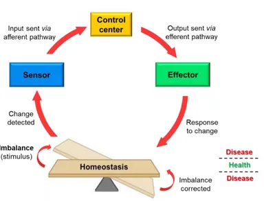
In general, the tendency to maintain a stable, relatively constant internal environment is called homeostasis and is actually a dynamic equilibrium. Biological systems require a carefully controlled homeostasis, which involves a great communication between sensors and effectors, through tightly regulatory networks. This communication is sustained by cell-cell interactions, secreted factors and extracellular matrix components, and tends to reach a balance that resists outside forces of changes. Maintenance of homeostasis depends on negative feedback loops, which act to oppose the stimulus that triggers them. Anything that interferes with the feedback mechanisms can disrupt homeostasis and, in the case of the human body, leads to diseases when the balance is not restored (Figure 1).
Figure 1. Homeostatic control mechanisms.
Any change will be detected by sensors, usually receptors, and relayed to a regulatory control center in the brain. It will process the information and activate the effector whose function is to oppose the stimulus by bringing the balance back.
In the human body, a constant number of cells and a proper organ function is preserved since cells can respond to exogeneous signals with proliferation, differentiation or cell death. Several intracellular signaling molecules transduce mitogenic signals from the environment and cell surface to the nucleus, regulating the normal function of cells. The
Page | 21 alteration or loss of such components cause continued signal transduction, even when an activating signal is absent. Consequently, cells escape from their signaling circuits and inappropriately proliferate, resist apoptosis, induce angiogenesis and invade distant organs. Many evidences demonstrate how cancer results from mutational events that interfere with the usual cooperative behavior of the cells, deregulating various cell signaling pathways that control cell function. (Beerenwinkel et al. 2007)
Knowing the signaling components and their integration in signaling networks might be a potential therapeutic strategy. (Bild et al. 2006)
STAT proteins have emerged as important transcription factors, which can also transduce signals. In particular, STAT3 has been found to play key roles in carcinogenesis and can be considered a potential target in developing strategies to prevent or treat cancer. (Xiong
et al. 2014)
1.2 The STAT proteins family
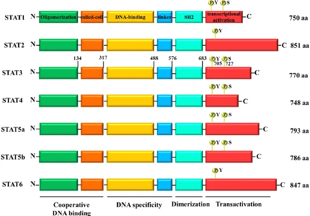
STAT proteins owe their name to their double function as signal transducer and activator of transcription. They were first identified by Darnell et al. in 1994 through the study of transcriptional activation in response to interferon alpha (IFN-α) and gamma (IFN-γ). The STAT family comprises seven members, namely STAT1, STAT2, STAT3, STAT4, STAT5a, STAT5b and STAT6 (Figure 2). Ranging in size from 750 to 850 amino acid residues (90-155 kDa), all STAT proteins share structurally and functionally conserved domains. The N-terminus contains an oligomerization domain and mediates the oligomerization of STAT dimers to tetramers. STAT tetramers contribute to stabilize the STAT-DNA binding and then increase the transcriptional activity. The region between residues 130 and 315 is a four-stranded helical coiled-coil domain that mediates the interaction of STAT with other helical proteins. The DNA-binding domain interacts with
specific DNA elements within STAT-sensitive promoter, ensuring the DNA-binding specificity of each STAT protein. The Src homology 2 (SH2) domain, preceded by the linker region, mediates the interaction of STATs with the receptor and Janus Kinases (JAKs), other than the formation of STAT dimers. The C-terminus containing a transcriptional activation domain (TAD) is the least conserved domain and provides functional specificity, interacting with co-activators and co-repressors. All STAT proteins have a critical tyrosine residue (Y) approximately at position 700 in the TAD. It is target of phosphorylation and essential for STAT activation and function. Phosphorylated STATs can dimerize, translocate in the nucleus and bind to DNA, allowing the transcription of their target genes. Except for STAT2 and STAT6, all STATs contain a critical serine residue (S); if phosphorylated it can improve the transcriptional activity. Inactive non-phosphorylated STATs are present in the cytoplasm of healthy cells and can be divided into two groups, depending on their different activation and function. STAT2, STAT4 and STAT6, activated through a small number of cytokines, play an important role in the development of T-cells and IFN-γ signaling. On the other hand, STAT1, STAT3 and STAT5 are activated in several tissue types through many ligands and are implicated in IFN-γ signaling, development of mammary glands and embryogenesis. Playing a key role in cell cycle progression and apoptosis, STAT3 and STAT5 are also involved in oncogenesis. (Lim and Cao, 2006; Lavecchia et al. 2011)
STAT proteins are all encoded by separate genes. In the human genome, they are organized in 3 different chromosomes: STAT1 and STAT4 are grouped on chromosome 2, whereas STAT2 and STAT6 on chromosome 12. STAT3, STAT5a and STAT5b are clustered on chromosome 17. (Copeland et al. 1995)
In particular, STAT3 gene is located on chromosome 17q21.31. (Choi et al. 1996) Among all STAT proteins, STAT3 is the most studied member, due to its role in the
Page | 23 development of carcinogenesis, other than its involvement in different pathological conditions, including inflammation. (Lin and Karin, 2007)
Figure 2. The STAT family members.
The N-terminal domain of STATs contains an oligomerization domain, followed by the coiled-coil domain and a central DNA-binding domain with a sequence specificity for a palindromic IFN-γ-activated sequence (GAS) element. The DNA-binding domain is connected to the SH2 domain by a linker region. The C-terminal domain has transactivation activity and presents conserved tyrosine and serine residues.
STAT3 is a 92 kDa protein, composed of 770 amino acids. Its TAD contains two important phosphorylation sites: the residue Y705, that promotes STAT3 activation and dimerization; and the residue S727, phosphorylated after tyrosine phosphorylation, that ensures the maximal transcriptional activity. (Wen et al. 1995)
Furthermore, STAT3 dimerization is regulated by reversible acetylation of single lysine residue (K685). (Yuan et al. 2005)
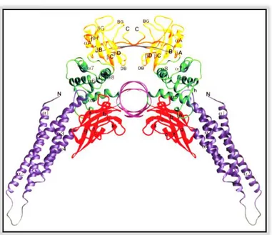
In 1998, Becker et al. first reported X-ray crystal structure of STAT3 homodimer bound to DNA (Figure 3).
Figure 3. Three-dimensional structure of STAT3 homodimer-DNA complex.
The N-terminal domain is shown in purple, the β-barrel domain in red, the linker region in green, and the SH2 domain and phosphotyrosine-containing region in yellow. Regions between helices α1 and α2, and residues from 689 to 701 are shown in grey. The figure represents the view along the DNA axis, with the dyad of the complex running vertically. (Figure by Becker et al. 1998)
1.3 STAT3 signal transduction: the canonical and the non-canonical
pathways
STAT3 activation can be triggered in different ways, through the tyrosine phosphorylation pathway (Figure 4). Receptor-associated JAKs are activated upon cytokine receptor binding (e.g. interleukin-6, IL-6) by cross-phosphorylation. The receptor of IL-6 is a complex containing IL-6 receptor subunit (IL6R) and IL-6 signal transducer glycoprotein 130 (gp130). JAKs are cytoplasmic tyrosine kinases that are inactive in unstimulated cells. The interaction of IL-6 with its receptor results in dimerization of gp130 in the cytoplasm and subsequent recruitment of JAKs. The aggregation of JAKs leads to self-activation by either auto- or trans-phosphorylation.
Page | 25 Consequently, the activated JAKs phosphorylate tyrosine residues on the cytoplasmic domain of the IL-6 receptor, which become docking elements for the SH2 domain of cytoplasmic STAT3 proteins. Being close to JAKs, STAT3 is phosphorylated at specific tyrosine residue (Y705) in the C-terminal portion and activated. The activation of STAT3 can be also initiated by the binding of cytokines and growth factors to their receptors, which have intrinsic tyrosine kinase activity. After ligand binding, the receptors dimerize, autophosphorylate each other and may directly activate STAT3 without JAKs recruitment. Furthermore, STAT3 can be phosphorylated and activated by constitutively active non-receptor tyrosine kinases, such as Src and Abl. Upon activation, STAT3 proteins form homo- or hetero-dimers through reciprocal interaction between the phosphotyrosine of one monomer and the SH2 domain on the partnering STAT3 protein. Then, STAT3 dimers translocate into the nucleus, where they bind to specific promoter elements of target genes and regulate gene expression. (Rane and Reddy, 2000; Levy and
Darnell, 2002)
The dynamic shuttling of STAT3 dimers between cytoplasmic and nuclear compartments is facilitated by importin-α/importin-β1/Ran complex and presence of nuclear localization signal (NLS). (Cimica et al. 2011) Although phosphorylation of STAT3 is important for its function as transcription factor, the translocation of STAT3 into the nucleus may be independent of its phosphorylation status. (Liu et al. 2005)
Among all STAT proteins, STAT3 plays a central role in several normal biological functions and carcinogenesis, since it critically regulates the transcription of a number of key genes involved in cell proliferation and survival, other than differentiation, apoptosis, angiogenesis, invasion and metastases. The upstream signals that triggers activation of STAT3 signaling via phosphorylation of conserved Y705 residue is known as canonical pathway of activation.
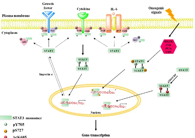
Figure 4. The classical canonical and non-canonical pathways of STAT3 activation. The canonical pathway of STAT3 activation depend on the phosphorylation of Y705 residue of STAT3, by both receptor-associated JAKs and receptors with intrinsic tyrosine kinase activity, upon binding of their specific ligands, as well as by non-receptor tyrosine kinases. The activated STAT3 dimerizes and translocates into the nucleus, where regulates the expression of target genes. The phosphorylation of S727 by various upstream kinases represent the non-canonical pathway of STAT3 activation. Apart from phosphorylation of Y705 and S727, STAT3 also undergoes acetylation at K685 residue, inducing STAT3 dimerization even in the absence of pY705 and pS727.
The non-canonical pathway of STAT3 signaling is independent of classical Y705 phosphorylation. STAT3 can be phosphorylated at another crucial residue, S727. Different converging kinases, such as MAPK, JNK, PKC family and CDK5, are responsible for phosphorylating STAT3 at S727. Several reports highlighted the independent role of pS727 and pY705 in regulating STAT3 function in cancer. Phosphorylated S727 either alone or together with pY705 activates the transcription of genes involved in cell proliferation, survival, DNA repair, drug resistance and some unknown genes.
Page | 27 STAT3 also undergoes acetylation at K685, induced by interaction of p300/CBP histone acetyl transferase protein with the C-terminus of STAT3. Upon K685 acetylation, STAT3 showed increased nuclear localization, DNA binding and enhanced transactivation activity. The acetylated STAT3 dimers interact with DNMT1 and methylate the promoter of tumor suppressor genes to inhibit its expression. (Dimri et al. 2017)
1.4 STAT3-regulated transcription in normal and cancer cells
STATs were originally considered as latent cytoplasmic molecules, activated through phosphorylation and acting as transcription factors with specific DNA binding activity in the nucleus. In particular, STAT3 dynamically translocates from the cytoplasm to the nucleus, maintaining a prominent nuclear presence. In normal cells, STAT3 activation and transcription are tightly regulated and transient. STAT3 was first discovered and characterized as critical mediators in the immune system. In fact, it was originally found to induce the expression of a limited set of target genes in response to IL-6 released during inflammation, interacting with the acute-phase response factor. (Lütticken et al. 1994;
Raz et al. 1994)
STAT3 is ubiquitously expressed in most tissues; therefore, its function was then widely studied in cell culture systems, showing the central role of STAT3 in a variety of signaling systems and responses. Several studies suggested that STAT3 is the main signal transducer downstream of gp130-like receptors, since it could stimulate the proliferation of B lymphocytes, activate terminal differentiation and growth arrest in monocytes, and maintain the pluripotency of embryonic stem cells. (Heinrich et al. 1998; Niwa et al.
1998; Raz et al. 1999; Matsuda et al. 1999)
STAT3 is also essential for the differentiation of helper T cells, implicated in a variety of autoimmune disease. (Yang et al. 2007)
Furthermore, IL-6-mediated activation of STAT3 is required for the maturation of dendritic cells. (Park et al. 2004)
Unlike all other members of the STAT gene family, the disruption of STAT3 gene causes embryonic lethality. (Takeda et al. 1997)
Despite STAT3 plays a crucial role in the early developmental process, the ablation of STAT3 in adult tissues causes unexpectedly mild phenotypes. Depending on the target tissue, STAT3 can modulate cell proliferation and survival, or apoptosis. (Levy and Lee,
2002; Gao et al. 2017)
For this reason, STAT3 is often correlated to carcinogenesis and it is considered as an oncogene (Figure 5).
Figure 5. STAT3-regulated transcription in normal and cancer cells.
Once in the nucleus, STAT3 dimers bind to specific DNA regions and mediate the expression of a variety of genes, in response to different stimuli in multiple cell types. The expression of these downstream genes regulates several processes both in normal and cancer cells, in which STAT3 signaling pathway is no longer transient but persistent.
If STAT3 activation is transient in normal cells, its persistent and aberrant activation was reported in malignant transformation and carcinogenesis (Figure 6).
Page | 29 Figure 6. STAT3 contribution to tumor growth and metastasis.
STAT3 promotes cell proliferation primarily by modulating the expression of key genes, correlated to tumor growth and involved in the regulation of the cell cycle. STAT3 contributes to malignancy by preventing apoptosis, through upregulation of several anti-apoptotic proteins. STAT3 signaling is implicated in cancer inflammation. STAT3 plays an important role in cell migration and contributes to angiogenesis. Persistent activation of STAT3 is often correlated to tumor invasion and metastasis.
The increased level of phosphorylated STAT3 is due mostly to the excess of growth factors and/or cytokines within the tumor microenvironment that activate STAT3 in a paracrine way. Activation of oncogenes and inactivation of tumor-suppressor genes, as well as deregulation of multiple potential upstream inputs and genetic events in neoplastic cells directly activate STAT3 or release inflammatory mediators in an autocrine manner. Through regulation of its target genes, constitutive activated STAT3 contributes to carcinogenesis mainly by triggering pro-proliferative and pro-survival signaling. STAT3 signaling is also involved in the prevention of apoptotic pathway and tumor inflammation. Furthermore, some STAT3 target genes contribute to tumor invasion, angiogenesis and
metastasis. Activated STAT3 is finally involved in the maintenance of cancer stem cells, also known as tumor initiating cells, able to generate diverse tumor cells and contribute to tumor heterogeneity. (Siveen et al. 2014; Xiong et al. 2014)
Due to its ability to regulate the expression of various genes involved in cell proliferation and differentiation, apoptosis, immune and inflammatory responses, angiogenesis, tumor invasion and metastasis, maintenance of cancer stem cells, STAT3 is integrally involved in carcinogenesis.
1.5 STAT3 as a novel molecular target for cancer prevention and
treatment
Increasing evidences confirm that STAT3 plays a critical role in the development and progression of multiple cancer types. Because it is constitutively activated during the growth and the invasiveness of a variety of cancers, STAT3 has emerged as an attracting novel target for cancer prevention and for the design and the development of new drugs potentially effective as chemotherapeutic agents.
The inhibition of STAT3 signaling provides a rational strategy to block carcinogenesis at early stage of tumor development. The discovery of STAT3 inhibitors is currently demanded for both cancer prevention and treatment. STAT3 was originally considered as non-targetable or undruggable and the development of STAT3 inhibitors was delayed, contributing to the lack of FDA-approved STAT3 inhibitors. Recent advances in cancer biology and drug discovery efforts has motivated researchers to develop STAT3 inhibitors as a novel generation of antitumor drugs.
CHAPTER 2:
STAT3 inhibitors for cancer prevention and therapy
2.1 Inhibition of STAT3 signaling pathway in cancer cells
Increasing evidences have shown STAT3 as a key cell signaling molecule in the proliferation and survival of cancer cells.
Since STAT3 is constitutively activated in several malignances, it is a promising drug target and the inhibition of its signaling pathway could be a potential strategy to prevent or treat cancer (Figure 7).
Figure 7. Endogenous and exogenous STAT3 inhibitors.
The endogenous inhibitors keep STAT3-mediated proliferation in equilibrium under normal conditions. The direct and indirect inhibition of STAT3 signaling pathway is a potential strategy for cancer prevention and treatment.
As already mentioned, in normal cells STAT3 activation and transcription is tightly controlled by an equilibrium between stimulation and inhibition.
If tyrosine kinase-mediated phosphorylation activates STAT3 monomers, in contrast, some phosphatases, such as cytoplasmic and nuclear phosphatase, downregulate its
Page | 33 activity. They repress JAK-STAT signaling through dephosphorylation of STAT3. Initially, the persistent activation of STAT3 automatically triggers regulatory feedback loops. For example, the expression of suppressor of cytokine signaling 3 (SOCS3) is induced by activated STAT3. It serves as a feedback inhibitor, preventing the persistent activation of STAT3 signaling pathway. (Munoz et al. 2014)
Furthermore, the protein inhibitor of activated STAT3 (PIAS3) deregulates cell proliferation by modulation of DNA binding transcription factors and their oncogenic activity. (Munoz et al. 2014)
Although there are not FDA-approved STAT3 inhibitors available for use to date, many molecules were found to be able to interfere with STAT3 signaling. The crucial role of STAT3 in cancer initiation, progression and metastasis has motivated many groups to design and develop approaches to inhibit STAT3 signaling. Two main strategies can be exploited to inhibit STAT3 signaling pathway:
• indirect inhibition: blocking the upstream regulators of the pathway, such as growth factors and cytokines, as well as kinases, involved in STAT3 phosphorylation, then in its activation;
• direct inhibition of STAT3 protein: with small molecule inhibitors targeting one of the STAT3 structural domains, such as SH2, DNA-binding and N-terminal domains. Interacting directly with STAT3, they inhibit phosphorylation, dimerization, nuclear translocation, DNA binding and transcription of target genes.
Many different classes of molecules have been investigated, including antisense oligonucleotides, small interfering RNA (siRNA), peptides and small molecule inhibitors. Results show a decrease in cell proliferation and invasiveness, as well as an increase in apoptosis.
2.2 Oligonucleotides and peptides
Antisense RNA and siRNA have been used to inhibit STAT3 gene expression by binding mRNA, resulting in subsequent degradation. Furthermore, decoy oligodeoxynucleotides can mimic the consensus STAT DNA binding sequence and it is used to compete for STAT3 DNA binding. The result is a downregulation of STAT3-dependent gene transcription. Most in vitro and in vivo studies using oligonucleotides and siRNA as STAT3 inhibitors are consistent and show the suppression of tumor cell growth and the induction of apoptosis. Phase 0, Phase 1, and Phase 1/2 trial studies of oligonucleotide inhibitors are still ongoing. (Groner et al. 2008; Munoz et al. 2014)
Peptides have been used to block reciprocal docking between SH2 domain of one STAT3 monomer and the pY705 motif on the partnering STAT3 monomer, preventing protein-protein dimerization. The isolated SH2 domain of STAT3, a short peptide of six amino acids, reduces the DNA binding of STAT3, while peptide aptamers, designed on the base of crystallography and structural analysis, interact with upstream receptors and functionally important domains of STAT3, preventing phosphorylation, dimerization and DNA binding. The use of pure peptide is limited by low biological activity, poor membrane permeability, modest stability, and relative non-specificity for STAT3, then peptidomimetic and nonpeptide approaches can overcome these limitations. (Groner et
al. 2008; Munoz et al. 2014)
2.3 Small molecule inhibitors
Small molecule inhibitors of STAT3 is the largest class of compounds used to prevent and treat cancer. They can act in both direct and indirect ways, inhibiting STAT3 phosphorylation, dimerization, nuclear translocation, and/or DNA binding. Multiple methods including high-throughput screening of large chemical libraries, virtual
Page | 35 screening and rational design based on peptides and peptidomimetic inhibitors, fragment-based drug design, and multiple ligand simultaneous docking (MLSD) were used to identify novel small molecule inhibitors of STAT3.
Many natural and synthetic agents showed significant inhibition of STAT3 activation and thereby suppression of tumor growth and metastasis. Unfortunately, a comparison across different classes of inhibitors is difficult since there is no uniformity in terms of assay systems used to evaluate their potency. (Debnath et al. 2012)
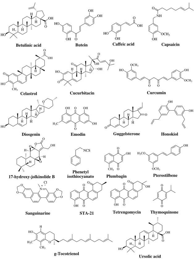
2.3.1 Natural inhibitors of STAT3 signaling pathway
A selection of natural products inhibiting STAT3 signal is shown in Figure 8. Mostly they inhibit the constitutive activation of STAT3 in a dose- and time-dependent manner, by inhibiting IL-6 and JAK-induced phosphorylation of STAT3, inducing ROS generation that blocks STAT3 signaling, blocking STAT3 translocation into the nucleus, and inhibiting the expression of STAT3 target genes.
Although natural products have the advantage to be generally well tolerated, many limitations hamper their application as effective chemopreventive and chemotherapeutic agents. In fact, they present non-specific effects on targeting STAT3, off-target effects, variable bioavailability and moderate efficacy. (Debnath et al. 2012; Siveen et al. 2014)
2.3.2 Synthetic agents inhibiting STAT3 signal
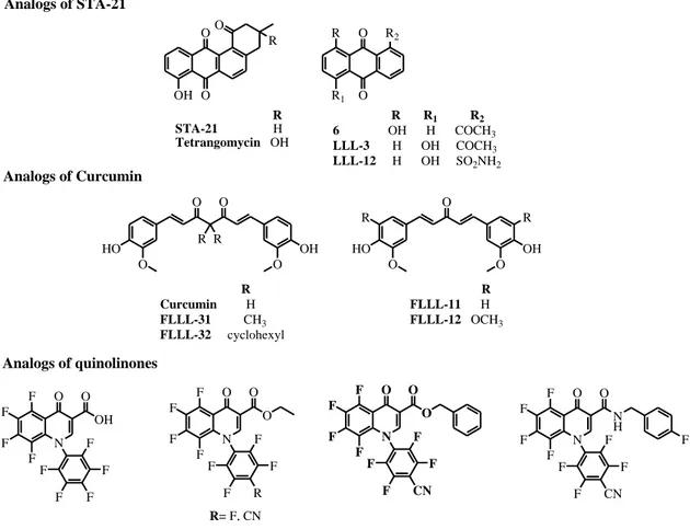
Rational structural modification in natural products might give novel drug candidates for cancer prevention and therapy (Figure 9). STA-21 is a natural STAT3 inhibitor discovered through virtual screening. It inhibits dimerization, nuclear translocation, DNA binding, and the expression of STAT3-regulated target genes in cancer cells with constitutively active STAT3. Several analogs of STA-21, with hydroxyl, acetyl and
sulfamidic groups, bind to the SH2 domain of STAT3. In particular, LLL-12 is more potent than the lead compound. (Debnath et al. 2012; Siveen et al. 2014)
HO H H H H O OH Betulinic acid OH HO O OH OH Butein OH HO O OH Caffeic acid OH NH O OCH3 Capsaicin Celastrol O HO CH3 H OH O H Cucurbitacin H H OH HO O O O OH OH OCH 3 HO O O OH OCH3 Curcumin H H Diosgenin H O H H HO O O OH OH OH H3C Emodin H H Guggelsterone O O H OH OH Honokiol 17-hydroxy-jolkinolide B O H O O H H O OH O O Plumbagin CH3 OH OCH3 Pterostilbene H3CO OH Sanguinarine O O N O O Cl O O Tetrengomycin O OH OH O O Thymoquinone O H H3C g-Tocotrienol CH3 HO HO H H H O OH Ursolic acid Phenetyl isothiocyanato NCS O O STA-21 O OH
Page | 37 Curcumin inhibits IL-6-induced STAT3 phosphorylation and nuclear translocation. Some analogs have been designed based on its structure. They also decrease phosphorylated STAT3 levels and induce apoptosis in a dose-dependent manner in several cancer cell lines. (Debnath et al. 2012)
Several quinolinones have been reported as JAK/STAT3 pathway inhibitors. These analogs inhibit STAT3 phosphorylation at Y705. The carboxylic analog is less potent than esters, whereas the introduction of a cyano group significantly increases the potency of quinolinones analogs. (Debnath et al. 2012)
N F F F F O O OH F F F F F N F F F F O O O F F R F F R= F, CN N F F F F O O O F F CN F F N F F F F O O N H F F CN F F F O O R R HO O O OH O HO O R R OH O R Curcumin H FLLL-31 CH3 FLLL-32 cyclohexyl R FLLL-11 H FLLL-12 OCH3 OH O OO R R STA-21 H Tetrangomycin OH R1 O O R R1 R2 6 OH H COCH3 LLL-3 H OH COCH3 LLL-12 H OH SO2NH2 R2 R Analogs of STA-21 Analogs of Curcumin Analogs of quinolinones
Figure 9. Synthetic analogs of natural STAT3 inhibitors.
2.3.3 Other small molecule inhibitors of STAT3 signaling pathway
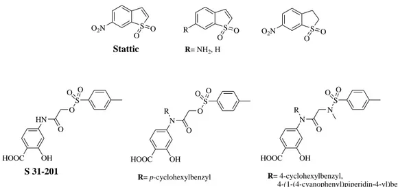
diverse chemical library as a STAT3 inhibitor, binding its SH2 domain, and inhibiting the proliferation of cancer cells. Stattic may lack target selectivity because of its simple chemical structure. Some analogs of Stattic have been synthesized in order to optimize the lead compound (Figure 10). Unfortunately, the attempt was unsuccessful, suggesting the importance of both nitro group and unsaturated five-membered ring for the inhibitory activity of Stattic. (Debnath et al. 2012; Siveen et al. 2014)
On the other hand, S 31-201 has been identified as STAT3 inhibitor through structural-based virtual screening of chemical libraries from NCI. It preferentially inhibits the DNA binding ability of STAT3 dimers, showing antiproliferative activity both in vitro and in
vivo studies. In an attempt to optimize compound S 31-201, structure-based modeling
resulted in several analogs with improved potency (Figure 10). (Debnath et al. 2012)
S O2N O O S R O O R= NH2, H S O2N O O Stattic HOOC OH HN O O S O O S 31-201 HOOC OH N O O S O O R R= p-cyclohexylbenzyl HOOC OH N O N S O O R R= 4-cyclohexylbenzyl, 4-(1-(4-cyanophenyl)piperidin-4-yl)benzyl, 4-(1-(4-cyanophenylsulfonyl)piperidin-4-yl)benzyl
Figure 10. Chemical structures of Stattic, S 31-201 and their analogs.
Using high-throughput and virtual screening, several drugs and additional compounds have been found to interfere directly and/or indirectly with STAT3 signaling pathway (Figure 11).
Page | 39 actual molecular targets. Therefore, it is too early to classify them as STAT3 inhibitors. (Debnath et al. 2012)
Figure 11. Additional compounds identified from high-throughput and virtual screening.
2.3.4 Inhibitors of JAK and Src kinases
STAT3 phosphorylation is essential for its activation and then for its signaling pathway. Inhibiting the upstream kinases such as JAK and Src, which phosphorylate STAT3 is a rational approach to inhibit STAT3 signal and have a chemopreventive and/or chemotherapeutic effect. Both JAK and Src inhibitors decrease phosphorylated STAT3 levels, causing suppression of tumor growth, induction of apoptosis, and inhibition of metastasis. New developed compounds show antiproliferative activity by inhibiting synergistically multiple cytoplasmic kinases. (Xiong et al. 2014)
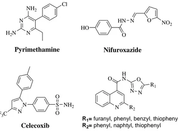
Few kinase inhibitors are shown in Figure 12.
Figure 12. Inhibitors of JAK and Src kinases.
N N NH2 H2N Cl Pyrimethamine HO HN O N O NO 2 Nifuroxazide N S Celecoxib N F3C NH2 O O N R2 O HN N N O R1
R1= furanyl, phenyl, benzyl, thiophenyl
R2= phenyl, naphtyl, thiophenyl
Cl N H O N S H N N N N N OH Dasatinib AG490 N H O CN OH OH N H N NH O HO Br 6-bromoindirubin
2.3.5 Small molecule inhibitors of STAT3 under clinical trial studies
STAT3 inhibitors have important features that certainly need further investigations, but the preliminary results are promising. Nowadays, few inhibitors are under evaluation in a number of early phase trials, in gastric cancer. (Cafferkey et Chau, 2016)
First, Kim et al. identified compound OPB-31121 (structure not disclosed) as STAT3 inhibitor by suppressing both constitutively activated and IL-6-mediated JAK/STAT signaling pathways. It reduces cell proliferation, induces apoptosis, and downregulates the expression of antiapoptotic proteins both in vitro and in vivo studies. OPB-31121 also has synergistic effect with known antitumor drugs, such as 5-FU and cisplatin. It binds to the SH2 domain of STAT3 in a unique way, compared to other inhibitors, as shown by computational docking and molecular dynamics simulation. (Brambilla et al. 2015) Currently, OPB-31121 is under the first experimental phase of clinical trials, to evaluate the safety and tolerability. Its administration is associated with mild side effects, including grade 1 to 2 nausea, vomiting, diarrhea, and fatigue. (Bendell et al. 2014)
Compound OPB-51602 (structure not disclosed) is also an oral direct inhibitor of STAT3.
Wong et al. investigated the maximum tolerated dose (MTD), safety, and
pharmacokinetics of OPB-51602 in a phase 1 study. It inhibits tyrosine and serine phosphorylation of STAT3 in vitro. The phase 1 clinical study confirms the safety of OPB-51602 in terms of toxicity. The most common adverse effects observed are again nausea, vomiting, diarrhea, anorexia, peripheral neuropathy, and fatigue. (Ogura et al.
2015)
Compound BBI608, also known as napabucasin, inhibits STAT3-regulated transcription (Figure 13). In preclinical studies, napabucasin exhibits its effect in various ways, including a dose-dependent decrease of antiapoptotic protein levels and suppression of genes important for cancer stemness. (Li et al. 2015)