The New England
Journal
of
Medicine
© C o py r ig ht , 2 0 0 1 , by t he Ma s s ac h u s e t t s Me d ic a l S o c ie t y
V O L U M E 3 4 4 JA N U A R Y 1 8 , 2 0 0 1 N U M B E R 3
A RANDOMIZED TRIAL OF THE DISCONTINUATION OF PRIMARY
AND SECONDARY PROPHYLAXIS AGAINST PNEUMOCYSTIS CARINII PNEUMONIA
AFTER HIGHLY ACTIVE ANTIRETROVIRAL THERAPY IN PATIENTS WITH HIV INFECTION
JUAN C. LOPEZ BERNALDODE QUIROS, M.D., JOSE M. MIRO, M.D., JOSE M. PEÑA, M.D., DANIEL PODZAMCZER, M.D., JUAN C. ALBERDI, M.D., ESTEBAN MARTÍNEZ, M.D., JAIME COSIN, M.D., XAVIER CLARAMONTE, M.D.,
JUAN GONZALEZ, M.D., PERE DOMINGO, M.D., JOSE L. CASADO, M.D., ESTEBAN RIBERA, M.D.,
ANDTHE GRUPODE ESTUDIODEL SIDA 04/98*
ABSTRACT
Background Prophylaxis against Pneumocystis ca-rinii pneumonia is indicated in patients with human immunodeficiency virus (HIV) infection who have less than 200 CD4 cells per cubic millimeter and in those
with a history of P. carinii pneumonia. However, it is
not clear whether prophylaxis can be safely discon-tinued after CD4 cell counts increase in response to highly active antiretroviral therapy.
Methods We conducted a randomized trial of the discontinuation of primary or secondary prophylaxis
against P. carinii pneumonia in HIV-infected patients
with a sustained response to antiretroviral therapy, defined by a CD4 cell count of 200 or more per cubic millimeter and a plasma HIV type 1 (HIV-1) RNA level of less than 5000 copies per milliliter for at least three months. Prophylactic treatment was restarted if the CD4 cell count declined to less than 200 per cubic mil-limeter.
Results The 474 patients receiving primary pro-phylaxis had a median CD4 cell count at entry of 342 per cubic millimeter, and 38 percent had detectable HIV-1 RNA. After a median follow-up period of 20 months (388 person-years), there had been no
epi-sodes of P. carinii pneumonia in the 240 patients who
discontinued prophylaxis (95 percent confidence inter-val, 0 to 0.85 episode per 100 person-years). For the 113 patients receiving secondary prophylaxis, the median CD4 cell count at entry was 355 per cubic millimeter, and 24 percent had detectable HIV-1 RNA. After a me-dian follow-up period of 12 months (65 person-years),
there had been no episodes of P. carinii pneumonia in
the 60 patients who discontinued prophylaxis (95 per-cent confidence interval, 0 to 4.57 episodes per 100 person-years).
Conclusions In HIV-infected patients receiving high-ly active antiretroviral therapy, primary and
second-ary prophylaxis against P. carinii pneumonia can be
safely discontinued after the CD4 cell count has in-creased to 200 or more per cubic millimeter for more than three months. (N Engl J Med 2001;344:159-67.)
Copyright © 2001 Massachusetts Medical Society.
From the Hospital Universitario Gregorio Marañón, Madrid (J.C.L.B.Q., J.C.); the Institut d’Investigacions Biomèdiques August Pi I Sunyer and Hospital Clinic Universitari, Barcelona (J.M.M., E.M., X.C.); the Ciudad Sanitaria La Paz, Madrid (J.M.P., J.G.); the Hospital de Bellvitge, Barcelona (D.P.); the Consejería de Sanidad Comunidad Autonoma de Madrid, Madrid (J.C.A.); the Hospital de Sant Pau, Barcelona (P.D.); the Hospital Ramón y Cajal, Madrid (J.L.C.); and the Hospital Universitari de la Vall d’Hebron, Barcelona (E.R.) — all in Spain. Address reprint requests to Dr. Lopez at the Division of Infectious Diseases, Hospital Gregorio Marañón, Dr. Es-querdo 46, 28007 Madrid, Spain, or at [email protected].
*Other members of the Grupo de Estudio del SIDA (GESIDA) are list-ed in the Appendix.
NEUMOCYSTIS carinii pneumonia was a common and often fatal infection in patients infected with the human immunodeficien-cy virus (HIV) in the early 1980s.1 Before the use of primary prophylaxis became standard, the proportion of patients with P. carinii pneumonia as the initial event defining the presence of the acquired immunodeficiency syndrome (AIDS) was 62 percent, and about 80 percent of patients with CD4 cell counts below 200 per cubic millimeter had this complica-tion.2,3 It was calculated that without secondary pro-phylaxis, 50 percent of patients would relapse within 24 weeks after an episode of P. carinii pneumonia.4 Chemoprophylaxis has been dramatically effective, and it is currently recommended for all patients with less than 200 CD4 cells per cubic millimeter.4,5 Trimeth-oprim–sulfamethoxazole is the first choice for pro-phylaxis, and it can be taken in double-strength form three times a week.6-8 However, adverse effects of tri-methoprim–sulfamethoxazole may occur in as many as 50 percent of patients so treated, and 30 percent will need to change their regimen for this reason.9,10 The alternatives include aerosolized pentamidine, dap-sone with or without pyrimethamine, and atova-quone.11,12
The use of highly active antiretroviral therapy has changed the course of HIV infection, resulting in a striking reduction in morbidity and mortality.13,14 The
The Ne w E n g l a nd Jo u r n a l o f Me d ic i ne
persistent suppression of HIV replication leads to a sustained increase in CD4 cells, even in patients with severe immunosuppression. There have been several reports of dramatic declines in the incidence of op-portunistic infections, such as P. carinii pneumonia, cytomegalovirus (CMV) retinitis, and Mycobacterium avium infections.15-17 Recently, observational and ret-rospective studies have suggested that P. carinii pro-phylaxis may be safely discontinued in patients re-ceiving highly active antiretroviral therapy who have improved immunologic function.18-23 A task force of the U.S. Public Health Service and the Infectious Dis-eases Society of America has recommended the dis-continuation of primary prophylaxis against P. carinii
pneumonia but recognizes that “the optimal criteria for discontinuation remain to be defined.”24
In a randomized multicenter trial, we tested the hypothesis that primary and secondary prophylaxis against P. carinii pneumonia can be safely discontin-ued in patients in whom highly active antiretroviral treatment results in immune reconstitution, as long as their CD4 cell counts remain at 200 or more per cubic millimeter.
METHODS
Patients
Patients were eligible for the study if they had had previous CD4 cell counts of less than 200 per cubic millimeter or had had a pre-vious episode of P. carinii pneumonia; if they were receiving treat-ment with any of the regimens accepted for prophylaxis against
P. carinii pneumonia; if they had a sustained response to highly
active antiretroviral therapy, defined by a CD4 cell count of 200 or more per cubic millimeter and a plasma HIV type 1 (HIV-1) RNA level of less than 5000 copies per milliliter for more than three months; and if they had a Karnofsky score higher than 80. Patients were excluded if they were under 18 years of age, if they were preg-nant, or if they had poor adherence to antiretroviral treatment. Study Design
The study was a randomized, nonblinded, multicenter trial that evaluated whether primary and secondary prophylaxis against
P. carinii pneumonia can be safely discontinued in HIV-infected
patients. Patients were recruited from 19 Spanish public hospitals; the staff at each had broad experience in the treatment and care of HIV-infected patients. The randomization, based on permuted blocks, was stratified according to center. The trial was approved by the institutional review boards of the participating hospitals, and all the patients gave written informed consent.
Patients were randomly assigned to continue or to discontinue prophylaxis against P. carinii pneumonia. Accepted regimens of prophylaxis were those recommended in the 1997 guidelines of the Public Health Service and the Infectious Diseases Society of Amer-ica.11 Accepted highly active antiretroviral therapy involved at least
three antiretroviral drugs, one of which was a protease inhibitor or a non-nucleoside reverse-transcriptase inhibitor. P. carinii pneu-monia was diagnosed either after microbiologic confirmation in res-piratory samples or when the clinical and radiographic presentation was strongly suggestive of P. carinii pneumonia and there was a response to treatment only with agents active against P. carinii. When the CD4 cell counts of patients assigned to discontinue pro-phylaxis fell below 200 per cubic millimeter, propro-phylaxis was im-mediately reinstituted, although the patients were kept in the study. An increase in the HIV-1 RNA level was not a criterion for re-starting prophylaxis.
Patients were evaluated at three-month intervals with a clinical assessment and laboratory monitoring that included measurements of CD4 cell counts and HIV-1 RNA levels, which were performed at each site. Lymphocyte subpopulations were measured at all cen-ters by three-color flow cytometry. HIV-1 RNA levels were de-termined by either a polymerase-chain-reaction assay (Amplicor HIV-1 Monitor Assay, Roche Molecular Systems, Somerville, N.J.) or a branched-chain DNA assay (Chiron, Emeryville, Calif.). When the study was designed, most of the hospitals used techniques with a limit of detection of 400 copies per milliliter for the polymerase-chain-reaction assay or 500 copies per milliliter for the branched-chain DNA assay. Although by the end of the study all of the hos-pitals were able to detect levels as low as 200 copies per milliliter with the polymerase-chain-reaction assay or less than 50 copies per milliliter with the branched-chain DNA assay, we kept 500 copies per milliliter as the limit of detection for the HIV-1 RNA level throughout the study.
End Points and Follow-up
The primary end point in the assessment of safety was the oc-currence of P. carinii pneumonia. The secondary end points were the development of an AIDS-defining event other than P. carinii pneumonia (a “C” event as defined by the Centers for Disease Con-trol and Prevention [CDC]), the occurrence of drug-related adverse effects, the development of non–AIDS-defining bacterial infections, changes in CD4 cell counts and HIV-1 RNA levels, and death. Pa-tients were removed from the study during follow-up if one of the following occurred: an AIDS-defining event (including P. carinii pneumonia), hypersensitivity to the prophylactic agents, discontin-uation of highly active antiretroviral therapy, or voluntary withdraw-al from the study. A fwithdraw-all in CD4 cell counts to under 200 per cubic millimeter was not a criterion for removal from the study. Statistical Analysis
We assumed that P. carinii pneumonia would develop in 5 per-cent of patients receiving primary prophylaxis during the 12 months of follow-up and in at least 15 percent of patients who discontin-ued prophylaxis.25 We estimated that at least 200 patients at risk
would be needed in each group for the study to be able to detect a 10 percent difference with 90 percent certainty and a 5 percent significance level. Ten percent of patients were expected to be lost to follow-up.
We assumed that P. carinii pneumonia would develop in 15 percent of patients receiving secondary prophylaxis during the first 12 months of follow-up, and in at least 60 percent of patients who discontinued prophylaxis.4 We estimated that at least 30
pa-tients at risk would be needed in each group to permit us to de-tect a 45 percent difference with 90 percent certainty and a 5 per-cent significance level. Ten perper-cent of patients were expected to be lost to follow-up.
An intention-to-treat analysis was performed. Medians and in-terquartile ranges (25th to 75th percentile) were used as measures of central tendency and dispersion. Confidence intervals for both groups were calculated with the use of Poisson distribution tables. For the base-line variables, comparisons between groups were made with the chi-square test for categorical variables and the Mann– Whitney nonparametric test for quantitative variables. Multivariate analysis of variance with repeated measures was used to compare CD4 cell counts at enrollment and at the first, second, third, and fourth follow-up visits. A polynomial contrast was used to model the within-group sum of squares, and a difference contrast was used to model the between-group sum of squares. All reported P values were two-sided.
RESULTS
Primary Prophylaxis
A total of 474 patients with no history of P. cari-nii pneumonia were enrolled in the study between January 1, 1998, and January 31, 1999. Of these,
D I S C O N T I N UAT I O N O F P N E U M O CYST I S C A R I N I I P R O P H Y L A X I S I N H I V- I N F EC T E D PAT I E N T S
*CDC denotes Centers for Disease Control and Prevention, HAART highly active antiretroviral therapy, and CI confidence interval.
†Category A includes patients who have had no HIV-related diseases; category B includes patients who have had HIV-related diseases that are not in category C; category C includes patients who have had HIV-related diseases that are considered to be AIDS defining.26
TABLE 1. MAIN CHARACTERISTICSOF PATIENTS DISCONTINUING PRIMARY PROPHYLAXISOR CONTINUING PRIMARY PROPHYLAXIS.*
CHARACTERISTIC GROUP DISCONTINUING PRIMARY PROPHYLAXIS (N=240) GROUP CONTINUING PRIMARY PROPHYLAXIS (N=234) CHARACTERISTIC GROUP DISCONTINUING PRIMARY PROPHYLAXIS (N=240) GROUP CONTINUING PRIMARY PROPHYLAXIS (N=234)
At base line At base line (cont.)
Age — yr Median Interquartile range Male sex — no. (%)
Mode of acquisition — no. (%) Intravenous drug use Homosexual activity Heterosexual activity Other
Time from diagnosis of HIV — yr Median Interquartile range CDC group — no. (%)† A-3 B-3 C-3 CD4 count — cells/mm3 Nadir Median Interquartile range At base line Median Interquartile range HIV-1 RNA <500 copies/ml — no. (%) Level if >500 copies/ml Median Interquartile range Time with CD4 »200/mm3
and HIV-1 RNA <5000/ml — mo Median
Interquartile range
Time receiving prophylaxis — mo Median
Interquartile range Time receiving HAART — mo
Median Interquartile range 36 33–41 175 (73) 125 (52) 48 (20) 57 (24) 10 (4) 7 4–9 110 (46) 46 (19) 84 (35) 113 56–156 342 277–440 197 (82) 1100 791–2455 9 5–14 34 19–49 15 10–59 36 33–40 169 (72) 131 (56) 42 (18) 59 (25) 2 (1) 8 5–11 98 (42) 40 (17) 96 (41) 98 44–147 329 268–407 199 (85) 2256 1448–2587 8 5–11 35 22–51 16 10–20
Treatment received — no. of patients Lamivudine Stavudine Indinavir Zidovudine Saquinavir Ritonavir Nelfinavir Didanosine Nevirapine Zalcitabine At follow-up Episodes of P. carinii pneumonia — no. Duration of follow-up after
randomization Months
Median Interquartile range Person-years
95% CI for no. of episodes/100 person-yr
99% CI for no. of episodes/100 person-yr
Duration of follow-up while CD4 »200/mm3
Months Median Interquartile range Person-years
95% CI for no. of episodes/100 person-yr
Duration of follow-up while CD4 <200/mm3
Months Median Interquartile range Person-years
95% CI for no. of episodes/100 person-yr “C” events 189 165 147 66 50 26 29 23 14 6 0 20 17–25 387.9 0–0.85 0–1.23 19 16–24 377.7 0–0.98 13 11–18 10.2 1 186 157 157 75 43 29 22 37 14 2 0 19 15–24 370.5 0–0.89 0–1.28 19 15–24 360.1 0–1.02 10 8–14 10.4 1
240 were randomly assigned to discontinue prophy-laxis and 234 to continue it. The groups were well balanced with regard to demographic characteristics (Table 1). Most of the patients were men and had at least a five-year history of HIV infection that includ-ed a long period with a CD4 cell count of less than 200 per cubic millimeter. One hundred twenty-one patients (54 in the group discontinuing prophylaxis and 67 in the group continuing prophylaxis) had a nadir CD4 cell count of no more than 50 per cubic millimeter. Ninety-one percent were receiving pro-phylaxis with trimethoprim–sulfamethoxazole. A to-tal of 472 patients were receiving highly active anti-retroviral therapy with a protease inhibitor and only
2 with a non-nucleoside reverse-transcriptase inhibi-tor. At enrollment, patients had had more than 200 CD4 cells per cubic millimeter and less than 5000 copies of HIV-1 RNA per milliliter for a median of 8 months (range, 3 to 72). A total of 172 patients had 200 to 299 CD4 cells per cubic millimeter at enroll-ment, and 169 were enrolled during the first 12 months of highly active antiretroviral therapy.
Of the 22 patients who dropped out of the study, 12 were lost to follow-up (7 assigned to discontinue prophylaxis and 5 assigned to continue it), 3 discon-tinued highly active antiretroviral therapy, 5 assigned to continue prophylaxis discontinued it after enroll-ment, and 2 in the group discontinuing prophylaxis
The Ne w E n g l a nd Jo u r n a l o f Me d ic i ne
decided to resume it because they were concerned about the risk of P. carinii pneumonia. There were no significant differences in base-line characteristics between the patients who dropped out of the study and those who remained in it. To our knowledge, only a single patient (in the group continuing prophylaxis) had P. carinii pneumonia after dropping out of the study.
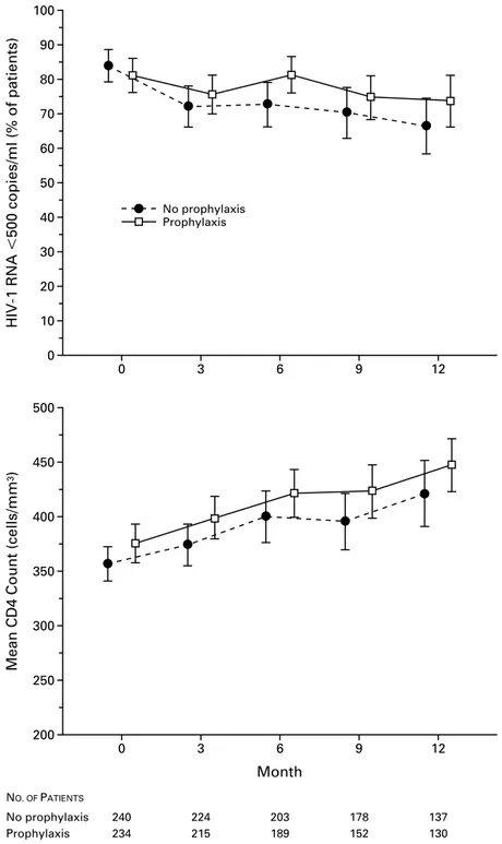
The median duration of follow-up was 20 months (range, 16 to 24). The CD4 cell counts and the pro-portion of patients with less than 500 copies of HIV-1 RNA per milliliter during follow-up were similar in the two groups (P=0.67 and P=0.41, respectively) (Fig. 1). In 21 patients (9 in the group discontinuing prophylaxis), CD4 cell counts fell below 200 per cubic millimeter, and prophylaxis had to be reintroduced for those in whom it had been discontinued. Ninety-two patients in the group discontinuing prophylaxis and 89 in the group continuing prophylaxis had more than 500 copies of HIV-1 RNA per milliliter during 136 person-years of follow-up (group discontinuing pro-phylaxis: median, 3250 copies per milliliter; range, 510 to 57,599; group continuing prophylaxis: medi-an, 3458 copies per milliliter; range, 515 to 61,057). During follow-up, the protease inhibitor was replaced with a non-nucleoside reverse-transcriptase inhibitor in 36 patients (17 in the group discontinuing prophy-laxis and 19 in the group continuing prophyprophy-laxis).
There were no episodes of P. carinii pneumonia in either group during follow-up — neither among those with a nadir CD4 cell count of less than 50 per cubic millimeter before enrollment (95 percent confidence interval, 0 to 4.6 episodes per 100 per-son-years for the group discontinuing prophylaxis vs. 0 to 4.3 for the group continuing prophylaxis; P= 0.48) nor among those with more than 500 copies of HIV-1 RNA per milliliter during follow-up (95 percent confidence interval, 0 to 4.0 episodes per 100 person-years for the group discontinuing prophylax-is vs. 0 to 4.0 for the group continuing prophylaxprophylax-is; P=0.63). Two patients (one in each group) had a “C” event, both with diagnoses of extrapulmonary tuberculosis. Two patients (one in each group) died of cancer (hepatic carcinoma and laryngeal carcinoma).
P. carinii pneumonia did not develop during
follow-up in any of the 21 patients whose CD4 cell counts fell below 200 per cubic millimeter.
Fourteen patients (seven in each group) had an infection during follow-up, including six with com-munity-acquired pneumonia. In all of these patients,
P. carinii was ruled out as the cause of the infection
by microbiologic methods. No patient received em-pirical anti–P. carinii treatment in therapeutic doses. Drug-related adverse effects occurred in 49 patients (23 in the group discontinuing prophylaxis and 26 in the group continuing prophylaxis); they were re-lated in most patients to the use of protease inhibitors and in 4 patients to the use of prophylactic agents
(3 of them discontinued prophylaxis). Finally, the antiretroviral treatment had to be modified in 78 pa-tients (41 in the group discontinuing prophylaxis and 37 in the group continuing prophylaxis), either be-cause of adverse effects of the antiretroviral drugs or because of virologic evidence of treatment failure. Secondary Prophylaxis
Between January 1, 1998, and June 30, 1999, 113 patients who had had a previous episode of P. carinii pneumonia were enrolled in the study. In 93 patients (82 percent), the infection had been diagnosed by microbiologic methods (48 in the group discontinu-ing prophylaxis and 45 in the group continudiscontinu-ing pro-phylaxis). Sixty patients were randomly assigned to discontinue prophylaxis. The characteristics of the pa-tients who were receiving secondary prophylaxis at study entry are shown in Table 2. Seventy-seven pa-tients (68 percent) had a nadir CD4 cell count of less than 50 per cubic millimeter, and 61 (54 percent) were enrolled more than two years after the initial episode of P. carinii pneumonia. Ninety-five patients were receiving prophylaxis with trimethoprim–sul-famethoxazole. In all patients, the initial highly ac-tive antiretroviral therapy included a protease inhibitor, which resulted in steady increases in the CD4 cell counts. Twenty-seven patients (24 percent) had more than 500 copies of HIV-1 RNA per milliliter during follow-up (median, 1730; range, 506 to 26,494). The changes in CD4 cell counts, the number of patients with undetectable HIV-1 RNA levels, and the num-ber of patients withdrawn from the study were sim-ilar in the group assigned to discontinue secondary prophylaxis and that assigned to continue prophy-laxis (Table 2).
Two patients withdrew from the group discontinu-ing prophylaxis (one stopped highly active antiretro-viral therapy, and the other decided to resume pro-phylaxis after Haemophilus influenzae pneumonia was diagnosed). Neither has had P. carinii pneumonia since they withdrew. After 65 person-years of follow-up, there were no episodes of P. carinii pneumonia or other “C” events in these patients (95 percent con-fidence interval for the incidence of P. carinii pneu-monia or other “C” events in the group discontinuing prophylaxis, 0 to 4.57 episodes per 100 person-years of follow-up; and in the group continuing prophylax-is, 0 to 5.19 episodes per 100 person-years). The high-ly active antiretroviral regimen was modified in five patients in each group because of virologic evidence of treatment failure or because of adverse effects. One patient in each group had an episode of bacterial pneu-monia.
DISCUSSION
This multicenter, randomized, nonblinded trial test-ed the safety of discontinuing primary and secondary prophylaxis against P. carinii pneumonia. We enrolled
D I S C O N T I N UAT I O N O F P N E U M O CYST I S C A R I N I I P R O P H Y L A X I S I N H I V- I N F EC T E D PAT I E N T S
Figure 1. Mean CD4 Cell Counts and Proportions of Patients Who Had Undetectable HIV-1 RNA Levels
at Base Line (Month 0) and during Follow-up, According to Whether They Were Assigned to Discon-tinue or ConDiscon-tinue Primary Prophylaxis against P. carinii Pneumonia.
The bars represent 95 percent confidence intervals. Only data for the first 12 months of follow-up are included because of the small number of patients followed for more than 1 year. The curves have been offset for ease of viewing; all measurements were made at three-month intervals.
200 500 0 12 250 300 350 400 450 3 6 9 Month NO. OF PATIENTS No prophylaxisG Prophylaxis 240G 234 224G 215 203G 189 178G 152 137G 130
Mean CD4 Count (cells/mm
3) 0 100 0 12 10 20 30 40 50 60 70 80 90 3 6 9
HIV-1 RNA <500 copies/ml (% of patients)
No prophylaxisG Prophylaxis
The Ne w E n g l a nd Jo u r n a l o f Me d ic i ne
more than 500 patients at 19 Spanish hospitals. The patients were representative of the HIV-infected pop-ulation in our country; that is, most of them were former intravenous drug users who had low CD4 cell counts and had been infected with HIV for a long period. Under these circumstances, P. carinii pneu-monia can be expected to develop in a proportion of patients not receiving prophylaxis. However, none of them had an episode of P. carinii pneumonia after the discontinuation of prophylaxis; this was the case even among those receiving secondary prophylaxis, those with low nadir CD4 cell counts, and those with detectable HIV-1 RNA levels during follow-up. These data suggest that both primary and secondary pro-phylaxis against P. carinii pneumonia can be safely discontinued in HIV-infected patients who have im-proved immunologic function while receiving highly active antiretroviral therapy, as long as the CD4 cell count has remained at 200 or more per cubic milli-meter for more than three months.
After the institution of highly active antiretroviral therapy, there is improvement in various immunolog-ic variables,27,28 and after several years immune recon-stitution may be achieved.29,30 The effect of therapy is reflected in a decrease in the incidence of opportun-istic infections and death in HIV-infected patients.15-17 There are few studies of the discontinuation of primary prophylaxis against P. carinii pneumonia in patients with improved immunologic function during highly active antiretroviral therapy. Most of the studies have been observational, and P. carinii pneumonia devel-oped in only one patient during follow-up.18-22 In a recent randomized trial in Italy in which primary pro-phylaxis against P. carinii was discontinued, no epi-sodes of P. carinii pneumonia were reported after a median follow-up of six months.31
All these data, as well as the results of our own study, support the recommendation of the CDC that primary prophylaxis be discontinued in patients who have a sustained increase in the CD4 cell count to 200 or more per cubic millimeter for at least three to six months. Although there are no guidelines for the re-introduction of prophylaxis against P. carinii pneu-monia, it is reasonable to resume it according to the criteria used for primary prophylaxis — i.e., when the CD4 cell count drops to less than 200 per cubic mil-limeter.
Among the patients in our study, 113 had had a previous episode of P. carinii pneumonia and were receiving secondary prophylaxis. It is well known that the risk of relapse after an initial episode is high with-out secondary prophylaxis; in these cases, the inci-dence of recurrent P. carinii pneumonia is 65 percent in patients who survive for more than 18 months.4,32 Indeed, the 1999 CDC guidelines do not recom-mend the discontinuation of secondary prophylaxis. There are few data regarding the discontinuation of prophylaxis in such patients, and most of the
avail-able data are from observational studies. An analysis of several European observational studies identified no cases of P. carinii pneumonia after 236 person-years of follow-up in 246 patients who discontinued prophylaxis.33 Taken together, these results and those of our study — in which patients receiving second-ary prophylaxis were randomly assigned to continue or discontinue it — suggest that prophylaxis can be discontinued even in patients who have had a previ-ous episode of P. carinii pneumonia. However, this group is at higher risk for P. carinii pneumonia than those receiving primary prophylaxis, and patients who discontinue secondary prophylaxis should remain un-der close medical supervision.
Only two of our patients were receiving a non-nucleoside reverse-transcriptase inhibitor at the time of enrollment. Most previous studies of immune re-constitution in HIV-infected patients have been per-formed with the use of a protease inhibitor, and a recent study suggests that these drugs may also have activity against P. carinii.34 For all these reasons, dis-continuation of prophylaxis should be undertaken cautiously when patients are receiving protease-inhib-itor–sparing regimens.
It is difficult to establish clear criteria for discon-tinuing prophylaxis against P. carinii pneumonia af-ter highly active antiretroviral treatment has begun. We know the importance of the CD4 cell count and the HIV-1 RNA level in the development of oppor-tunistic infections.1,35-37 After the initiation of highly active antiretroviral therapy, there have been reports of opportunistic infections developing during the first two or three months, especially in patients with less than 50 CD4 cells per cubic millimeter.38,39 For these reasons, our inclusion criteria for the discon-tinuation of prophylaxis against P. carinii pneumonia required that patients receive triple therapy resulting in an increase in the CD4 cell count to 200 or more per cubic millimeter and total or partial suppression of viral replication for at least three months. New stud-ies should be undertaken to determine whether it is safe to discontinue prophylaxis when only one or two of these criteria are met.
It has been suggested that patients receiving high-ly active antiretroviral therapy that results in an in-crease in CD4 cell counts to 200 or more per cubic millimeter, but only partial suppression of viral repli-cation, may not be as well protected as patients with full viral suppression.40 Thirty-eight percent of our patients who discontinued either primary or secondary prophylaxis had more than 500 copies of HIV-1 RNA per milliliter at some point during follow-up, and nei-ther P. carinii pneumonia nor any onei-ther opportun-istic infections developed in any of these patients. Our data, as well as data from other studies,41,42 suggest that the HIV-1 RNA level is less predictive of the evolution of AIDS in patients receiving highly active antiretroviral therapy who have CD4 cell counts of
D I S C O N T I N UAT I O N O F P N E U M O CYST I S C A R I N I I P R O P H Y L A X I S I N H I V- I N F EC T E D PAT I E N T S
*HAART denotes highly active antiretroviral treatment, and CI confidence interval.
TABLE 2. MAIN CHARACTERISTICSOF PATIENTS DISCONTINUING SECONDARY PROPHYLAXIS OR CONTINUING SECONDARY PROPHYLAXIS.*
CHARACTERISTIC GROUP DISCONTINUING SECONDARY PROPHYLAXIS (N=60) GROUP CONTINUING SECONDARY PROPHYLAXIS (N=53) At base line Age — yr Median Interquartile range 37 33–39 36 32–40
Male sex — no. (%) 45 (75) 41 (77)
Mode of acquisition — no. (%) Intravenous drug use Homosexual activity Heterosexual activity Other 25 (42) 13 (22) 20 (33) 2 (3) 24 (45) 10 (19) 15 (28) 4 (8) CD4 count — cells/mm3 Nadir Median Interquartile range 32 14–82 26 10–57 At base line Median Interquartile range 355 280–447 350 266–426 HIV-1 RNA <500 copies/ml — no. (%) Level if >500 copies/ml Median Interquartile range 52 (86) 3161 2273–3942 46 (87) 2170 1000–2828 Time from P. carinii pneumonia to enrollment
— mo Median Interquartile range 26 18–41 27 10–37 Time with CD4+ »200/mm3 and HIV-1 RNA
<5000/ml — mo Median Interquartile range 9 6–14 7 4–16 Time receiving HAART — mo
Median Interquartile range 19 13–24 18 13–25 Treatment received — no. of patients
Lamivudine Stavudine Indinavir Zidovudine Ritonavir Nelfinavir Didanosine Saquinavir Nevirapine 47 42 31 16 15 10 6 9 6 47 31 39 22 3 8 5 5 4 At follow-up
CD4 cell count during follow-up — cells/mm3
Month 3 Median Interquartile range Month 6 Median Interquartile range Month 9 Median Interquartile range Month 12 Median Interquartile range 408 320–520 430 364–533 476 368–628 491 404–632 370 273–473 380 292–471 400 280–506 513 416–578
Episodes of P. carinii pneumonia — no. 0 0
Duration of follow-up after randomization Months
Median Interquartile range Person-years
95% CI for no. of episodes/100 person-yr 99% CI for no. of episodes/100 person-yr
12 10–16 65.4 0–4.57 0–7.29 11 10–15 57.6 0–5.19 0–8.28 “C” events — no. 0 0
The Ne w E n g l a nd Jo u r n a l o f Me d ic i ne
200 or more per cubic millimeter than in patients not receiving highly active antiretroviral therapy.
In conclusion, the results of this randomized study suggest that primary and secondary prophylaxis against
P. carinii pneumonia may be safely discontinued
dur-ing highly active antiretroviral therapy when the CD4 cell count has remained above 200 cells per cubic millimeter for more than three months — even in pa-tients with incomplete suppression of viral replication. In the absence of further data, it seems prudent to re-institute prophylaxis when the CD4 cell count drops below 200 per cubic millimeter.
Supported by the Grupo de Estudio del SIDA de la Sociedad Española de Enfermedades Infecciosas y Microbiología Clínica (GESIDA/SEIMC), by the National AIDS Plan Secretariat of the Spanish Ministry of Health, and by DuPont Pharma Laboratories.
Presented in part at the 6th Conference on Retrovirus and Opportunistic Infections, Chicago, January 31–February 4, 1999, and at the 39th Intersci-ence ConferIntersci-ence on Antimicrobial Agents and Chemotherapy, American So-ciety for Microbiology, San Francisco, September 26–29, 1999.
We are indebted to Thomas O’Boyle for his assistance with the English version of the manuscript.
APPENDIX
The members of the GESIDA 04/98 Study Group were as follows: J. Berenguer, P. Miralles, and B. Padilla, Hospital Gregorio Marañón, Madrid; B. Gómez and J.M. Gatell, Institut d’Investigacions Biomèdiques August Pi I Sunyer and Hospital Clinic Universitari, Barcelona; J.R. Arribas and J.J. Vazquez, Hospital La Paz, Madrid; M. Santín, E. Ferrer, and F. Gu-diol, Ciutat Sanitaria de Bellvitge, L’Hospitalet; A. Pahissa, Hospital Gen-eral Vall d’Hebron, Barcelona; J. Arrizabalaga, J.A. Iribarren, and M.A. von Wichmann, Hospital Nuestra Señora de Aránzazu, Donostia; P. Viciana, Hospital Virgen del Rocio, Seville; F. Laguna and E. Valencia, Hospital Carlos III, Instituto de Salud Carlos III, Madrid; R. Rubio and F. Pulido, Hospital 12 de Octubre, Madrid; F. Dronda, A. Antela, and S. Moreno, Hospital Ramón y Cajal, Madrid; G. Sirera and B. Clotet, Hospital Ger-mans Trias i Pujol, Badalona; C. Barros, Hospital General de Móstoles, Madrid; D. Dalmau and X. Martinez-Lacasa, Mutua de Terrassa, Terrassa; H. Knobel, Hospital de Nuestra Señora del Mar, Barcelona; K. Aguirre-bengoa and M. Montejo, Hospital de Cruces, Vizcaya; J. Sola, Hospital de Navarra, Pamplona; J. Gómez, Hospital Severo Ochoa, Madrid; J. Sanz, Hospital Principe de Asturias, Madrid; and V. de Miguel, Agencia de En-sayos Clínicos de GESIDA/SEIMC, Madrid — all in Spain. The members of the GESIDA 04/98 steering committee were J.C. Lopez, J.M. Miro, J.M. Peña, D. Podzamczer, X. Claramonte, J.C. Alberdi, and V. de Miguel.
REFERENCES
1. Phair J, Muñoz A, Detels R , et al. The risk of Pneumocystis carinii
pneu-monia among men infected with human immunodeficiency virus type 1. N Engl J Med 1990;322:161-5.
2. Selik RM, Starcher ET, Curran JW. Opportunistic diseases reported in
AIDS patients: frequencies, associations, and trends. AIDS 1987;1:175-82.
3. Guidelines for prophylaxis against Pneumocystis carinii pneumonia for
persons infected with human immunodeficiency virus. JAMA 1989;262: 335-9.
4. Montaner JS, Lawson LM, Gervais A, et al. Aerosol pentamidine for
secondary prophylaxis of AIDS-related Pneumocystis carinii pneumonia: a randomized, placebo-controlled study. Ann Intern Med 1991;114:948-53.
5. Fischl MA, Dickinson GM, La Voie L. Safety and efficacy of
sulfameth-oxazole and trimethoprim chemoprophylaxis for Pneumocystis carinii pneumonia in AIDS. JAMA 1988;259:1185-9.
6. Raviglione MC, Nsah EN, Cortes H, Mariuz P, Sanjana V. Intermittent
co-trimoxazole prophylaxis against Pneumocystis carinii pneumonia. Lancet 1990;336:180.
7. Podzamczer D, Salazar A, Jimenez J, et al. Intermittent
trimethoprim-sulfamethoxazole compared with dapsone-pyrimethamine for the simulta-neous primary prophylaxis of Pneumocystis pneumonia and toxoplasmosis in patients infected with HIV. Ann Intern Med 1995;122:755-61.
8. Schneider MME, Hoepelman AIM, Eeftinck Schattenkerk JKM, et al.
A controlled trial of aerosolized pentamidine or
trimethoprim–sulfameth-oxazole as primary prophylaxis against Pneumocystis carinii pneumonia in patients with human immunodeficiency virus infection. N Engl J Med 1992;327:1836-41.
9. Bozzette SA, Forthal D, Sattler FR , et al. The tolerance for zidovudine
plus thrice weekly or daily trimethoprim-sulfamethoxazole with and with-out leucovorin for primary prophylaxis in advanced HIV disease. Am J Med 1995;98:177-82.
10. Martin MA, Cox PH, Beck K, Styer CM, Beall GN. A comparison of
the effectiveness of three regimens in the prevention of Pneumocystis carinii pneumonia in human immunodeficiency virus-infected patients. Arch In-tern Med 1992;152:523-8.
11. 1997 USPHS/IDSA guidelines for the prevention of opportunistic
in-fections in persons infected with human immunodeficiency virus. MMWR Morb Mortal Wkly Rep 1997;46(RR-12):1-46.
12. El-Sadr WM, Murphy RL, Yurik TM, et al. Atovaquone compared
with dapsone for the prevention of Pneumocystis carinii pneumonia in pa-tients with HIV infection who cannot tolerate trimethoprim, sulfonamides, or both. N Engl J Med 1998;339:1889-95.
13. Gulick RM, Mellors JW, Havlir D, et al. Treatment with indinavir,
zi-dovudine, and lamivudine in adults with human immunodeficiency virus infection and prior antiretroviral therapy. N Engl J Med 1997;337:734-9.
14. Cameron DW, Heath-Chiozzi M, Danner S, et al. Randomised
pla-cebo-controlled trial of ritonavir in advanced HIV-1 disease. Lancet 1998; 351:543-9.
15. Mocroft A, Vella S, Benfield TL, et al. Changing patterns of mortality
across Europe in patients infected with HIV-1. Lancet 1998;352:1725-30.
16. Palella FJ Jr, Delaney KM, Moorman AC, et al. Declining morbidity
and mortality among patients with advanced human immunodeficiency vi-rus infection. N Engl J Med 1998;338:853-60.
17. Forrest DM, Seminari E, Hogg RS, et al. The incidence and spectrum
of AIDS-defining illnesses in persons treated with antiretroviral drugs. Clin Infect Dis 1998;27:1379-85.
18. Furrer H, Egger M, Opravil M, et al. Discontinuation of primary
pro-phylaxis against Pneumocystis carinii pneumonia in HIV-1–infected adults treated with combination antiretroviral therapy. N Engl J Med 1999;340: 1301-6.
19. Kirk O, Lundgren JD, Pedersen C, Nielsen H, Gerstoft J. Can
chemo-prophylaxis against opportunistic infections be discontinued after an in-crease in CD4 cells induced by highly active antiretroviral therapy? AIDS 1999;13:1647-51.
20. Schneider MM, Borleffs JC, Stolk RP, Jaspers CA, Hoepelman AI.
Discontinuation of prophylaxis for Pneumocystis carinii pneumonia in HIV-1-infected patients treated with highly active antiretroviral therapy. Lancet 1999;353:201-3.
21. Weverling GJ, Mocroft A, Ledergerber B, et al. Discontinuation of
Pneumocystis carinii pneumonia after start of highly active antiretroviral
therapy in HIV-1 infection. Lancet 1999;353:1293-8.
22. Soriano V, Dona C, Rodríguez-Rosado R , Barreiro P,
Gonzalez-Lahoz J. Discontinuation of secondary prophylaxis for opportunistic infec-tions in HIV-infected patients receiving highly active antiretroviral therapy. AIDS 2000;14:383-6.
23. Lopez JC, Peña JM, Miro JM, Podzamczer D, GESIDA 04/98 Study
Group. Discontinuation of PCP prophylaxis is safe in HIV-infected pa-tients with immunological recovery with HAART: preliminary results of an open, randomized and multicentric clinical trial (GESIDA 04/98). In: Program and abstracts of the Sixth Conference on Retrovirus and Oppor-tunistic Infections, Chicago, January 31–February 4, 1999. Alexandria, Va.: Foundation for Human Retrovirology, 1999:206. abstract.
24. 1999 USPHS/IDSA guidelines for the prevention of opportunistic
infections in persons infected with human immunodeficiency virus: U.S. Public Health Service (USPHS) and Infectious Diseases Society of America (IDSA). MMWR Morb Mortal Wkly Rep 1999;48(RR-10):1-59, 61-6.
25. Hardy WD, Feinberg J, Finkelstein DM, et al. A controlled trial of
tri-methoprim–sulfamethoxazole or aerosolized pentamidine for secondary prophylaxis of Pneumocystis carinii pneumonia in patients with the ac-quired immunodeficiency syndrome: AIDS Clinical Trials Group protocol 021. N Engl J Med 1992;327:1842-8.
26. 1993 Revised classification system for HIV infection and expanded
surveillance case definition for AIDS among adolescents and adults. MMWR Morb Mortal Wkly Rep 1992;41(RR-17):1-13.
27. Gorochov G, Neumann AU, Kereveur A, et al. Perturbation of CD4+
and CD8+ T-cell repertoires during progression to AIDS and regulation of the CD4+ repertoire during antiviral therapy. Nat Med 1998;4:215-21.
28. Valdez H, Smith K, Lederman M, et al. Response to immunization
with recall antigens and neoantigens after 48 weeks of HAART. In: Pro-grams and abstracts of the Sixth Conference on Retrovirus and
Opportun-D I S C O N T I N UAT I O N O F P N E U M O CYST I S C A R I N I I P R O P H Y L A X I S I N H I V- I N F EC T E Opportun-D PAT I E N T S
istic Infections, Chicago, January 31–February 4, 1999. Alexandria, Va.: Foundation for Human Retrovirology, 1999:130. abstract.
29. Gea-Banacloche JC, Lane HC. Immune reconstitution in HIV
infec-tion. AIDS 1999;13:Suppl A:S25-S38.
30. Carcelain G, Li T, Autran B. Immune reconstitution under highly
ac-tive antiretroviral therapy. AIDS Rev 1999;1:51-6.
31. Mussini C, Pezzotti P, Govoni A, et al. Discontinuation of primary
prophylaxis for Pneumocystis carinii pneumonia and toxoplasmic encepha-litis in human immunodeficiency virus type I-infected patients: the changes in opportunistic prophylaxis study. J Infect Dis 2000;181:1635-42.
32. Huang L, Stansell JD. Pneumocystis carinii pneumonia. In: Sande MA,
Volberding PA, eds. The medical management of AIDS. 6th ed. Philadel-phia: W.B. Saunders, 1999:305-30.
33. Ledergerber B, Mocroft A, Reiss P, et al. Discontinuation of
second-ary prophylaxis against Pneumocystis carinii pneumonia in patients with HIV infection who have a response to antiretroviral therapy. N Engl J Med 2001;344:168-74.
34. Atzori C, Angeli E, Mainini A, Agostoni F, Micheli V, Cargnel A. In
vitro activity of human immunodeficiency virus protease inhibitors against
Pneumocystis carinii. J Infect Dis 2000;181:1629-34.
35. Stansell JD, Osmond DH, Charlebois E, et al. Predictors of
Pneu-mocystis carinii pneumonia in HIV-infected persons. Am J Respir Crit Care
Med 1997;155:60-6.
36. Mellors JW, Rinaldo CR Jr, Gupta P, White RM, Todd JA, Kingsley
LA. Prognosis in HIV-1 infection predicted by the quantity of virus in plasma. Science 1996;272:1167-70. [Erratum, Science 1997;275:14.]
37. Romeu J, Balague M, Ruiz L, et al. Short-term risk for AIDS-indicator
diseases predicted by plasma HIV-1 RNA and CD4+ lymphocytes. Scand J Infect Dis 1999;31:37-42.
38. Jacobson MA, Zegans M, Pavan PR , et al. Cytomegalovirus retinitis
after initiation of highly active antiretroviral therapy. Lancet 1997;349: 1443-5.
39. Michelet C, Arvieux C, Francois C, et al. Opportunistic infections
oc-curring during highly active antiretroviral treatment. AIDS 1998;12:1815-22.
40. Masur H, Kaplan J. Does Pneumocystis carinii prophylaxis still need to
be lifelong? N Engl J Med 1999;340:1356-8.
41. Moore RD, Keruly JC, Chaisson RE. Decline in CMV and other
op-portunistic diseases with combination antiretroviral therapy. In: Program and abstracts of the Fifth Conference on Retrovirus and Opportunistic In-fections, Chicago, February 1–5, 1998. Alexandria, Va.: Foundation for Retrovirus and Human Health, 1999:113. abstract.
42. Kaplan JE, Hanson DL, Dworkin MS, Jones JL. HIV plasma RNA,
an independent predictor of opportunistic infections in HIV-infected per-sons. In: Program and abstracts of the 39th Interscience Conference on Antimicrobial Agents and Chemotherapy, San Francisco, September 26– 29, 1999. Washington, D.C.: American Society for Microbiology, 1999: 124. abstract.
Copyright © 2001 Massachusetts Medical Society.
FULL TEXT OF ALL JOURNAL ARTICLES ON THE WORLD WIDE WEB Access to the complete text of the Journal on the Internet is free to all subscribers. To use this Web site, subscribers should go to the Journal’s home page (www.nejm.org) and register by entering their names and subscriber numbers as they appear on their mailing labels. After this one-time registration, subscribers can use their passwords to log on for electronic access to the entire Journal from any computer that is connected to the Internet. Features include a library of all issues since January 1993, a full-text search capacity, a personal archive for saving articles and search results of interest, and free software for downloading articles so they can be printed in a format that is virtually identical to that of the typeset pages.