VIEWS & REVIEWS OPEN ACCESS
Stridor in multiple system atrophy
Consensus statement on diagnosis, prognosis, and treatment
Pietro Cortelli, MD, PhD,* Giovanna Calandra-Buonaura, MD, PhD,* Eduardo E. Benarroch, MD, Giulia Giannini, MD, Alex Iranzo, MD, Phillip A. Low, MD, Paolo Martinelli, MD, Federica Provini, MD, PhD, Niall Quinn, MD, Eduardo Tolosa, MD, PhD, Gregor K. Wenning, MD, PhD, Giovanni Abbruzzese, MD, Pamela Bower, Enrico Alfonsi, MD, Imad Ghorayeb, MD, PhD, Tetsutaro Ozawa, MD, PhD,
Claudio Pacchetti, MD, Nicol`o Gabriele Pozzi, MD, Claudio Vicini, MD, Angelo Antonini, MD, PhD, Kailash P. Bhatia, MD, Jacopo Bonavita, MD, Horacio Kaufmann, MD, Maria Teresa Pellecchia, MD, PhD, Nicole Pizzorni, MSc, Antonio Schindler, MD, François Tison, MD, PhD, Luca Vignatelli, MD, PhD, and Wassilios G. Meissner, MD, PhD Neurology
®
2019;93:630-639. doi:10.1212/WNL.0000000000008208 Correspondence Prof. Cortelli [email protected]Abstract
Multiple system atrophy (MSA) is a neurodegenerative disorder characterized by a combination of autonomic failure, cerebellar ataxia, and parkinsonism. Laryngeal stridor is an additional feature for MSA diagnosis, showing a high diagnostic positive predictive value, and its early occurrence might contribute to shorten survival. A consensus definition of stridor in MSA is lacking, and disagreement persists about its diagnosis, prognosis, and treatment. An International Consensus Conference among experts with methodological support was convened in Bologna in 2017 to define stridor in MSA and to reach consensus statements for the diagnosis, prognosis, and treatment. Stridor was defined as a strained, high-pitched, harsh respiratory sound, mainly in-spiratory, occurring only during sleep or during both sleep and wakefulness, and caused by laryngeal dysfunction leading to narrowing of the rima glottidis. According to the consensus, stridor may be recognized clinically by the physician if present at the time of examination, with the help of a witness, or by listening to an audio recording. Laryngoscopy is suggested to exclude mechanical lesions or functional vocal cord abnormalities related to different neurologic con-ditions. If the suspicion of stridor needs confirmation, drug-induced sleep endoscopy or video polysomnography may be useful. The impact of stridor on survival and quality of life remains uncertain. Continuous positive airway pressure and tracheostomy are both suggested as symp-tomatic treatment of stridor, but whether they improve survival is uncertain. Several research gaps emerged involving diagnosis, prognosis, and treatment. Unmet needs for research were identified.
*These authors contribute equally to the manuscript.
From the IRCCS (P.C., G.C.-B., G.G., F.P., L.V.), Istituto delle Scienze Neurologiche di Bologna, Bologna, Italy; Dipartimento di Scienze Biomediche e Neuromotorie (P.C., G.C.-B., G.G., P.M., F.P.), Universit`a di Bologna, Bologna, Italy; Department of Neurology (E.E.B., P.A.L.), Mayo Clinic, Rochester, MN; Multidisciplinary Sleep Unit (A.I.), Neurology Service, Hospital Clinic de Barcelona, IDIBAPS CIBERNED, Barcelona, Spain; UCL Queen Square Institute of Neurology (N.Q.), Queen Square, London; Parkinson’s Disease and Movement Disorders Unit (E.T.), Neurology Service, Hospital Clinic de Barcelona; Institut d’Investigacions Biom`ediques August Pi i Sunyer (IDIBAPS) (E.T.), University of Barcelona (UB), and Centre for Networked Biomedical Research on Neurodegenerative Diseases (CIBERNED), Barcelona, Spain; Department of Neurology (G.K.W.), Innsbruck Medical University, Innsbruck, Austria; Department of Neuroscience (G.A.), Rehabilitation, Ophthalmology, Genetics and Maternal Child Health, University of Genoa, Genoa, Italy; The Multiple System Atrophy Coalition, Inc. (P.B.), Charlotte, NC; Neurophysiopathology Unit (E.A.), IRCCS“C. Mondino” Foundation, Pavia, Italy; Department of Clinical Neurophysiology (I.G.), CHU de Bordeaux, Bordeaux, France; Universit´e de Bordeaux (I.G.), Institut de Neurosciences Cognitives et Int´egratives d’Aquitaine, Bordeaux, France; CNRS (I.G.), Institut de Neurosciences Cognitives et Int´egratives d’Aquitaine, Bordeaux, France; Department of Neurology (T.O.), Uonuma Institute of Community Medicine, Niigata University Medical and Dental Hospital, Minami Uonuma, Niigata, Japan; Parkinson’s Disease and Movement Disorders Unit (C.P., N.G.P.), IRCCS “C. Mondino” Foundation, Pavia; Dipartimento di Medicina Specialistica (C.V.), Diagnostica e Sper-imentale (DIMES), University of Bologna, Bologna, Italy; Dipartimento di Scienze biomediche e chirurgico specialistiche (C.V.), University of Ferrara, Ferrara, Italy; Department of Neurosciences (A.A.), University of Padua, Padua, Italy; Department of Clinical and Motor Neuroscience (A.P.B.), UCL Institute of Neurology, National Hospital for Neurology and Neurosurgery, Queen Square, London; Spinal Unit (J.B.), Montecatone Rehabilitation Institute, Imola, Italy; Department of Neurology (H.K.), New York University School of Medicine, New York, NY; Department of Medicine (M.T.P.), Surgery and Dentistry“Scuola Medica Salernitana”, University of Salerno, Salerno, Italy; “Luigi Sacco” Department of Biomedical and Clinical Sciences (N.P., A.S.), University of Milan, Milan, Italy; Service de Neurologie (F.T., W.G.M.), CRMR Atrophie Multisyst´ematis´ee, CHU Bordeaux, Bordeaux, France; Univ. de Bordeaux (F.T., W.G.M.), Institut des Maladies Neurod´eg´en´eratives, Bordeaux, France; and Department Medicine (W.G.M.), University of Otago, Christchurch, and New Zealand Brain Research Institute, Christchurch, New Zealand.
Go to Neurology.org/N for full disclosures. Funding information and disclosures deemed relevant by the authors, if any, are provided at the end of the article. The Article Processing Charge was funded by IRCCS Istituto delle Scienze Neurologiche di Bologna.
This is an open access article distributed under the terms of the Creative Commons Attribution-NonCommercial-NoDerivatives License 4.0 (CC BY-NC-ND), which permits downloading and sharing the work provided it is properly cited. The work cannot be changed in any way or used commercially without permission from the journal.
Multiple system atrophy (MSA) is a progressive neurodegen-erative disorder characterized by a variable combination of autonomic failure, cerebellar ataxia, and parkinsonian features, typically poorly responsive to levodopa. The diagnostic criteria define 3 degrees of certainty for diagnosis, possible, probable, and definite, and 2 phenotypes, parkinsonian (MSA-P) and cerebellar (MSA-C), according to the predominant feature at the time of evaluation.1,2Causes of death in MSA commonly include bronchopneumonia, urosepsis, or sudden death that often occurs during sleep.2
Several sleep-related breathing disorders, including stridor and central and obstructive sleep apneas, frequently occur in MSA.3,4Stridor has been included in the diagnostic criteria as additional feature for the diagnosis of possible MSA, showing a high diagnostic positive predictive value.5–7A recent study has suggested that early stridor onset is an independent risk factor for shorter survival8; however, its prognostic role remains controversial.9–11This may be a consequence of the distinct design, population characteristics, and MSA diagnostic cer-tainty (clinical vs autopsy based) across studies. Furthermore, the lack of a universal definition for stridor and a gold standard for its diagnostic assessment may explain result heterogeneity. Two main options have been suggested for treating stridor: tracheostomy or continuous positive airway pressure (CPAP).5 Tracheostomy is currently preferred in the advanced disease stage for severe stridor and in case of stridor during wakefulness with immobile vocal cords on laryngoscopy.5CPAP as a non-invasive therapy can be used for mild and moderate intensity stridor occurring during sleep and related obstructive apneas. However, guidelines for stridor management are lacking, and only a few studies have assessed the role of stridor treatment on survival.8,12–14 The “Istituto di Ricovero e Cura a Carattere Scientifico delle Scienze Neurologiche di Bologna” (IRCCS-ISNB) promoted an international consensus conference among experts in thefield with methodological support, convened in Bologna, Italy, on October 6 and 7, 2017. The aims of the conference were to (1) determine criteria for the diagnosis of stridor and consequently define stridor in MSA, (2) define the prognostic value of stridor on MSA survival, (3) suggest ther-apeutic options for stridor, and (4) provide statements for future research after systematically reviewing evidence and identifying unmet needs for clinical practice and research gray zones.
Methods
The method was inspired by the US National Institutes of Health Consensus Development Program15 and adapted
from the Methodological Handbook of the Italian National Guideline System.16The consensus conference method is recommended for addressing important clinical questions in the face of limited good quality evidence. The main outcome, a consensus statement, represents the collective opinions of an expert panel, derived from systematic re-view and discussion of available evidence.17The organizer of the Bologna Consensus Conference was the IRCCS-ISNB, Italy. Planning and execution of the project was performed in 4 phases: (1) assignment, (2) scoping, (3) assessment, and (4) the consensus conference itself. All activities, from conception to realization, were completed between February and October 2017. During the same period, a parallel project on dysphagia in patients with MSA was performed.
In (1) the assignment phase, the entities and their roles were defined, and participants were nominated and invited. Four entities were appointed: (1) the Scientific Committee (6 members) planned and organized the whole project, nomi-nated the Consensus Development Panel and Workgroup members, and chose the questions to be answered by the Workgroup; (2) the Technical Committee (2 members) established methods and rules of the Consensus Conference, assisted with defining questions, and performed the sys-tematic review with evidence mapping; (3) 1 Workgroup of experts (8 members) focused on stridor, synthesized and integrated information from the systematic review before the consensus conference, provided shared answers to the pro-posed questions, and presented their findings during the Consensus Conference, including research gaps and a pro-posal for future research; and (4) the Consensus De-velopment Panel (8 members) chaired the Consensus Conference, established presentation procedures, and pro-videdfinal statements.
In (2) the scoping phase, the scope and the protocol for the systematic review (registered on PROSPERO database, PROSPERO 2018 CRD42018079084)18and the protocol for the conference were defined. The Scientific Committee identified the topics and together with the Technical Committee formulated the questions to be addressed. The questions were framed according to the Problem/Patient/ Population, Intervention/Indicator, Comparison, Out-come model.19
In (3) the assessment phase, the Technical Committee per-formed a systematic review with evidence mapping to assess the state of knowledge on stridor in MSA. The systematic review
Glossary
CPAP = continuous positive airway pressure; DISE = drug-induced sleep endoscopy; IRCCS-ISNB = Istituto di Ricovero e Cura a Carattere Scientifico delle Scienze Neurologiche di Bologna; MSA = multiple system atrophy; MSA-C = MSA-cerebellar; MSA-P = MSA-parkinsonian; VPSG = video polysomnography.
was performed following accepted criteria for the good conduct and reporting of systematic reviews18and reported according to Preferred Reporting Items for Systematic Reviews and Meta-Analyses guidelines.20The descriptive map of available research evidence was performed by adapting the methodology reported by the Global Evidence Mapping Initiative,21which involved detailed coding of included studies and in-depth syntheses of the available research. Studies eligible for inclusion were pub-lished studies of any kind of design reporting original data on subjects with MSA having stridor during sleep, dealing with diagnosis, prognosis, and/or treatment. Studies published in abstract form, narrative review, or concept papers were ex-cluded. Published studies were identified from the National Library of Medicine’s MEDLINE database, Elsevier’s EMBASE database, and the Cochrane Central Register of Controlled Trials by means of specific search strategies, using a combina-tion of exploded MeSH terms and free text combing the con-cepts of stridor in sleep and MSA (see protocol on PROSPERO for details). Reference lists of identified articles were reviewed to find additional references. All abstracts or full articles without electronic abstracts were reviewed independently by 2 reviewers to identify potentially relevant studies. Each study was classified according to various descriptors, including topic domain, sam-ple size, design, presence of diagnostic criteria of the stridor, and level of evidence according to the Classification of Evidence Schemes of the Clinical Practice Guideline Process Manual of the American Academy of Neurology (2011).22 Briefly, each study was graded according to its risk of bias from Class I (highest quality) to Class IV (lowest quality). Risk of bias was judged by assessing specific quality elements (i.e., study design, patient spectrum, data collection, and masking) for each clinical topic (diagnostic accuracy, prognostic accuracy, and therapeu-tic). Disagreement between the 2 reviewers was resolved by discussion.
The Technical Committee then sent to the Workgroup experts a detailed summary of the systematic review with evidence mapping, the questions, and the abstracts of the most prominent studies, classified by topic and quality. The workgroup produced draft answers to be discussed during the Consensus Conference.
The Consensus Conference (4) was held over 2 days (Oc-tober 6 and 7, 2017) in Bologna. On thefirst day, the Con-sensus Development Panel established the rules for the open discussion meetings, appraised the state of knowledge on stridor in MSA and the preliminary answers provided by experts, and proposed future strategies for the publication of the consensus statements. During a contemporaneous closed meeting, the experts independently discussed and reached final answers to the questions assigned to them. Finally, an open discussion was held in which experts presented their findings and all participants debated openly to reach con-sensus regarding each topic and the need for further research. On the second day, the Consensus Development Panel drafted a summary of thefindings in a closed session. The chairperson then reported thefindings in an open session that
included the consensus conference participants and other members of the scientific community and officials from the organizing institution. Finally, experts gave a presentation on needs for future research.
Results
Systematic review with evidence mapping
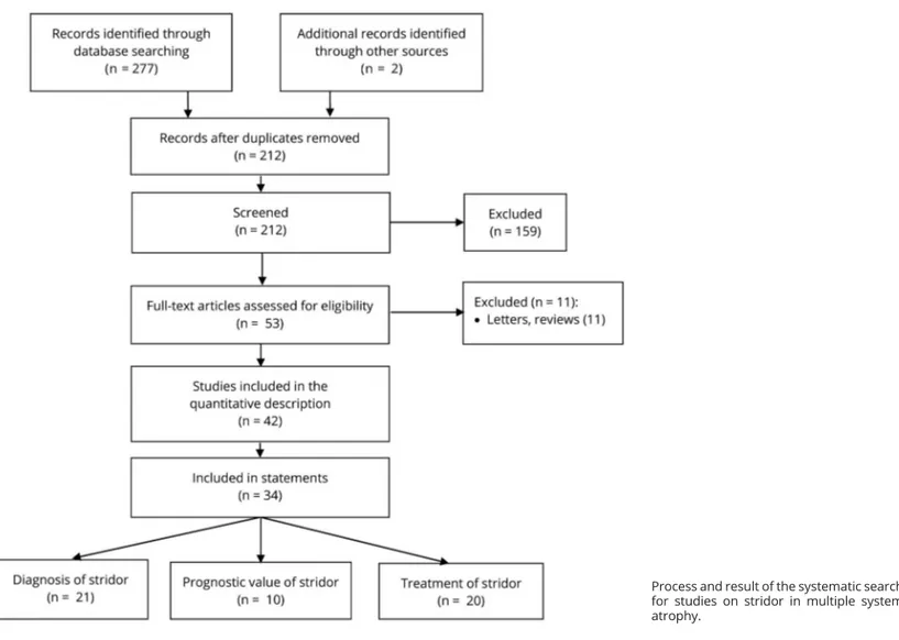
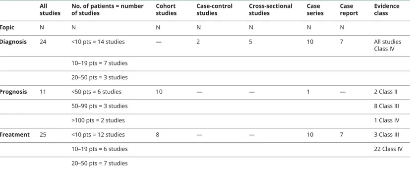
The literature search was performed in July 2017 and retrieved 212 citations after duplicate removal (figure). Each retrieved article was screened to assess potential relevance, and 53 were reviewed from the full text for inclusion. A total of 42 studies finally met the prespecified inclusion criteria, and 34 were used as basis for the statements. The majority of studies on diagnosis and treatment were of Class IV quality; those on prognosis of Class III (tables 1–4). Most studies on diagnosis and treatment included fewer than 10 patients and were case series or case reports as design. The cohort design was used in the majority of prognosis studies.
Diagnosis of stridor
Stridor is a respiratory disturbance that in MSA typically occurs during sleep and might develop at any time point in the disease process. According to clinical and clinicopathologic studies, stridor prevalence in MSA ranges from 12% to 42%8–12,23–25and is similar in MSA-C and MSA-P.8,9,11,12In 2 studies, 4%–5.2% of patients presented stridor as an initial manifestation of MSA.8,26 The clinical diagnosis of stridor remains challenging. The presence of a nighttime witness is typically necessary to suspect stridor because patients may be unaware of it. A high-pitched sound or heavy snoring are the symptoms frequently reported in patients who eventually turn out to have stridor, illustrating the problem of the differential diagnosis with snoring and ob-structive sleep apnea syndrome, which are 2 other frequent sleep-related breathing disorders in MSA.3,4,10,24,27–29 The term“peculiar snoring” was initially used to describe the distinctive noise occurring in MSA due to vibration of the vocal folds in inspiration, with a fundamental acoustic frequency of 260–330 Hz, different from that of ordinary soft palate snor-ing.30 Subsequently, only a single study has analyzed the acoustic features of stridor in 22 patients with MSA by means of the Multi-Dimensional Voice Program.31This study showed that stridor can be decomposed into rhythmic and semi-rhythmic waveforms. In both cases, it comprises formats and harmonics, whose presence suggests an origin in the vocal cords. In contrast, the sound analysis of snoring that was available for 18 of these patients revealed an irregular-shaped sound with no formats and harmonics.
Studies with video polysomnography (VPSG), which include audio recording and concurrent evaluation of vocal cord motion by fiberoptic laryngoscopy, showed that the high-pitched sound identified as stridor in patients with MSA was associated with impaired vocal cord abduction, paradoxical adduction, or both during inspiration and expiration, leading
to narrowing of the rima glottidis. This indicates that in-spiratory vibration of the narrowed vocal cord folds causes stridor.32–38
In 1 VPSG study exploring breathing activity and EMG ac-tivity of the respiratory muscles, stridor was accompanied by overactivation of intercostalis and diaphragmatic muscles. In this study, the observation of tonic and subcontinuous muscle recruitment with phasing out of thoracic as opposed to ab-dominal respiratory traces with paradoxical inward move-ments of the abdominal wall during inspiration was suggestive of paradoxical breathing.37
Further VPSG studies showed that other sleep-related re-spiratory disturbances such as snoring, central and obstructive sleep apneas, and breathing rate abnormalities (i.e., a patho-logic breathing rate increase during non-REM and sleep) may occur in patients with MSA with and without stridor.12,24,33,37–39
Laryngoscopy during wakefulness in patients with MSA with stridor, performed to exclude secondary causes or functional vocal cord abnormalities related to other neurologic con-ditions, can reveal bilateral or unilateral impairment of vocal
cord abduction of varying severity or normal vocal cord motility.27,29,32,33,35,36,39–44Conversely, impairment of vocal cord motility during wakefulness was also observed in patients with MSA without stridor during sleep.27
Studies with drug-induced sleep endoscopy (DISE) dem-onstrated impaired abduction or paradoxical adduction of the vocal cords in patients with MSA with stridor who had normal vocal cord motility on awake laryngoscopy.32,33,35–37,41–44
Finally, a few studies have performed EMG of laryngeal muscles during wakefulness and drug-induced sleep. Patients with MSA with stridor could present normal EMG activity of adductor and abductor laryngeal muscles during quiet breathing and inspiration. Alternatively, they could show a neurogenic pattern of muscle unit action potential analysis of these muscles associated with tonic activity of adductor muscles during quiet breathing and paradoxical activity during inspiration. During drug-induced sleep, the main patterns were persistent tonic activity or paradoxical activation of laryngeal adductor muscles during inspiration.35–38,44–46
FigurePreferred Reporting Items for Systematic Reviews and Meta-Analyses flow diagram
Process and result of the systematic search for studies on stridor in multiple system atrophy.
Statements on the diagnosis of stridor Clinical features suggesting the presence of stridor
Statements are based on core literature consisting of Class IV level studies10,12,24,31and expert opinion.
c Stridor is suspected when a high-pitched breathing sound
is emitted by the patient during sleep or while awake, or when reported by caregivers.
c Stridor is probably underrecognized because patients and
caregivers may be unaware of its presence, especially when it occurs at night.
c Recognition could be possible by the patient or
caregiver after imitation of stridor by the physician (see supplementary sound track file, links.lww.com/ WNL/A973).
Home audio recording to support the diagnosis of stridor
Statement is based on expert opinion because literature on the use of home audio recording to support the diagnosis of stridor (differential diagnosis of stridor from snoring) is lacking.
c Patients and caregivers should be encouraged to audio
record episodes of suspected stridor.
VPSG to support the diagnosis of stridor
Statements are based on core literature consisting of Class IV level studies10,12,24,31,33,37–39and expert opinion.
c VPSG including audio is not necessary if the physician
has already diagnosed stridor.
c VPSG including audio may demonstrate stridor and its
inspiratory nature.
c VPSG can characterize other sleep sounds.
Laryngoscopy for assessing stridor
Statements are based on core literature consisting of Class IV level studies27,29,32–37,39–44and expert opinion.
c Laryngoscopy can exclude mechanical lesions (e.g.,
masses and scars) or functional vocal cord abnormalities related to different neurologic conditions (central or peripheral disorders).
c Laryngoscopy may reveal vocal cord motility impairment
in patients with MSA with stridor.
c If awake laryngoscopy is normal, DISE might be considered
if the suspicion of sleep-related stridor needs confirmation.
Other investigations for assessing stridor
Statements are based on core literature consisting of Class IV level studies35–38,44–46and expert opinion.
c There is no evidence that other investigations are useful. c EMG of the laryngeal muscles may show denervation or
abnormal hyperactivity.
Conclusion on diagnostic criteria for stridor and definition of stridor in MSA
c Stridor in MSA is a strained, high-pitched, harsh
respiratory sound, mainly inspiratory, caused by laryngeal dysfunction leading to narrowing of the rima glottidis. It may occur only during sleep or it may be present both during sleep and wakefulness.
c Stridor may be recognized clinically if present at the time
of neurologic examination, with the help of a witness, or by listening to an audio recording.
c Laryngoscopy is suggested to exclude mechanical lesions
(e.g., masses and scars) or functional vocal cord abnormalities related to different neurologic conditions (central or peripheral disorders).
Table 1Descriptive features of eligible studies on stridor in MSA
All studies
No. of patients = number of studies Cohort studies Case-control studies Cross-sectional studies Case series Case report Evidence class Topic N N N N N N N
Diagnosis 24 <10 pts = 14 studies — 2 5 10 7 All studies
Class IV
10–19 pts = 7 studies 20–50 pts = 3 studies
Prognosis 11 <50 pts = 6 studies 10 — — 1 — 2 Class II
50–99 pts = 3 studies 8 Class III
>100 pts = 2 studies 1 Class IV
Treatment 25 <10 pts = 12 studies 8 — — 10 7 3 Class III
10–19 pts = 6 studies 22 Class IV
20–50 pts = 7 studies
Abbreviation: MSA = multiple system atrophy; pts = patients.
c If awake laryngoscopy is normal and the suspicion of
sleep-related stridor needs confirmation, the following additional evaluations might be considered: (1) DISE
and (2) VPSG to document the inspiratory nature of the sound, the presence of expiratory intercostalis activation, or the presence of associated sleep breathing disorders.
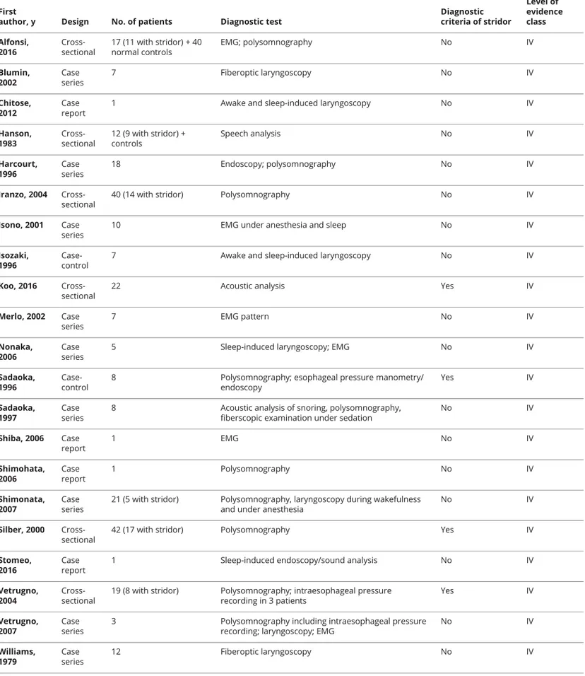
Table 2 Studies that form the basis of the statements on diagnosis with their level of evidence
First
author, y Design No. of patients Diagnostic test
Diagnostic criteria of stridor Level of evidence class Alfonsi, 2016 Cross-sectional 17 (11 with stridor) + 40 normal controls EMG; polysomnography No IV Blumin, 2002 Case series 7 Fiberoptic laryngoscopy No IV Chitose, 2012 Case report
1 Awake and sleep-induced laryngoscopy No IV
Hanson, 1983 Cross-sectional 12 (9 with stridor) + controls Speech analysis No IV Harcourt, 1996 Case series 18 Endoscopy; polysomnography No IV Iranzo, 2004 Cross-sectional
40 (14 with stridor) Polysomnography No IV
Isono, 2001 Case series
10 EMG under anesthesia and sleep No IV
Isozaki, 1996
Case-control
7 Awake and sleep-induced laryngoscopy No IV
Koo, 2016 Cross-sectional
22 Acoustic analysis Yes IV
Merlo, 2002 Case series 7 EMG pattern No IV Nonaka, 2006 Case series
5 Sleep-induced laryngoscopy; EMG No IV
Sadaoka, 1996
Case-control
8 Polysomnography; esophageal pressure manometry/ endoscopy Yes IV Sadaoka, 1997 Case series
8 Acoustic analysis of snoring, polysomnography, fiberscopic examination under sedation
No IV Shiba, 2006 Case report 1 EMG No IV Shimohata, 2006 Case report 1 Polysomnography No IV Shimonata, 2007 Case series
21 (5 with stridor) Polysomnography, laryngoscopy during wakefulness and under anesthesia
No IV
Silber, 2000 Cross-sectional
42 (17 with stridor) Polysomnography Yes IV
Stomeo, 2016
Case report
1 Sleep-induced endoscopy/sound analysis No IV
Vetrugno, 2004
Cross-sectional
19 (8 with stridor) Polysomnography; intraesophageal pressure recording in 3 patients Yes IV Vetrugno, 2007 Case series
3 Polysomnography including intraesophageal pressure recording; laryngoscopy; EMG
No IV Williams, 1979 Case series 12 Fiberoptic laryngoscopy No IV
Each study was classified according to various descriptors, including topic domain, sample size, design, presence of diagnostic criteria of the syndrome, and level of evidence according to the Classification of Evidence Schemes of the Clinical Practice Guideline Process Manual of the American Academy of Neurology (2011). Each study was graded according to its risk of bias from Class I to Class IV (with I highest quality and IV lowest quality). Risk of bias was judged by assessing specific quality elements (i.e., study design, patient spectrum, data collection, and masking) for each clinical topic (diagnostic accuracy, prognostic accuracy, and treatment).
Prognostic value of stridor
Retrospective cohort studies have reported conflicting results on the prognostic value of stridor.47Seven studies did notfind an association between the presence of stridor during the disease course and shortened survival.8,9,11,14,48–50In most of these studies, stridor was clinically suspected without in-strumental confirmation. In contrast, 1 study showed shorter survival in patients with MSA with stridor after VPSG re-cording, but not from disease onset.10 Finally, the largest study with VPSG found that early onset of stridor (within 3 years from motor or autonomic symptom onset) was an in-dependent predictor of shorter survival.8Based on an analysis with the Multi-Dimensional Voice Program, 1 study reported that acoustic features of stridor may affect survival in MSA.31
Statements on the prognostic value of stridor Effect of stridor on survival
Statements regarding the effect of stridor on survival in MSA are based on core literature consisting of Class II/III level studies,8–11,14,31,48–50a systematic review,47and expert opinion.
c Whether stridor affects survival is uncertain.
c Stridor within 3 years of motor or autonomic symptom
onset may shorten survival. However, identification of stridor onset may be difficult.
c Whether specific features of stridor affect survival remains
to be determined.
c Stridor during wakefulness is widely considered to reflect
a more advanced stage of the disease than stridor occurring during sleep.
Effect of stridor on well-being or health-related quality of life
Literature on the effect of stridor on well-being or health-related quality of life is lacking. Statements are based on expert opinion.
c Stridor can be distressing for patients and caregivers. c The impact on health-related quality of life remains to be
determined.
Treatment of stridor
Four retrospective studies reported that the treatment of stridor improves survival.8,12–14 Stridor treatment mainly comprised CPAP or tracheostomy.10,27,29,32,33,35,39,51,52In 3 studies (<15 patients), CPAP initially eliminated stridor in almost all patients,12,13,51but the long-term symptomatic ef-fect remains unknown.
Three studies reported survival in patients with MSA treated with CPAP. In 1 small study, CPAP had no effect on sur-vival.10 In another prospective cohort study, patients with MSA with stridor receiving CPAP (n = 13) had similar me-dian survival compared with a group of patients with MSA without stridor (n = 26).12Sudden death was reported in 2 of 13 patients following CPAP initiation.13
Classic tracheostomy is usually the surgical procedure of choice for stridor. This involves the positioning of a fenes-trated cannula, maintained closed during the day to allow phonation.53 Alternative techniques such as skin-lined tra-cheostomy have recently been proposed for the treatment of severe stridor in MSA.54Skin-lined tracheostomy offers sev-eral advantages, such as a greater opening of the stoma, higher stability over time, less risk of granulation tissue, and re-versibility. In addition, it does not require a cannula during the night, and the stoma is easy to plug during the day.
Three studies focused on the role of tracheostomy on survival. In the largest retrospective study (n = 42 with stridor), patients treated with tracheostomy had longer overall disease
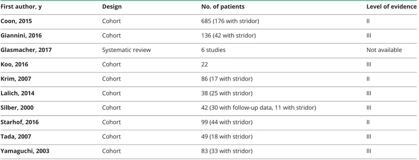
Table 3Studies that form the basis of the statements on prognosis with their level of evidence
First author, y Design No. of patients Level of evidence
Coon, 2015 Cohort 685 (176 with stridor) II
Giannini, 2016 Cohort 136 (42 with stridor) III
Glasmacher, 2017 Systematic review 6 studies Not available
Koo, 2016 Cohort 22 III
Krim, 2007 Cohort 86 (17 with stridor) II
Lalich, 2014 Cohort 38 (25 with stridor) III
Silber, 2000 Cohort 42 (30 with follow-up data, 11 with stridor) III
Starhof, 2016 Cohort 99 (44 with stridor) II
Tada, 2007 Cohort 49 (18 with stridor) III
Yamaguchi, 2003 Cohort 83 (33 with stridor) III
Each study was classified according to various descriptors, including topic domain, sample size, design, presence of diagnostic criteria of the syndrome, and level of evidence according to the Classification of Evidence Schemes of the Clinical Practice Guideline Process Manual of the American Academy of Neurology (2011). Each study was graded according to its risk of bias from Class I to Class IV (with I highest quality and IV lowest quality). Risk of bias was judged by assessing specific quality elements (i.e., study design, patient spectrum, data collection, and masking) for each clinical topic (diagnostic accuracy, prognostic accuracy, and treatment).
duration, longer disease duration after stridor onset, and longer disease duration after treatment compared with those treated with CPAP.8Another study showed that tracheos-tomy may reduce the risk of death and of sudden death in patients with MSA with stridor.11One study reported that 2 of the 4 patients with tracheostomy died 1 year after the sleep evaluation, whereas the other 2 were alive 1.9 and 7 years later.10
Single case reports have described the use of posterior cordotomy and arytenoidectomy,28,43,44,55 and botulinum toxin relieved dystonic stridor in 3 of 4 patients 1 month after inoculation.45
Statements on the treatment of stridor Treatment for symptomatic control of stridor
Statements are based on core literature consisting of Class III/ IV level studies8,10–14,27,29,32,33,35,39,49,51,52and expert opinion.
c Ventilation during sleep (CPAP) can be useful in the
symptomatic control of stridor.
c Consider ventilation during sleep (CPAP) as afirst-line
symptomatic therapy.
c Tracheostomy bypasses upper airway obstruction at
laryngeal level and relieves distressing stridor. Tracheos-tomy is effective in the symptomatic control of stridor.
c Persistent and severe stridor may require tracheostomy.
CPAP for improving survival of patients with stridor
Statement is based on core literature consisting of Class III/IV level studies.8,10,12,13
c Whether CPAP improves survival in patients with MSA
with stridor is uncertain.
Tracheostomy for improving survival of patients with stridor
Statement is based on core literature consisting of Class III/IV level studies.8,10,11
c Tracheostomy might improve survival in patients with
stridor.
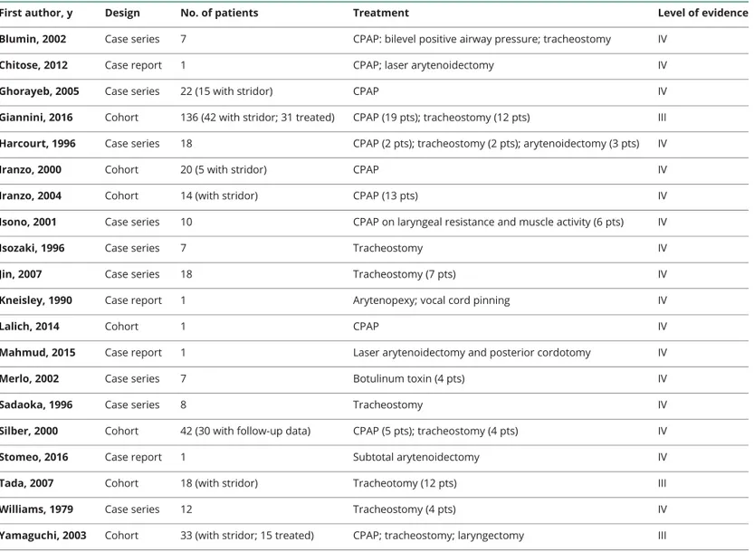
Table 4 Studies that form the basis of the statements on therapy with their level of evidence
First author, y Design No. of patients Treatment Level of evidence
Blumin, 2002 Case series 7 CPAP: bilevel positive airway pressure; tracheostomy IV
Chitose, 2012 Case report 1 CPAP; laser arytenoidectomy IV
Ghorayeb, 2005 Case series 22 (15 with stridor) CPAP IV
Giannini, 2016 Cohort 136 (42 with stridor; 31 treated) CPAP (19 pts); tracheostomy (12 pts) III
Harcourt, 1996 Case series 18 CPAP (2 pts); tracheostomy (2 pts); arytenoidectomy (3 pts) IV
Iranzo, 2000 Cohort 20 (5 with stridor) CPAP IV
Iranzo, 2004 Cohort 14 (with stridor) CPAP (13 pts) IV
Isono, 2001 Case series 10 CPAP on laryngeal resistance and muscle activity (6 pts) IV
Isozaki, 1996 Case series 7 Tracheostomy IV
Jin, 2007 Case series 18 Tracheostomy (7 pts) IV
Kneisley, 1990 Case report 1 Arytenopexy; vocal cord pinning IV
Lalich, 2014 Cohort 1 CPAP IV
Mahmud, 2015 Case report 1 Laser arytenoidectomy and posterior cordotomy IV
Merlo, 2002 Case series 7 Botulinum toxin (4 pts) IV
Sadaoka, 1996 Case series 8 Tracheostomy IV
Silber, 2000 Cohort 42 (30 with follow-up data) CPAP (5 pts); tracheostomy (4 pts) IV
Stomeo, 2016 Case report 1 Subtotal arytenoidectomy IV
Tada, 2007 Cohort 18 (with stridor) Tracheotomy (12 pts) III
Williams, 1979 Case series 12 Tracheostomy (4 pts) IV
Yamaguchi, 2003 Cohort 33 (with stridor; 15 treated) CPAP; tracheostomy; laryngectomy III
Abbreviation: CPAP = continuous positive airway pressure; pts = patients.
Each study was classified according to various descriptors, including topic domain, sample size, design, presence of diagnostic criteria of the syndrome, and level of evidence according to the Classification of Evidence Schemes of the Clinical Practice Guideline Process Manual of the American Academy of Neurology (2011). Each study was graded according to its risk of bias from Class I to Class IV (with I highest quality and IV lowest quality). Risk of bias was judged by assessing specific quality elements (i.e., study design, patient spectrum, data collection, and masking) for each clinical topic (diagnostic accuracy, prognostic accuracy, and treatment).
Other treatment options for stridor
Statement is based on core literature consisting of Class IV level studies including case reports.28,43–45,55
c There is insufficient evidence for minimally invasive
procedures and botulinum toxin injections for the symptomatic treatment of stridor.
Research needs
The present Consensus Conference represents thefirst effort to systematically revise the literature and to provide state-ments on stridor, a specific feature of MSA, that could affect the disease course. However, despite the importance of this topic, the majority of studies available in the literature were of Class III–IV quality, leading also to statements based on ex-pert opinions. Furthermore, several research gaps emerged during the consensus meeting concerning diagnosis, prog-nosis, and treatment for stridor.
One main challenge is the diagnosis of stridor. Whether the imitation of stridor by the physician during follow-up visits is sufficient to correctly and earlier identify stridor or whether other tools, such as specific questionnaires or a home audio recording, may improve diagnostic accuracy is unknown. This point could be of crucial importance if the negative prognostic role of early stridor onset is confirmed. It has also to be established when the use of VPSG is necessary for the diagnosis of stridor. For these reasons, a questionnaire for detecting stridor should be de-veloped and its diagnostic accuracy be compared with VPSG in a multicenter prospective study. Similarly, the diagnostic accu-racy of sound imitation by the physician or home audio re-cording should be evaluated. A smartphone application could be developed to automatically recognize the stridor sound. Finally, the place of DISE for early stridor detection requires further investigation, and the method to measure the progression of stridor over time should be standardized.
The relationship between stridor and other breathing disorders (i.e., central apneas and breathing rate abnormalities) and their respective contributions to disease prognosis and survival should be determined through a multicenter prospective study. VPSG should be used to determine stridor and breathing disorders in this study. Moreover, further studies could contribute to elucidate the role on survival of specific characteristic obtained from awake laryngoscopy, DISE, laryngeal EMG (denervation/muscle hy-peractivity), and acoustic recordings. In addition, the contribu-tion of stridor to patients and caregiver quality of life is unknown. To guide the physician for the treatment of stridor, randomized controlled trials comparing the efficacy of CPAP and trache-ostomy for different degrees of stridor are warranted. Studies using laryngoscopy and/or laryngeal EMG should be conducted to identify specific characteristics that may predict treatment tolerance and response. Moreover, the usefulness of CPAP for severe stridor, as well as technical aspects including titration, patient compliance, and the timing of follow-up need to be
determined. Finally, the usefulness of bilevel PAP should be compared with CPAP. Similarly to CPAP, interventional studies should also compare skin-lined vs conventional tracheostomy. Literature revision and the emergence of several research gaps on diagnosis, prognosis, and treatment for stridor rise the need of prospective multicenter studies on large samples, and with a randomized controlled design concerning its treat-ment, to provide high level of evidence in the role of stridor in MSA course.
Author contributions
Scientific Committee: P. Cortelli (Chair), G. Calandra-Buonaura, F. Provini, P. Martinelli, A. Iranzo, and P. A. Low. Technical Committee: L. Vignatelli and G. Giannini. Consen-sus Panel: G. Abbruzzese (Chairperson), P. Bower, P. Cortelli, P. Martinelli, N. Quinn, E. E. Benarroch, E. Tolosa, and G. K. Wenning. Stridor Workgroup: E. Alfonsi, G. Calandra-Buonaura, I. Ghorayeb, W. G. Meissner (Speaker), T. Ozawa, C. Pacchetti, N. G. Pozzi, and C. Vicini. Dysphagia Workgroup: A. Antonini, K. Bhatia, J. Bonavita, H. Kaufmann (Speaker), M. T. Pellecchia, N. Pizzorni, A. Schindler, and F. Tison.
Acknowledgment
The authors thank Maria Camerlingo (Agenzia sanitaria e sociale regionale, Regione Emilia-Romagna) for assisting in the search strategy.
Study funding
This study was funded by IRCCS, Istituto delle Scienze Neurologiche di Bologna, Bologna, Italy.
Disclosure
P. Cortelli and G. Calandra-Buonaura report no disclosures relevant to the manuscript. E. Benarroch: Section Editor, Neurology. G. Giannini and A. Iranzo report no disclosures relevant to the manuscript. P. Low: supported by the NIH (P01NS44233, U54NS065736, R01 FD004789, and R01 NS092625) and Mayo Funds. P. Martinelli reports no dis-closures relevant to the manuscript. F. Provini has received honoraria for speaking engagements or consulting activities from Sanofi, Bial, Fidia, Vanda Pharmaceuticals, Zambon, Eisai Japan, and Italfarmaco. N. Quinn reports no disclosures relevant to the manuscript. E. Tolosa received honoraria for consultancy from Novartis, Teva, Bial, Accorda, Boehringer Ingelheim, UCB, Solvay, Lundbeck, and Biogen and has re-ceived funding for research from Spanish Network for Re-search on Neurodegenerative Disorders (CIBERNED)–Instituto Carlos III (ISCIII) and The Michael J. Fox Foundation for Parkinson’s Research (MJFF). G. Wenning, G. Abbruzzese, P. Bower, E. Alfonsi, I. Ghorayeb, T. Ozawa, C. Pacchetti, N. Pozzi, C. Vicini, A. Antonini, K. Bhatia, J. Bonavita, H. Kaufmann, M. Pellecchia, N. Pizzorni, A. Schindler, F. Tison, L. Vignatelli, and W. Meissner report no disclosures. Outside the present work, W. Meissner has received fees for editorial activities with Springer, for con-sultancy activities from Affiris, Biohaven, Lundbeck, and
Sanofi, teaching honoraria from UCB and MDS, and research support from the Michael J Fox Foundation, the University Hospital Bordeaux, the French Health Ministry, the European Community, ANR, ARAMISE, PSP France, MSA Coalition, ARAMISE, and LABEX Excellence Initiative. Go to Neurol-ogy.org/N for full disclosures.
Publication history
Received by Neurology March 22, 2019. Accepted in final form June 20, 2019.
References
1. Gilman S, Wenning GK, Low PA, et al. Second consensus statement on the diagnosis of multiple system atrophy. Neurology 2008;71:670–676.
2. Fanciulli A, Wenning GK. Multiple-system atrophy. N Engl J Med 2015;372: 249–263.
3. Abbott SM, Videnovic A. Sleep disorders in atypical parkinsonism. Mov Disord Clin Pract 2014;1:89–96.
4. Iranzo A. Sleep and breathing in multiple system atrophy. Curr Treat Options Neurol 2007;9:347–353.
5. Ozawa T, Sekiya K, Aizawa N, Terajima K, Nishizawa M. Laryngeal stridor in multiple system atrophy: clinicopathological features and causal hypotheses. J Neurol Sci 2016; 361:243–249.
6. Osaki Y, Ben-Shlomo Y, Lees AJ, Wenning GK, Quinn NP. A validation exercise on the new consensus criteria for multiple system atrophy. Mov Disord 2009;24: 2272–2276.
7. K¨ollensperger M, Geser F, Seppi K, et al. Redflags for multiple system atrophy. Mov Disord 2008;23:1093–1099.
8. Giannini G, Calandra-Buonaura G, Mastrolilli F, et al. Early stridor onset and stridor treatment predict survival in 136 patients with MSA. Neurology 2016;87: 1375–1383.
9. Coon EA, Sletten DM, Suarez MD, et al. Clinical features and autonomic testing predict survival in multiple system atrophy. Brain 2015;138:3623–3631. 10. Silber MH, Levine S. Stridor and death in multiple system atrophy. Mov Disord 2000;
15:699–704.
11. Tada M, Onodera O, Tada M, et al. Early development of autonomic dysfunction may predict poor prognosis in patients with multiple system atrophy. Arch Neurol 2007; 64:256–260.
12. Iranzo A, Santamaria J, Tolosa E, et al. Long-term effect of CPAP in the treatment of nocturnal stridor in multiple system atrophy. Neurology 2004;63:930–932. 13. Ghorayeb I, Yekhlef F, Bioulac B, Tison F. Continuous positive airway pressure for
sleep-related breathing disorders in multiple system atrophy: long-term acceptance. Sleep Med 2005;6:359–362.
14. Yamaguchi M, Arai K, Asahina M, Hattori T. Laryngeal stridor in multiple system atrophy. Eur Neurol 2003;49:154–159.
15. Nair R, Aggarwal R, Khanna D. Methods of formal consensus in classification/ diagnostic criteria and guideline development. Semin Arthritis Rheum 2011;41: 95–105.
16. Candiani G, Colombo C, Daghini R, et al. Manuale metodologico: come organizzare una conferenza di consenso. Istituto Superiore di Sanit`a, Sistema Nazionale Linee Guida SNLG, Roma, Novembre 2009 [online]. Available at: medicinanarrativa.eu/ wp-content/uploads/consensus_conference-1.pdf. Accessed September 29, 2018. 17. American College of Chest Physicians. Consensus Statement Methodology [online].
Available at: chestnet.org/Guidelines-and-Resources/About-CHEST-Guidelines/ Guideline-Development. Accessed February 28, 2019.
18. Centre for Reviews and Dissemination. Systematic Reviews: CRD’s guidance for undertaking systematic reviews in health care. 2009. Available at: york.ac.uk/crd/ guidance/. Accessed February 28, 2019.
19. CEBM- Centre for Evidence-Based Medicine, PICO Structure. Available at: cebm. net/2014/06/asking-focused-questions/. Accessed February 28, 2019.
20. Moher D, Liberati A, Tetzlaff J, Altman DG, Group P. Preferred reporting items for systematic reviews and meta-analyses: the PRISMA statement. BMJ 2009;339:b2535. 21. Bragge P, Clavisi O, Turner T, Tavender E, Collie A, Gruen RL. The Global Evidence Mapping Initiative: scoping research in broad topic areas. BMC Med Res Methodol 2011;11:92.
22. AAN (American Academy of Neurology). Clinical Practice Guideline Process Man-ual, 2011. St. Paul: The American Academy of Neurology; 2011. Available at tools.aan. com/globals/axon/assets/9023.pdf. Accessed September 29, 2018.
23. Wenning GK, Ben Shlomo Y, Magalhães M, Daniel SE, Quinn NP. Clinical features and natural history of multiple system atrophy. An analysis of 100 cases. Brain 1994; 117(pt 4):835–845.
24. Vetrugno R, Provini F, Cortelli P, et al. Sleep disorders in multiple system atrophy: a correlative video-polysomnographic study. Sleep Med 2004;5:21–30.
25. Figueroa JJ, Singer W, Parsaik A, et al. Multiple system atrophy: prognostic indicators of survival. Mov Disord 2014;29:1151–1157.
26. Uzawa A, Sakakibara R, Tamura N, et al. Laryngeal abductor paralysis can be a solitary manifestation of multiple system atrophy. J Neurol Neurosurg Psychiatry 2005;76: 1739–1741.
27. Williams A, Hanson D, Calne DB. Vocal cord paralysis in the Shy-Drager syndrome. J Neurol Neurosurg Psychiatry 1979;42:151–153.
28. Kneisley LW, Rederich GJ. Nocturnal stridor in olivopontocerebellar atrophy. Sleep 1990;13:362–368.
29. Blumin JH, Berke GS. Bilateral vocal fold paresis and multiple system atrophy. Arch Otolaryngol Head Neck Surg 2002;128:1404–1407.
30. Kakitsuba N, Sadaoka T, Kanai R, Fujiwara Y, Takahashi H. Peculiar snoring in patients with multiple system atrophy: its sound source, acoustic characteristics, and diagnostic significance. Ann Otol Rhinol Laryngol 1997;106:380–384.
31. Koo DL, Lee JY, Joo EY, Hong SB, Nam H. Acoustic characteristics of stridor in multiple system atrophy. PLoS One 2016;11:e0153935.
32. Isozaki E, Naito A, Horiguchi S, Kawamura R, Hayashida T, Tanabe H. Early diagnosis and stage classification of vocal cord abductor paralysis in patients with multiple system atrophy. J Neurol Neurosurg Psychiatry 1996;60:399–402.
33. Sadaoka T, Kakitsuba N, Fujiwara Y, Kanai R, Takahashi H. Sleep-related breathing disorders in patients with multiple system atrophy and vocal fold palsy. Sleep 1996;19: 479–484.
34. Sadaoka T, Kakitsuba N, Fujiwara Y, Kanai R, Takahashi H. Limitation of vocal fold abduction only during sleep in a patient with Shy-Drager syndrome. Am J Otolaryngol 1997;18:145–147.
35. Isono S, Shiba K, Yamaguchi M, et al. Pathogenesis of laryngeal narrowing in patients with multiple system atrophy. J Physiol (Lond) 2001;536:237–249.
36. Nonaka M, Imai T, Shintani T, Kawamata M, Chiba S, Matsumoto H. Non-invasive positive pressure ventilation for laryngeal contraction disorder during sleep in mul-tiple system atrophy. J Neurol Sci 2006;247:53–58.
37. Vetrugno R, Liguori R, Cortelli P, et al. Sleep-related stridor due to dystonic vocal cord motion and neurogenic tachypnea/tachycardia in multiple system atrophy. Mov Disord 2007;22:673–678.
38. Alfonsi E, Terzaghi M, Cosentino G, et al. Specific patterns of laryngeal electromy-ography during wakefulness are associated to sleep disordered breathing and noc-turnal stridor in multiple system atrophy. Parkinsonism Relat Disord 2016;31: 104–109.
39. Harcourt J, Spraggs P, Mathias C, Brookes G. Sleep-related breathing disorders in the Shy-Drager syndrome. Observations on investigation and management. Eur J Neurol 1996;3:186–190.
40. Hanson DG, Ludlow CL, Bassich CJ. Vocal fold paresis in Shy-Drager syndrome. Ann Otol Rhinol Laryngol 1983;92:85–90.
41. Shimohata T, Nakayama H, Shinoda H, et al. Multiple system atrophy with pro-gressive nocturnal hypoxemia: case report with polysomnography and continuous positive airway pressure treatment. Eur Neurol 2006;56:258–260.
42. Shimohata T, Shinoda H, Nakayama H, et al. Daytime hypoxemia, sleep-disordered breathing, and laryngopharyngealfindings in multiple system atrophy. Arch Neurol 2007;64:856–861.
43. Chitose S, Kikuchi A, Ikezono K, Umeno H, Nakashima T. Effect of laser arytenoi-dectomy on respiratory stridor caused by multiple system atrophy. J Clin Sleep Med 2012;8:713–715.
44. Stomeo F, Rispoli V, Sensi M, Pastore A, Malagutti N, Pelucchi S. Subtotal aryte-noidectomy for the treatment of laryngeal stridor in multiple system atrophy: pho-natory and swallowing results. Braz J Otorhinolaryngol 2016;82:116–120. 45. Merlo IM, Occhini A, Pacchetti C, Alfonsi E. Not paralysis, but dystonia causes stridor
in multiple system atrophy. Neurology 2002;58:649–652.
46. Shiba K, Isono S. Tracheostomy abolishes paradoxical activation of the vocal cord adductor in multiple system atrophy. Auris Nasus Larynx 2006;33:295–298. 47. Glasmacher SA, Leigh PN, Saha RA. Predictors of survival in progressive supranuclear
palsy and multiple system atrophy: a systematic review and meta-analysis. J Neurol Neurosurg Psychiatry 2017;88:402–411.
48. Krim E, Yekhlef F, Chrysostome V, Ghorayeb I, Tison F. Multiple system atro-phy: prognostic factors in the“MSA- Aquitaine” cohort. Rev Neurol 2007;163: 54–65.
49. Lalich IJ, Ekbom DC, Starkman SJ, Orbelo DM, Morgenthaler TI. Vocal fold motion impairment in multiple system atrophy. Laryngoscope 2014;124:730–735. 50. Starhof C, Korbo L, Funch Lassen C, Winge K, Friis S. Clinical features in a Danish
population-based cohort of probable Multiple System Atrophy patients. Neuro-epidemiology 2016;46:261–267.
51. Iranzo A, Santamaria J, Tolosa E. Continuous positive air pressure eliminates noc-turnal stridor in multiple system atrophy. Barcelona Multiple System Atrophy Study Group. Lancet 2000;356:1329–1930.
52. Jin K, Onodera H, Chida K, Abe N, Itoyama Y. Spade-shaped vocal cord in a multiple system atrophy patient with nocturnal stridor. Intern Med 2007;46:921. 53. Kuhlo W, Doll E, Franck MC. Successful management of Pickwickian syndrome using
long-term tracheostomy [in German]. Dtsch Med Wochenschr 1969;94:1286–1290. 54. Campanini A, De Vito A, Frassineti S, Vicini C. Role of skin-lined tracheotomy in obstructive sleep apnoea syndrome: personal experience. Acta Otorhinolaryngol Ital 2004;24:68–74.
55. Mahmud A, Strens LH, Tedla M. Laser arytenoidectomy and posterior cordotomy in a patient with bilateral vocal cord paralysis due to multiple system atrophy. BMJ Case Rep 2015;2015:1–3.
DOI 10.1212/WNL.0000000000008208
2019;93;630-639
Neurology
Pietro Cortelli, Giovanna Calandra-Buonaura, Eduardo E. Benarroch, et al.
treatment
Stridor in multiple system atrophy: Consensus statement on diagnosis, prognosis, and
This information is current as of September 30, 2019
Services
Updated Information &
http://n.neurology.org/content/93/14/630.full
including high resolution figures, can be found at:
References
http://n.neurology.org/content/93/14/630.full#ref-list-1
This article cites 50 articles, 9 of which you can access for free at:
Subspecialty Collections
http://n.neurology.org/cgi/collection/prognosis
Prognosis
http://n.neurology.org/cgi/collection/multiple_system_atrophy
Multiple system atrophy
following collection(s):
This article, along with others on similar topics, appears in the
Permissions & Licensing
http://www.neurology.org/about/about_the_journal#permissions
its entirety can be found online at:
Information about reproducing this article in parts (figures,tables) or in
Reprints
http://n.neurology.org/subscribers/advertise
Information about ordering reprints can be found online:
ISSN: 0028-3878. Online ISSN: 1526-632X.
Wolters Kluwer Health, Inc. on behalf of the American Academy of Neurology.. All rights reserved. Print 1951, it is now a weekly with 48 issues per year. Copyright Copyright © 2019 The Author(s). Published by
® is the official journal of the American Academy of Neurology. Published continuously since