Keywords:
ADP-ribosylating toxins; NAD; NAD-glycohydrolase; Neisseria meningitidis; PathogenesisAbbreviations: NAD: β-Nicotinamide Adenine Nucleotide; NAM:
Nicotinamide; ARTs: ADP-Ribosyltransferases; NADase: NAD-Glycohydrolase; ε-NAD: N6-Etheno-NAD; IPTG:Isopropyl-1-β-D-Thiogalactopyranoside; B-PER: Bacterial Protein Extraction Reagent; PMSF: Phenyl Methyl Sulfonyl Fluoride; Ni-NTA: Nickel-Nitriloacetic Acid Matrix.
Introduction
Neisseria meningitidis is a Gram-negative opportunistic human specific pathogen, capable of life-threatening infections like meningitis and septicemia [1], responsible for significant worldwide morbidity and mortality [2,3]. Most of the bacterial pathogens possess a large number of toxic products, which target and kill host cells. Among these proteins, ADP-ribosylating toxins represent one of the main virulence factors of pathogens. ADP-ribosylating toxins catalyze the enzymatic ADP-ribosylation reaction [4] in which a single ADP-ribose moiety is transferred from NAD onto critical amino-acid residues of specific protein targets with the simultaneous release of nicotinamide (NAM). Physiological substrates of ADP-ribosylating toxins include elongation factor-2 [5,6], GTP-binding proteins [7], actin [8] and members of the antimicrobial peptide family [9]. We have identified an ORF in the N. meningitidis genome that codes for the ADP-ribosyltransferase (ART) NarE, which shares the molecular signature and biochemical features with the ART from V. cholerae and E. coli [10]. In analogy with these two toxins, NarE can ADP-ribosylate arginine, hydrolyze NAD in free ADP-ribose and NAM [11], and ADP-ribosylate itself [12]. A recently discovered feature of NarE is the presence of an iron-sulfur center (Fe-S) that we identified for the first time in a member of the ART family [13]. The iron coordination site has been resolved and the amino acids, two cysteines (C67 and C128) and two histidines (H46 and H57) that bind iron, have been identified [14]. The lack of assembly of the iron-binding site strongly reduced the transferase activity of NarE, while it affected the hydrolase activity only marginally [13]. These observations suggested that the Fe-S cluster and iron could have a role
*Corresponding author: Enrico Balducci, School of Biosciences and Veterinary Medicine, University of Camerino, via Gentile III da Varano, 62032 Camerino, Italy, Tel: +39-0737-402710; Fax +39-0737-636216; E-mail: [email protected] Received: August 20, 2015; Accepted: October 28, 2015; Published October 30, 2015
Citation: Del Vecchio M, Balducci E (2015) Opposite Regulatory Effects of Iron Ions on the In Vitro Catalytic Activities of NarE, the Toxin-like ADP-ribosyltransferase from Neisseria meningitidis. Biochem Anal Biochem 4: 214. doi:10.4172/2161-1009.1000214
Copyright: © 2015 Del Vecchio M, et al.. This is an open-access article distributed under the terms of the Creative Commons Attribution License, which permits unrestricted use, distribution, and reproduction in any medium, provided the original author and source are credited.
Abstract
NarE, the mono ADP-ribosyltransferase identified in Neisseria meningitidis, catalyzes three enzymatic reactions. NarE transfers a single ADP-ribose unit to guanidine compounds, hydrolyses NAD in nicotinamide and free ADP-ribose, and ADP-ribosylates itself. We have previously shown that NarE contains an iron-sulfur cluster by biophysical and biochemical analyses. The presence of a structured and stable iron-sulfur cluster is essential for ADP-ribosyltransferase but not for NAD-glycohydrolase activity. We report here that ferric, but not ferrous, ions stimulated the ADP-ribosyltransferase activity. On the contrary ferrous, but not ferric, ions activated NAD-glycohydrolase activity. These iron effects were reversed when enzymatic reactions were run in the presence of the iron-chelator O-phenantroline. In the presence of either ferric or ferrous ions there was an increase of the Vmax both for transferase and NADase
activity while the Km value for NAD was unaltered. The presence of 10 mM Fe3+ increased the ADP-ribosyltransferase
activity when we mutated residues not involved in cluster, while is ineffective when residues involved in cluster are mutated. Similar results were obtained with NAD-glycohydrolase activity. The results presented here demonstrate that iron, which plays an important role in metabolism, can modulate NarE activities depending on its oxidation state. This novel observation could be relevant in the context of Neisseria meningitidis infection.
Opposite Regulatory Effects of Iron Ions on the In Vitro Catalytic Activities of
NarE, the Toxin-like ADP-ribosyltransferase from Neisseria meningitidis
Mariangela Del Vecchio1 and Enrico Balducci2*
1Novartis Vaccines and Diagnostics S.r.l, a GlaxoSmithKline Vaccines company Via Fiorentina 1, 53100 Siena, Italy 2School of Biosciences and Veterinary Medicine, University of Camerino, via Gentile III da Varano, 62032 Camerino, Italy
in the catalytic mechanism of NarE [13]. It is well established that the success in survival and colonization of N. meningitidis depends on its ability to acquire iron from the host [15]. Moreover, iron regulates the expression of a number of genes involved in N. meningitidis virulence [16]. Since NarE contains iron we hypothesized that this metal could modulate the NarE transferase activity. Following this lead, we assessed the effects that iron ions exert on NarE catalytic activities in vitro. While the ADP-ribosyltranferase activity is enhanced by the presence of ferric ions (Fe3+), NAD-glycohydrolase (NADase) is enhanced by
ferrous ions (Fe2+). Kinetic parameters calculated in the presence of
Fe3+ or Fe2+ indicated that Km for NAD remained unaltered while the
Vmax relative to transferase or NADase activities consistently increased in the presence of Fe3+ or Fe2+ respectively. The results we have shown
previously [13] are expanded in the present study, in which we describe a selective activation of NarE catalytic activities by ferric and ferrous ions.
Material and Methods
Chemicals
[adenine-U-14C] NAD (274 mCi/mmol), [carbonyl-14C] NAD (53
mCi/mmol) were purchased from Amersham; MHAB N45 filter plates from Millipore; isopropyl-1-thio-β-D-galactopyranoside (IPTG) from Calbiochem; Bradford reagent for protein quantification and Dowex AG1-X2 were purchased from Bio-Rad. Bacterial Protein Extraction
Reagent (B-PER) was from Pierce while SimplyBlue Safe Stain from Invitrogen. All other reagents used in this study were purchased from Sigma-Aldrich.
Overexpression and purification of recombinant wild type
and mutant NarE
Recombinant NarE was produced growing Escherichia coli BL21 (DE3) transformed with pET21b+ plasmid carrying the narE gene [11] 18 h at 37°C. Recombinant NarE was purified as previously described [13]. Briefly, expression of narE gene was induced by addition of 1 mM IPTG. After 3 h induction at 25°C, cells were harvested by centrifugation and resuspended in B-PER using a ratio of 10 ml for 3 g of pellet in the presence of 0.1 mM MgCl2, 100 units of DNase I, and 1 mg/ml of chicken egg lysozyme. The supernatant obtained after centrifugation of the mixture was loaded onto metal nickel-affinity chromatography column (Ni-NTA; Pharmacia; 1.5 ml matrix/liter of culture volume). Bound proteins were eluted with buffer A (50 mM sodium phosphate buffer pH 7.4, 300 mM NaCl) containing 250 mM imidazole. The purification steps were carried out mainly under anaerobic conditions inside a portable glove box saturated with a mixture of N2 and H2. When the purification was carried out in aerobic conditions, the protein was used shortly after purification, to prevent iron-cluster disruption by air.
Site-directed mutagenesis
To mutate amino-acids residues of N. meningitidis NarE a PCR-based method was used. Oligonucleotides indicated below carrying a single nucleotide mismatch in the underlined positions were used to generate the mutants:
C4S, AGAGGCATTAGTTCCCAACAAGATGAG; C67S, TCTATATGACGGATCTTATATATCTACGA;
C128S, AGCTGAAGATTCTGGCTGGCTGTATTCCTGAAG; C130S, AGCTGAAGATTGTGGCTCTATTCCTGAAG.
After mutagenesis and DNA sequencing, all of the generated mutants were cloned into pET21b+, expressed and purified following
the procedure previously described [13].
Enzymatic assays
ADP-ribosyltransferase activity was measured using the standard assay described previously [17]. Briefly, NarE was incubated in a final volume of 0.3 ml, containing 50 mM potassium phosphate (pH 7.5), 20 mM agmatine as ADP-ribose acceptor and 0.1 mM
[adenine-U-14C]NAD (0.05 µCi). After incubation at 30°C, duplicate samples (100
µl) were applied to 1 ml columns of Dowex AG 1-X2. [adenine- 14C]
ADP-ribosylagmatine was eluted for radioassays with 5 ml of H2O and the radioactivity counted in a Packard Top counter. In alternative, the transferase activity of NarE was tested monitoring the enzymatic transfer of ADP-ribose to poly-arginine by a filter plate-based assay as described [18]. Assays were performed in a total volume of 0.3 ml with 0.6 mg of poly-arginine and 0.1 mM [carbonyl-14C]NAD (0.05
μCi). After incubation at 30°C, samples were precipitated with 0.3 ml of 50% (w/v) TCA for 30 min on ice. Samples (100 µl) were placed in quadruplicate in single wells of a mixed cellulose esters filter-plate under vacuum conditions. The unreacted [adenine-U-14C]NAD was
washed out with 10 volumes of 5% (w/v) TCA and the incorporated radioactivity measured in a Packard Top counter.
NADase activity was measured with an assay based on NAM release [19]. This assay was carried out in 50 mM potassium phosphate buffer,
pH 7.5, 0.1 mM [carbonyl-14C] NAD (0.05 μCi) in a total volume of 0.3
ml. Samples (100 μl) after incubation at 30 °C were applied in duplicate to 1-ml column of Dowex AG1-X2 and 14C-nicotinamide eluted with
5 ml of H2O for liquid scintillation counting. As an alternate method, NADase activity was evaluated with a fluorimetric assay as described by Barrio [20]. The reaction was started with the appropriate amount of purified NarE in a final volume of 0.7 ml buffered with 20 mM Tris/ HCl pH 7.0 containing 10 μM 1, N6-etheno-NAD (ε-NAD). Cleavage
of ε-NAD, which causes a 10-fold fluorescence enhancement, was continuously followed at 37°C in a Perkin Elmer spectrofluorometer at an emission wavelength of 410 nm and an excitation of 300 nm. NADase activity was expressed as ∆ relative fluorescence/min.
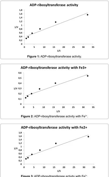
Iron effects were assayed in the transferase and NADase standard mixture assays above described with 0-25 mM of the following salts, Fe2Cl3 and FeCl3. In these experiments the enzyme preparation was extensively dialyzed against 50 mM potassium phosphate buffer (pH 7.4) before the assay (Figures 1-3).
Determination of kinetic parameters
To determine the kinetic constants we used the standard radioactive assay above described. Six concentrations of unlabelled NAD were
Figure 1: ADP-ribosyltransferase activity.
Figure 2: ADP-ribosyltransferase activity with Fe3+.
added for final concentration of 32.2, 16, 8, 4, 2, 1 and 0.5 mM. Kinetic parameters were calculated from Lineweaver-Burk plots according to H.I. Segel, Enzyme Kinetics: Behavior and Analysis of Rapid Equilibrium and Steady-state Enzyme Systems. Wiley-Interscience, New York, NY, 1975. Steady-state kinetics was also determined in the presence of 10 mM FeCl3 or Fe2Fe3* The enzyme used for the determination of kinetic analyses was previously dialyzed against 50 mM potassium phosphate buffer.
Protein assay
Protein concentration was determined using the Bradford assay kit (Bio-Rad) with BSA (Pierce) as a standard [21].
Results
Ferric ions stimulate NarE transferase activity
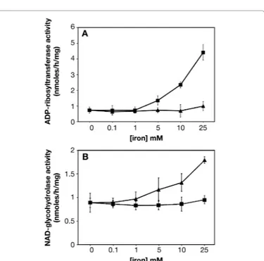
In our recent study we have reported that the ADP-ribosyltransferase of N. meningitidis, NarE, is an iron-containing enzyme [13]. Since the presence of iron in NarE was analyzed using Electron Paramagnetic Resonance spectroscopy (EPR), which identifies paramagnetic metal in proteins, we questioned whether the presence of ferric ions could have some effects on the catalytic activities of NarE. Therefore, we tested the NarE transferase activity in the presence of 10 mM FeCl3 or for comparison, 10 mM Fe2Cl3. As shown in Figure 4, Fe3+ increased
the transferase activity of NarE by 3 fold. This activation was ferric-ion specific since additferric-ion of 10 mM Fe2Cl3 had no effect. Members of the bacterial ART family such as Cholera and Escherichia coli toxins are activated by Mg2+ ions [22]; therefore, we also performed the
experiments in the presence of other trivalent (Cr, V) and bivalent (Ca, Mn, Li, Co, Ni, Zn, Mg) ions. However, none of the cations used showed any effects (data not shown). Then, we determined the ART activity of NarE with concentrations of Fe3+ ranging from 0.1 mM to
25 mM. As shown in Figure 5A, the effect was observed at 5 mM Fe3+
but not at lower concentrations and was more pronounced at 10 and 25 mM (Figure 5A).
Ferrous ions stimulate NarE NAD-glycohydrolase activity
Since NarE possesses also NADase activity, we tested whether ironions affected this enzymatic activity. NADase activity of NarE was unaltered in the presence of various concentrations of Fe3+ (Figure
5B). Of interest Fe2+, which was ineffective on ART activity (Figure
5A), enhanced the NADase activity starting at 5 mM Fe2+ up to 25
mM (Figure 5B) suggesting an asymmetrical activation of the catalytic activities of NarE. To confirm that the oxidation state of iron has a key role in the regulation of the catalytic activities of NarE, we performed experiments in the presence of 1 mM Na2S2O4 (sodium dithionite), a potent reducing agent. As shown in Table 1, Na2S2O4 enhanced the NADase activity while reducing the transferase activity thus reinforcing the hypothesis that the redox state of the iron ions is a key factor for the modulation of the catalytic activities of NarE. No effect on auto-ADP-ribosylation activity of NarE, were observed at any of conditions and concentrations of Fe2+ and Fe3+ used (data not shown) (Figure 6-8).
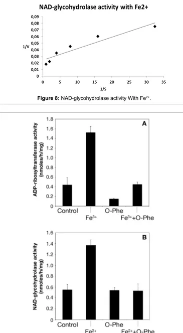
The iron-chelating agent O-phenantroline reduces the
acti-vating effect of iron
Both transferase and NADase NarE catalytic activities were assayed by a radioactive assay that implies the use of an anion-exchange resin to separate the unreacted labeled substrate from the product. The anionic interaction of the resin could interfere with the quantification of the enzymatic activities. Therefore, to ensure that our results were not influenced by our analytical method of analysis we assayed NarE transferase activity by using a filter plate-based assay [18] and the NADase activity with a fluorimetric assay [20]. NarE transferase activity Figure 4: Ferric ion enhances ADP-ribosyltransferase activity. NarE (10 μg),
extensively dialyzed with 50 mM potassium phosphate buffer pH 7.5 before the assay, was incubated for 18 h at 30°C in 50 mM potassium phosphate buffer, pH 7.5 with 0.1 mM [adenine-14C]NAD (0.05 μCi) and with or without (control) 10 mM of Fe3+ or Fe2+ in a total volume of 0.3 ml. Samples (100 μl) after incubation at 30°C were applied to 1-ml column of Dowex AG 1-X2, the ADP-ribosylagmatine formed was eluted with 5 ml of H2O for liquid scintillation counting. Activity is expressed as percentage of control. Data shown are the mean ± SD of three independent assays, with each activity measurement performed in duplicate.
Figure 5: Dose-response curve of ADP-ribosyltransferase and NAD-glycohydrolase activities in the presence of iron. (A) Affinity purified NarE was incubated for 18 h at 30°C in 50 mM potassium phosphate buffer with 0.1 mM [adenine-U-14C]NAD (0.05 μCi) and 20 mM agmatine as ADP-ribose acceptor in the presence of different concentrations of Fe3+ () or different concentrations of Fe2+ () as specified in the Figure in a final volume of 0.3 ml. (B) NADase activity was assayed in the same conditions above described in the absence of the ADP-ribose acceptor and with 0.1 mM [carbonyl-14C] NAD (0.05 µCi) replacing [adenine-U-14C]NAD (0.05 μCi) in the presence of the concentrations of Fe3+ () or Fe2+ (/) used above in a final volume of 0.3 ml. ADP-ribosylagmatine or 14C-nicotinamide were purified by AG1-X2 and quantified by scintillation counting. Data are expressed as ADP-ribosylagmatine formed or nicotinamide released per mg of protein. Data presented are the means ± SD of three independent assays, with each activity measurement performed in duplicate.
was increased by the addition of 10 mM Fe3+ (1.47 ± 0.21 vs. 0.49 ± 0.03
nmol/h/mg) while NarE NADase activity was increased by the addition of 10 mM Fe2+ (0.94 ± 0.22 vs. 0.35 ± 0.15 ∆F/min) confirming our
previous results. To further confirm that the activation of transferase or NADase activity depends on the presence of Fe3+or Fe2+ in the assay
mixture, we used O-phenantroline, which is a specific iron-chelator that decreases NarE transferase activity [13]. As shown in Figure 9A, O-phenantroline decreased the effect of Fe3+ on transferase activity.
Similarly the presence of the iron chelator considerably reduced the effect of Fe2+ on NADase activity (Figure 9B), reinforcing the hypothesis
of an iron-mediated activation.
Iron enhances maximum velocity of NarE keeping unaltered
affinity for NAD
In order to get insights into the mechanism of iron-mediated ART activity (nmol/h/mg)
W/o Na2S2O4 0.47 ± 0.064
Na2S2O4 0.16 ± 0.028
NADase activity (nmol/h/mg )
W/o Na2S2O4 0.35 ± 0.023
Na2S2O4 0.69 ± 0.120
Table 1: The ADP-ribosyltransferase activity of purified and extensively dialyzed NarE (13 µg) was assayed with a filter-based assay. Assays were performed in a total volume of 0.3 ml with 0.6 mg of poly-arginine and 0.1 mM [carbonyl-14C]NAD (0.05 μCi) in the presence of 1 mM Na2S2O4 . Samples of 100 µl after incubation at 30°C, were precipitated with 0.3 ml of 50% (w/v) TCA for 30 min on ice and placed in single wells of a mixed cellulose esters filter-plate under vacuum conditions. To eliminate the unreacted [adenine-U-14C]NAD filters were extensively washed with 5% (w/v) TCA and the incorporated radioactivity measured in a Packard Top counter. Data shown represent the mean ± SD of two independent experiments performed in quadruplicate. NADase activity was assayed at 30°C for 18 h with 0.1 mM [carbonyl-14C]NAD in a final volume of 0.3 ml in 50 mM potassium phosphate buffer (pH 7.5) with or without 1 mM Na2S2O4 . At the end of incubation 100 µl sample were applied to AG1-X2 columns and 14C-nicotinamide eluted by water and counted. The values presented represent the means ± SD of three independent assays.
Figure 6: NAD-glycohydrolase activity.
Figure 7: NAD-glycohydrolase activity With Fe3+.
Figure 8: NAD-glycohydrolase activity With Fe2+.
Figure 9: ADP-ribosyltransferase and NAD-glycohydrolase activities in the presence of iron-chelating agent and iron. (A) The ADP-ribosyltransferase activity of purified NarE (15 μg) was assayed with a filter-based assay [18]. Affinity purified NarE was incubated for 18 hr at 30°C with 0.6 mg of poly-arginine as acceptor molecule and 0.1 mM [adenine-U-14C]NAD (0.05 μCi) in a final volume of 0.3 ml in the presence of 10 mM of Fe3+ with or without 10 mM O-phenantroline. After TCA precipitation, samples (100 μl) were applied under vacuum condition in each well of a mixed cellulose ester filter plate and washed with 10 vol. of 5% TCA. The incorporated radioactivity was measured in a Packard Top counter. Each activity measurement was run in duplicate and analyzed with four replicates. (B) To test NADase activity was used the same enzyme preparation and the radioactive assay described in Experimental procedures. Enzyme activity was assayed at 30°C for 18 h with 0.1 mM [carbonyl-14C]NAD in a final volume of 0.3 ml in 50 mM potassium phosphate buffer (pH 7.5) in the presence of 10 mM Fe2+ with or without 10 mM O-phenantroline. At the end of incubation 100 μl sample were applied to AG1-X2 columns and 14C-nicotinamide eluted by water and counted. The values presented represent the means ± SD of three independent assays. activation we performed a kinetic analysis of NarE catalytic activities using the radioactive assays. NAD was the variable substrate for both transferase and NADase activities and agmatine the fixed substrate in the transferase reaction. Table 2 lists the kinetic data determined
for NarE in the absence and in the presence of 10 mM Fe2+and Fe3+.
For comparative purposes, we listed the Vmax ratio compared to the control value. The close Km values (Table 2) indicated that the NAD binding is similar either during the catalysis of the transfer of ADP-ribose to agmatine used as ADP-ADP-ribose acceptor or during the catalysis of NAD hydrolysis to ADP-ribose and NAM. Strikingly, the Vmax values determined increased when iron ions, Fe3+ for transferase and
Fe2+ for NADase activity, were added to the assay mixture. The catalytic
efficiency with respect to NAD substrate measured as Kcat (turnover number) and ratio between Kcat and Km indicated a higher efficiency for both transferase and NADase reaction when NarE activities were quantified in the presence of iron.
Role of cluster in iron-dependent activation of NarE catalysis
We have previously shown that NarE coordinates a single iron ion through C67 and C128, which are involved in the cluster formation and are crucial for transferase activity. We used characterized NarE mutants [13], to assess the relevance of a functionally structured cluster in the iron-mediated activation of NarE. As shown in Figure 10A, the presence of 10 mM Fe3+ increased the transferase activity of the C11Sand C130S similarly to that of wt. On the contrary, no activation was detected with the C67S and C128S mutants. In addition, to assess how the conservative mutation influences the ability of NarE to hydrolyze NAD we measured the NADase activity of the four mutants in the presence of Fe2+. As shown in Figure 4B, the NADase activity of C11S
and C130S was enhanced in the presence of iron. The mutants C67S and C128S, which do not keep a structured cluster but are still able to hydrolyze NAD [13], were not affected by the presence of 10 mM Fe2+(Figure 10B).
Discussion
We have previously shown that NarE, a member of the ART family, is an iron-binding protein and the presence of iron is essential for its transferase activity [13]. In this paper we present data showing a selective and iron oxidation-state dependent activation of the catalytic activities of NarE. The action of iron was reversible suggesting that the direct binding of iron leads to the changes of the catalytic properties. The generation of functional information on NarE catalysis is a key point to uncover the pathogenic role of NarE since this protein belongs to the ART family, whose members are usually associated with bacterial pathogenesis [23]. Cytotoxicity exerted by bacterial ARTs is mediated mainly by ADP-ribosylation of target proteins [24]. Besides, bacterial and mammalian ARTs can also possess NADase activity [19],
whose physiological function is not clear, and auto-ADP-ribosylation activity [25]. Since in our previous study we identified a paramagnetic iron in NarE, we hypothesized that Fe3+ could also have a role in the
regulation of NarE activities. In agreement with this hypothesis, Fe3+
but not Fe2+ induced a concentration-dependent increase in the ART
activity. In metal binding sites containing cysteines and histidines Zn2+ can overlap iron, as demonstrated by modeling experiments [14].
Therefore, in addition to other cations including Mg2+, which have
positive effects on bacterial ARTs [22,26], we paid particular attention to Zn2+. However, the many experiments carried out in the presence of
Zn2+ in different experimental conditions showed no activating effects,
while this cation at any concentrations used, precipitated NarE (data not shown). The reason for this precipitation effect is unclear however, since NarE natively binds iron Zn2+ ions could be not physiologically
relevant. Of interest and somewhat unexpectedly, the addition of Fe2+
to assay mixture induced an activating effect on glycohydrolase but not on transferase activity (Figure 5). Since we commonly use an anion exchange column to separate the radioactive product from the unreacted substrate, we were concerned that the activation effect could be an artifact. However, this was not the case since alternative assays, which do not imply the use of ionic interactions, gave identical results. The velocity of an enzyme-catalyzed reaction could be influenced by an increased affinity of the enzyme for the substrate or by an increased concentration of active enzyme. In the case of NarE the presence of iron enhanced the Vmax for both catalytic activities without affecting the Km for NAD indicating that the presence of the cation increases the concentration of a catalytically active form of NarE. This latter was reinforced by values of Kcat and ratio of Kcat and Km that indicated ART activity Km (mM) Vmax (nmoles/h/mg) compared to controlVmax
____ 0.56 ± 0.03 5.7 ± 0.4 ____
Fe2+ 0.54 ± 0.09 5.6 ± 0.4 1
Fe3+ 0.58 ± 0.06 18.3 ± 0.1 3.2
NADase activity Km (mM) Vmax (nmoles/h/mg)
Vmax compared to control
____ 0.48 ± 0.01 22.9 ± 0.2 ____
Fe2+ 0.45 ± 0.02 63.4 ± 0.3 2.8
Fe3+ 0.45 ± 0.03 23.1 ± 0.3 1
Table 2: Km and Vmax values of NarE were determined using 30 µg of NarE and 20 mM of agmatine as ADP-ribose acceptor and variable NAD (32.2, 16, 8, 4, 2 and 1 mM) for 18 h at 30°C for the transferase activity in the presence or FeCl3 as indicated in the figure. Kinetic parameters for NADase were determined as described above omitting agmatine from the standard assay in the presence of 10 mM Fe2Cl3. Kinetic parameters were calculated from Lineweaver-Burk plots according to H.I. Segel, Enzyme Kinetics: Behavior and Analysis of Rapid Equilibrium and Steady-state Enzyme Systems. Wiley-Interscience, New York, NY, 1975. Data represent the mean ± SD of three independent experiments performed in duplicate.
Figure 10: Effect of iron on the ADP-ribosyltransferase and NAD-glycohydrolase activities of NarE mutants. NarE mutants were tested for their ability to transfer ADP-ribose moiety and release NAM with (grey bars) and without (black bars) iron. (A) ADP-ribosyltransferase activity was measured for 18 h at 30°C under the same conditions of previous experiments and as described under Experimental procedures in the presence of 10 mM Fe3+. (B) NADase was quantified with radioactive assay previously described in the same condition used for transferase activity in the presence of 10 mM Fe2+. Data are expressed as ADP-ribosylagmatine formed or NAM release per mg of purified protein. Error bars represent the standard deviation of three independent experiments.
a higher efficient enzyme. Site-directed mutagenesis and structural studies have shown the strong influence of C67 and C128substitution in transferase activity and Fe3+ was unable to enhance the activity. These
two mutants exhibited only modest reduction of NADase activity but the addition of Fe2+ to the assay mixture failed to enhance the
NAD-glycohydrolase activity. This latter observation suggests that the presence of a cluster coordinated with iron ions is necessary to exert the activating effects both of the transferase and NADase activities. A multi-step process involving different proteins is necessary to integrate clusters into proteins [27], in bacteria iron-sulfur clusters due to their sensitivity act as regulators of iron availability allowing bacteria to modify their gene expression profile in response to modified environmental conditions [28]. Moreover, clusters have also a key role in determining the structure and stability of a protein. In this paper we show that they can influence the enzymatic activity of a protein. The modification of the redox state of the iron as it occurs with the addition of Fe2+ or in the
presence of a reducing agent that reduces the iron already present in the cluster, likely alters the structure of the protein impeding the access of the ADP-ribose acceptor forcing the enzyme to transfer the ADP-ribose to water. Although the pathogenetic effects of ADP-ribosylating toxins are associated mainly with their transferase activity, toxic effects could also be linked to NADase activity as it occurs in Group A streptococci (GAS) where the toxin possess NADase activity [29]. Iron is a necessary element for survival of N. meningitidis into the host and is implicated in the regulation of the expression of a number of bacterial genes involved not only in metabolism but also in virulence [30].
Conclusions
The direct involvement of iron as regulator of NarE catalytic activities, as shown here appears to be a regulatory mechanism that could provide the missing link in the cellular control of the catalytic activities of NarE. This novel observation can open new possibilities in the participation of this enzyme to N. meningitidis physiology. N. meningitidis possesses a great ability to colonize different niches in the human body, characterized either by reducing or oxidizing conditions. Therefore, the ability of NarE to modulate its catalytic activities according to the redox state of iron could be instrumental for N. Meningitidis pathogenesis.
Acknowledgements.
Special thanks go to Giorgio Corsi for his precious artwork, to Elisabetta Soldaini for helpful discussion, valuable advice and assistance and Fabio Concetti for technical support. Novartis Vaccines & Diagnostics, a GlaxoSmithKline Vaccines company financially supported the work.
References
1. Johnson AP (1983) The pathogenic potential of commensal species of Neisseria. J Clin Pathol 36: 213-223.
2. Schwartz B, Moore PS, Broome CV (1989) Global epidemiology of meningococcal disease. Clin Microbiol Rev 2: 118-124.
3. Schuchat A, Robinson K, Wenger JD, Harrison LH, Farley M, et al. (1997) Bacterial meningitis in the United States in 1995. Active Surveillance Team. N Engl J Med 337: 970-976.
4. Ueda K, Hayaishi O (1985) ADP-ribosylation. Annu Rev Biochem 54: 73-100. 5. Honjo T, Nishizuka Y & Hayaishi O (1968) Diphtheria toxin-dependent
adenosine diphosphate ribosylation of aminoacyl transferase II and inhibition of protein synthesis. J Biol Chem 24: 3553–3555.
6. Iglewski BH, Kabat D (1975) NAD-dependent inhibition of protein synthesis by Pseudomonas aeruginosa toxin,. Proc Natl Acad Sci U S A 72: 2284-2288. 7. Locht C, Keith JM (1986) Pertussis toxin gene: nucleotide sequence and
genetic organization. Science 232: 1258-1264.
8. Aktories K, Wegner A (1989) ADP-ribosylation of actin by clostridial toxins. J Cell Biol 109: 1385-1387.
9. Castagnini M, Picchianti M, Talluri E, Biagini M, Del Vecchio M, et al. (2012) Arginine-specific mono ADP-ribosylation in vitro of antimicrobial peptides by ADP-ribosylating toxins. PLoS One 7: e41417.
10. Masignani V, Balducci E, Serruto D, Veggi D, Aricò B, et al. (2004) In silico identification of novel bacterial ADP-ribosyltransferases. Int J Med Microbiol 293: 471-478.
11. Masignani V, Balducci E, Di Marcello F, Savino S, Serruto D, et al. (2003) NarE: a novel ADP-ribosyltransferase from Neisseria meningitidis. Mol Microbiol 50: 1055-1067.
12. Picchianti M, Del Vecchio M, Di Marcello F, Biagini M, Veggi D, et al. (2013) Auto ADP-ribosylation of NarE, a Neisseria meningitidis ADP-ribosyltransferase, regulates its catalytic activities. FASEB J 27: 4723-4730.
13. Del Vecchio M, Pogni R, Baratto MC, Nobbs A, Rappuoli R, et al. (2009) Identification of an iron-sulfur cluster that modulates the enzymatic activity in NarE, a Neisseria meningitidis ADP-ribosyltransferase. J Biol Chem 284: 33040-33047.
14. Koehler C, Carlier L, Veggi D, Balducci E, Di Marcello F, et al. (2011) Structural and biochemical characterization of NarE, an iron-containing ADP-ribosyltransferase from Neisseria meningitidis. J Biol Chem 286: 14842-14851. 15. Rohde KH, Dyer DW (2003) Mechanisms of iron acquisition by the human
pathogens Neisseria meningitidis and Neisseria gonorrhoeae. Front Biosci 8: d1186-1218.
16. Chen CY, Genco CA, Rock JP, Morse SA (1989) Physiology and metabolism of Neisseria gonorrhoeae and Neisseria meningitidis: implications for pathogenesis. Clin Microbiol Rev 2 Suppl: S35-40.
17. Moss J, Stanley SJ (1981) Histone-dependent and histone-independent forms of an ADP-ribosyltransferase from human and turkey erythrocytes. Proc Natl Acad Sci U S A 78: 4809-4812.
18. Balducci E (2005) A filter plate-based assay for mono ADP-ribosyltransferases [corrected]. Anal Biochem 344: 278-280.
19. Moss J, Manganiello VC, Vaughan M (1976) Hydrolysis of nicotinamide adenine dinucleotide by choleragen and its A protomer: possible role in the activation of adenylate cyclase. Proc Natl Acad Sci U S A 73: 4424-4427.
20. Barrio JR, Secrist JA 3rd, Leonard NJ (1972) A fluorescent analog of nicotinamide adenine dinucleotide. Proc Natl Acad Sci U S A 69: 2039-2042. 21. Bradford MM (1976) A rapid and sensitive method for the quantitation of
microgram quantities of protein utilizing the principle of protein-dye binding. Anal Biochem 72: 248-254.
22. Moss J, Stanley SJ, Burns DL, Hsia JA, Yost DA, et al. (1983) Activation by thiol of the latent NAD glycohydrolase and ADP-ribosyltransferase activities of Bordetella pertussis toxin (islet-activating protein). J Biol Chem 258: 11879-11882.
23. Wreggett KA (1986) Bacterial toxins and the role of ADP-ribosylation. J Recept Res 6: 95-126.
24. Glowacki G, Braren R, Firner K, Nissen M, Kühl M, et al. (2002) The family of toxin-related ecto-ADP-ribosyltransferases in humans and the mouse. Protein Sci 11: 1657-1670.
25. Weng B, Thompson WC, Kim HJ, Levine RL, Moss J (1999) Modification of the ADP-ribosyltransferase and NAD glycohydrolase activities of a mammalian transferase (ADP-ribosyltransferase 5) by auto-ADP-ribosylation. J Biol Chem 274: 31797-31803.
26. Moss J, Stanley SJ, Morin JE, Dixon JE (1980) Activation of choleragen by thiol: protein disulfide oxidoreductase. J Biol Chem 255: 11085-11087. 27. Beinert H, Holm RH, Münck E (1997) Iron-sulfur clusters: nature's modular,
multipurpose structures. Science 277: 653-659.
28. Miller HK, Auerbuch V (2015) Bacterial iron-sulfur cluster sensors in mammalian pathogens. Metallomics 7: 943-956.
29. Bricker AL, Carey VJ, Wessels MR (2005) Role of NADase in virulence in experimental invasive group A streptococcal infection. Infect Immun 73: 6562-6566.
30. Perkins-Balding D, Ratliff-Griffin M, Stojiljkovic I (2004) Iron transport systems in Neisseria meningitidis. Microbiol Mol Biol Rev 68: 154-171.