Review
The chick embryo chorioallantoic membrane (CAM). A multifaceted
experimental model
Domenico Ribatti
⁎
Department of Basic Medical Sciences, Neurosciences and Sensory Organs, University of Bari Medical School, Bari, Italy National Cancer Institute“Giovanni Paolo II”, Bari, Italy
a b s t r a c t
a r t i c l e i n f o
Article history: Received 11 March 2016
Received in revised form 7 May 2016 Accepted 9 May 2016
Available online 10 May 2016
During avian development the mesodermal layers of the allantois and chorion fuse to form the chorioallantoic membrane (CAM). This structure rapidly expands generating a rich vascular network that provides an interface for gas and waste exchange. The CAM allows to study tissue grafts, tumor growth and metastasis, wound healing, drugs delivery and toxicologic analysis, and angiogenic and anti-angiogenic molecules. The CAM is relatively sim-ple, quick, and low-cost model that allows screening of a large number of pharmacological samples in a short time; does not require administrative procedures for obtaining ethics committee approval for animal experimen-tation. Moreover, being naturally immunodeficient, the chick embryo may receive transplantations from differ-ent tissues and species, without immune responses.
© 2016 Elsevier Ireland Ltd. All rights reserved. Keywords: Angiogenesis Anti-angiogenesis Tissue grafts Tumor growth Wound healing Contents
1. Embryonic development and morphological structure . . . 70
2. Physiological functions. . . 71
3. In ovo and ex ovo methods . . . 72
4. Tissue grafts . . . 72
5. Tumor growth and metastasis . . . 73
6. Wound healing . . . 74
7. Drugs delivery and toxicology studies . . . 74
8. Angiogenic and anti-angiogenic molecules . . . 74
9. Genomics . . . 75
10. Pros and contra . . . 75
Acknowledgments . . . 76
References. . . 76
1. Embryonic development and morphological structure
The allantois of the chick embryo appears at about 3.5 days of incu-bation as an evagination from the ventral wall of the endodermal hind gut. It pushes out of the body of the embryo into the extraembryonic coelom. Its proximal portion (allantoic stalk) lies parallel and just caudal to the yolk sac, and when the distal portion grows clear of the embryo it
becomes enlarged (allantoic vesicle). The allantoic vesicle enlarges very rapidly from days 4–10 of incubation. During this process, the mesoder-mal layer of the allantois fuses with the adjacent mesodermesoder-mal layer of the chorion to form the chorioallantoic membrane (CAM) (Fig. 1). In this double layer an extremely rich vascular network develops which is connected to embryonic circulation by two allantoic arteries and one allantoic vein.
Fuchs and Lindenbaum (1988)described six or seven generations of branches of the allantoic artery. Thefirst five or six are located in a plane parallel to the CAM surface and deep to the vein, which has a similar dis-tribution. Thefifth and sixth generations change direction, passing
⁎ Department of Basic Medical Sciences, Neurosciences and Sensory Organs, University of Bari Medical School, Policlinico - Piazza G. Cesare, 11, 70124 Bari, Italy.
E-mail address:[email protected].
http://dx.doi.org/10.1016/j.mod.2016.05.003
0925-4773/© 2016 Elsevier Ireland Ltd. All rights reserved.
Contents lists available atScienceDirect
Mechanisms of Development
almost vertically in the two-dimensional capillary plexus.De Fouw et al. (1989)have shown rapid extension of the CAM surface from 6 cm2at day 6 to 65 cm2at day 14. The number of feed vessels increased (2.5-and 5-fold for precapillary (2.5-and postcapillary vessels), predominantly due to growth and remodeling after day 10.
According toSchlatter et al. (1997), CAM vascularization undergoes three phases of development with both sprouting and intussusceptive microvascular growth (IMG). In the early phase multiple capillary sprouts invade the mesenchyme, fuse, and form the primary capillary plexus. During the second phase , sprouts are no longer present and have been replaced by tissue pillars expression of IMG. During the late phase, the growing pillars increase in size to form intercapillary meshes. Immature blood vessels scattered in the mesoderm grow very rapid-ly until day 8 and give rise to a capillary plexus, which comes to be inti-mately associated with the overlying chorionic epithelial cells and mediates gas exchange with the outer environment (Fig. 2). At day 14,
basal lamina (Ausprunk et al., 1974). By day 8, the CAM displays small, thin-walled capillaries beneath the chorionic epithelium, and other ves-sels in the mesodermal layer, whose walls have a layer of mesenchymal cells surrounding the endothelium and are completely wrapped by a basal lamina together with the endothelial cells (Ausprunk et al., 1974). On days 10–12, the capillaries are now near the surface of the chorionic epithelium (Fig. 3). The mesodermal vessels are now distinct arterioles and venules. The walls of arterioles contain one or two layers of mesenchymal cells and increased amounts of connective tissue sur-rounding them. Venules are surrounded by an incomplete investment of mesenchymal cells, which are to be developing smooth muscle cells and the walls of arterioles also develop a distinct adventitia containing fibroblast-like cells.
We have demonstrated that endogenousfibroblast growth factor-2 (FGF-2) is present in elevated amounts in the CAM from day 6 to day18 of incubation, maximal concentrations being observed between day 10 and day 14. Moreover, neutralizing antibodies to FGF-2 inhibit vessel growth, confirming a role of FGF-2 in vascularization of the CAM (Ribatti et al., 1995).Baum et al. (2010)reported an endogenous expression of vascular endothelial growth factor-A (VEGF-A) with two peaks at day 8–9 and 11–12. We have more recently found two peaks in VEGF-A expression at day 7 and day 18 (Marinaccio et al., 2013) (Fig. 4). A high expression level of hypoxia inducible factor 1 alpha (HIF-1α), VEGF and VEGF receptor-2 (VEGFR-2) at day 11 correlates with a peak in angiogenic process at this stage of incubation (Baum et al., 2010; Marinaccio et al., 2013; Makanyna et al., 2016).
CAM arterioles and venules are accompanied by a pair of intercon-nected lymphatics. Veins are also associated with lymphatics, and larger veins are surrounded by a lymphatic plexus (Oh et al., 1997). The lym-phatic endothelial cells of the differentiated CAM specifically express vascular endothelial growth factor receptor (VEGFR)-3, whereas ex-pression of VEGFR-2 is found in both its blood vascular and its lymphatic endothelial cells (Wilting et al., 1996). The application of VEGF-C on the CAM induces the development of lymphatic vessels (Oh et al., 1997; Papoutsi et al., 2001). The lymphatics o are located immediately adja-cent to the larger blood vessels and the expression of VEGF-C in the blood vascular wall serves for the patterning of lymphatics. The homeo-box gene Prox-1, specifically expressed in lymphatics, has been demon-strated also in the CAM's lymphatics (Cimpean et al., 2010).Papoutsi et al. (2000)have therefore grown VEGF-C-expressing human A375 melanoma cells on the CAM, and demonstrated that a great number of melanoma cells invaded the Prox-1-positive chicken lymphatics. More-over, lymphangiogenesis was inhibited when melanoma cells were transfected with cDNA encoding soluble VEGFR-3.
The extracellular matrix of the CAM modifies its composition terms of expression offibronectin, laminin, and collagen type IV, and in distri-bution of specific glycosaminoglycans, favoring the angiogenic process that occurs in the space between the chorionic epithelium and the me-sodermal blood vessels (Ausprunk, 1986; Ribatti et al., 1998).
2. Physiological functions
At the beginning of the development of the chick embryo, before day 6 of incubation, the CAM performs gas exchange function through the area vasculosa (Romanoff, 1960). Thereafter, the rich vascularization and the position of the allantois immediately subjacent to the porous shell confer a respiratory function on the highly vascularized CAM. In addition to the respiratory interchange of oxygen and carbon dioxide,
Fig. 1. Macroscopic in ovo features of the chick chorioallantoic membrane (CAM) at day 5 of incubation.
Reproduced fromRibatti, 2014.
Fig. 2. A semithin section of the CAM, showing the chorionic epithelium (CH), the intermediate vascularized mesenchyme (M), and the deep allantoic epithelium (AL). Reproduced fromRibatti, 2014.
the allantois also serves as a reservoir for the waste products excreted by the embryo, mostly urea atfirst, and chiefly uric acid later.
Moreover, CAM is involved in sodium and chloride transport from the allantoic cavity where urinary waste products are discharged, and mobilization of calcium form the shell to start bone mineralization, at an impressive rate of 100 nmol of calcium per hour for 1 cm2of the CAM surface. Typical chorionic villous cavity calcium transporting cells become recognizable by day 12 (Makanyna et al., 2016). Apoptosis was increased in both the chorion and allantois at day 18 of incubation and some apoptotic cells were also recognizable in the mesenchyme (Makanyna et al., 2016).
3.In ovo and ex ovo methods
Fertilized chicken eggs staged according to Hamburger and Hamil-ton (Hamburger and Hamilton, 1951) are placed in an incubator as soon as embryogenesis starts and are kept under constant humidity at 37 °C. On day 3, a square window is opened in the shell after removal of 2–3 ml of albumen to detach the CAM from the shell itself and the un-derlying CAM vessels are disclosed. The window is sealed with a glass and incubation goes on until the day of experiment. This technique may preserve a more physiological environment; in fact, it has the ad-vantage of high viability in long term incubation and using this tech-nique the embryos develop through hatching. However, it limits the observation and the area for grafting, transplantation, andfine micro-dissection.
The embryo and its extraembryonic membranes may be transferred to a Petri dish on day 3 or 4 of incubation and CAM develops at the top as
aflat membrane and reaches the edge of the dish to provide a two-dimensional monolayer onto which multiple grafts can be placed (Auerbach et al., 1974). This system has several advantages as the ac-cessibility of the embryo is greatly improved outside of the shell. Shell-less culture is much more amenable to live imaging than in ovo techniques. However, long term viability is often lower in shell-less cultures and great attention must be paid to preventing the embryo from drying out.
In the original description of embryos cultured in Petri dishes, there was a 50% loss in thefirst three days after cracking due to the frequent rupture of the yolk membrane, with 80% of those which survive to day 7 until day 16 (Auerbach et al., 1974). The ex ovo method is preferred to the in vivo method because it allows the quantification of the response over a wider area of the CAM; large number of samples can be tested at any one time; the time required for a response to occur is shorter.
4. Tissue grafts
Transplantation studies are used for a variety of purposes, such as determining inductive/suppressive interaction, and when combined with some form of cell tracing, can be used to determine migration and growth patterns. When a small fragment of an adult chicken organ was transplanted onto the CAM, it greatly stimulated growth of the homologous embryonic organs (Danchakoff, 1918; Ebert, 1954). Al-logenic and xenogenic tissue were successfully grafted on the CAM, such as liver, endometrium, adrenal gland, cerebellum, or intact skin. The formation of peripheral anastomoses between pre-existing donor and host vessels is the most common mechanism involved in the revas-cularization of embryonic grafts (Ausprunk et al., 1975). Moreover, the CAM may provide the explants with nutrients, growth factors, as well as stem cells derived from other parts of the developing chicken em-bryo. In this context,Maeda and Noda (2003)have demonstrated that the growth of epiphyseal cartilage is not suppressed in the presence of perichondrium when bone is cultured on the CAM, suggesting that CAM and/or other chicken cells can partially substitute for the perichon-drium in this system. Adult human kidney derived cells were grafted onto the CAM, and after a week, grafts self-organized into tubular struc-tures (Noiman et al., 2011).
Grafting of chick cells into quail embryos or vice versa is the most common form of chimera generation in the avians. A variety of spe-cies-specific antibodies have been generated including the Quail/Chick perinuclear antibody (QCPN) which reacts to quail cells and QH1, an an-tibody that recognizes a glycoprotein on the surface of quail endothelial cells and does not cross-react with chick endothelium (Pardanaud et al., 1996; Peault et al., 1983).
Acellular matrix can be transplanted onto the CAM without rejection and provide a matrix where cell growth, angiogenesis, and differentia-tion can occur. We have implanted acellular matrix obtained from brain, aorta, femur, esophagus, diaphragm, and have obtained in all the experimental conditions a strong angiogenic response (Fig. 5) (Conconi et al., 2004, 2005, 2009; Marzaro et al., 2006; Pardanaud et al., 1996; Peault et al., 1983; Ribatti et al., 2003). More recently, we have investigated the angiogenic properties of decellularized laryngeal and tracheal matrices (Baiguera et al., 2011; Haag et al., 2012).
Fig. 3. Electron microscopy micrography of the CAM at 15th day of incubation, showing numerous capillaries located between the chorionic (CH) epithelial cells. Reproduced fromRibatti et al., 1998.
Fig. 4. Relative expression of VEGF-A in the developing chorioallantoic membrane at ED7, 12, 14 of incubation in comparison to expression at E18 assumed as reference stage. * pb 0.05 vs CAM at ED7. List of abbreviations: CAM, chorioallantoic membrane; VEGF-A, vascular endothelial growth factor-A; ED, embryonic day.
5. Tumor growth and metastasis
In 1911, Rous and Murphy demonstrated the growth of the Rous sar-coma transplanted onto the CAM and Murphy described the effects of mouse and rat tumors transplantations on the CAM (Murphy, 1913, 1914a, 1914b; Rous, 1911). Starting from these observations, the CAM has been established as an experimental system for research in tumor biology The behavior of chicken, mouse, and human tumor cells and tis-sues implanted on the CAM surface was compared and was evaluated their growth, histological features, viability after re-transplantation in its original host and the effects on the chick embryo (Karnofsky et al., 1952). Human tumor transplanted and re-transplanted on CAM retain and express human antigens after several passages (Korngold and Lipari, 1955). The presence of metastases into the embryo was evaluat-ed after 10 days of tumor implantation on the CAM and the inhibitory capacity of chemotherapeutic agents on their growth was tested (Harris, 1958). The changes in the CAM adjacent to the tumor implanta-tion site, consisting in fibroblast proliferation, keratinization and stratification of the chorionic epithelium was also evaluated (Kaufman et al., 1956). Moreover, the CAM has been used as a test system for tumor chemosensitivity (Kunzi-Rapp et al., 1992) for the study of
tumor invasion and metastasis and of neovascularization of heterolo-gous normal and neoplastic implants (Auerbach et al., 1976; Quigley and Armstrong, 1998).
Grafting of tumors onto the CAM allows us to study the morpholog-ical aspects of the interactions of the tumors with the blood vessels of the host and to examine the identity of the vessels that supply the grafts. The formation of peripheral anastomoses between host and pre-existing donor vessels is the main and the most common mechanism in-volved in the revascularization of the graft of an embryonic organ onto the CAM, whereas sprouting of CAM-derived vessels into the trans-plants only occurs in the grafts of tumor tissue (Ausprunk and Folkman, 1976; Ausprunk et al., 1975).
All studies of mammalian neoplasms in the CAM have utilized solid tumors and cell suspensions derived from solid tumors. Compared with mammals' models, where tumor growth often takes between 3 and 6 weeks, assays using chick embryos are faster. Between 2 and 5 days after tumor cell inoculation, the tumor xenografts become visible and are supplied with vessels of CAM origin. Tumors grafted onto the CAM remain nonvascularized for few days, after which they can be penetrated by new blood vessels and begin a phase of rapid growth (Fig. 6).
Tumor cells can be identified in the CAM, as well as in the internal or-gans of the embryo, such as lungs, liver, and brain (Bobek et al., 2004). Viral nanoparticles have been used to visualize newly formed vascula-ture in expanding tumors (Leong et al., 2010), and high-resolution im-aging of CAM-supported human tumors reveal fluid and small molecule dynamics within tumors (Cho et al., 2011).
Fergelot et al. (2013)validated the CAM as a suitable model for studying the development of human clear cell renal carcinoma, with the aim to obtain a more complete characterization of the vessel pheno-type. Intratumor vessels were stained with anti-desmin antibodies or with anti-CD31 to detect human vessels and co-labeled with SNA1 isolectin to detect chick vessels. CD31 antibodies only recognized ves-sels of human origin and SNA isolectin vesves-sels of chicken origin, while anti-desmin antibodies labeled vessels of both species. The vessels were mostly hybrid as shown by the presence of SNA1 positive endo-thelial cells in the close vicinity of CD31 positive cells.
Other studies have focused on the invasion of the chorionic epitheli-um and the blood vessels by tepitheli-umor cells (Armstrong et al., 1982; Dagg et al., 1956; Kim et al., 1998; Scher et al., 1976). The cells invade the vascularized mesenchymal connective tissue below the chorionic epi-thelium (Fig. 7). The cancer cells migrate through the mesenchyme and attach to arterioles, and migrate to the vicinity of preexisting vessels (Koop et al., 1994). The changes in morphology of cancer cells arrested in the CAM microcirculation can be readily observed by in vivo micros-copy and most of them survive without significant cell damage
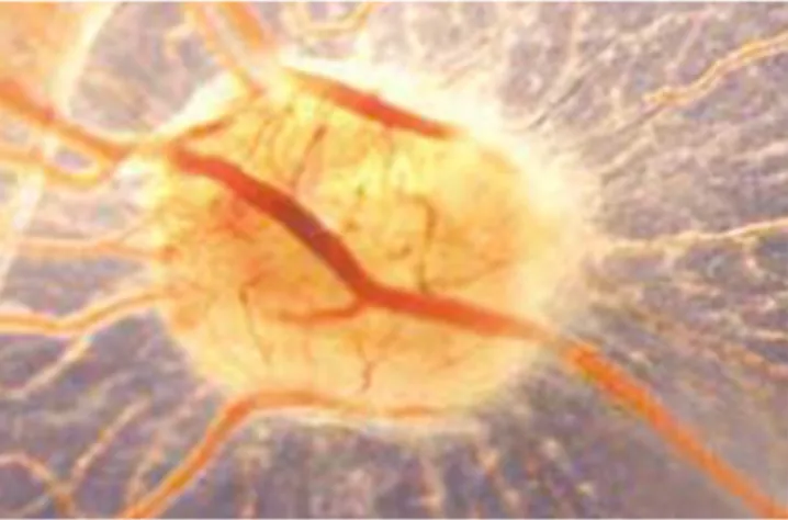
Fig.5. Macroscopic picture of implant of an aortic acellular matrix surrounded by allantoic vessels developing radially toward it (I) in a spoke-wheel pattern (a). The vasoproliferative response is comparable to that induced by a gelatin sponge (S) soaked with FGF-2.
Reproduced fromConconi et al., 2004.
Fig. 6. A 12-day-old CAM incubated on day 8 for 4 days with bioptic specimen of a tumor xenograft, showing numerous host vessels around and inside the graft.
(Chambers et al., 1992). Within few days after inoculation of human tumor cells on the CAM, tumor cells can be identified in portions of the CAM distant from the inoculation site, as well as in internal organs. Ten minutes after the intravenous injection offluorescent labeled B16-F10 melanoma cells, the cell arrested into the capillary bed, and six hours later, tumor cells changed their shape and spread in close contact with the capillary wall (Shioda et al., 1997). Moreover, the detection in the CAM of disseminated cells by Alu-PCR makes it possible to quantita-tively assess metastasis to organs that are colonized by as few as 25 cells (Zijlstra et al., 2002). More recently, quantitative methods for analyzing tumor angiogenesis by polymer perfusion followed by micro CT scans have been developed (Ames et al., 2016).
The major advantages of the CAM model as an experimental animal model of metastasis are: (a) the chick embryo is naturally immunode fi-cient and can accept cancer cells regardless of their origin without im-mune response; (b) the changes in morphology of cancer cells arrested in the CAM microcirculation can be readily observed by in vivo microscopy (Chambers et al., 1992); (c) most cancer cells arrested in the CAM microcirculation survive without significant cell damage, and complete extravasation (Chambers et al., 1992).
6. Wound healing
The CAM has been used as in vivo model to study wound repair (Ribatti et al., 1996). This model consistently reproduces all the phases observed in adult wound healing, including re-epithelization, angiogen-esis, inflammation, and fibronectin deposition, resulting in scar forma-tion (Ribatti et al., 1996). Histological examination of the CAM during wound healing demonstrated hyperplasia of the chorionic epithelium in the area involved in the repair process and an inflammatory infiltrate consisting mainly of macrophages. Also, about three times as many micro-vessels andfibroblasts were present in the mesenchyme of the wounded area with respect to the adjacent control regions.
The inhibition of FGF-2 after CAM wounding results in decreased wound healing by inhibiting microvessel andfibroblast density. Con-versely, the application of FGF-2 accelerates wound repair such that healing occurs 24 h earlier as compared to control wounds by stimulat-ing angiogenesis,fibroblast proliferation, and macrophage infiltration (Ribatti et al., 1999).
The U.S. Food and Drug Administration (FDA) has approved prod-ucts pre-clinically evaluated in the CAM assay and released for the treat-ment of chronic cutaneous ulcer and burn wounds (FDA Wound Healing Clinical Focus Group, 2001).
Kilarski et al. (2009)have developed a method to investigate granu-lation formation in the CAM, and the role of invadingfibroblasts and blood vessels in this process. Tissue tension generated by activated fi-broblasts or myofibroblasts during wound contraction mediated and di-rected translocation of the vasculature, which can be expanded secondarily by elongation and vessel enlargement, andfinally, through splitting and sprouting.
7. Drugs delivery and toxicology studies
Drug topically applied to the CAM can reach the systemic circulation, after absorption through the membrane and affect the development of the chick embryo. During the development of drug delivery systems, chick embryos can be used to evaluate the activity or toxicity of a drug in both the CAM and CAM-grafted tumors, as well as on the develop-ment of the embryo. Toxicity of drugs or carriers on chick embryos can be evaluated in terms of embryo death and adverse effects on the CAM, including inflammation and neovascularization. Drug delivery systems can be topically applied on the CAM or injected into the amni-on. When tumors are grafted on the CAM, anticancer therapies can be evaluated.
Ex ovo cultivated chicken embryos can be used for investigation of toxicity of different substances, as the influence of nicotine and cigarette smoke in developing chicken embryos (Hamamichi and Nishigori, 2001). In addition, the effects of acute glucose toxicity have been assessed in shell-less chick embryo cultures (Datar and Bhonde, 2005). Moreover, the CAM has been accepted as a substitute for the Draize test on rabbits for the testing of irritation potential of chemicals (Kishore et al., 2008; Yan et al., 2007).
8. Angiogenic and anti-angiogenic molecules
A variety of compounds have been reported to stimulate and inhibit angiogenesis in the CAM. They include growth factors, hormones, natu-ral molecules, anti-cancer agents, gases, organo-metallic compounds,
Fig. 7. Pentraxin-3 (PTX3) overexpression inhibits the invasive potential of melanoma B16-F10 cells. A, mock (black bar) and hPTX3-B16-F10 cells (open bar) were grafted onto the CAM at day 8. After 4 days, S-100–positive melanoma cells invading the CAM mesenchyme were counted. Representative images of sections cut parallel to the surface of the CAM are shown in right panels (CH, chorionic epithelium; M, mesenchyme; T, tumor cells). The number of hPTX3-overexpressing cells invading the CAM mesenchyme is significantly decreased when compared with mock cells.
inhibition of the basal angiogenesis, and another one, which evaluate the inhibition of an angiogenic stimulus previously applied to the CAM. An angiogenic response occurs 72–96 h after stimulation in the form of an increased vessel density around the implant, with the vessels radi-ally converging toward the center like spokes in a wheel. Conversely, when an angiostatic compound is tested, the vessels become less dense around the implant after 72–96 h, and eventually disappear. When the substance is inoculated into the cavity of allantoic vesicle, then the angiogenic or anti-angiogenic response affects the CAM vessels as a whole.
Several qualitative, quantitative, and semi-quantitative techniques, including blood vessel length, diameter, density, vessel branch points, total area of the CAM, have been described for assessment of angiogen-esis (Ribatti et al., 2010). New imaging technologies allow us to visualize vascular perfusion and vascularization, as well as, new contrast and im-aging agents that selectively label developing vessels allow for the selec-tive visualization of vascular structures at microscopic level (Jilani et al., 2003; Leong et al., 2010; Lewis et al., 2006; MacDonald et al., 1992).
Many techniques can be applied within the constraints of paraffin and plastic embedding, including histochemistry and immunohisto-chemistry. Electron microscopy can also be used in combination with light microscopy.
9. Genomics
Genomics and bioinformatics tools are currently available for a vari-ety of model systems, and avian embryology has begun to adopt these techniques. Comparative genomic revealed that the chick genome is three times smaller than the one of both human and mouse, but con-tains approximately the same number of genes (Burt, 2005). Modern advances, including the creation of the annotated chick genome (Wallis et al., 2004), gene expression profiling, live imaging, improved somatic transgenesis, and gene specific attenuation of RNA levels, have also added to the classical strengths of the avian embryo. 70 million bp of the chicken sequence is highly conserved with humans both within coding gene segments and outside coding regions (ICGSC, 2004). The complete characterization of the chick embryo genome (www.nhgri.gov/11510730) will be helpful to synthesize a broad panel of antibodies with high specificity for chicken tissues, for blood and lymphatic endothelial cells and stroma components. This aspect could be useful to better characterize the interactions between implanted human and/or mouse tumors and chicken tissues.
Gene expression profiling associated with the physiological CAM de-velopment as well as with the angiogenic switch during tumor progres-sion has been reported (Javerzat et al., 2009; Saidi et al., 2008). Engraftment of human tumor tissue onto the CAM, followed by transcriptomic analyses with both human and chicken microarrays, en-ables the gene signatures of both the host stroma and the human tumor to be distinguished.Soulet et al. (2010)by using an experimental model of granulation tissue formation in the CAM, performed wounding of the chicken CAM and compared gene expression to normal CAM at the same stage of development. Bioinformatics analysis lead to the identi fi-cation of several new genes with an endothelial cell signature. More-over,Soulet et al. (2013)applied in vivo biotinylation combined with high-resolution mass spectrometry and bioinformatic analyses to study the vascular and matrix proteome in the CAM. More recently, Exertier et al. (2014)performed an Affymetrix gene screening for VEGF-A-induced genes during CAM vascularization. A total of 53 single
the gene product that is expressed directly by CAM cells, and makes fea-sible the study of the effects of intracellular or membrane-bound pro-teins as well as of dominant-negative gene products. This approach will shed new lights on the study of the metastatic process by using the CAM assay.
10. Pros and contra
The CAM is relatively simple, quick, and low-cost model that allows screening of a large number of pharmacological samples in a short time. The CAM does not require administrative procedures for obtaining ethics committee approval for animal experimentation, because the chick embryo is not considered as living animal until day 17 of develop-ment in most countries. The CAM is not innervated, and experidevelop-ments are terminated before the development of centers in the brain associat-ed with pain perception, making this a system not requiring animal ex-perimentation permissions.
The Institutional Animal Care and Use Committee (IACUC), an Asso-ciation of New England Medical Center and Tufts (IACUC, 2001), as well as the National Institutes of Health, USA (National Institute of Health, 1991), established that a chick embryo that has not reached the 14th day of its gestation period would not experience pain and can therefore be used for experimentation without any ethical restrictions or prior protocol approval.
Early lymphoid cells deriving from the yolk sac and spleen are usual-ly recognizable in the thymus on day 8 and in the bursa of Fabricius on day 11 (Leene et al., 1973). Thymus cells are present by day 11 and cell-mediated immunity has been demonstrated by day 13–14 (Solomon, 1971). By day 12, mononuclear phagocytes are found in the yolk sac, spleen, bursa, gut, thymus and in the liver (Janse and Jeurissen, 1991). The chick embryo cannot mount an‘immune’ response to foreign tumor cells until day 12, but it can respond to tumor cells by infiltration of monocytes and inflammatory-like cells such as avian heterophils. Being naturally immunodeficient, the chick embryo may receive trans-plantations from different tissues and species, without immune re-sponses. Moreover, CAM allows a rapid vascularization, development of tumor cells or bioptic specimens placed on its surface, and allows to observe and analyze the real time changes in morphology of cancer cells in its microcirculation.
The main limitation of CAM assays is a nonspecific inflammatory re-action, which can occur if experiments extend after 15 days of incuba-tion. Examination of histological CAM sections help to detect the presence of a perivascular inflammatory infiltrate, together with a hy-perplastic reaction of the chorionic epithelium. A nonspecific inflamma-tory response is much less likely when the test material is grafted as soon as the CAM begins to develop while the host's immune system is relatively immature (Leene et al., 1973). Real neovascularization can hardly be distinguished from a falsely increased vascular density due to rearrangement of existing vessels (Knighton et al., 1991), and timing of the CAM angiogenic response is essential. Many studies determine angiogenesis after 24 h, when there is no angiogenesis, but only vasodi-lation. The CAM is also extremely sensitive to modification by environ-mental factors, such as changes in oxygen tension, which make the sealing of the opening in the shell critical, pH, osmolarity, and the amount of keratinization (Auerbach et al., 2000).
Another drawback is due to the low number of reagents compatible with avian species including , antibodies, cytokines, and primers which work in the chick.
Acknowledgments
This work was supported by European Union Seventh Framework Programme (FP7/2007-2013) under grant agreement no. 278570 to DR.
References
Ames, J.J., Henderson, T., Liaw, L., Brooks, P.C., 2016. Methods for analyzing tumor angio-genesis in the chick chorioallantoic membrane model. Methods in Molecular Biology. Springer Science\mathplus Business Media, pp. 255–269http://dx.doi.org/10.1007/ 978-1-4939-3444-7_22.
Armstrong, P.B., Quigley, J.P., Sidebottom, E., 1982.Transepithelial invasion and intramesenchymal infiltration of the chick embryo chorioallantois by tumor cell lines. Cancer Res. 42, 1826–1837.
Auerbach, R., Kubai, L., Knighton, D., Folkman, J., 1974. A simple procedure for the long-term cultivation of chicken embryos. Dev. Biol. 41, 391–394.http://dx.doi.org/10. 1016/0012-1606(74)90316-9.
Auerbach, R., Kubai, L., Sidky, Y., 1976.Angiogenesis induction by tumors, embryonic tis-sues, and lymphocytes. Cancer Res. 36, 3535–3540.
Auerbach, R., Akhtar, N., Lewis, R.L., Shinners, B.L., 2000. Angiogenesis assays: problems and pitfalls. Cancer Metastasis Rev.http://dx.doi.org/10.1023/A:1026574416001. Ausprunk, D.H., 1986. Distribution of hyaluronic acid and sulfated glycosaminoglycans
during blood-vessel development in the chick chorioallantoic membrane. Am. J. Anat. 177, 313–331.http://dx.doi.org/10.1002/aja.1001770304.
Ausprunk, D.H., Folkman, J., 1976.Vascular injury in transplanted tissues. Fine structural changes in tumor, adult, and embryonic blood vessels. Virchows Arch. B Cell Pathol. 31–44.
Ausprunk, D.H., Knighton, D.R., Folkman, J., 1974. Differentiation of vascular endothelium in the chick chorioallantois: a structural and autoradiographic study. Dev. Biol. 38, 237–248.http://dx.doi.org/10.1016/0012-1606(74)90004-9.
Ausprunk, D.H., Knighton, D.R., Folkman, J., 1975.Vascularization of normal and neoplas-tic tissues grafted to the chick chorioallantois. Role of host and preexisting graft blood vessels. Am. J. Pathol. 79, 597–618.
Baiguera, S., Gonfiotti, A., Jaus, M., Comin, C.E., Paglierani, M., Gaudio, C.D., Bianco, A., Ribatti, D., Macchiarini, P., 2011. Development of bioengineered human larynx. Bio-materials 32, 4433–4442.http://dx.doi.org/10.1016/j.biomaterials.2011.02.055. Baum, O., Suter, F., Gerber, B., Tschanz, S.A., Buergy, R., Blank, F., Hlushchuk, R., Djonov, V.,
2010.VEGF-A promote intussusceptive angiogenesis in the developing chicken cho-rioallantoic membrane. Microcirculation 7, 447–457.
Bobek, V., Plachy, J., Pinterova, D., Kolostova, K., Boubelik, M., Jiang, P., Yang, M., Hoffman, R.M., 2004. Development of a greenfluorescent protein metastatic-cancer chick-em-bryo drug-screen model. Clin. Exp. Metastasis 21, 347–352.http://dx.doi.org/10. 1023/b:clin.0000046138.58210.31.
Burt, D.W., 2005. Chicken genome: current status and future opportunities. Genome Res. 15, 1692–1698.http://dx.doi.org/10.1101/gr.4141805.
Chambers, A.F., Schmidt, E.E., MacDonald, I.C., Morris, V.L., Groom, A.C., 1992. Early steps in hematogenous metastasis of B16F1 melanoma cells in chick embryos studied by high-resolution intravital videomicroscopy. J. Natl. Cancer Inst. 84, 797–803.http:// dx.doi.org/10.1093/jnci/84.10.797.
Cho, C.-F., Ablack, A., Leong, H.-S., Zijlstra, A., Lewis, J., 2011. Evaluation of nanoparticle up-take in tumors in real time using intravital imaging. J. Vis. Exp.http://dx.doi.org/10. 3791/2808.
Cimpean, A.M., Seclaman, E., Ceauşu, R., Gaje, P., Feflea, S., Anghel, A., Raica, M., Ribatti, D., 2010.VEGF-A/HGF induce Prox-1 expression in the chick embryo chorioallantoic membrane lymphatic vasculature. Clin. Exp. Med. 10, 169–172.
Conconi, M.T., Nico, B., Mangieri, D., Tommasini, M., di Liddo, R., Parnigotto, P.P., Nussdorfer, G.G., Ribatti, D., 2004. Angiogenic response induced by acellular aortic matrix in vivo. Anat. Rec. 281A, 1303–1307.http://dx.doi.org/10.1002/ar.a.20137. Conconi, M.T., Nico, B., Rebuffat, P., Crivellato, E., Parnigotto, P.P., Nussdorfer, G.G., Ribatti,
D., 2005. Angiogenic response induced by acellular femoral matrix in vivo. J. Anat. 207, 79–83.http://dx.doi.org/10.1111/j.1469-7580.2005.00427.x.
Conconi, M.T., Bellini, S., Teoli, D., de Coppi, P., Ribatti, D., Nico, B., Simonato, E., Gamba, P.G., Nussdorfer, G.G., Morpurgo, M., Parnigotto, P.P., 2009. In vitro and in vivo eval-uation of acellular diaphragmatic matrices seeded with muscle precursors cells and coated with VEGF silica gels to repair muscle defect of the diaphragm. J. Biomed. Mater. Res. Part A 89A, 304–316.http://dx.doi.org/10.1002/jbm.a.31982.
Dagg, C.P., Karnofsky, D.A., Roddy, J., 1956.Growth of transplantable human tumors in the chick embryo and hatched chick. Cancer Res. 16, 589–594.
Danchakoff, V., 1918. Equivalence of different hematopoietic anlages. (By method of stim-ulation of their stem cells). II. Grafts of adult spleen on the allantois and response of the allantoic tissues. Am. J. Anat. 24, 127–189.http://dx.doi.org/10.1002/aja. 1000240202.
Datar, S., Bhonde, R.R., 2005. Shell-less chick embryo culture as an alternative in vitro model to investigate glucose-induced malformations in mammalian embryos. Rev. Diabet. Stud. 2, 221.http://dx.doi.org/10.1900/rds.2005.2.221.
De Fouw, D.O., Rizzo, V.J., Steinfeld, R., Feinberg, R.N., 1989.Mapping of the microcircula-tion in the chick chorioallantoic membrane during normal angiogenesis. Microvasc. Res. 38, 136–147.
Ebert, J.D., 1954. The effects of chorioallantoic transplants of adult chicken tissues on ho-mologous tissues of the host chick embryo. Proc. Natl. Acad. Sci. 40, 337–347.http:// dx.doi.org/10.1073/pnas.40.5.337.
Exertier, P., Javerzat, S., Wang, B., Franco, M., Herbert, J., Platonova, N., Winandy, M., Pujol, N., Nivelles, O., Ormenese, S., Godard, V., Becker, J., Bicknell, R., Pineau, R., Wilting, J.,
Bikfalvi, A., Hagedorn, M., 2014.Impaired angiogenesis and tumor development by inhibition of the mitotic kinesin Eg5. Oncotarget 4, 2302–2316.
FDA Wound Healing Clinical Focus Group, 2001. Guidance for industry: chronic cutaneous ulcer and burn wounds-developing products for treatment. Wound Repair Regen. 9, 258–268.http://dx.doi.org/10.1046/j.1524-475x.2001.00258.x.
Fergelot, P., Bernhard, J.C., Soulet, F., Kilarski, W.W., Léon, C., Courtois, N., Deminière, C., Herbert, J.M., Antczak, P., Falciani, F., Rioux-Leclercq, N., Patard, J.J., Ferrière, J.M., Ravaud, A., Hagedorn, M., Bikfalvi, A., 2013.The experimental renal cell carcinoma model in the chick embryo. Angiogenesis 16, 181–194.
Fuchs, A., Lindenbaum, E.S., 1988.The two- and three-dimensional structure of the micro-circulation of the chick chorioallantoic membrane. Acta Anat. 131, 271–275.
Haag, J., Baiguera, S., Jungebluth, P., Barale, D., Gaudio, C.D., Castiglione, F., Bianco, A., Comin, C.E., Ribatti, D., Macchiarini, P., 2012. Biomechanical and angiogenic proper-ties of tissue-engineered rat trachea using genipin cross-linked decellularized tissue. Biomaterials 33, 780–789.http://dx.doi.org/10.1016/j.biomaterials.2011.10.008. Hamamichi, S., Nishigori, H., 2001. Establishment of a chick embryo shell-less culture
sys-tem and its use to observe change in behavior caused by nicotine and substances from cigarette smoke. Toxicol. Lett. 119, 95–102. http://dx.doi.org/10.1016/s0378-4274(00)00300-3.
Hamburger, V., Hamilton, H.L., 1951. A series of normal stages in the development of the chick embryo. J. Morphol. 88, 49–92.http://dx.doi.org/10.1002/jmor.1050880104. Harris, J.J., 1958. The human tumor grown in the egg. Ann. N. Y. Acad. Sci. 76, 764–774.
http://dx.doi.org/10.1111/j.1749-6632.1958.tb54894.x.
Hen, G., Yosefi, S., Shinder, D., Or, A., Mygdal, S., Condiotti, R., Galun, E., Bor, A., Sela-Donenfeld, D., Friedman-Einat, M., 2012. Gene transfer to chicks using lentiviral vec-tors administered via the embryonic chorioallantoic membrane. PLoS ONE 7, e36531.
http://dx.doi.org/10.1371/journal.pone.0036531.
IACUC, 2001.Policy on Protocol Approval for Use of Chicken Embryos and Eggs. An Asso-ciation of New England Medical Center and Tufts.
ICGSC, 2004.Sequence and comparative analysis of the chicken genome provide unique perspectives on vertebrate evolution. Nature 432, 695–716.
Janse, E.M., Jeurissen, S.H.M., 1991. Ontogeny and function of two non-lymphoid cell pop-ulations in the chicken embryo. Immunobiology 182, 472–481.http://dx.doi.org/10. 1016/s0171-2985(11)80211-1.
Javerzat, S., Franco, M., Herbert, J., Platonova, N., Peille, A.-L., Pantesco, V., Vos, J.D., Assou, S., Bicknell, R., Bikfalvi, A., Hagedorn, M., 2009. Correlating global gene regulation to angiogenesis in the developing chick extra-embryonic vascular system. PLoS ONE 4, e7856.http://dx.doi.org/10.1371/journal.pone.0007856.
Jilani, S.M., Murphy, T.J., Thai, S.N.M., Eichmann, A., Alva, J.A., Iruela-Arispe, M.L., 2003. Se-lective binding of lectins to embryonic chicken vasculature. J. Histochem. Cytochem. 51, 597–604.http://dx.doi.org/10.1177/002215540305100505.
Karnofsky, D.A., Ridgway, L.P., Patterson, P.A., 1952. Tumor transplantation to the chick embryo. Ann. N. Y. Acad. Sci. 55, 313–329.http://dx.doi.org/10.1111/j.1749-6632. 1952.tb26547.x.
Kaufman, N., Kinney, T.D., Mason, E.J., Prieto, L.C., 1956.Maintenance of human neoplasm on the chick chorioallantoic membrane. Am. J. Pathol. 32, 271–285.
Kilarski, W.W., Samolov, B., Petersson, L., Kvanta, A., Gerwins, P., 2009. Biomechani-cal regulation of blood vessel growth during tissue vascularization. Nat. Med. 15, 657–664.
Kim, J., Yu, W., Kovalski, K., Ossowski, L., 1998. Requirement for specific proteases in can-cer cell intravasation as revealed by a novel semiquantitative PCR-based assay. Cell 94, 353–362.http://dx.doi.org/10.1016/s0092-8674(00)81478-6.
Kishore, A.S., Surekha, P.A., Sekhar, P.V.R., Srinivas, A., Murthy, P.B., 2008. Hen egg chorio-allantoic membrane bioassay: an in vitro alternative to draize eye irritation test for pesticide screening. Int. J. Toxicol. 27, 449–453. http://dx.doi.org/10.1080/ 10915810802656996.
Knighton, D.R., Fiegel, V.D., Phillips, G.D., 1991.The assay of angiogenesis. Prog. Clin. Biol. Res. 365, 291–299.
Koop, S., Khokha, R., Schmidt, E.E., MacDonald, I.C., Morris, V.L., Chambers, A.F., Groom, A.C., 1994.Overexpression of metalloproteinase inhibitor in B16F10 cells does not af-fect extravasation but reduces tumor growth. Cancer Res. 54, 4791–4797.
Korngold, L., Lipari, R., 1955.Tissue antigens of human tumors grown in rats, hamsters, and eggs. Cancer Res. 15, 159–161.
Kunzi-Rapp, K., Schneckenburger, H., Westphal-Frösch, C., 1992. Test system for human tumor cell sensitivity to drugs on chicken chorioallantoic membranes. In Vitro Cell. Dev. Biol. Anim. 28, 565–566.http://dx.doi.org/10.1007/bf02631021.
Leene, W., Duyzings, M.J.M., Steeg, C., 1973. Lymphoid stem cell identification in the de-veloping thymus and bursa of Fabricius of the chick. Z. Zellforsch. Mikrosk. Anat. 136, 521–533.http://dx.doi.org/10.1007/bf00307368.
Leong, H.S., Steinmetz, N.F., Ablack, A., Destito, G., Zijlstra, A., Stuhlmann, H., Manchester, M., Lewis, J.D., 2010. Intravital imaging of embryonic and tumor neovasculature using viral nanoparticles. Nat. Protoc. 5, 1406–1417.http://dx.doi.org/10.1038/nprot.2010. 103.
Lewis, J.D., Destito, G., Zijlstra, A., Gonzalez, M.J., Quigley, J.P., Manchester, M., Stuhlmann, H., 2006. Viral nanoparticles as tools for intravital vascular imaging. Nat. Med. 12, 354–360.http://dx.doi.org/10.1038/nm1368.
MacDonald, I.C., Schmidt, E.E., Morris, V.L., Chambers, A.F., Groom, A.C., 1992. Intravital videomicroscopy of the chorioallantoic microcirculation: a model system for studying metastasis. Microvasc. Res. 44, 185–199. http://dx.doi.org/10.1016/0026-2862(92)90079-5.
Maeda, Y., Noda, M., 2003.Coordinated development of embryonic long bone on chorio-allantoic membrane in ovo prevents perichondrium-derived suppressive signals against cartilage growth. Bone 32, 27–34.
Makanyna, A.N., Dimova, I., Koller, T., Styp-Rekowska, B., Djonov, V., 2016.Dynamics of developing chick chorioallantoic membrane assessed by stereology, allometry, im-munohistochemistry and molecular analysis. PLoS ONE 11 (4), e152821.
Murphy, J.B., 1914b. Studies in tissue specificity : ii. The ultimate fate of mammalian tissue implanted in the chick embryo. J. Exp. Med. 19, 181–186.http://dx.doi.org/10.1084/ jem.19.2.181.
National Institute of Health, 1991. The public health service responds to commonly asked questions. ILAR News 33.4. Office of Laboratory Animal Welfare , pp. 68–70http:// grants.nih.gov/grants/olaw/references/ilar91.htm.
Marinaccio, C., Nico, B., Ribatti, D., 2013.Differential expression of angiogenic and anti-an-giogenic molecules in the chick embryo chorioallantoic membrane and selected or-gans during embryonic development. Int. J. Dev. Biol. 57, 907–916.
Noiman, T., Buzhor, E., Metsuyanim, S., Harari-Steinberg, O., Morgenshtern, C., Dekel, B., Goldstein, R.S., 2011.A rapid in vivo assay system for analyzing the organogenetic ca-pacity of human kidney cells. Organogenesis 7 (2), 140–144.
Oh, S.-J., Jeltsch, M.M., Birkenhäger, R., McCarthy, J.E.G., Weich, H.A., Christ, B., Alitalo, K., Wilting, J., 1997. VEGF and VEGF-C: specific induction of angiogenesis and lymphangiogenesis in the differentiated avian chorioallantoic membrane. Dev. Biol. 188, 96–109.http://dx.doi.org/10.1006/dbio.1997.8639.
Rous P, M.J., 1911.Tumor implantation in the developing embryo. J. Am. Med. Assoc. 56, 741.
Papoutsi, M., Siemeister, G., Weindel, K., Tomarev, S.I., Kurz, H., Schächtele, C., Martiny-Baron, G., Christ, B., Marmé, D., Wilting, J., 2000.Active interaction of human A375 melanoma cells with the lymphatics in vivo. Histochem. Cell Biol. 114, 373–385.
Papoutsi, M., Tomarev, S.I., Eichmann, A., Prols, F., Christ, B., Wilting, J., 2001. Endogenous origin of the lymphatics in the avian chorioallantoic membrane. Dev. Dyn. 222, 238–251.http://dx.doi.org/10.1002/dvdy.1187.
Pardanaud, L., Luton, D., Prigent, M., Bourcheix, L.M., Catala, M., Dieterlen-Lievre, F., 1996.
Two distinct endothelial lineages in ontogeny, one of them related to hemopoiesis. Development 122, 1363–1371.
Peault, B.M., Thiery, J.P., Douarin, N.M.L., 1983. Surface marker for hemopoietic and endo-thelial cell lineages in quail that is defined by a monoclonal antibody. Proc. Natl. Acad. Sci. 80, 2976–2980.http://dx.doi.org/10.1073/pnas.80.10.2976.
Quigley, J.P., Armstrong, P.B., 1998.Tumor cell intravasation alu-cidated: the chick em-bryo opens the window. Cell 94, 281–284.
Ribatti, D., 2014. The chick embryo chorioallantoic membrane as a model for tumor biol-ogy. Exp. Cell Res. 328, 314–324.http://dx.doi.org/10.1016/j.yexcr.2014.06.010. Ribatti, D., Urbinati, C., Nico, B., Rusnati, M., Roncali, L., Presta, M., 1995.Endogenous basic
fibroblast growth factor is implicated in the vascularization of the chick embryo cho-rioallantoic membrane. Dev. Biol. 170, 39–49.
Ribatti, D., Vacca, A., Ranieri, G., Sorino, S., Roncali, L., 1996. The chick embryo chorioallan-toic membrane as an in vivo wound healing model. Pathol. Res. Pract. 192, 1068–1076.http://dx.doi.org/10.1016/s0344-0338(96)80050-1.
Ribatti, D., Bertossi, M., Nico, B., Vacca, A., Ria, R., Riva, A., Roncali, L., Presta, M., 1998.Role of basicfibroblast growth factor in the formation of the capillary plexus in the chick embryo chorioallantoic membrane. An in situ hybridization, immunohistochemical and ultrastructural study. J. Submicrosc. Cytol. Pathol. 30, 127–136.
Ribatti, D., Nico, B., Vacca, A., Roncali, L., Presta, M., 1999.Endogenous and exogenous fi-broblast growth factor-2 modulate wound healing in the chick embryo chorioallanto-ic membrane. Angiogenesis 3, 89–95.
Ribatti, D., Conconi, M.T., Nico, B., Baiguera, S., Corsi, P., Parnigotto, P.P., Nussdorfer, G.G., 2003. Angiogenic response induced by acellular brain scaffolds grafted onto the chick embryo chorioallantoic membrane. Brain Res. 989, 9–15.http://dx.doi.org/10. 1016/s0006-8993(03)03225-6.
chymal transition in melanoma cells. Mol. Cancer Ther. 12, 2760–2771.http://dx. doi.org/10.1158/1535-7163.mct-13-0487.
Saidi, A., Javerzat, S., Bellahcene, A., De Vos, J., Bello, L., Castronovo, V., Deprez, M., Loiseau, H., Bikfalvi, A., Hagedorn, M., 2008.Experimental anti-angiogenesis causes upregula-tion of genes associated with poor survival in glioblastoma. Int. J. Cancer 122, 2187–2198.
Scher, C.D., Haudenschild, C., Klagsbrun, M., 1976. The chick chorioallantoic membrane as a model system for the study of tissue invasion by viral transformed cells. Cell 8, 373–382.http://dx.doi.org/10.1016/0092-8674(76)90149-5.
Schlatter, P., Konig, M.F., Karlsson, L.M., Burri, P.H., 1997.Quantitative study of intussus-ceptive capillary growth in the chorioallantoic membrane (CAM) of the chicken em-bryo. Microvasc. Res. 54, 65–73.
Shioda, T., Munn, L.L., Fenner, M.H., Jain, R.K., Isselbacher, K.J., 1997.Early events of metas-tasis in the microcirculation involve changes in gene expression of cancer cells. Track-ing mRNA levels of metastasizTrack-ing cancer cells in the chick embryo chorioallantoic membrane. Am. J. Pathol. 150, 2099–2112.
Solomon, J.B., 1971.Lymphocytopoiesis and ontogeny of defined immunity in birds. Fetal and Neonatal Immunology, Frontiers of Biology, Monograph 20. Plenum Press, New York.
Soulet, F., Kilarski, W.W., Antczak, P., Herbert, J., Bicknell, R., Falciani, F., Bikfalvi, A., 2010.
Gene signatures in wound tissue as evidenced by molecular profiling in the chick em-bryo model. BMC Genomics 11, 495.
Soulet, F., Kilarski, W.W., Roux-Dalvai, F., Herbert, J.M., Sacewicz, I., Mouton-Barbosa, E., Bicknell, R., Lalor, P., Monsarrat, B., Bikfalvi, A., 2013.Mapping the extracellular and membrane proteome associated with the vasculature and the stroma in the embryo. Mol. Cell. Proteomics 12, 2293–2312.
Wallis, J.W., Aerts, J., Groenen, M.A.M., Crooijmans, R.P.M.A., Layman, D., Graves, T.A., Scheer, D.E., Kremitzki, C., Fedele, M.J., Mudd, N.K., Cardenas, M., Higginbotham, J., Carter, J., McGrane, R., Gaige, T., Mead, K., Walker, J., Albracht, D., Davito, J., Yang, S.-P., Leong, S., Chinwalla, A., Sekhon, M., Wylie, K., Dodgson, J., Romanov, M.N., Cheng, H., de Jong, P.J., Osoegawa, K., Nefedov, M., Zhang, H., McPherson, J.D., Krzywinski, M., Schein, J., Hillier, L., Mardis, E.R., Wilson, R.K., Warren, W.C., 2004. A physical map of the chicken genome. Nature 432, 761–764.http://dx.doi.org/10. 1038/nature03030.
Wilting, J., Birkenhäger, R., Eichmann, A., Kurz, H., Martiny-Baron, G., Marmé, D., McCarthy, J.E.G., Christ, B., Weich, H.A., 1996. VEGF121 induces proliferation of vascu-lar endothelial cells and expression offlk1 without affecting lymphatic vessels of the chorioallantoic membrane. Dev. Biol. 176, 76–85.http://dx.doi.org/10.1006/dbio. 1996.9993.
Yan, X., Piterski, C., Nitka, S., 2007. Evaluation of the hen's egg test-chorioallantonic mem-brane (CAM) method in prediction of the eye irritation potential formulated personal wash products. Cutan. Ocul. Toxicol. 26, 25–36. http://dx.doi.org/10.1080/ 15569520601183815.
Zijlstra, A., Mellor, R., Panzarella, G., Aimes, R.T., Hooper, J.D., Marchenko, N.D., Quigley, J.P., 2002.A quantitative analysis of rate-limiting steps in the metastatic cascade using human-specific real-time polymerase chain reaction. Cancer Res. 62, 7083–7092.