UNIVERSITÀ DEGLI STUDI DELLA TUSCIA DI VITERBO
DIPARTIMENTO DI AGROBIOLOGIA ED AGROCHIMICA
CORSO DI DOTTORATO DI RICERCA In “GENETICA E BIOLOGIA CELLULARE”
-XXII Ciclo-
DISSEZIONE GENETICA DELLA SPERMATOGENESI IN DROSOPHILA
MELANOGASTER: CARATTERIZZAZIONE GENETICA, CITOLOGICA E
MOLECOLARE DI UN GRUPPO DI MUTANTI MASCHIO-STERILI COINVOLTI NEL
CICLO CELLULARE MEIOTICO
BIO/18
Coordinatore: Prof. GIORGIO PRANTERA
Firma ………..
Tutor: Prof. GIORGIO PRANTERA
Firma………
Dottoranda: SILVIA VOLPI
ABSTRACT 1
RIASSUNTO 3 1. INTRODUCTION 5 1.1 Drosophila melanogaster spermatogenesis 5 1.2 Drosophila as model system for studying spermatogenesis process 8 1.3 Male meiotic prophase and prometaphase: how primary spermatocytes get ready to enter meiosis 9 1.4 Homologous chromosomes pairing during male meiosis in Drosophila 11
1.5 Genetic control of male meiosis in Drosophila 13
1.6 Spindle formation and function during male meiotic divisions in Drosophila 19
1.7 Central spindle assembly and its implications in cytokinesis process 23
1.8 Inheritance of mitochondria in Drosophila male meiosis 28
1.9 Basal body formation and nucleus anchoring to the axoneme 30
2. RATIONALE 32
3. MATERIALS AND METHODS 34
3.1 Drosophila strains and mapping experiments 34
3.2 Analysis of live and fixed testes contents 35
3.3 Preparation of mitotic chromosome squashes from larval brains 35
3.4 Immunofluorescence staining of male meiosis 36
3.5 Twine sequencing 37
3.6 Protein extraction of mutant and wildtype testes 39
3.7 Bi-dimensional isoelectrofocusing SDS-PAGE (2D-IEF-SDS-PAGE) of mutant and wildtype testes 39 3.8 Statistical analysis 40 3.9 Protein identification by MS/MS 40 4. RESULTS 42 4.1 12-228; 50-38; 60-40; 54-23; 55-99; 27-76 characterization 43 4.1.1 Mapping results 43
4.1.2 Sequencing of twine in non-complementing mutants: 12-228; 60-40; 50-38; 54-23 45
4.2 71-43 characterization 49
4.2.1 Recombination mapping and chromosome cleaning 49
4.2.2 Deficiency mapping 50
4.2.6 Towards the identification of the 71-43 gene 71
4.2.6i Genetic approach 71
4.2.6ii Proteomic approach 71
4.3 50-38 cytological characterization 75
5. DISCUSSION 84
ABSTRACT
My research was focused on a set of ethyl methanesulfonate (EMS)-induced male sterile recessive mutations mapping to chromosomes 2 and 3 in Drosophila melanogaster. The fruitfly is an amenable system to study spermatogenesis process for several reasons: ease of cytological analysis; availability of mutations at any step; and highly improved methods of genetic and molecular investigation. My study consisted in the genetic, molecular and cytological dissection of two groups of Drosophila male sterile mutants: twine-noncomplementing and twine-complementing mutants.
Twine is a gene mapping on the left arm of chromosome 2 and encoding for a phosphatase
responsible for the triggering of the Cdc2-CyclinB complex, underlying meiotic entry. tweHB5 loss of function mutation causes the primary spermatocytes to fail both meiotic divisions, yet allowing some further spermatid differentiation. However, the ensuing phenotype is sterile, since the sperms are not mobile. The set of EMS-induced male sterile mutations I’ve been handling showed a mutant phenotype resembling that of tweHB5, thus, implying the possibility that the twine-noncomplementing mutations were allelic to twine. Mapping of such mutations by meiotic recombination together with sequencing of twine region in the genome of these flies suggested that
12-228, 60-40 and 54-23 mutations had two mutations on chromosome 2 selected for male sterility:
one accounting for male sterility and the other one on twine gene, underlying the meiotic phenotype. The just mentioned results were also supported by the “cleaning” of the chromosome 2, carried out in these mutants by meiotic recombination. Different were the outcomes from 50-38 stock, another twine-noncomplementing mutation. In this case, recombination mapping and twine sequencing as well as chromosome “cleaning” suggested the presence of a single mutation on chromosome 2 of these mutants, close to twine gene, underlying both male sterility and meiotic phenotype. A detailed cytological analysis of this mutant by antibodies against some basic effectors of meiotic cell cycle, like -tubulin to detect meiotic spindle, lamin for nuclear envelope and Spd2 for centrosomes, proved the failure of nuclear envelope breakdown and spindle assembly besides defects in number and localization of centrosomes in primary spermatocytes. Such anomalies underlie the failure of meiosis in 50-38 male mutants. Cytological characterization also revealed defects during the later stages of spermatogenesis, consisting into mislocalization of basal body that prevented a proper cyst polarization.
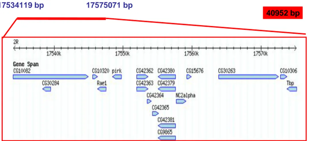
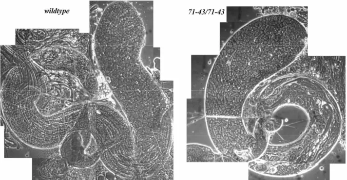
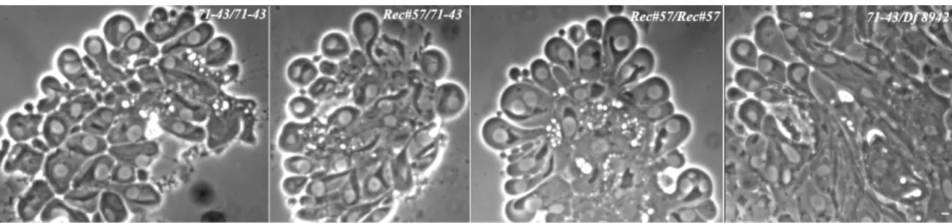
Among the twine-complementing mutations, one, named 71-43, resulted extremely interesting because of the following features. Mapping data by recombination and deficiency, and chromosome “cleaning” placed it on the distal portion of the right arm of the chromosome 2 as the only mutation present on this chromosome, responsible for meiotic and sterile phenotype. Cytological dissection of the spermatogenesis process in these mutants by means of antibodies provided the evidence of a
panoply of anomalies, starting from young primary spermatocytes until to spermatids. In particular, defects of nuclear envelope, centrosomes and meiotic spindle were detected by immunofluorescence experiments in the early stages of male germ line development as well as impairment of nucleus-basal body docking, cyst polarization and spermatid individualization processes in the later stages. The failure of assembly of a canonical meiotic cell cycle machinery accounted for the execution of only one, as well defective, meiotic division in 71-43 male mutants.
71-43 homozygous mutant females showed reduced fertility, thus indicating that the function
performed by this gene in oogenesis is likely redundant. A lower mitotic index in mutant larval brains in comparison with wildtype, pointed out that 71-43 acts also in mitosis, even though its action should be dispensable since no delayed development nor lethality are apparent. Towards the molecular identification of the gene mutated in 71-43 stock, different approaches were followed, both via forward and reverse genetics. Complementation analysis of males heterozygous for 71-43 mutation and a mutated allele of most of the genes embedded into the region where 71-43 was identified to lie, allowed to rule out all the genes so far tested as candidates for 71-43 locus. Proteomics approach, consisted in a comparative study of protein profiles of 71-43 homozygote vs
71-43 heterozygote testes, underlined the presence of some proteins misregulated in 71-43 mutants,
whose genes however map outside the 71-43 genetic interval. Such an analysis, however, provided intriguing clues about the putative pathways where 71-43 may act and, therefore, new roads to pursue.
RIASSUNTO
La mia ricerca nel corso del dottorato si è concentrata su un gruppo di mutazioni recessive maschio-sterili, indotte da etil metansulfonato (EMS), localizzate sui cromosomi 2 e 3 in Drosophila
melanogaster. Il moscerino della frutta è un sistema modello per lo studio del processo della
spermatogenesi per varie ragioni: la facilità di indagine citologica, la disponibilità di mutazioni ad ogni stadio e di metodi di indagine molecolare e genetica altamente sviluppati. La mia analisi è consistita nella dissezione genetica, molecolare e citologica di due gruppi di mutanti maschio-sterili di Drosophila: quelli che non complementano twine e quelli che, invece, lo complementano. Twine è un gene che risiede sul braccio sinistro del cromosoma 2 e codifica per una fosfatasi responsabile dell’attivazione del complesso Cdc2-CiclinaB, alla base dell’entrata in meiosi. La mutazione a perdita di funzione tweHB5 rende gli spermatociti primari incapaci di eseguire entrambe le divisioni meiotiche, permettendo tuttavia un certo differenziamento degli spermatidi. Il fenotipo che ne risulta è, comunque, sterile in quanto gli spermatozoi prodotti non sono mobili. Il gruppo di mutazioni recessive maschio-sterili, indotte da EMS, sul quale ho concentrato la mia ricerca, mostrava un fenotipo simile a quello dei mutanti tweHB5, suggerendo, quindi, la possibilità che tali mutazioni fossero alleli del gene twine. Esperimenti di mappatura per ricombinazione meiotica insieme al sequenziamento della regione twine nel genoma dei mutanti 12-228, 60-40 e 54-23, hanno suggerito la presenza di due mutazioni sul cromosoma 2 di questi mutanti, selezionate per il fenotipo maschio sterile: una responsabile della sterilità maschile e l’altra a carico della sequenza genica di twine, alla quale si deve il fenotipo meiotico. I risultati appena menzionati erano anche supportati dai dati di “ripulitura” del cromosoma 2 eseguita in questi mutanti mediante ricombinazione meiotica. Diversi erano i risultati ottenuti dallo stock 50-38, ovvero un’altra mutazione che non complementa twine. In questo caso, i dati di mappatura per ricombinazione e del sequenziamento di twine, insieme a quelli ottenuti dalla “ripulitura” del cromosoma, indicavano la presenza di una singola mutazione sul cromosoma 2 di questi mutanti, molto vicina al gene twine, che determina sia il fenotipo maschio-sterile che meiotico. Una dettagliata analisi citologica di questo mutante attraverso anticorpi in grado di rilevare alcuni componenti chiave del ciclo cellulare meiotico, quali la -tubulina per il fuso meiotico, la lamina per la membrana nucleare ed Spd2 per i centrosomi, evidenziava l’incapacità, da un lato, di disassemblaggio della membrana nucleare e, dall’altro, di assemblaggio del fuso meiotico, oltre a difetti nel numero e nella localizzazione dei centrosomi negli spermatociti primari. Tali anomalie sono alla base della mancata esecuzione di entrambe le divisioni meiotiche nei maschi mutanti 50-38. La caratterizzazione citologica mostrava anche difetti durante gli stadi tardivi della spermatogenesi, consistenti nella errata localizzazione del corpo basale, che impedisce di eseguire una corretta polarizzazione della cisti.
Tra le mutazioni che complementano twine, una, chiamata 71-43, risultava estremamente interessante per via delle seguenti caratteristiche. I risultati della mappatura genica, effettuata sia tramite ricombinazione meiotica che attraverso l’uso di deficienze, e della “ripulitura” del cromosoma, posizionavano la mutazione 71-43 sulla porzione distale del braccio destro del cromosoma 2 come unica mutazione presente su tale cromosoma, a cui si deve il fenotipo meiotico e maschio-sterile. La dissezione citologica del processo della spermatogenesi, eseguita per mezzo di anticorpi, ha permesso di riscontrare la presenza, in questi mutanti, di anomalie a partire dai primi stadi della spermatogenesi fino a quelli più tardivi. In particolare, gli esperimenti di immunofluorescenza hanno evidenziato dei difetti di membrana nucleare, di centrosomi e di fuso meiotico nelle prime fasi dello sviluppo delle cellule germinali maschili così come aberrazioni nei processi che caratterizzano le fasi finali del differenziamento degli spermatidi, quali l’ancoraggio nucleo-corpo basale, la polarizzazione delle cisti e l’individualizzazione degli spermatozoi. L’incapacità di assemblaggio di un canonico macchinario del ciclo cellulare meiotico giustificava l’esecuzione di una sola divisione meiotica, peraltro non corretta, nei mutanti 71-43. Femmine mutanti 71-43 mostravano ridotta fertilità, indicando quindi che la funzione espletata da questo gene nell’oogenesi è probabilmente ridondante. Un indice mitotico inferiore nei cervelli larvali dei mutanti rispetto a quello dei selvatici ha suggerito, inoltre, che 71-43 agisce anche in mitosi, sebbene la sua azione sembrerebbe essere dispensabile, in quanto né ritardato sviluppo né letalità sono visibili. Differenti approcci, basati sia su metodi genetici classici che innovativi, sono stati seguiti verso l’identificazione molecolare del gene mutato nel ceppo 71-43. L’analisi per complementazione di maschi eterozigoti per la mutazione 71-43 ed un allele mutato della maggior parte dei geni che risiedono nella regione in cui il locus 71-43 mappa, ha permesso di scartare tutti quelli finora testati come potenziali candidati per tale locus. L’approccio proteomico, fondato sullo studio comparativo dei profili di espressione proteica dei testicoli degli omozigoti 71-43 vs gli eterozigoti, ha mostrato l’esistenza di alcune proteine misregolate nei mutanti 71-43, i cui geni, comunque, risiedono al di fuori dell’intervallo genico di 71-43. Tale analisi ha fornito, tuttavia, intriganti indizi riguardo ai putativi processi in cui il gene 71-43 può agire e, quindi, nuove strade da percorrere.
1. INTRODUCTION
1.1 DROSOPHILA MELANOGASTER SPERMATOGENESIS
The Drosophila testis is a long tube with a straight apical and a coiled basal end (Fig.1). The different stages are laid out in chronological order from its apical tip to its basis, making it easy to discern distinct stages of germ cell development. Male germ line stem cells reside in the germinal proliferation center, at the tip of the testis (white arrow, Fig.1). Following an asymmetrical division, each of them generates another stem cell and a primary spermatogonium, encysted by two-somatically derived cyst cells. The spermatogonium undertakes four rounds of mitotic division to generate 16 interconnected cells, that then enter into a so called “growth period” (brackets, Fig1 and Scheme 1). Upon this premeiotic G2 phase, spermatogonia display a 25-fold increase in their nuclear volume, due to the transcription of many genes required in the subsequent meiotic stages. In
Drosophila most transcription is shut off upon entry into the meiotic divisions, differently from
mammals, where transcription is readily detected, by 3H-Uridine incorporation, in early spermatids and continues until chromatin compaction (Monesi, 1965). Until very recently, there was only data from experiments by Olivieri and Olivieri in 1965, where no radiographic labelling of nascent transcripts was revealed in Drosophila spermatids. Hence, transcription of genes required for spermatid differentiation occurs in primary spermatocytes, and the transcripts are stored in the cytoplasm for later use. Last year, White-Cooper’s group provided some evidence about the transcription of 24 genes, named “comets” and “cups”, in the mid-elongation spermatids, just before histone to protamine chromatin transition, also showing a curious localization of these transcripts at the extreme distal ends of the elongating cells (Barreau et al., 2008).
There is no meiotic recombination in male Drosophila, so after homologous chromosome pairing, most of the classic stages of meiotic prophase are not apparent in these cells. As the spermatogonial cells exit from the growth phase they develop into primary spermatocytes which undergo two meiotic divisions (large arrowhead, Fig.1 and Scheme 1), resulting in 64 haploid onion-stage spermatids. Cysts of haploid early round spermatids at the ‘onion stage’ are identified easily by their regular array of phase-light, spherical nuclei paired with phase-dark, nucleus similar size, mitochondrial derivatives or Nebenkernes, containing multiple layers of mitochondrial membranes (black arrow, Fig.1 and Scheme 1). Such a stage is an excellent indicator of any lack of fidelity in the meiotic divisions, as far as both chromosomal segregation and cytokinesis anomalies. The former by the nucleus size, since it’s directly proportional to DNA content; the latter by the evenly mitochondria partition between the daughter cells, since each cell after meiotic division is
characterized by the same mitochondria amount, visible as a same-sized spherical Nebenkern per spermatid.
The final stage of spermatogenesis consists in the elongation of spermatids, with dramatic morphological changes to transform themselves into fully elongated cells of 1.8 mm in length (small arrowheads, Fig.1 and Scheme 1). Besides tail formation, consisting into mitochondrial derivative and axoneme elongation, sperm maturation also involves nucleus conformational change from round to needle shape, through the switch from a nucleosome- to a protamine-based chromatin structure, determining a high chromatin condensation and compaction (Rathke C. et al., 2007). Their heads are oriented towards the basal end of the tube, with the sperm tails pushing up the testis towards the apical tip.Since cytokinesis is incomplete during gonial and meiotic divisions, the 64 haploid spermatids are connected among each other by cytoplasmic bridges, and they need to individualize in order to become functional. The individualization process involves a coordinated movement of actin-based ‘investment cones’ from the nuclei along the tails, expelling waste cytoplasm into a ‘waste bag’ (Fabrizio et al., 1998). Mature sperms coil and pass into the seminal vesicle where they acquire motility. On mating they move into the ejaculatory duct, where they are mixed with seminal fluid produced by the accessory glands, and then transferred into the female (Fuller, 1993).
Fig.1 wildtype Drosophila melanogaster testis viewed by phase contrast microscopy (by Fuller M.T., 1998)
spermatocytes at the growth phase. The large arrowhead near the start of the coiled part of the testis shows cells entering meiotic divisions, whereas the black arrow to the opposite side points out cysts of haploid early round spermatids at the “onion stage”. Small arrowheads along the testis indicate bundles containing 64 elongating spermatids, filling the testis up the lumen.
Scheme 1. wildtype spermatogenesis in Drosophila melanogaster (by Fuller M.T., 1998).
A spermatogonium undergoes four rounds of mitotic amplification divisions with incomplete cytokinesis. The resulting 16 interconnected cells enter the primary spermatocyte period of growth and gene expression. For simplicity, only one of the 16 primary spermatocytes in a cyst is shown. All 16 primary spermatocytes exit the cell growth and transcription program and enter meiotic divisions, resulting in a cyst of 64 interconnected, onion-stage, haploid, round spermatids. Each spermatid in an onion-stage cyst has the same sized nucleus (white sphere) and the same sized mitochondrial derivative (black sphere). The haploid spermatids remain interconnected and proceed through the dramatic morphological changes of spermiogenesis, eventually, with the production of fully elongated individualized sperms.
1.2 DROSOPHILA AS MODEL SYSTEM FOR STUDYING SPERMATOGENESIS PROCESS
Drosophila is a particularly suitable organism to undertake the genetic dissection of the complex
process of germ cell meiosis and differentiation that culminates in mature sperm formation. The reasons are several:
1. the ease to cytologically analyse such a process and discern distinct stages of germ cell development, since each different stage is laid out in chronological order from the apical tip of the testis to the basis.
2. The presence or absence of a germ line, the number and quality of mitotic and meiotic divisions, and the production of motile sperm can all be scored simply using phase contrast optics.
3. Sterile mutations can be easily generated at any step, identified and investigated, providing the starting point for molecular biological studies. To date, a number of genome-wide genetic screens for male sterile mutations have been carried out, making available a lot of mutations affecting different stages.
4. The availability of the complete DNA sequence of the Drosophila genome (from 1998) has been enormously improved the identification of genes required for spermatogenesis.
5. Spermatogenesis process has shown a remarkable degree of evolutionary conservation, so that the data obtained in Drosophila may have a broad applicability to other systems, including humans.
1.3 MALE MEIOTIC PROPHASE AND PROMETAPHASE: HOW PRIMARY SPERMATOCYTES GET READY TO ENTER MEIOSIS
As soon as a spermatogonial cell comes out from gonial division program, switches to a growth and gene expression program, entering a very long G2 interphase (it approximately lasts 3.5 days in
D.melanogaster) (Lindsley and Tokuyasu, 1980), that can be considered a meiotic prophase. During
this period, the cell transcribes most of the genes required for the further meiotic and post-meiotic stages (see above) and starts to get ready for cellular division. Chromatin appears as three masses or clumps, closely apposed to the inner nuclear envelope. The two larger masses represent the two somatically-paired autosomes of Drosophila genome (2 and 3 chromosomes), whereas the less dense chromatin mass corresponds to the X and Y chromosomes and the tiny fourth chromosomes (generally visible as dots) (Cenci et al., 1994). At this time are also apparent the Y chromosomes loops. In 1916 Bridges demonstrated that Y chromosome is not basic for vitality since flies with X/0 karyotype are vital, phenotipically male but completely sterile individuals. This means that Y chromosome carries genes required only for male fertility. Three of the six “fertility factors” (kl-5, kl-3 and ks-1) present in Y chromosome form lampbrush-like loops in Drosophila melanogaster primary spermatocytes, representing the cytological manifestation of their activity (Bonaccorsi et al., 1998). One major characteristic for all loops is that they are bound by several proteins, coded by autosomal genes, whose mutations alter loops morphology and provoke sterility. Some of them also have pleiotropic effects on meiosis and post-meiotic development (Ceprani et al., 2008). Y loops disintegrate during meiotic prophase pointing out the end of the growth phase and the beginning of the first meiotic division. While chromatin is organized into three clumps, representing an analogous state of interphase chromatin, the centrosomes, by now duplicated and consequently each formed by a pair of centrioles, migrate from plasma membrane to nuclear membrane where they start to nucleate astral microtubules and move towards the opposite poles of the nucleus. Cells enter prometaphase during which, concomitantly to spindle formation, there is a progressive chromatin condensation and the nuclear envelope breakdown, necessary to allow microtubules to make contact with chromosome kynetochores. Nuclear membrane is constituted by an inner envelope, made of lamins, which contact bivalents, located underneath nuclear envelope. As the bivalents start to congregate to metaphase plate, nuclear envelope disaggregates probably subsequently lamina phosphorylation that brings about nuclear lamina depolymerisation with consequent nuclear envelope breakdown. Lamin polymerisation will take place at telophase when nuclear envelope again forms around each of the two separated nuclei. In 1998, it was hypothesized a functional role for nuclear lamina in maintaining and alterating the three-dimensional genome structure (Gigliotti et
al., 1998). An important contribute in such a function is given by nuclear pore complex too, as proved by looking at the effects of mutations on the gene Nup154, homologous to rat Nup155 and yeast Nup170 and Nup157 nucleoporins. Nup154 male mutants are sterile and show meiotic arrest at the transition from prophase to prometaphase with alterated nuclear envelope structure in proximity to nuclear pores. Nuclear membrane of primary spermatocytes formed small evaginations near nuclear pores, that presumably detached in the surrounding cytoplasm as distinct vesicles, a phenotype reminiscent of the herniations described in nuclear envelopes of some nucleoporin yeast mutants. Meiotic arrest and further abnormalities in Nup154 mutants are justified by the authors as consequence of a misregulated nuclear import and export of factors underlying meiosis and spermatid development (Gigliotti et al., 1998). In support of this theory there is very recent data about Drosophila mutants of D1 gene, encoding Importin a protein which functions as an
adapter to link cargoes to import across nuclear pores from cytoplasm to nucleus through importin /1 heterodimer. In Drosophila there are three importin genes, D1, D2, D3. The last one
required for general viability, whereas D1 and D2 required almost exclusively for
gametogenesis: D1 affects more severely spermatogenesis than D2, exhibiting completely
sterility of D1 null mutants. These sterile males have numerous large round spermatid cysts, only partially elongated and not organized into bundles, also showing abnormal large round shape nuclei instead of needle-shape. Such a phenotype suggests a direct or indirect role for D1 in chromatin condensation, by means of nuclear import of chromatin remodelling factors (Ratan et al., 2008). The importance of a regulated nucleocytoplasmic trafficking for driving a correct male germ cell differentiation is highlighted in mammals as well. The mammalian testis-specific nucleoporin BS-63 has, as binding partner in a yeast 2-hybrid assay, an AT-hook domain chromatin remodelling factor, aF10, suggesting that factors like aF10 may access the nucleus through direct interaction with nucleoporins. It’s definitely evident that regulation of nucleocytoplasmic shuttling and, thereby, of the access of critical factors to the nucleus is a fundamental means of driving male germ line differentiation together with regulation of gene transcription (Hogarth et al., 2005).
1.4 HOMOLOGOUS CHROMOSOMES PAIRING DURING MALE MEIOSIS IN
DROSOPHILA
In sexual reproduction, the accurate segregation of homologous chromosomes during meiosis is a crucial step. Meiotic pairing of homologs is essential for their proper orientation along the metaphase plate and their subsequent disjunction during anaphase I. In Drosophila females, there are two systems ensuring segregation: a chiasmata system and the so-called “distributive system” (Day and Grell, 1976) that guarantee the segregation of achiasmata homologous chromosomes. The former is based on the formation of chiasmata, physical structures assembled at sites of reciprocal exchange. These structures are the last remaining sites of physical attachment between homologs at meiosis I metaphase, providing the strategy used by recombination-proficient organisms to ensure the maintenance of homolog associations until that stage.
The latter system is presumably necessary for the small fourth chromosome that never recombines in Drosophila females. According to Grell, the choice of partners in the distributive system is not determined by homology but rather by the availability, size and shape of these chromosomes. Different is the situation in Drosophila males, where meiosis occurs in the absence of recombination and of a recognizable synaptonemal complex (SC) (Baker and Hall, 1976). Moreover, mutations affecting distributive segregation in females have no effect on meiosis I in males, thus suggesting the existence of a distinct pathway for homolog segregation in males (Moore et al., 1994). The isolation of mutants that alter the segregation of specific chromosomes has revealed the presence of different segregation systems in males. One example may be represented by teflon mutants where pairing, specifically between autosomal bivalents, is defective with subsequent failure of unpaired autosomes to orient to opposite poles resulting in nondisjunction phenomenon. Conversely, tef mutation doesn’t affect sex chromosomes segregation (Tomkiel et al., 2001). Accordingly, there is a distinction between sex chromosomes and autosomes pairing and segregation. Pairing of sex chromosomes requires specific sequences from the rDNA gene repeats present in the heterochromatin of the X and Y chromosomes (McKee and Karpen, 1990), as proved by the failure to pair and the high degree of nondisjunction of sex chromosomes deleted for the rDNA locus. Pairing of autosomes, on the other hand, requires multiple regions of homology in the euchromatin, at least for the initiation of meiotic pairing, originally suggested by Metz to take place during the last gonial mitotic division (Metz, 1926). Upon this stage, the heterochromatic regions would be associated non-specifically. According to the model proposed by Vazquez and collaborators in 2002, heterochromatic associations would be responsible for the maintenance of
homolog pairing during late G2. Separation of chromosomes into distinct territories, each corresponding to a set of paired homologs, underneath the nuclear envelope in mid-G2, would disrupt non-specific heterochromatic interactions between nonhomologous chromosomes while preserving interactions between heterochromatic regions on homologous chromosomes. Separation of homologs and sister chromatids along the euchromatic chromosome arms also occurs together with separation of homologous centromeres while sister centromeres remain tightly paired until the second meiotic division. Therefore, despite the associations along the euchromatic autosomal arms and at the centromeres are loosen, homologous chromosomes remain in close physical proximity within each territory during late spermatocyte development, through heterochromatic associations and probably the contribute of some internal nuclear structure of the envelope into attaching and ensuring correct meiotic pairing (Vazquez et al., 2002). This theory still seems to be confirmed, since the latest data showed by Tsai and McKee at the Drosophila Conference 2009 would not prove the pairing of 9, tested by FISH, autosomal heterochromatic sequences, during mid and late G2.
Although control of meiosis I appears to be quite different between males and females, processes that ensure sister chromatid adhesion and segregation at meiosis II, instead, seem to be under common control in the two sexes. This is exemplified by the mei-S332 and ord mutations which affect the control of sister chromatid separation in both sexes (Lee and Orr-Weaver, 2001).
1.5 GENETIC CONTROL OF MALE MEIOSIS IN DROSOPHILA
The genetic control of spermatogenesis in Drosophila can be divided in three main programmatic switches. The first checkpoint occurs when a stem cell has to decide if maintaining the stem cell fate or switching into the spermatogenesis program. Upon the growth phase there is the second regulatory point, when the spermatogonial cells shift from a program of active mitotic divisions into one of growth and gene expression. The third checkpoint is when the primary spermatocyte exits the extended G2 phase to enter the first meiotic division. At this stage, two classes of genes have been shown by previous works to be required for meiotic entry - the so-called “Spermatocyte arrest class” and the “Twine class”-.
Spermatocyte Arrest genes coordinately control meiosis and spermiogenesis. In meiotic-arrest mutant testes, primary spermatocytes fail to initiate transcription of many genes whose products are required after meiosis as well as fail to express some meiotic gene products. Thus, mutant testes contain morphologically normal spermatogenic cells up to mature primary spermatocytes, which then arrest and fail to initiate either the meiotic divisions or spermatid differentiation, filling testes with primary spermatocytes arrested at the end of growth phase, without the presence of any meiotic and post-meiotic cells. Hence, it’s evident that the meiotic cell cycle progression and the onset of spermatid differentiation both require the action of this class of genes. Arrest Class genes have been, currently, subdivided, according to their molecular targets and specific role in promoting transcription, into two classes: always early (aly)-class and cannonball (can)-class (Lin et al., 1996; White-Cooper et al., 1998). The former is constituted by 5 genes: aly, encoding one of the two
Drosophila homologues of the C.elegans gene lin-9; cookie monster (comr); achinthya/vismay
(achi/vis) and matotopetli (topi), both encoding sequence-specific DNA-binding proteins; tombola (tomb), encoding a chromatin-associated protein. Tomb, Achi/Vis and Topi presumably enter the nucleus independently and localize to chromatin by means of sequence-specific DNA-binding activity, while Aly and Comr become or remain nuclear as a complex. Nuclear Aly and Comr bind to and stabilise Tomb, then interact with Topi and Achi/Vis by forming a Drosophila testis-specific meiotic arrest complex (tMAC) (Beall et al., 2007) to facilitate cooperative DNA binding. Since the
aly-class gene products accumulate on chromatin in transcriptionally active regions of primary
spermatocytes, their function strictly depends on the chromatin localisation where they act alterating chromatin structure, in order to permit the high levels of transcription necessary for spermatocytes. aly class is required for transcription of meiotic control genes like boule, twine and
On the other hand, can-class is constituted by genes codifying testis-specific TBP-associated factors (tTAFs), paralogs of generally expressed TAF subunits of transcription factor IID (TFIID). Recruitment of TFIID to sequences near the transcriptional start site is thought to, in turn, recruit and/or stabilise PolII binding in the preinitiation complex. The multisubunit TFIID complex contains the TATA-binding protein (TBP) and 12-14 TBP-associated factors (TAFs). Recently, alternative forms of TBP and tissue-specific TAfs have been described in a number of organisms,
Drosophila as well, and proposed to play a role in selective activation of cell-type specific gene
expression programs during cellular differentiation. In Drosophila melanogaster, 5 tTAFs have been so far described: cannonball (can), encoding a paralog of dTAF5, expressed only in male germ cells; meiosis I arrest (mia), paralog of dTAF6; spermatocyte arrest (sa), paralog of dTAF8;
no hitter (nht), paralog of dTAF4; ryan express (rye), paralog of dTAF12. These genes are required
either to allow translation of the meiotic gene twine or to stabilise twine protein in mature primary spermatocytes, apart from allowing the transcription of spermatid differentiation genes. Several evidences support the existence of a testis-specific TFIID, an alternative TAF-containing protein complex, proposed to regulate a testis-specific gene expression program in primary spermatocytes, required for terminal differentiation of male germ cells. The action mechanism hypothesized for the
can-class meiotic-arrest genes is the following: they activate transcription in two ways, globally by
sequestering the PRC1 complex, (Polycomb Repression Complex) away from active chromatin by means of a re-localisation of this repressor to the nucleolus in spermatocytes, thus, implicating subnuclear architecture in the regulation of terminal differentiation, and locally by the exclusion of PRC1 at target promoters. tTAFs binding to target promoters reduces Polycomb binding and promotes the recruitment of Trithorax (Trx) complex, an activator complex, with consequent accumulation of H3K4me3, a mark of transcriptional active chromatin (Hiller et al., 2001; Hiller et al., 2004; Perezgasga et al., 2004; Chen et al., 2005; Wright et al., 2006; Jiang et al., 2007; Metcalf and Wassarman, 2007; Kolthur-Seetharam et al., 2008). Similarly to tTAFs, TRF2 or TLF (TBP-like factor) may be the major transcription-initiating factor of the testis-specific TFIID complex in primary spermatocytes, since its high-level expression during the growth phase in mature spermatocytes is coincident with a decrease of TBP expression. Moreover, trf2 mutants show pre-meiotic chromatin condensation defects together with abnormal chromosome congregation, implying an essential role for TRF2 to allow primary spermatocytes to enter the first meiotic division (Kopytova et al., 2006). Some russian researchers also found a protein, Modulo, the
Drosophila homologue of nucleolin, that presumably cooperates with testis-specific TFIID to
induce expression of spermatid differentiation genes, such as 2-tubulin, and whose itself
the G2/M meiotic transition through post-transcriptional regulation of Cdc25/Twine (see below). Thus, Modulo plays an important role in integration of meiosis and spermatid differentiation in
Drosophila spermatogenesis, providing a mechanistic link between these two pathways
(Mikhaylova et al., 2006). A strongly reminiscent phenotype of spermatocyte-arrest mutants is observed in off-schedule (ofs) mutants testes even if with some differences. ofs mutants exhibit the lack of meiosis and of any significant differentiation together with a significant cell size defect. However, there are some differences with arrest-class, for example in Cyclin A accumulation. Indeed, Cyclin A in spermatocyte-arrest class persists in the cytoplasm without translocating to the nucleus, whereas in ofs mutants the nuclear translocation of Cyclin A is precocious and then its degradation is delayed. Differently from Cyclin A, nuclear Boule accumulation is delayed in G2 with consequent delay in Twine accumulation. Several key meiotic regulators show advanced or delayed patterns in these mutants, hence the name off-schedule. This gene is homologous to eukaryotic translation initiation factor eIF4G. Because translation is primarily regulated at initiation, eIF4G is instrumental in determining the translational capacity of a cell and thus its ability to accumulate mass. Therefore, ofs mutant phenotype suggests that sufficient cell mass must accumulate before spermatocytes execute meiosis and differentiation as if mass accumulation were used to time the G2/M transition. Spermatocytes monitor growth during G2 as one way to ensure that execution of the meiotic divisions is timed co-ordinately with spermatid differentiation (Franklin-Dumont et al., 2007; Baker and Fuller, 2007). A similar system exists during the mitotic cell cycle in yeast: for cells to maintain the same average size overall several divisions, there are control points acting during G1 predominantly in budding yeast and G2 in fission yeast, to allow cell cycle progression only when the cell reached a threshold size (Rupes, 2002).
Mutations that result in a uniform block of the progression of spermatogenesis are rare among male-sterile mutants in D.melanogaster. For the most male male-sterile mutants, spermatogenesis proceeds despite defects in one or even several morphogenetic events, leading to the production of aberrant spermatids, suggesting that the majority of events in spermatogenesis proceed by independent pathways. Twine-class mutations, which I introduced before, belong to this context, since mutant spermatocytes skip several crucial events of meiotic divisions, however they initiate and enact spermatid differentiation. Both flagellar elongation and condensation and shaping of spermatid nuclei proceed despite failure to execute previous meiotic divisions, with the final production of immotile sperm bundles, unable to fecundate. Genes so far identified belonging to twine-class are:
Dmcdc2, twine (twe), pelota (pelo) and boule (bol). Dmcdc2 is required for mitosis; mutations result
in developmental defects and larval lethality (Stern et al., 1993). To investigate its role in meiosis, a temperature-sensitive Dmcdc2 allele has been engineered making possible to study the meiotic
function in Dmcdc2ts mutants (Sigrist et al., 1995). Dmcdc2 codifies for the cdc2 protein kinase, well known to form the Cdc2-Cyclin B complex, underlying the G2/M transition. Indeed, entry into mitosis is triggered by the activation of M cyclin/cdc2 kinase complexes. The activity of these complexes is controlled by a cycle of phosphorylation and dephosphorylation of the cdc2 kinase (reviewed by Nurse, 1990). Phoshorylation is mediated by Wee1 (on tyrosine 15 in S.pombe and threonine 14 in higher eukaryotes) and inhibits cdc2 activity (Gould et al., 1990; Krek and Nigg, 1991a.b), while the removal of these inhibitory phosphates by cdc25 phosphatase, activates the M-cyclin/ cdc2 kinase complex (Dumphy and Newport, 1989; Gautier et al., 1989; Labbe et al., 1989). Inactivation of this complex is required for exit from mitosis and needs the M cyclin destruction by a mechanism of ubiquitin-dependent proteolysis during metaphase (Glotzer et al., 1991). Studies in a variety of distantly related organisms have emphasized the similarities in the regulation of mitotic and meiotic M-phase. According to this, the entry into meiosis in Drosophila is regulated by the same mechanism based on the activation by de-phosphorylation of cdc2-Cyclin B complex. The phosphatase responsible to remove inhibitory phosphates on cdc2 kinase in male and female meiosis is codified by twine/cdc25 gene. The cdc25 gene was initially identified in
Schizosaccharomyces pombe (Russel and Nurse, 1986) and later its Drosophila homolog was found
at the string locus (Edgar and O’Farrell, 1989). Mutations in the cdc25 and in string block the cell cycle at G2-phase; moreover in Drosophila the timing of the embryonic cell divisions is also affected (Edgar and O’Farrell, 1990). In Drosophila, a second cdc25 homolog was identified in 1992 (Courtot et al., 1992). This gene was twine, which, differently from string, is required in meiosis but not in mitosis. The twine gene is transcribed relatively early during the long G2 phase, but twine mRNA accumulation is not sufficient to promote entry into the meiotic divisions, differently from that observed in early embryos where the transcription of string/cdc25 gene is both necessary and sufficient to drive the G2/M transition (Edgar and O’Farrell, 1989, 1990). TWINE is activated by POLO kinase thus triggering cdc2-Cyclin B complex. This complex itself, once activated, phosphorylates and activates its same activator (TWINE) and inhibits its repressor (Wee1), by a positive feedback loop. Mutations in twine result in male and female sterility but do not affect the embryonic development or viability of the fly (Alphey et al., 1992; White-Cooper et al., 1993). Surprisingly in female mutants entry into the first meiotic division occurs at the correct stage of oogenesis. The defect starts after entry into meiosis, in mature stage-14 oocytes, when oogenesis should arrest at metaphase of meiosis I, to then normally re-start after egg-activation, which occurs during egg laying. In twine females, mutant oocytes don’t arrest at metaphase I but continue to progress through aberrant cell cycles during which chromosomes are replicated and segregated irregularly. This phenotype suggests that twine function in females is required to
maintain the arrest of oocyte at metaphase I by keeping Dmcdc2 protein de-phosphorylated and thereby active (White-Cooper et al., 1993).
The pelota gene encodes a RNA-binding protein, S.cerevisiae DOM34 homolog, required not only for male gametogenesis but also for eye development, thereby for mitosis (Eberhart and Wasserman, 1995). In contrast boule is expressed only in the testis and boule mutations affect only meiosis. Boule protein has a conserved RNA-binding domain that has been shown to directly bind
twine mRNA. By means of this interaction, Boule exerts a translational control of Twine, as
supported by a markedly reduced Twine protein expression in a genetic background deficient in
boule. Also, although the male sterile phenotype produced by mutation of the pelota gene closely
resembles that produced in twine or boule mutants, the 2-twine transgene does not restore meiotic
entry in a pelota male, whereas it does in boule mutants (Maines and Wasserman, 1999). Boule belongs to the DAZ (Deleted in Azoospermia) gene family and one member of this family in humans- human BOULE- was shown having a very similar function as the Drosophila homolog as a regulator of meiosis, because spermatogenesis was rescued in boule mutant flies made transgenic for the human BOULE gene (Luetjens et al., 2004).
The loss-of-function phenotype of twe, pelo, Dmcdc2 and bol alleles is characterized by failure into execution of both meiotic divisions, however they are still able to undergo some spermatid differentiation. In all these null mutants, spermatogenesis is normal until late meiotic prophase, at which point the cell cycle arrests. Chromosomes are partially condensed but never move away from the nuclear periphery of primary spermatocytes towards the center of nucleus. Nucleolus regularly breaks down. In contrast, while in a wildtype scenary the nuclear envelope breaks down during prophase and nuclear lamins disperse, forming diffuse clouds around the chromatin to then associate again with the reformed nuclear envelope after meiosis, in twine-class mutants the nuclear envelope does not breakdown (Eberhart and Wasserman, 1995). It’s, however, noteworthy to say that White-Cooper and colleagues reported in 1993 contrasting data, showing that nuclear envelope breakdown still occurs in tweHB5 (null allele) male mutants (White-Cooper et al., 1993). Another crucial event of meiotic cell division, lacking in twine-class mutants, is the spindle formation. Normally, during the late meiotic prophase, the centrosomes separate and move towards the opposite poles of the cell; after they reach the poles, the cytoplasmic tubulin network, previously formed, breaks down and a spindle is formed. In these mutants, centrosomes separate but don’t complete their migration and they nucleate no significant asters. Thereby, a spindle is never observed (White-Cooper et al., 1993; Eberhart and Wasserman, 1995). Despite the meiotic defects just mentioned, some post-meiotic differentiation occurs, such as nebenkern and tail elongation and nuclear shaping of spermatid heads. Two lines of evidence prove that spermatocyte-arrest mutants
arrest at a precocious point of cell cycle in comparison with twine-class mutants. As mentioned before, nucleolus breaks down in twine-class mutants, whereas a distinct nucleolus is still visible in primary spermatocytes of meiotic arrest mutants. Another hint is represented by the nuclear presence of Cyclin A in twine-class mutants where it remains until degradation at onion-stage spermatid, conversely to a still cytoplasmic localization in primary spermatocytes of arrest-class mutants. Furthermore, the phenotype of twine; sa and twine; can double mutants is similar to sa/Df and can/can phenotype, indicating that can and sa are epistatic to twine (Lin et al., 1996).
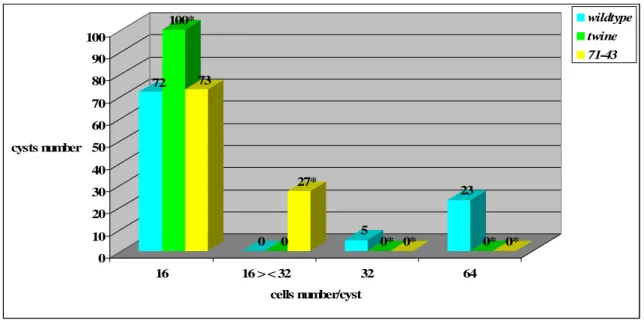
From the literature, hypomorphic twine (tweZO758) and pelota (pelotaweak) alleles are known. tweZO758 homozygotes execute a single division, the first meiotic division, resulting in cysts of 32 cells, in contrast with 16 cell-cysts of tweHB5 null mutants. Spermatocytes from tweZO758 / tweZO758 males with only a single wildt-ype copy of boule fail to enter meiosis, resembling phenotype of flies homozygous for loss-of-function twine alleles (Maines and Wasserman, 1999). pelotaweak homozygote mutants show some meioses, albeit aberrant. Mutant spermatids contain two or four nuclei in a single cell, indicating a failure of cytokinesis in one or both meiotic divisions. In some cells, the nuclei are of different sizes, a sign of uneven chromosome complements, thus reflecting defects in chromosome segregation. Defects within a cyst are usually heterogeneous, with some cells displaying meiotic defects while others appear wild-type or are arrested before meiosis I. The presence of such anomalies during meiotic cell divisions in weak pelota alleles is explained by Eberhart and Wasserman as direct consequence of aberrant regulation and initiation of meiotic divisions rather than the possibility that pelota plays separate roles in the meiotic entry and in specific meiotic processes such as spindle formation and cytokinesis (Eberhart and Wasserman, 1995).
The regulation of second meiotic division is still not well understood. No genes have been identified for which loss-of-function mutations specifically block MII. There is evidence, however, that
Dmcdc2, twine and roughex (rux) play a critical role in control of MII.
Dmcdc2 mutant males that carry two copies of the Dmcdc2ts transgene are sterile at all temperatures, with defects in meiosis. At 18°C -permissive temperature- they execute MI but fail in MII, producing 32-cell spermatid cysts. When shifted as adults to 27°C –nonpermissive temperature- they fail to execute either meiotic division (Sigrist et al., 1995).
As far as the role of twine in the regulation of MII, in twine mutants, background expression of
string from a heat-shock promoter in late primary spermatocytes, is sufficient to rescue entry into
MI, but not MII (Sigrist et al., 1995). Moreover, in females, twine mutants execute MI but not MII, suggesting that twine play a unique role in MII (Courtot et al., 1992; White-Cooper et al., 1993). Since flies with two copies of the Dmcdc2ts transgene execute MI but fail in MII, the level of kinase
activity may be of particular importance for entry into MII and Twine phosphatase would play a basic task for maintaining this level of cdc2 kinase activity.
Roughex gene acts as negative regulator of MII (Gonczy et al., 1994). An increase in roughex gene
dosage blocks MII while allowing normal execution of MI. Reciprocally, germ cells from rux mutants attempt to execute an extra MII-like division. Thereby, these data strongly suggest that rux negatively regulates MII, with excess rux preventing MII and insufficient rux permitting an additional MII. In rux mutants there is an increase in Cyclin A levels prior to MI. Since lowering the dose of either twine or cyclin A suppresses the extra division, consistent with the hypothesis that the excess cdc2 activity is responsible for the extra meiotic division in the rux mutants, presumably
rux acts by limiting the amount of cdc2 activity available for MII.
1.6 SPINDLE FORMATION AND FUNCTION DURING MALE MEIOTIC DIVISIONS IN
DROSOPHILA
During spermatocyte growth phase, centrosomes dissociate from the nuclear membrane and become associated with the spermatocyte cell membrane. As the cell enters meiotic prophase, centrosomes migrate back to the nuclear membrane and nucleate two asters while the network of microtubules, previously formed around the nucleus, disappears. In late prophase, the asters, initially located on the same side of the nucleus, migrate to the opposite poles where they radiate spindle fibers that reach the bivalents. Nuclei become irregularly shaped and are surrounded by structures corresponding to a system of parafusorial and astral membranes that surround the spindle and the poles during male meiosis. By the following prometaphase, the bivalents are connected to the spindle poles by kinetochore microtubules and start to congress to a metaphase plate. In addition to pole-to-chromosome microtubules, meiotic spindle shows bundles of microtubules that do not encounter the chromosomes, instead they originate from the poles, run outside the metaphase plate, ending either in the opposite pole or distally to the metaphase plate. As spermatocyte enter anaphase, the spindle organization changes: the density of microtubules in the region between the segregating sets of chromosomes –central spindle- (for more details see below) progressively increases, whereas the microtubules located in proximity of chromosomes decrease in number. Centrosome separation occurs early and two distinct centrosomes are already visible in late anaphase. At telophase, the dense bundle of microtubules in the central spindle is squeezed, eventually attaining an hourglass shape. The spindle organization is similar in meiotic second
division. The only difference concerns centrosome composition. At the first meiotic division each centrosome is composed by a pair of centrioles, whereas the second meiotic spindle is built with centrosomes which contain each a single centriole, since there is no centrosome duplication before meiosis-II (Cenci et al., 1994).
There is an open debate about the role of centrosomes in spindle assembly. While some evidence from several systems supports the need for centrosomes, other reports suggest that they are dispensable (commentary by Gonzalez et al., 1998). Drosophila female meiosis, for example, is a case in which, under natural conditions, the spindle is built in the absence of centrosomes, thereby, in female meiotic spindle, microtubules originate from around the chromatin and its poles are anastral and not contain centrioles (Huettner, 1933; Theurkauf and Hawley, 1992). The phenotypes of several mutants which alter the centrosome cycle have revealed different mechanisms of spindle assembly. As an example, there is the testis-specific isoform of centrosomin (cnn), expressed during spermatogenesis and closely associated with the centrioles. Mutations on cnn gene result in karyokinesis as well as cytokinesis defects deriving from abnormal spindles. Furthermore, the cytokinesis damage is more severe than karyokinesis, suggesting a more susceptibility of cytokinesis to reduced centrosomin function. This result is consistent with the finding that centrosomes are not absolutely required for chromosome segregation, as supported by Drosophila female meiosis and studies of micromanipulation by Church et al. (1986), who demonstrated that chromosome pairs relocated from the nucleus to the spermatocyte cytoplasm are capable of organizing minispindles around themselves with no centrosomes at the poles. Similar experiments have been conducted by Rebollo et al. (2004) to study the contribution of centrosomes in spindle organization in Drosophila spermatocytes. They took advantage of two experimental conditions that inhibit the natural process of centriole migration from the plasma membrane to the interior of the cell that takes place at the onset of meiosis. Under such conditions, the centrosomes organize asters but these are kept at the plasma membrane, away from the nuclear region. In these cells, microtubules can be seen grow from the remnants of the fenestrated nuclear envelope (NE) and to assemble into anastral bipolar spindles in a centrosome-independent manner. However, these anastral spindles could sustain only some degree of chromosome segregation. The model of spindle assembly during meiosis I in Drosophila spermatocytes proposed by the researchers is that, at the beginning, microtubule bundles of centrosomal origin, connecting centrosomes to chromosomes, develop. Immediately after, noncentrosomal microtubules start to polymerise in association with the remnants of the NE. The fully mature spindle would contain a spindle-shaped structure made of microtubules of noncentrosomal origin embedded in another spindle-shape array made of two overlapping asters. Thus, according to this theory, acentrosomal microtubules are nucleated on the
inner side of the remnants of the NE and not around the chromosomes. This is, on a side, in contrast with what seen in asterless mutants, where, despite the absence of asters, spermatocytes assemble a peculiar anastral spindle by means of meiotic chromosomes that act as microtubule-organizing centers, thus promoting formation of bipolar minispindles associated with individual bivalents (Bonaccorsi et al., 1998). On the other side, the Rebollo’s model is consistent with the phenotype of
fusolo and solofuso mutants, where secondary spermatocytes are produced, devoid of chromosomes,
yet able to give rise to bipolar spindles that undergo all the morphological transformations required for a correct cell division, such as central spindle formation. This strongly argues that in Drosophila spermatocytes, spindle formation and dynamics are controlled by chromosome-independent factors (Bucciarelli et al., 2003).
Failure in centrosome segregation may result in different phenotypes as evidenced by some mutants. In ms(1)516 mutants, for example, failure in centrosome segregation gives rise to a monoastral bipolar spindle (Lifschytz and Meyer, 1977), whereas in polo (Llamazares et al., 1991) and aurora (Glover et al., 1995) (serine-threonine kinases) mutants, the impairment of centrosome segregation determines the formation of monopolar spindles, where microtubules radiate from the single pole which contains the two unsegregated centrosomes.
Centrosomes or MTOC (microtubule organising centers) consist of a pair of centrioles surrounded by a cloud of pericentriolar material, whose components change according to cell-type and development in Drosophila (Gonzalez et al., 1998). Centrioles are not essential for microtubule nucleation; rather they appear to organize pericentriolar components into a cohesive focus at spindle poles. Microtubules are nucleated within the pericentriolar cloud, their minus ends within or near the pericentriolar material and their plus ends extending into the cytoplasm (Wilson et al., 1997). -tubulin is one of the pericentriolar components. Antibodies against --tubulin recognise two orthogonal rods located near the surface of the cell in mature primary spermatocytes. During meiosis the same antibody recognises a dot-like centrosome at the poles of the meiotic spindle and the rod-like structures are no longer present (Wilson et al., 1997). Twine function seems to be required for -tub23C reorganization and/or recruitment to centriole pairs specifically at the G2/M transition, when -tubulin is visible as a dot at each spindle pole (Wilson et al., 1997). Mutations in -tub23C (germ-line specific isoform) impair the assembly of meiotic bipolar spindles. Centrosomes associate with a large number of astral microtubules that later get organized into a conical structure never observed in wildtype cells. Both meiotic divisions fail to be accomplished, as evidenced by the presence of spermatid cysts containing only 16 cells, with each spermatid having a single large nebenkern associated with several nuclei of different sizes. By observing the
behaviour of these mutants, it seems like the depletion of -tubulin does not prevent microtubule polymerization or stabilization (Sampaio et a., 2001).
Like other Drosophila mutants that disrupt spindle assembly during male meiosis, -tub23C does
not result in meiotic arrest. On the contrary, besides the absence of a bipolar spindle, the chromosome cycle proceeds until abnormal spermatids are formed but prometaphase lasts about 50% longer than in wildtype cells. Same case as it’s seen in wildtype spermatocytes following microtubule depolymerisation with colchicine. Both examples, as well as the delay of the onset of anaphase resulting from a relatively high number of misaligned chromosomes, provide evidence substantiating the activity of a feeble meiotic spindle checkpoint in Drosophila male meiosis (Rebollo and Gonzalez, 2000).
DSpd2 is another centrosomal protein, recently shown to be enriched at both the centrioles and the pericentriolar material (PCM). Its function is to maintain the cohesion between the two centrioles and to mediate the recruitment of additional PCM components, such as -tubulin, DGrip91 and D-TACC. Null mutations in DSpd2 don’t affect vitality of the flies but, instead, provoke sterility in both sexes. Astral microtubule nucleation is suppressed in both neuroblasts and spermatocytes even if a more severe phenotype is seen in the testis (Giansanti et al., 2008). DGrip 91, just mentioned, is a gamma-ring protein (Grip) forming, together with DGrip 84 and -tubulin, the -tubulin small complex (-TuSC) that, in turn, is a component of the larger -TuRC (-tubulin ring complex), present in the pericentriolar material in Drosophila. -TuSC displays an open ring-like structure, resembling a lock washer, capped on a side by other Grips, like Grip71, Grip75, Grip128 and Grip163. The activity of -TuRC is to nucleate the microtubule assembly within the centrosome and to keep the microtubule minus ends capped by cap components, preventing depolymerisation of the microtubules. Also, the -TuRC cap structure is dispensable for microtubule function, as cap Grip mutants provide TuRC functionality sufficient for the viability. However, they are required for anchoring the TuRC at specialized MTOCs, (in the germline, for example) allowing microtubules to tightly associate with specific MTOCs, as suggested by the failure of alignment of the nuclei at one end of the sperm bundle, (a phenomenon due to the interaction between nuclei and axoneme microtubules), owing to the loss of -tubulin association with spermatid nuclei, in Grip128 and
Grip75 mutants (Vogt et al., 2006).
1.7 CENTRAL SPINDLE ASSEMBLY AND ITS IMPLICATIONS IN CYTOKINESIS PROCESS
During late anaphase, a dense body of overlapping antiparallel microtubules (MTs), that forms in the region between the two daughter nuclei migrating to the opposite poles, gets evident. This bundle of microtubules has been termed as “central spindle” or “midbody” and its middle part as “central spindle midzone”. The midzone, in particular, is associated with a proteinaceous matrix, the so-called “Flemming body” (Zeitlin and Sullivan, 2001), which prevents tubulin immunostaining, thus forming a dark band. About the origin of the central spindle microtubules, there are controversial theories. One of these relies on the presence of transient microtubule organizing centers between the two daughter nuclei, that would de novo nucleate new microtubules which then will be part of central spindle (Vogt et al., 2006). Studies on -tubulin localization have provided support for this possibility, since this protein has been found at the minus ends of central spindle MTs (Julian et al., 1993). On the other side, there is the hypothesis according to which the central spindle would consist of MTs dissociated from the spindle poles, with the plus ends in the midzone and the minus ends terminated near the chromosomes migrating to the opposite cell poles. Thereby, these microtubules would be the remnants of the interpolar MTs nucleated by the centrosomes. This is consistent with the presence of Asp (Abnormal Spindle) protein on the borders of the central spindle region, that correspond to the minus ends of microtubules released from the spindle poles, where Asp previously was localized. To strengthen this theory there are the central spindle defects evident in asp mutant spermatocytes (Riparbelli et al., 2002). On the contrary, data by asterless mutants, previously showed, argue for a self-organization of the central spindle since it regularly assembles in the absence of the asters (Bonaccorsi et al., 1998). Furthermore, there is one more model for the central spindle microtubule composition proposed by Inoue et al. (2004), where the central spindle would consist of two distinct set of MTs, “peripheral” astral MTs and “interior” MTs. The former corresponds to the peripheral MTs of the asters, which dynamically probe the cytoplasm as they extend along the cell periphery and are thought contact the cortex and bundle at the location of the future cleavage site. These MTs translocate toward the spindle equator and form a series of overlapping bundles, a structure termed “peripheral central spindle”. The latter, instead, is confined within the “spindle envelope”, the highly fenestrated remains of the nuclear membrane that persists in Drosophila cells. These interior MTs are proposed to elongate and then to be released from the spindle poles to form the overlapping bundles of the “interior central spindle”. The two MT populations are biochemically distinct, as the Orbit/Mast protein (a MT-associated protein) strongly accumulates in the region of interior MTs, the reason why orbit mutants fail to
form stable interior central spindles, whereas peripheral MTs normally contact the cortex, generating the signal for cleavage furrow. Although cleavage furrow initiates, it ultimately regresses in these mutants, allowing to postulate a role for peripheral MTs in initiating furrow formation where they contact the equatorial cortex and bundle, and a second function for the interior MTs into stabilizing and propagating the furrow (Inoue et al., 2004). The fact that MTs of central spindle are implicated into furrow positioning and, thus, cytokinesis, introduces another strongly debated issue: the relationship between the central spindle and the cytokinesis structures.
Some results from micromanipulation experiments by Rappaport (Rappaport, 1961) support a role for the asters into stimulating cytokinesis according to a model where an organizing factor is directed along astral microtubules to concentrate at the point at which the plus ends of the microtubules contact the cell cortex. Furthermore, experiments where asters are kept in the cytoplasm and anastral spindle form, the orientation of the cleavage furrow correlates tightly with the position of the two asters and not at all with the orientation of the spindle. Hence, the asters seem to contribute to specify the place of furrow (Rebollo et al., 2004). Contrasting data come from Bonaccorsi et al., (1998), who show normal cell contraction in asterless mutants as well as in
DSpd2 mutants where nonetheless the defects in asters nucleation, a regular furrow ingression is
visible (Giansanti et al., 2008).
Metaphase chromosomes has been implicated in signalling cytokinesis as well, but the behaviour of the Drosophila secondary spermatocytes in fusolo mutants argue against the need of chromosomes for cytokinesis to take place. Indeed, these mutant spermatocytes II, even in the absence of chromosomes, assemble a regular contractile apparatus and undergo cytokinesis (Bucciarelli et al., 2003). The other structure suggested to be required for cytokinesis is the central spindle. Both results from asl and fsl mutants suggest that central spindle is necessary and sufficient to stimulate the cytokinetic process, ruling out the involvement of either astral microtubules or chromosomes. A possible explanation to justify the different results obtained about the presumed factor underlying cytokinesis signalling is that these difference may depend on the cell type. Hence, in large cells, such as those used in the micromanipulation experiments by Rappaport, where the central spindle is far from the cell cortex, astral microtubules may play a role into inducing furrow. Conversely, in
D.melanogaster spermatocytes it may be the central spindle having this role (Saint and Somers,
2003. The analysis of mutations that impair both central spindle formation and cytokinesis processes reveals a cooperative interaction between central spindle and cytokinetic apparatus components. The impairment of either of these structures prevents the formation of the other, thus displaying an interdependence with each other (Gatti et al., 2000). This the case, for example, of