Evidence-Based Consensus and Systematic Review on Reducing the
Time to Diagnosis of Duchenne Muscular Dystrophy
Annemieke Aartsma-Rus, PhD
1, Madhuri Hegde, PhD, FACMG
2, Tawfeg Ben-Omran, MD, FRCPS, FCCMG, FACMG
3,
Filippo Buccella, PharmD
4, Alessandra Ferlini, MD, PhD
5, Pia Gallano, PhD
6, R. Rodney Howell, MD
7,
France Leturcq, PharmD
8, Ann S. Martin, MS, CGC
9, Anna Potulska-Chromik, MD, PhD
10, Jonas A. Saute, MD, PhD
11,
Wolfgang M. Schmidt, PhD
12, Thomas Sejersen, MD, PhD
13, Sylvie Tuffery-Giraud, PhD
14, Zehra Oya Uyguner, PhD
15,
Luci A. Witcomb, PhD
16, Shu Yau, PhD, FRCPath
17, and Stanley F. Nelson, MD
18D
uchenne muscular dystrophy (DMD) is a severe,pro-gressive, X-linked, recessive neuromuscular disease that affects approximately 1 in 5000 live male births.1 Pa-tients with DMD experience progressive muscle weakness, owing to the absence of functional dystrophin protein, and typi-cally experience delayed walking, difficulty running or climb-ing stairs, and frequent falls.2,3However, several nonmotor signs and symptoms (eg, behavioral issues, neurocognitive defi-cits, and speech delay) also can be associated with the disease.3 Boys with DMD are generally diagnosed between 4 and 5 years of age, when their physical development begins to diverge more clearly from that of their peers,3-6and, with corticosteroid treat-ment, typically lose ambulation at a median age of about 12.0-14.0 years.7However, this can vary by country.
The accurate and early diagnosis of DMD plays a crucial role in the effective management of patients: it has the potential to lead to earlier intervention; appropriate genetic counsel-ing; treatment with mutation-specific therapies (where appli-cable); and appropriate assignment to clinical trials. However, reports indicate that significant delays in diagnosis of DMD persist.4-6,8It has been shown that in the absence of a family history of DMD, there is a delay of approximately 1 year from onset of earliest symptoms to the first assessment by a health-care professional.4A further delay of approximately 1 year from this first assessment to referral to a neurologist or a neuro-muscular specialist is observed (mean age at diagnosis± SD: 4.9± 1.7 years).4Findings by Vry et al6suggest that this could be shorter, with a mean delay of just over 1 year from first symptoms to diagnosis (mean delay± SD: 1.3 ± 1.8 years). Overall, these findings suggest that, on average, there has been little to no reduction in the age at diagnosis for patients with DMD over the last 30 years.9,10
Once DMD is suspected, genetic testing is required to obtain a complete diagnosis.3,11,12However, results from a recent survey of 41 delegates from Europe, Turkey, and India (primarily child neurologists and clinical/molecular geneticists) revealed that there may be issues relating to the genetic diagnosis for DMD.
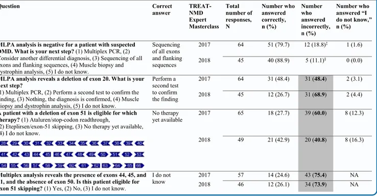
For example, although 100% of delegates understood the importance of genetic testing for DMD, more than 10% did not perform additional genetic tests if deletion/duplication testing was negative.11Survey results from the 2017 and 2018 TREAT-NMD Expert Masterclasses on DMD, attended by more than 100 delegates combined (primarily from pediatrics and neurology backgrounds from 27 and 20 countries, respec-tively), showed that some delegates experienced difficulties in-terpreting DMD genetic test results (Table I; available at
www.jpeds.com) and subsequently were not always aware of
whether patients were eligible for treatment with mutation-specific therapies. Together, these issues highlight the need for shorter times to diagnosis for patients with DMD and clearer recommendations for DMD genetic testing to ensure com-plete genetic assessment is performed to reach an accurate genetic diagnosis. This review, supported by a systematic lit-erature search, presents expert consensus on ways of reduc-ing the time to diagnosis of DMD.
Methods
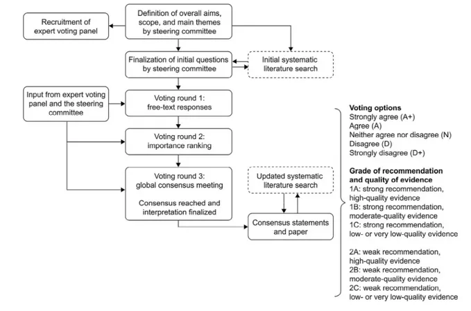
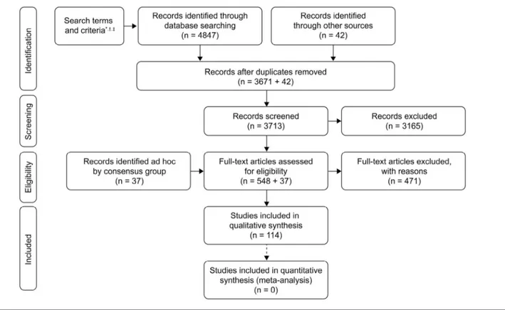
The Delphi Consensus Initiative presented here is focused on how to reduce the time to diagnosis of DMD. The develop-ment process for this initiative is summarized inFigure 1. The steering committee comprised 3 experts in the field of human genetics, specializing in the diagnosis of DMD and/or inter-pretation of genetic mutations: S. F. Nelson (nonvoting chair), A. Aartsma-Rus (voting chair), and M. Hegde (voting co-chair). After an initial meeting of the steering committee, 14 experts in the field were invited to form the expert voting panel. The panel comprised primarily medical geneticists specializ-ing in the diagnosis of patients with DMD; however, 2 child neurologists, 1 patient advocate, and 1 genetic counselor also were invited. All members of the expert voting panel and the 2 voting co-chairs voted anonymously on the statements to reach consensus. A systematic literature review was also per-formed to support development of the consensus statements
BMD Becker muscular dystrophy CK Creatine kinase
DMD Duchenne muscular dystrophy
MLPA Multiplex ligation-dependent probe amplification NBS Newborn screening
NGS Next-generation sequencing
Detailed affiliations, funding, and conflicts of interest disclosure available at www.jpeds.com
0022-3476/$ - see front matter. © 2018 The Authors. Published by Elsevier Inc. This is an open access article under the CC BY-NC-ND license (http://creativecommons.org/licenses/by-nc-nd/4.0/).
(PRISMA flow diagram;Figure 2andTable II[available at
www.jpeds.com]). The evidence was graded using the Grading
of Recommendations Assessment, Development, and Evalu-ation system (Figure 1). Further information regarding the de-velopment process is provided in theAppendix(available at
www.jpeds.com). The grading of evidence for the statements
was reviewed and agreed on by the expert voting panel (Table III).
Discussion
Consensus Statements
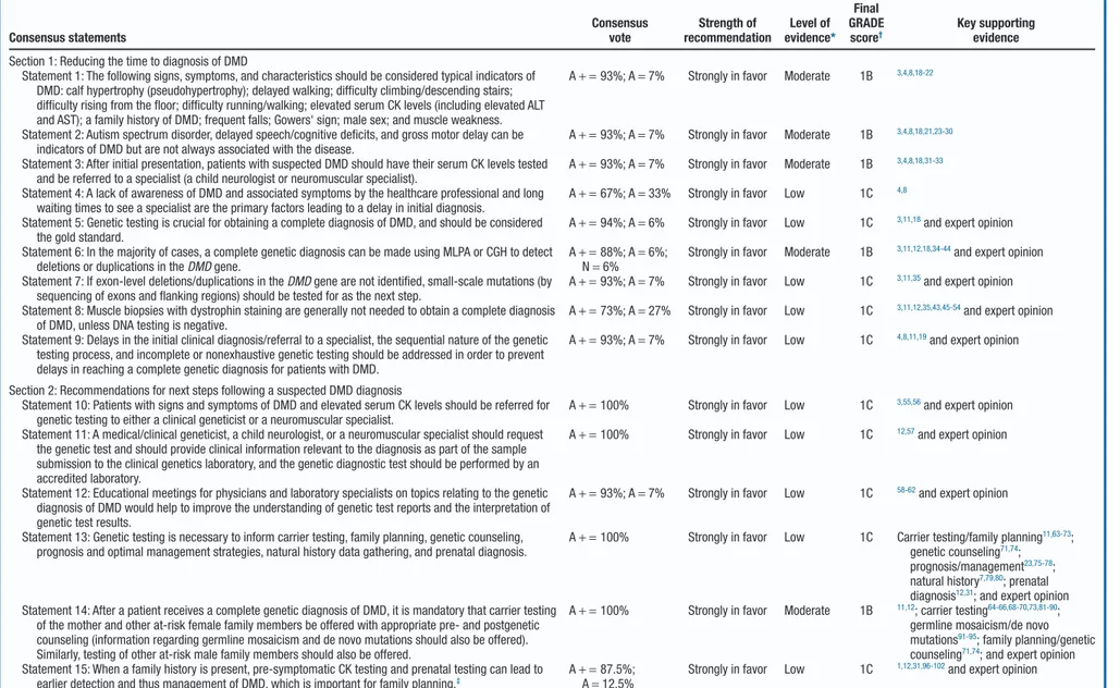
The 15 consensus statements are presented inTable III. A summary of the supporting information is provided herein, with a full discussion included in theAppendix. Several other more general statements were discussed, but only those most pertinent to reducing the time to diagnosis of DMD are pre-sented here.
Section 1: Reducing the Time to Diagnosis of DMD
Signs and symptoms of DMD (motor and nonmotor) are pre-sented in the 2018 care guidelines for DMD.3The consensus group agreed on a series of motor signs and symptoms (Table III) that are typically observed in patients with DMD (supported by the literature; Statement 1). It also should be noted that patients are sometimes referred because of el-evated alanine transaminase or aspartate transaminase levels,3
and that this finding also should require a high index of sus-picion from physicians. The consensus group agreed on a number of nonmotor signs and symptoms that may also act as indicators of DMD (autism spectrum disorder, delayed speech/cognitive deficits, and gross motor delay; Statement 2) but are not always associated with the disease and may need wider clinical assessment. As such, these symptoms may some-times be overlooked, and as a result, patients could be re-ferred incorrectly, for example to a physical, occupational, or speech therapist.4As a result, the time to diagnosis for these patients can be delayed. The consensus group therefore agreed that for patients presenting with cognitive or developmental deficits, DMD should be considered as part of the differen-tial diagnosis.
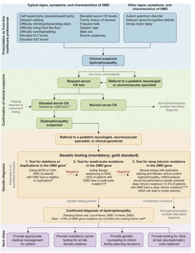
Patients presenting with the motor and nonmotor signs and symptoms of DMD as per Statements 1 and 2 should imme-diately have their serum creatine kinase (CK) levels tested and be referred to a child neurologist or a neuromuscular specialist.3 A marked increase in serum CK, defined as>2000 IU/L,31 should prompt further investigation for DMD. However, el-evated CK levels within the range of approximately 500-1200 U/L (1.5 times the upper limit of normal for men), even if asymptomatic, may be indicative of other neuromuscular disorders that require further assessment.103It has been shown that once a patient has had a serum CK test, the time to reach a complete diagnosis of DMD is relatively short (mean age± SD [range] at first CK test, 4.7± 1.7 [0.3-8.6] years, n = 151; and at complete diagnosis, 4.9± 1.7 [0.3-8.8] years, n = 154).4
Figure 1. Delphi Consensus Initiative development process following the steps outlined by Rosenfeld et al13and summary of
Table III. Summary of statements and recommendations for reducing the time to diagnosis of DMD Consensus statements Consensus vote Strength of recommendation Level of evidence* Final GRADE score† Key supporting evidence Section 1: Reducing the time to diagnosis of DMD
Statement 1: The following signs, symptoms, and characteristics should be considered typical indicators of DMD: calf hypertrophy (pseudohypertrophy); delayed walking; difficulty climbing/descending stairs; difficulty rising from the floor; difficulty running/walking; elevated serum CK levels (including elevated ALT and AST); a family history of DMD; frequent falls; Gowers' sign; male sex; and muscle weakness.
A+ = 93%; A = 7% Strongly in favor Moderate 1B 3,4,8,18-22
Statement 2: Autism spectrum disorder, delayed speech/cognitive deficits, and gross motor delay can be indicators of DMD but are not always associated with the disease.
A+ = 93%; A = 7% Strongly in favor Moderate 1B 3,4,8,18,21,23-30
Statement 3: After initial presentation, patients with suspected DMD should have their serum CK levels tested and be referred to a specialist (a child neurologist or neuromuscular specialist).
A+ = 93%; A = 7% Strongly in favor Moderate 1B 3,4,8,18,31-33
Statement 4: A lack of awareness of DMD and associated symptoms by the healthcare professional and long waiting times to see a specialist are the primary factors leading to a delay in initial diagnosis.
A+ = 67%; A = 33% Strongly in favor Low 1C 4,8
Statement 5: Genetic testing is crucial for obtaining a complete diagnosis of DMD, and should be considered the gold standard.
A+ = 94%; A = 6% Strongly in favor Low 1C 3,11,18and expert opinion
Statement 6: In the majority of cases, a complete genetic diagnosis can be made using MLPA or CGH to detect deletions or duplications in the DMD gene.
A+ = 88%; A = 6%; N= 6%
Strongly in favor Moderate 1B 3,11,12,18,34-44and expert opinion
Statement 7: If exon-level deletions/duplications in the DMD gene are not identified, small-scale mutations (by sequencing of exons and flanking regions) should be tested for as the next step.
A+ = 93%; A = 7% Strongly in favor Low 1C 3,11,35and expert opinion
Statement 8: Muscle biopsies with dystrophin staining are generally not needed to obtain a complete diagnosis of DMD, unless DNA testing is negative.
A+ = 73%; A = 27% Strongly in favor Low 1C 3,11,12,35,43,45-54and expert opinion
Statement 9: Delays in the initial clinical diagnosis/referral to a specialist, the sequential nature of the genetic testing process, and incomplete or nonexhaustive genetic testing should be addressed in order to prevent delays in reaching a complete genetic diagnosis for patients with DMD.
A+ = 93%; A = 7% Strongly in favor Low 1C 4,8,11,19and expert opinion
Section 2: Recommendations for next steps following a suspected DMD diagnosis
Statement 10: Patients with signs and symptoms of DMD and elevated serum CK levels should be referred for genetic testing to either a clinical geneticist or a neuromuscular specialist.
A+ = 100% Strongly in favor Low 1C 3,55,56and expert opinion
Statement 11: A medical/clinical geneticist, a child neurologist, or a neuromuscular specialist should request the genetic test and should provide clinical information relevant to the diagnosis as part of the sample submission to the clinical genetics laboratory, and the genetic diagnostic test should be performed by an accredited laboratory.
A+ = 100% Strongly in favor Low 1C 12,57and expert opinion
Statement 12: Educational meetings for physicians and laboratory specialists on topics relating to the genetic diagnosis of DMD would help to improve the understanding of genetic test reports and the interpretation of genetic test results.
A+ = 93%; A = 7% Strongly in favor Low 1C 58-62and expert opinion
Statement 13: Genetic testing is necessary to inform carrier testing, family planning, genetic counseling, prognosis and optimal management strategies, natural history data gathering, and prenatal diagnosis.
A+ = 100% Strongly in favor Low 1C Carrier testing/family planning11,63-73;
genetic counseling71,74;
prognosis/management23,75-78;
natural history7,79,80; prenatal
diagnosis12,31; and expert opinion
Statement 14: After a patient receives a complete genetic diagnosis of DMD, it is mandatory that carrier testing of the mother and other at-risk female family members be offered with appropriate pre- and postgenetic counseling (information regarding germline mosaicism and de novo mutations should also be offered). Similarly, testing of other at-risk male family members should also be offered.
A+ = 100% Strongly in favor Moderate 1B 11,12; carrier testing64-66,68-70,73,81-90;
germline mosaicism/de novo mutations91-95; family planning/genetic
counseling71,74; and expert opinion
Statement 15: When a family history is present, pre-symptomatic CK testing and prenatal testing can lead to earlier detection and thus management of DMD, which is important for family planning.‡
A+ = 87.5%; A= 12.5%
Strongly in favor Low 1C 1,12,31,96-102and expert opinion
ALT, alanine aminotransferase; AST, aspartate aminotransferase; CGH, comparative genome hybridization; CK, creatine kinase; DMD, Duchenne muscular dystrophy; GRADE, Grading of Recommendations Assessment, Development, and Evaluation.
*The level of evidence for most of the statements was graded as either low or moderate, owing to the fact that most of the studies included here are observational in nature rather than randomized controlled trials (due to the nature of this initiative). When there were multiple corroborative supporting observational studies, we have selected “moderate” for quality of evidence.
†Consensus: A+ = strongly agree; A = agree; N = neither agree nor disagree; D = disagree; D + = strongly disagree. Grade of recommendation: 1A = strong recommendation, high-quality evidence; 1B = strong recommendation, moderate-quality evidence; 1C = strong = weak recommendation, high-quality evidence; 2B = weak recommendation, moderate-quality evidence; 2C = weak recommendation, low-quality or very low-quality evidence.
y 2019
WORKSHOP/SYMPOSIUM
SUMMAR
307 Consensus and Systematic Review on Reducing the T ime to Diagnosis of Duchenne DystrophyIndeed, patients who are identified by an incidental finding of elevated CK (before onset of signs and symptoms) can be di-agnosed earlier.8The consensus group agreed that a lack of awareness of the signs and symptoms of DMD and delays in the time taken to see a specialist are the primary factors con-tributing to a delay in the initial diagnosis. In support of this, a retrospective chart review of 156 patients without a family history of DMD using the Muscular Dystrophy Surveillance, Tracking and Research Network (MD STARnet) showed, in a subset of patients (n= 127), that 63.8% (81/127) were seen by a pediatrician/family practitioner at their first evaluation and that a CK test was ordered as a result of the first evaluation in only 34.6% (44/127) of cases.4This finding indicates a need to increase awareness of DMD among front-line healthcare professionals.
The consensus group agreed that genetic testing is crucial to obtain a complete diagnosis of DMD. The DMD gene is one of the largest known human genes (2.2 Mb), containing 79 exons34,104with a relatively high mutation rate (~30% of cases are caused by a de novo mutation).11,45,105The approximate dis-tribution of mutations in the DMD gene is as follows: dele-tion of 1 or more exons, 68%; duplicadele-tion of 1 or more exons, 11%; small-scale mutations, 20% (small-scale deletions, 5%; small-scale insertions, 2%; splice-site, 3%; nonsense, 10%; mis-sense, 0.4%); and deep intronic mutations, 0.3%.35This dis-tribution is supported by a number of studies.23,36,37,46,106-110It is therefore practical to test for DMD gene mutations in order of frequency. The consensus group agreed on 3 statements that outline the recommended steps needed to reach a complete genetic diagnosis of DMD (Figure 3).4,35,46First, multiplex ligation-dependent probe amplification (MLPA) or compara-tive genome hybridization should be used to screen for dele-tions or duplicadele-tions in the DMD gene. These tests are recommended because they can screen all 79 exons of the DMD gene; however, it should be noted that there are several limi-tations associated with MLPA. For example, point mulimi-tations or polymorphisms along the probe hybridization region can present as single-exon deletions when using this method, and thus a second confirmatory test (usually Sanger sequencing) is required.11,12,31However, if the mutation is identified and cor-relates with the severity of symptoms (eg, DMD or Becker mus-cular dystrophy [BMD]), no further genetic testing is typically required. If exonic deletions/duplications in the DMD gene are not identified, small-scale mutations should be tested for by sequencing exons and flanking intronic regions. If clinical signs and genetic testing are conclusive, in most cases, muscle bi-opsies are not needed. Nevertheless, if no mutation is de-tected after DNA analysis, it is possible that the patient may have a large rearrangement, such as an inversion (or translo-cation in females11), a deep intronic mutation that affects splic-ing, or an alternative diagnosis.12Inversions and translocations are challenging to detect with most conventional genetic analy-ses, because they do not affect copy number.111Deep intronic mutations often can be detected by next-generation sequenc-ing if the full genomic sequence of the gene is available37,112; however, it can be difficult to predict the impact of intronic mutations (or variations) on mRNA. If a muscle biopsy with
dystrophin staining confirms dystrophinopathy, mRNA analy-sis should be performed to identify any impact on mRNA splicing11,12,45,113that escapes detection by both MLPA and DNA sequencing. RNA analysis is also crucial to determine the con-sequence of the mutation on the mRNA and can be consid-ered for discordant phenotypes. Muscle biopsy remains a relevant diagnostic tool, especially for mutation types in which the mutation is poorly predictive of disease progression (for instance, a deletion of exons 3-7 can cause both BMD and DMD phenotypes), or for determining dystrophin expres-sion in boys with unexpectedly mild disease progresexpres-sion.11We note that there are several databases that can help physicians to determine the severity of individual and rare causal muta-tions in the DMD gene (Appendix). In addition to consult-ing these databases, physicians enterconsult-ing new data on genotype and phenotype are warranted to help inform future diagnoses. The consensus group agreed that delays in the initial clini-cal diagnosis or referral to a specialist, the sequential nature of the genetic testing process, and incomplete or nonexhaustive genetic testing should be addressed to prevent delays in diag-nosis. In addition, if the ordering physician is not sufficiently informed or trained to understand the hierarchy of tests needed to provide a complete genetic diagnosis for DMD, the testing process could be terminated prematurely (as shown elsewhere).11It would therefore be helpful if the responsibil-ity for these decisions was integrated with the primary testing laboratory’s operating procedures. The laboratory would thus be obligated to contact the ordering physician to discuss the next level of testing required either to obtain a complete genetic diagnosis of DMD or to exclude DMD from the diagnosis.
Section 2: Recommendations for Next Steps Following a Suspected DMD Diagnosis
A patient with signs and symptoms of DMD and elevated CK levels should be referred to a clinical geneticist/neuromuscular specialist during the genetic testing process, because these in-dividuals are best placed to provide accurate interpretation of genetic test results55and can help to avoid diagnostic delay (Table III).3
In 2010, best practice guidelines for the molecular diagno-sis of DMD/BMD were developed using a consensus-building approach.12More recently, general standards and guidelines for the interpretation of sequence variants have been published by the American College of Medical Genetics and Genomics and the Association for Molecular Pathology.57
The consensus group agreed that a clinical geneticist usually is responsible for the interpretation of the genetic test results but that the process should be shared between the clinical ge-neticist, the physician who ordered the test (typically a child neurologist or neuromuscular specialist), and/or a genetic counselor.
Although improving the clarity of genetic test reports and directing physicians to the appropriate specialists for assis-tance would ultimately ensure a quicker and more accurate genetic diagnosis for patients, it is also important for physi-cians (nongeneticists) to understand genetic test results, because this will have a direct impact on patient management. The
Figure 3. Diagnostic steps for reaching a complete diagnosis of DMD. ALT, alanine aminotransferase; AST, aspartate amino-transferase; NGS, next-generation sequencing. *Elevated CK levels within the range of approximately 500-1200 U/L (1.5 times the upper limit of normal for men), even if asymptomatic, may be indicative of other neuromuscular disorders that require further
assessment.103†Point mutations or polymorphisms along the probe hybridization region can present as single-exon deletions
when using MLPA and thus a second confirmatory test (typically Sanger sequencing) may be required.11,12,31‡Testing of at-risk
consensus group therefore agreed that educational meetings for physicians and laboratory specialists on topics relating to the genetic diagnosis of DMD would help to improve physi-cians’ understanding of genetic tests and the interpretation of the results.
The consensus group also agreed on 3 statements to high-light the next steps that should be taken after a complete di-agnosis has been reached. These statements highlight the importance of carrier testing, genetic counseling, family-planning decisions, and prenatal diagnosis. It is estimated that approximately one-third of patients with DMD develop the disease owing to de novo mutations and that the remaining two-thirds inherit the mutation from carrier mothers.11,63,64If the mutation identified in the affected patient is not carried by the mother in her somatic cell line, the mother may be a germline mosaic and should be provided with counseling about the risk of having a second son with the disease12or having a daughter that is a carrier.11Female carriers are typically as-ymptomatic because DMD is an X-linked inherited disease; however, carriers can develop mild clinical symptoms, includ-ing muscle weakness and scoliosis,65,66 and are at an in-creased risk of cardiomyopathy.67-69Despite this, approximately one-third of potential carriers are likely unaware of their carrier status.64,70 Genetic testing is thus important for family-planning decisions and counseling of other family members.71,74 Genetic information also is used for genotype–phenotype correlations,7,75,76,79,114so that patients can be provided with prog-nostic information and be offered appropriate medical management.11,23,77,78
As per the 2010 best practice guidelines for the molecular diagnosis of DMD/BMD, prenatal testing is advised to be carried out only for at-risk male pregnancies (those with a family history of the disease).12Prenatal screening is not cur-rently recommended for female fetuses, because it is not yet possible to determine whether a female heterozygote for a DMD mutation will exhibit any signs of disease.12However, the con-sensus group agreed that when a family history is present, pre-natal testing leads to earlier detection of DMD and is thus important for family-planning decisions. Recommendations regarding prenatal diagnosis will depend on country-specific legislation.12,31The consensus group also agreed that in the pres-ence of a family history of disease, presymptomatic CK testing would lead to earlier detection and thus earlier management of patients with DMD, but would be dependent on country-specific legislation.
The 195th European Neuromuscular Centre International Workshop (2012) report presented discussions on newborn screening (NBS) for DMD; the meeting was attended by 21 experts from 7 countries. It was discussed that the introduc-tion of NBS for DMD using CK testing would help to detect the disease earlier in patients and reduce the risk of having additional children with DMD.1 A pilot study in the US demonstrated the feasibility of a 2-tier NBS system for DMD using dried blood spots to test CK levels, followed by DMD gene testing96; however, there are many complexities involved with NBS programs, some of which are discussed in the
Appendix.
Conclusions
Delays in the diagnosis of DMD have remained relatively un-changed over the last 30 years,9,10despite advances in our un-derstanding of the natural history and improvements in genetic testing. Delays occur early in the diagnostic pathway, because of a lack of awareness of DMD and its signs and symptoms among families, and, more pertinently, among front-line health-care professionals.4Issues relating to the understanding of genetic testing required to obtain a complete diagnosis of DMD have been highlighted (Table I).11Patients presenting with the typical motor signs and symptoms of DMD, as well as the less well-recognized neurocognitive deficits, developmental delays, and elevated liver enzymes, should be immediately referred to a specialist (child neurologist or neuromuscular specialist) and should have their serum CK levels measured. Patients with a marked elevation in serum CK should be referred to a clini-cal geneticist as soon as possible, and the full range of sequen-tial genetic tests offered to provide a complete diagnosis. After diagnosis, it is mandatory to offer carrier testing to mothers and other at-risk female relatives. By highlighting these issues and providing an in-depth discussion of the DMD diagnos-tic pathway, we hope that patients will be diagnosed earlier, care provided as soon as possible, and personalized interven-tion provided for eligible patients.■
This process was conducted and managed by an independent third-party agency (PharmaGenesis London, London, United Kingdom), and we acknowledge the support of Sally Janani, MSc, and Nicola Baines, BSc (PharmaGenesis London) for their support with the Delphi con-sensus process. Both were funded by PTC Therapeutics Inc.
Submitted for publication Jun 27, 2018; last revision received Sep 27, 2018; accepted Oct 24, 2018
Reprint requests: Stanley F. Nelson, MD, Department of Human Genetics, University of California, Box 957088, 5506A Gonda Center, Los Angeles, CA 90095. E-mail:[email protected]
Disclaimer
The Delphi Consensus statements presented here are based on the opinions of carefully selected experts in the field and are for information and educational purposes only. The state-ments may reflect gaps in current knowledge, but, where pos-sible, have been supported by relevant literature. These statements do not reflect clinical practice guidelines or legal standards of care and, as such, do not include all potential di-agnostic or management steps. The responsible physician, in light of all of the circumstances presented by the individual patient, must determine the appropriate treatment, diagno-sis, and management.
Endorsements
Endorsed by the European Paediatric Neurology Society (EPNS), the Muscular Dystrophy Association (MDA), Duchenne Parent Project Italy, Parent Project Muscular Dys-trophy (PPMD), and TREAT-NMD.
References
1. Ellis JA, Vroom E, Muntoni F. 195th ENMC International Workshop: newborn screening for Duchenne muscular dystrophy December 14-16, 2012, Naarden, The Netherlands. Neuromuscul Disord 2013;23:682-9.
2. Hoffman EP, Brown RH Jr, Kunkel LM. Dystrophin: the protein product of the Duchenne muscular dystrophy locus. Cell 1987;51:919-28.
3. Birnkrant DJ, Bushby K, Bann CM, Apkon SD, Blackwell A, Brumbaugh D, et al. Diagnosis and management of Duchenne muscular dystrophy, part 1: diagnosis, and neuromuscular, rehabilitation, endocrine, and gas-trointestinal and nutritional management. Lancet Neurol 2018;17:251-67.
4. Ciafaloni E, Fox DJ, Pandya S, Westfield CP, Puzhankara S, Romitti PA, et al. Delayed diagnosis in Duchenne muscular dystrophy: data from the Muscular Dystrophy Surveillance, Tracking, and Research Network (MD STARnet). J Pediatr 2009;155:380-5.
5. D’Amico A, Catteruccia M, Baranello G, Politano L, Govoni A, Previtali SC, et al. Diagnosis of Duchenne Muscular Dystrophy in Italy in the last decade: critical issues and areas for improvements. Neuromuscul Disord 2017;27:447-51.
6. Vry J, Gramsch K, Rodger S, Thompson R, Steffensen BF, Rahbek J, et al. European cross-sectional survey of current care practices for Duchenne muscular dystrophy reveals regional and age-dependent differences. J Neuromuscul Dis 2016;3:517-27.
7. Bello L, Morgenroth LP, Gordish-Dressman H, Hoffman EP, McDonald CM, Cirak S, et al. DMD genotypes and loss of ambulation in the CINRG Duchenne natural history study. Neurology 2016;87:401-9.
8. van Ruiten HJ, Straub V, Bushby K, Guglieri M. Improving recogni-tion of Duchenne muscular dystrophy: a retrospective case note review. Arch Dis Child 2014;99:1074-7.
9. Crisp DE, Ziter FA, Bray PF. Diagnostic delay in Duchenne’s muscular dystrophy. JAMA 1982;247:478-80.
10. Firth MA. Diagnosis of Duchenne muscular dystrophy: experiences of parents of sufferers. Br Med J (Clin Res Ed) 1983;286:700-1.
11. Aartsma-Rus A, Ginjaar IB, Bushby K. The importance of genetic di-agnosis for Duchenne muscular dystrophy. J Med Genet 2016;53:145-51.
12. Abbs S, Tuffery-Giraud S, Bakker E, Ferlini A, Sejersen T, Mueller CR. Best practice guidelines on molecular diagnostics in Duchenne/Becker muscular dystrophies. Neuromuscul Disord 2010;20:422-7.
13. Rosenfeld RM, Nnacheta LC, Corrigan MD. Clinical consensus state-ment developstate-ment manual. Otolaryngol Head Neck Surg 2015;153:S1-14.
14. Guyatt GH, Cook DJ, Sackett DL, Eckman M, Pauker S. Grades of rec-ommendation for antithrombotic agents. Chest 1998;114:441S-444S.
15. Guyatt GH, Oxman AD, Kunz R, Falck-Ytter Y, Vist GE, Liberati A, et al. GRADE: going from evidence to recommendations. BMJ 2008;336:1049-51.
16. Guyatt GH, Oxman AD, Vist GE, Kunz R, Falck-Ytter Y, Alonso-Coello P, et al. GRADE: an emerging consensus on rating quality of evi-dence and strength of recommendations. BMJ 2008;336:924-6.
17. Guyatt GH, Oxman AD, Vist GE, Kunz R, Falck-Ytter Y, Schunemann HJ. GRADE: what is “quality of evidence” and why is it important to clinicians? BMJ 2008;336:995-8.
18. Bushby K, Finkel R, Birnkrant DJ, Case LE, Clemens PR, Cripe L, et al. Diagnosis and management of Duchenne muscular dystrophy, part 1: diagnosis, and pharmacological and psychosocial management. Lancet Neurol 2010;9:77-93.
19. Wong SH, McClaren BJ, Archibald AD, Weeks A, Langmaid T, Ryan MM, et al. A mixed methods study of age at diagnosis and diagnostic odyssey for Duchenne muscular dystrophy. Eur J Hum Genet 2015;23:1294-300.
20. Na SJ, Kim WJ, Kim SM, Lee KO, Yoon B, Choi YC. Clinical, immuno-histochemical, Western blot, and genetic analysis in dystrophinopathy. J Clin Neurosci 2013;20:1099-105.
21. Dey S, Senapati AK, Pandit A, Biswas A, Guin DS, Joardar A, et al. Genetic and clinical profile of patients of Duchenne muscular dystrophy: expe-rience from a tertiary care center in Eastern India. Indian Pediatr 2015;52:481-4.
22. Holtzer C, Meaney FJ, Andrews J, Ciafaloni E, Fox DJ, James KA, et al. Disparities in the diagnostic process of Duchenne and Becker muscu-lar dystrophy. Genet Med 2011;13:942-7.
23. Magri F, Govoni A, D’Angelo MG, Del Bo R, Ghezzi S, Sandra G, et al. Genotype and phenotype characterization in a large dystrophinopathic cohort with extended follow-up. J Neurol 2011;258:1610-23.
24. Ricotti V, Mandy WP, Scoto M, Pane M, Deconinck N, Messina S, et al. Neurodevelopmental, emotional, and behavioural problems in Duchenne muscular dystrophy in relation to underlying dystrophin gene muta-tions. Dev Med Child Neurol 2016;58:77-84.
25. Sarrazin E, von der Hagen M, Schara U, von Au K, Kaindl AM. Growth and psychomotor development of patients with Duchenne muscular dys-trophy. Eur J Paediatr Neurol 2014;18:38-44.
26. Colombo P, Nobile M, Tesei A, Civati F, Gandossini S, Mani E, et al. As-sessing mental health in boys with Duchenne muscular dystrophy: emo-tional, behavioural and neurodevelopmental profile in an Italian clinical sample. Eur J Paediatr Neurol 2017;21:639-47.
27. Latimer R, Street N, Conway KC, James K, Cunniff C, Oleszek J, et al. Secondary conditions among males with Duchenne or Becker muscu-lar dystrophy. J Child Neurol 2017;32:663-70.
28. Mirski KT, Crawford TO. Motor and cognitive delay in Duchenne mus-cular dystrophy: implication for early diagnosis. J Pediatr 2014;165:1008-10.
29. Snow WM, Anderson JE, Jakobson LS. Neuropsychological and neurobehavioral functioning in Duchenne muscular dystrophy: a review. Neurosci Biobehav Rev 2013;37:743-52.
30. Vicari S, Piccini G, Mercuri E, Battini R, Chieffo D, Bulgheroni S, et al. Implicit learning deficit in children with Duchenne muscular dystro-phy: evidence for a cerebellar cognitive impairment? PLoS ONE 2018;13:e0191164.
31. Araujo APQC, Carvalho AAS, Cavalcanti EBU, Saute JAM, Carvalho E, Franca MCJ, et al. Brazilian consensus on Duchenne muscular dystro-phy. Part 1: diagnosis, steroid therapy and perspectives. Arq Neuropsiquiatr 2017;75:104-13.
32. Hughes BP. Serum enzyme changes in muscle disease and their rela-tion to tissue change. Proc R Soc Med 1963;56:179-82.
33. Strehle EM, Straub V. Recent advances in the management of Duchenne muscular dystrophy. Arch Dis Child 2015;100:1173-7.
34. Hegde MR, Chin EL, Mulle JG, Okou DT, Warren ST, Zwick ME. Microarray-based mutation detection in the dystrophin gene. Hum Mutat 2008;29:1091-9.
35. Bladen CL, Salgado D, Monges S, Foncuberta ME, Kekou K, Kosma K, et al. The TREAT-NMD DMD Global Database: analysis of more than 7,000 Duchenne muscular dystrophy mutations. Hum Mutat 2015;36:395-402.
36. Juan-Mateu J, Gonzalez-Quereda L, Rodriguez MJ, Baena M, Verdura E, Nascimento A, et al. DMD mutations in 576 dystrophinopathy fami-lies: a step forward in genotype-phenotype correlations. PLoS ONE 2015;10:e0135189.
37. Zhong J, Xu T, Chen G, Liao H, Zhang J, Lan D. Genetic analysis of the dystrophin gene in children with Duchenne and Becker muscular dys-trophies. Muscle Nerve 2017;56:117-21.
38. Lalic T, Vossen RH, Coffa J, Schouten JP, Guc-Scekic M, Radivojevic D, et al. Deletion and duplication screening in the DMD gene using MLPA. Eur J Hum Genet 2005;13:1231-4.
39. Manjunath M, Kiran P, Preethish-Kumar V, Nalini A, Singh RJ, Gayathri N. A comparative study of mPCR, MLPA, and muscle biopsy results in a cohort of children with Duchenne muscular dystrophy: a first study. Neurol India 2015;63:58-62.
40. Khordadpoor-Deilamani F, Akbari MT, Nafissi S, Zamani G. Dystrophin gene mutation analysis in Iranian males and females using multiplex polymerase chain reaction and multiplex ligation-dependent probe amplification methods. Genet Test Mol Biomarkers 2011;15:893-9.
41. Dastur RS, Kachwala MY, Khadilkar SV, Hegde MR, Gaitonde PS. Identification of deletions and duplications in the Duchenne muscular dystrophy gene and female carrier status in western India using combined methods of multiplex polymerase chain reaction and mul-tiplex ligation-dependent probe amplification. Neurol India 2011;59:803-9.
42. Bovolenta M, Neri M, Fini S, Fabris M, Trabanelli C, Venturoli A, et al. A novel custom high density-comparative genomic hybridization array detects common rearrangements as well as deep intronic mutations in dystrophinopathies. BMC Genomics 2008;9:572.
43. Baskin B, Gibson WT, Ray PN. Duchenne muscular dystrophy caused by a complex rearrangement between intron 43 of the DMD gene and chromosome 4. Neuromuscul Disord 2011;21:178-82.
44. Ishmukhametova A, Khau Van Kien P, Mechin D, Thorel D, Vincent MC, Rivier F, et al. Comprehensive oligonucleotide array-comparative genomic hybridization analysis: new insights into the molecular pathology of the
DMD gene. Eur J Hum Genet 2012;20:1096-100.
45. Santos R, Goncalves A, Oliveira J, Vieira E, Vieira JP, Evangelista T, et al. New variants, challenges and pitfalls in DMD genotyping: implications in diagnosis, prognosis and therapy. J Hum Genet 2014;59:454-64.
46. Tuffery-Giraud S, Miro J, Koenig M, Claustres M. Normal and altered pre-mRNA processing in the DMD gene. Hum Genet 2017;136:1155-72.
47. Roucher Boulez F, Menassa R, Streichenberger N, Manel V, Mallet-Motak D, Morel Y, et al. A splicing mutation in the DMD gene de-tected by next-generation sequencing and confirmed by mRNA and protein analysis. Clin Chim Acta 2015;448:146-9.
48. Magri F, Del Bo R, D’Angelo MG, Govoni A, Ghezzi S, Gandossini S, et al. Clinical and molecular characterization of a cohort of patients with novel nucleotide alterations of the dystrophin gene detected by direct sequencing. BMC Med Genet 2011;12:37.
49. Niba ETE, Nishida A, Tran VK, Vu DC, Matsumoto M, Awano H, et al. Cryptic splice activation but not exon skipping is observed in minigene assays of dystrophin c.9361+ 1G>A mutation identified by NGS. J Hum Genet 2017;62:531-7.
50. Wang Z, Lin Y, Qiu L, Zheng D, Yan A, Zeng J, et al. Hybrid minigene splicing assay verified the pathogenicity of a novel splice site variant in the dystrophin gene of a Chinese patient with typical Duchenne mus-cular dystrophy phenotype. Clin Chem Lab Med 2016;54:1435-40.
51. Baskin B, Stavropoulos DJ, Rebeiro PA, Orr J, Li M, Steele L, et al. Complex genomic rearrangements in the dystrophin gene due to replication-based mechanisms. Mol Genet Genomic Med 2014;2:539-47.
52. Khelifi MM, Ishmukhametova A, Khau Van Kien P, Thorel D, Mechin D, Perelman S, et al. Pure intronic rearrangements leading to aberrant pseudoexon inclusion in dystrophinopathy: a new class of mutations? Hum Mutat 2011;32:467-75.
53. Greer K, Mizzi K, Rice E, Kuster L, Barrero RA, Bellgard MI, et al. Pseudoexon activation increases phenotype severity in a Becker mus-cular dystrophy patient. Mol Genet Genomic Med 2015;3:320-6.
54. Zaum AK, Stuve B, Gehrig A, Kolbel H, Schara U, Kress W, et al. Deep intronic variants introduce DMD pseudoexon in patient with muscu-lar dystrophy. Neuromuscul Disord 2017;27:631-4.
55. Williams MS. Genetics and managed care: policy statement of the Ameri-can College of Medical Genetics. Genet Med 2001;3:430-5.
56. Baars MJ, Henneman L, Ten Kate LP. Deficiency of knowledge of ge-netics and genetic tests among general practitioners, gynecologists, and pediatricians: a global problem. Genet Med 2005;7:605-10.
57. Richards S, Aziz N, Bale S, Bick D, Das S, Gastier-Foster J, et al. Stan-dards and guidelines for the interpretation of sequence variants: a joint consensus recommendation of the American College of Medical Ge-netics and Genomics and the Association for Molecular Pathology. Genet Med 2015;17:405-24.
58. Thurston VC, Wales PS, Bell MA, Torbeck L, Brokaw JJ. The current status of medical genetics instruction in US and Canadian medical schools. Acad Med 2007;82:441-5.
59. Greb AE, Brennan S, McParlane L, Page R, Bridge PD. Retention of medical genetics knowledge and skills by medical students. Genet Med 2009;11:365-70.
60. Klitzman R, Chung W, Marder K, Shanmugham A, Chin LJ, Stark M, et al. Attitudes and practices among internists concerning genetic testing. J Genet Couns 2013;22:90-100.
61. Burke S, Stone A, Bedward J, Thomas H, Farndon P. A “neglected part of the curriculum” or “of limited use”? Views on genetics training by nongenetics medical trainees and implications for delivery. Genet Med 2006;8:109-15.
62. Shields AE, Burke W, Levy DE. Differential use of available genetic tests among primary care physicians in the United States: results of a na-tional survey. Genet Med 2008;10:404-14.
63. Lee T, Takeshima Y, Kusunoki N, Awano H, Yagi M, Matsuo M, et al. Differences in carrier frequency between mothers of Duchenne and Becker muscular dystrophy patients. J Hum Genet 2014;59:46-50.
64. Helderman-van den Enden ATJM, van den Bergen JC, Breuning MH, Verschuuren JJGM, Tibben A, Bakker E, et al. Duchenne/Becker mus-cular dystrophy in the family: have potential carriers been tested at a mo-lecular level? Clin Genet 2011;79:236-42.
65. Juan-Mateu J, Rodriguez MJ, Nascimento A, Jimenez-Mallebrera C, Gonzalez-Quereda L, Rivas E, et al. Prognostic value of X-chromosome inactivation in symptomatic female carriers of dystrophinopathy. Orphanet J Rare Dis 2012;7:82.
66. Papa R, Madia F, Bartolomeo D, Trucco F, Pedemonte M, Traverso M, et al. Genetic and early clinical manifestations of females heterozygous for Duchenne/Becker muscular dystrophy. Pediatr Neurol 2016;55:58-63.
67. McCaffrey T, Guglieri M, Murphy AP, Bushby K, Johnson A, Bourke JP. Cardiac involvement in female carriers of Duchenne or Becker muscu-lar dystrophy. Muscle Nerve 2017;55:810-8.
68. Schelhorn J, Schoenecker A, Neudorf U, Schemuth H, Nensa F, Nassenstein K, et al. Cardiac pathologies in female carriers of Duchenne muscular dystrophy assessed by cardiovascular magnetic resonance imaging. Eur Radiol 2015;25:3066-72.
69. Florian A, Rosch S, Bietenbeck M, Engelen M, Stypmann J, Waltenberger J, et al. Cardiac involvement in female Duchenne and Becker muscular dystrophy carriers in comparison to their first-degree male relatives: a comparative cardiovascular magnetic resonance study. Eur Heart J Cardiovasc Imaging 2016;17:326-33.
70. Bogue L, Peay H, Martin A, Lucas A, Ramchandren S. Knowledge of carrier status and barriers to testing among mothers of sons with Duchenne or Becker muscular dystrophy. Neuromuscul Disord 2016;26:860-4.
71. Plumridge G, Metcalfe A, Coad J, Gill P. Parents’ communication with siblings of children affected by an inherited genetic condition. J Genet Couns 2011;20:374-83.
72. Hoffman EP, Arahata K, Minetti C, Bonilla E, Rowland LP. Dystrophinopathy in isolated cases of myopathy in females. Neurology 1992;42:967-75.
73. Bogue L, Ramchandren S. Outdated risk assessment in a family with Duchenne dystrophy: implications for duty to reassess. Neurol Genet 2016;2:e103.
74. Lehmann A, Speight BS, Kerzin-Storrar L. Extended family impact of genetic testing: the experiences of X-linked carrier grandmothers. J Genet Couns 2011;20:365-73.
75. Hightower RM, Alexander MS. Genetic modifiers of Duchenne and facioscapulohumeral muscular dystrophies. Muscle Nerve 2018;57:6-15.
76. Pons R, Kekou K, Gkika A, Papadimas G, Vogiatzakis N, Svingou M, et al. Single amino acid loss in the dystrophin protein associated with a mild clinical phenotype. Muscle Nerve 2017;55:46-50.
77. Taylor PJ, Betts GA, Maroulis S, Gilissen C, Pedersen RL, Mowat DR, et al. Dystrophin gene mutation location and the risk of cognitive im-pairment in Duchenne muscular dystrophy. PLoS ONE 2010;5:e8803.
78. Ricotti V, Jagle H, Theodorou M, Moore AT, Muntoni F, Thompson DA. Ocular and neurodevelopmental features of Duchenne muscular
dystrophy: a signature of dystrophin function in the central nervous system. Eur J Hum Genet 2016;24:562-8.
79. van den Bergen JC, Ginjaar HB, Niks EH, Aartsma-Rus A, Verschuuren JJ. Prolonged ambulation in Duchenne patients with a mutation ame-nable to exon 44 skipping. J Neuromuscul Dis 2014;1:91-4.
80. Barp A, Bello L, Politano L, Melacini P, Calore C, Polo A, et al. Genetic modifiers of Duchenne muscular dystrophy and dilated cardiomyopa-thy. PLoS ONE 2015;10:e0141240.
81. Imbornoni L, Price ET, Andrews J, Meaney FJ, Ciafaloni E, Cunniff C. Diagnostic and clinical characteristics of early-manifesting females with Duchenne or Becker muscular dystrophy. Am J Med Genet A 2014;164A:2769-74.
82. Mercier S, Toutain A, Toussaint A, Raynaud M, de Barace C, Marcorelles P, et al. Genetic and clinical specificity of 26 symptomatic carriers for dystrophinopathies at pediatric age. Eur J Hum Genet 2013;21:855-63.
83. Lang SM, Shugh S, Mazur W, Sticka JJ, Rattan MS, Jefferies JL, et al. Myo-cardial fibrosis and left ventricular dysfunction in Duchenne muscular dystrophy carriers using cardiac magnetic resonance imaging. Pediatr Cardiol 2015;36:1495-501.
84. Nozoe KT, Akamine RT, Mazzotti DR, Polesel DN, Grossklauss LF, Tufik S, et al. Phenotypic contrasts of Duchenne muscular dystrophy in women: two case reports. Sleep Sci 2016;9:129-33.
85. Parent JJ, Moore RA, Taylor MD, Towbin JA, Jefferies JL. Left ventricu-lar noncompaction cardiomyopathy in Duchenne muscuventricu-lar dystrophy carriers. J Cardiol Cases 2015;11:7-9.
86. Cheng VE, Prior DL. Peripartum cardiomyopathy in a previously as-ymptomatic carrier of Duchenne muscular dystrophy. Heart Lung Circ 2013;22:677-81.
87. Martinez HR, Pignatelli R, Belmont JW, Craigen WJ, Jefferies JL. Child-hood onset of left ventricular dysfunction in a female manifesting carrier of muscular dystrophy. Am J Med Genet A 2011;155A:3025-9.
88. Yilmaz A, Gdynia HJ, Ludolph AC, Klingel K, Kandolf R, Sechtem U. Images in cardiovascular medicine. Cardiomyopathy in a Duchenne mus-cular dystrophy carrier and her diseased son: similar pattern revealed by cardiovascular MRI. Circulation 2010;121:e237-9.
89. Walcher T, Kunze M, Steinbach P, Sperfeld AD, Burgstahler C, Hombach V, et al. Cardiac involvement in a female carrier of Duchenne muscu-lar dystrophy. Int J Cardiol 2010;138:302-5.
90. McGowan R, Challoner BR, Ross S, Holloway S, Joss S, Wilcox D, et al. Results of Duchenne muscular dystrophy family screening in practice: leaks rather than cascades? Clin Genet 2013;83:187-90.
91. Bermudez-Lopez C, Garcia-de Teresa B, Gonzalez-del Angel A, Alcantara-Ortigoza MA. Germinal mosaicism in a sample of families with Duchenne/Becker muscular dystrophy with partial deletions in the DMD gene. Genet Test Mol Biomarkers 2014;18:93-7.
92. Garcia S, de Haro T, Zafra-Ceres M, Poyatos A, Capilla JA, Gomez-Llorente C. Identification of de novo mutations of Duchenne/Becker muscular dystrophies in southern Spain. Int J Med Sci 2014;11:988-93.
93. Strmecki L, Hudler P, Benedik-Dolnicar M, Komel R. De novo muta-tion in DMD gene in a patient with combined hemophilia A and Duchenne muscular dystrophy. Int J Hematol 2014;99:184-7.
94. Luna-Angulo AB, Gomez-Diaz B, Escobar-Cedillo RE, Anaya-Segura MA, Estrada-Mena FJ, Lopez-Hernandez LB. A new de novo mutation in a non-hot spot region at the DMD gene in a Mexican family. Genet Couns 2014;25:429-32.
95. Li T, Zhang ZJ, Ma X, Lv X, Xiao H, Guo QN, et al. Prenatal diagnosis for a Chinese family with a de novo DMD gene mutation: a case report. Medicine (Baltimore) 2017;96:e8814.
96. Mendell JR, Shilling C, Leslie ND, Flanigan KM, al-Dahhak R, Gastier-Foster J, et al. Evidence-based path to newborn screening for Duchenne muscular dystrophy. Ann Neurol 2012;71:304-13.
97. Moat SJ, Bradley DM, Salmon R, Clarke A, Hartley L. Newborn bloodspot screening for Duchenne muscular dystrophy: 21 years experience in Wales (UK). Eur J Hum Genet 2013;21:1049-53.
98. Ke Q, Zhao ZY, Griggs R, Wiley V, Connolly A, Kwon J, et al. Newborn screening for Duchenne muscular dystrophy in China: follow-up diag-nosis and subsequent treatment. World J Pediatr 2017;13:197-201.
99. Wood MF, Hughes SC, Hache LP, Naylor EW, Abdel-Hamid HZ, Barmada MM, et al. Parental attitudes toward newborn screening for Duchenne/Becker muscular dystrophy and spinal muscular atrophy. Muscle Nerve 2014;49:822-8.
100. Lillie SE, Tarini BA, Janz NK, Zikmund-Fisher BJ. Framing optional genetic testing in the context of mandatory newborn screening tests. BMC Med Inform Decis Mak 2015;15:50.
101. Helderman-van den Enden AT, Madan K, Breuning MH, van der Hout AH, Bakker E, de Die-Smulders CE, et al. An urgent need for a change in policy revealed by a study on prenatal testing for Duchenne muscu-lar dystrophy. Eur J Hum Genet 2013;21:21-6.
102. Wang H, Xu Y, Liu X, Wang L, Jiang W, Xiao B, et al. Prenatal diagno-sis of Duchenne muscular dystrophy in 131 Chinese families with dystrophinopathy. Prenat Diagn 2017;37:356-64.
103. Moghadam-Kia S, Oddis CV, Aggarwal R. Approach to asymptomatic creatine kinase elevation. Cleve Clin J Med 2016;83:37-42.
104. The European Bioinformatics Institute (EMBL-EBI). EBI Search: gene & protein summary for dystrophin.https://www.ebi.ac.uk/s4/ summary/molecular/gene?term=dystrophin&classification=9606&tid =protOrthDMDHUMAN. Accessed November 26, 2018.
105. Grimm T, Kress W, Meng G, Muller CR. Risk assessment and genetic counseling in families with Duchenne muscular dystrophy. Acta Myol 2012;31:179-83.
106. Takeshima Y, Yagi M, Okizuka Y, Awano H, Zhang Z, Yamauchi Y, et al. Mutation spectrum of the dystrophin gene in 442 Duchenne/Becker mus-cular dystrophy cases from one Japanese referral center. J Hum Genet 2010;55:379-88.
107. Rani AQ, Sasongko TH, Sulong S, Bunyan D, Salmi AR, Zilfalil BA, et al. Mutation spectrum of dystrophin gene in Malaysian patients with Duchenne/Becker muscular dystrophy. J Neurogenet 2013;27:11-5.
108. Zimowski JG, Massalska D, Holding M, Jadczak S, Fidzianska E, Lusakowska A, et al. MLPA based detection of mutations in the dystrophin gene of 180 Polish families with Duchenne/Becker muscular dystro-phy. Neurol Neurochir Pol 2014;48:416-22.
109. de Almeida PA, Machado-Costa MC, Manzoli GN, Ferreira LS, Ro-drigues MC, Bueno LS, et al. Genetic profile of Brazilian patients with dystrophinopathies. Clin Genet 2017;92:199-203.
110. Ramos E, Conde JG, Berrios RA, Pardo S, Gomez O, Mas Rodriguez MF. Prevalence and genetic profile of Duchene and Becker muscular dys-trophy in Puerto Rico. J Neuromuscul Dis 2016;3:261-6.
111. Griffiths AJF, Gelbart WM, Miller JH, Lewontin RC. Chromosomal rearrangements. In: Modern genetic analysis. 1999 https:// www.ncbi.nlm.nih.gov/books/NBK21367/. Accessed November 26, 2018.
112. Wang Y, Yang Y, Liu J, Chen XC, Liu X, Wang CZ, et al. Whole dystrophin gene analysis by next-generation sequencing: a comprehensive genetic diagnosis of Duchenne and Becker muscular dystrophy. Mol Genet Ge-nomics 2014;289:1013-21.
113. Cummings BB, Marshall JL, Tukiainen T, Lek M, Donkervoort S, Foley AR, et al. Improving genetic diagnosis in Mendelian disease with transcriptome sequencing. Sci Transl Med 2017;9:eaal5209.
114. Bello L, Kesari A, Gordish-Dressman H, Cnaan A, Morgenroth LP, Punetha J, et al. Genetic modifiers of ambulation in the Cooperative In-ternational Neuromuscular Research Group Duchenne natural history study. Ann Neurol 2015;77:684-96.
Detailed affiliations
From the1Department of Human Genetics, Leiden University Medical Center, Leiden, The Netherlands; 2Department of Human Genetics, Emory University School of Medicine/School of Biological Sciences, Georgia Institute of Technology/Perkin Elmer Genetics, Atlanta, GA;
3Clinical and Metabolic Genetics, Department of Pediatrics, Hamad Medical Corporation, Doha, Qatar;
4Duchenne Parent Project Italy, Rome;
5Unit of Medical Genetics, University of Ferrara, Ferrara, Italy;
6U705 CIBERER, Servei de Genetica, Hospital de Sant Pau, Barcelona, Spain;
7Miller School of Medicine, University of Miami, Miami, FL;
8Department of Genetics and Molecular Biology, Hospitalier Universitaire Paris Centre, Cochin Hospital, Paris, France;
9Parent Project Muscular Dystrophy, Hackensack, NJ; 10Department of Neurology, Medical University of Warsaw, Warsaw, Poland;
11Medical Genetics and Neurology Services, Hospital de Clinicas de Porto Alegre/Internal Medicine Department, Universidade Federal do Rio Grande do Sul, Porto Alegre, Brazil;
12Neuromuscular Research Department, Medical University of Vienna, Vienna, Austria;
13Department of Women’s and Children’s Health, Karolinska Institute/Astrid Lindgrens Barnsjukhus, Karolinska
University Hospital, Stockholm, Sweden;
14Laboratory of Rare Genetic Diseases (LGMR), University of Montpellier, Montpellier, France;
15Department of Medical Genetics, Istanbul Medical Faculty, Istanbul University, Istanbul, Turkey;
16PharmaGenesis London;
17Viapath Analytics, Guy’s Hospital, London, United Kingdom; and
18Department of Human Genetics, University of California, Los Angeles, CA
Funding and Conflicts of Interest Disclosure
This initiative was funded by PTC Therapeutics Inc; however, neither PTC Therapeutics Inc nor any other commercial entity was involved in (1) the study design or the voting
rounds, (2) the collection, analysis and interpretation of data or the development of the consensus statements, (3) the writing of the report, or (4) the decision to submit the paper for publication. The consensus process was administered by PharmaGenesis London, a third-party agency, which was funded by PTC Therapeutics Inc under the guidance of the steering committee. No honoraria were provided to the steering committee or the expert voting panel for participat-ing in this initiative.
A.A-R. is employed by Leiden University Medical Center (LUMC), which has patents on exon skipping technology, some of which have been licensed to BioMarin and subsequently sublicensed to Sarepta Therapeutics. As co-inventor of some of these patents, A.A-R. is entitled to a share of royalties. Re-muneration for consultancy is paid to LUMC. LUMC also received speaker honoraria from BioMarin Pharmaceuticals and PTC Therapeutics. M.H. is employed by PerkinElmer Ge-netics and receives royalty payments from Agilent Technolo-gies and Oxford Gene TechnoloTechnolo-gies for next-generation sequencing products and has received speaker honoraria from BioMarin, Genzyme, Pharmaceuticals, and PTC Therapeu-tics. F.B. is a community pharmacy owner, has acted as a consultant for PTC Therapeutics and Santhera Pharmaceuti-cals, and has received honoraria related to advisory board par-ticipation or speaker activities. A.F. received honoraria via the Ferrara Hospital for participation in clinical trials and also served as a PTC Diagnostic Board member and a Sarepta Eu-ropean Scientific Board member, and received honoraria related to the board participation or speaker activities. R.H. receives speaking fees from Sanofi-Genzyme. F.L. served on a scien-tific advisory board for PTC Therapeutics Inc in France and also received honoraria related to speaker activities. A.M. re-ceived speaker honoraria from BioMarin and Sarepta Thera-peutics. A.P-C. is employed by the Medical University of Warsaw and received honoraria for lectures or consultancy from Allergan, Biogen, Kedrion, Novo-Nordisk, PTC Therapeutics (clinical trial investigator), Sanofi-Genzyme, and Teva Phar-maceutical Industries. J.S. received research funding from PTC Therapeutics. W.S. received honoraria for serving as a con-sultant from PTC Therapeutics and Santhera Pharmaceuti-cals. T.S. received research grants from Sanofi-Genzyme, and honoraria for lectures or consultancy from Biogen, BioMarin, PTC Therapeutics, and Sanofi-Genzyme. S.T-G. served on a scientific advisory board for PTC Therapeutics Inc in France, and remuneration for this activity was paid to the University of Montpellier. The University of Montpellier also received speaker honoraria from PTC Therapeutics Inc. Z.U. received honoraria from PTC Therapeutics Inc. L.W. is an employee of PharmaGenesis London, London, United Kingdom, and was funded by PTC Therapeutics Inc to provide medical writing and editorial support (including to perform the systematic review of the literature) for this initiative under the guidance of the steering committee. S.N. is employed by UCLA and received honoraria from PTC Therapeutics, Sanofi-Genzyme, Sarepta Therapeutics, and Solid Biosciences for DMD-related advisory panels. T.B-O, P.G., and S.Y. declare no con-flicts of interest.
Appendix
IntroductionExample survey results from the 2017 and 2018 TREAT-NMD Expert Masterclasses regarding the genetic diagnosis of patients with DMD are presented in Table I, available at
www.jpeds.com.
Methods
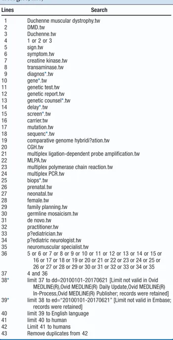
Systematic Literature Searches. Search terms were
identi-fied by the steering committee and literature searches per-formed in Ovid (2017 Ovid Technologies, Inc, New York, New York) by PharmaGenesis London, London, United Kingdom, to screen the MEDLINE and EMBASE databases. Search terms (title/abstract) were: Duchenne muscular dystrophy, DMD or Duchenne AND any of the following search terms: sign; symptom; creatine kinase; transaminase; diagnos*; gene*; genetic test*; genetic report*; genetic counsel*; delay*; screen*; carrier; mutation; sequenc*; comparative genome hybridi?ation; CGH; multiplex ligation-dependent probe amplification; MLPA; mul-tiplex polymerase chain reaction; mulmul-tiplex PCR; biops*; pre-natal; neopre-natal; female; family planning; germline mosaicism; de novo; practitioner; p?ediatrician; p?ediatric neurologist; neu-romuscular specialist. The “?” and “*” functions searched for spelling variations and variations of the word ending, respec-tively. Search results were limited to: articles published from January 1, 2010, to April 8, 2018; English; studies of humans; and full journal articles. The full electronic search strategy is provided inTable II, available atwww.jpeds.com. PharmaGenesis London exported the initial raw results and highlighted articles of potential relevance using a traffic light system (red—unlikely to be relevant; amber—potentially rel-evant; green—likely to be relevant). The 3 members of the steering committee then reviewed the relevance of the sup-porting literature. Any articles included by the authors that were not identified by the systematic literature review were categorized as ad hoc in the PRISMA flow diagram inFigure 2, available at www.jpeds.com. Data were then extracted by PharmaGenesis London, and used, under the guidance of the steering committee, to draft questions to aid the develop-ment of the Consensus Statedevelop-ments.
Development of the Consensus Statements via Iterative Voting. Voting round 1 included 29 questions drafted by the
steering committee based on initial review of the literature, to which the consensus group provided free-text responses. The results were collated by PharmaGenesis London to ensure voter anonymity, and the anonymized results were reviewed by the nonvoting chair. During each voting round, the consensus group was encouraged to comment on the wording and content of the questions, themes, and statements. After each round, the steering committee made changes to the statements to reflect the feedback received.
Voting round 2 included 20 questions formulated using the responses from round 1. The consensus group then rated the importance of, or agreement with, the themes identified
during round 2 (using a ranking or 5-point Likert scale). Voting rounds 1 and 2 were conducted using SurveyMonkey (https://www.surveymonkey.co.uk/).
Voting round 3 was developed in advance of a 1-day con-sensus meeting held in May 2017 in Copenhagen, Denmark, and the draft statements were circulated to the consensus group before the meeting. This round comprised 24 statements for which the consensus group had to rate their level of agree-ment using a 5-point Likert scale (“strongly agree” [A+];“agree” [A]; “neither agree nor disagree” [N]; “disagree” [D]; or “strongly disagree” [D+];Figure 1.1-4) using anonymized electronic keypads. Consensus was defined a priori as at least 75% agree-ment (either “strongly agree” [A+] or “agree” [A]). Each ques-tion was introduced by the nonvoting chair. If consensus was not reached after the first round of voting, alterations to the statement were made based on discussions before the next round of voting was conducted.
The 3 members of the steering committee then graded the level of evidence and strength of recommendations support-ing each statement ussupport-ing the Gradsupport-ing of Recommendations Assessment, Development, and Evaluation system (Figure 1). The grading was reviewed and agreed on by the expert voting panel (Table III).
Additional Supporting Information for the Consensus Statements. Additional supporting information from the
sys-tematic literature review for each consensus statement is de-tailed below.
Section 1: Reducing the Time to Diagnosis of DMD (Table III).
Statement 1. The following signs, symptoms, and
character-istics should be considered typical indicators of DMD: calf hy-pertrophy (pseudohyhy-pertrophy); delayed walking; difficulty climbing/descending stairs; difficulty rising from the floor; dif-ficulty running/walking; elevated serum CK level (including elevated alanine transaminase and aspartate transaminase); a family history of DMD; frequent falls; Gowers’ sign; male sex; and muscle weakness.
Vote A: + =93%;A=7%;grade of recommendation B:1
Discussion
In support of this statement (Table III), a retrospective chart review of 156 boys with DMD (no family history of disease) using the Muscular Dystrophy Surveillance, Tracking, and Re-search Network (MD STARnet) assessed the range of signs and symptoms first reported to healthcare providers by patients who were later diagnosed with DMD (n= 111).5In patients aged 3 to<5 years, the signs and symptoms included calf hyper-trophy, 7.1%; difficulty rising from the floor, 23.8%; diffi-culty climbing, 31.0%; frequent falls/clumsiness, 33.3%; difficulty running/walking, 38.1%; and muscle weakness, 40.5%.5These symptoms, with the exception of muscle weak-ness, difficulty running/walking, and difficulty climbing, were the most frequently reported within this age range compared
with younger or older patients with DMD.5Muscle weakness and difficulty climbing were reported more frequently in older patients (≥5 years old), and difficulty running/walking was more frequently reported in patients aged 1.5 to<3 years and pa-tients aged≥5 years, as the symptom(s) first reported to health-care providers.5
These findings are supported by several other studies. First, a mixed-methods study of parents’ and patients’ experiences of the diagnosis of DMD in Australia reported that the initial symptoms noted by parents (n= 62) (by >20% of parents) were calf hypertrophy, 42%; complaining of tired legs, 26%; diffi-culties with walking, 35%; frequent falls, 44%; tiring easily, 37%; difficulties running, 65%; and difficulties climbing stairs, 61%.6 Similarly, a case note review of 20 boys with DMD in the United Kingdom showed that 20% (8/20) of boys reported “diffi-culty with steps,” and 20% (8/20) of boys experienced “falls” as part of their initial symptom profile. Three of the 20 boys experienced delayed first walking (later than the 18-month mile-stone), and 4 of the boys were diagnosed from an incidental finding of elevated serum CK level.7A retrospective review of 24 patients with DMD in Korea reported that their initial symp-toms were: difficulty rising from the floor, 16.7%; lower-extremity muscle weakness, 90.0%; family history of DMD, 29.2%; and calf hypertrophy, 87.5%. Mean serum CK levels also were elevated (14 144 IU/L).8In addition, a study of tients with DMD from eastern India showed that of the 81 pa-tients assessed, 100% had lower-limb weakness, 97.5% had neck-muscle weakness, 93.8% had calf hypertrophy, 70.4% ex-hibited Gowers’ sign, and 27.1% had a family history of DMD.9 Lastly, a retrospective study of medical records for 540 pa-tients in the US with DMD showed that papa-tients with a family history of disease are typically seen and diagnosed at an earlier age than those without a family history (with family history vs without family history: age at initial evaluation, 30.8 months vs 56.8 months; P< .001; age at CK measurement, 35.1 months vs 64.0 months; P< .001).10Age at genetic testing did not differ significantly between the 2 groups (56.2 months vs 64.3 months, respectively).
Commentary
The consensus group agreed that patients were sometimes identified inadvertently as a result of high serum CK levels, detected as part of routine screening or assessments unre-lated to a diagnosis of DMD. It also was discussed that a family history of DMD would act as a strong indicator of DMD, par-ticularly in a male patient presenting with signs and symp-toms of the disease.
Statement 2. Autism spectrum disorder, delayed speech/
cognitive deficits, and gross motor delay can be indicators of DMD but are not always associated with the disease.
Vote A: + =93%;A=7%;grade of recommendation B:1
Discussion
In support of this statement, a European study of 4 centers found that 26% (34/130) of patients with DMD were reported
to have an intellectual disability. Of 87 patients who com-pleted the full neurodevelopmental assessment, 21% (18/87) scored in the autistic spectrum disorder range, 24% (21/87) showed clinical hyperactivity, and 44% (38/87) had severe dif-ficulties with inattention.11Similarly, a study in Germany of 263 patients with DMD showed that 30% of patients experi-enced a delay in gross motor development and that approxi-mately 40% of patients showed cognitive impairment (learning difficulty, 26%; intellectual disability, 17%).12A smaller case note review of 20 boys with DMD (without a family history of disease) in the United Kingdom showed that speech delay was a presenting feature in 25% of the cohort and that this was identified in 45% of the patients at the time of diagnosis.7 A study of patients with DMD from eastern India showed that approximately one-third of boys with DMD had mild intel-lectual disability (IQ 38-63).9A recent study of 209 caregiv-ers of boys with DMD identified through MD STARnet showed that cognitive deficits were reported in 38.4% of boys.13 Fur-thermore, a chart review study of 179 boys with DMD (1989-2012) showed that delayed walking and cognitive impairment were correlated (P≤ .0001).14It also has been shown that boys with DMD have a reduced rate of implicit learning com-pared with boys with typical development, even in the absence of global intellectual disability.15
The location of the DMD gene mutation also has been shown to correlate with the severity of cognitive impairment. For example, an observational study of 47 Italian boys with DMD showed that Full-Scale IQ scores correlated with the location of the dystrophin gene mutation; mutations in the distal region of the DMD gene were associated with more severe cognitive deficits.16Patients with point mutations in the DMD gene ex-hibited a higher degree of cognitive impairment than those with deletions or duplications (P= .005). In addition, patients with mutations in the distal region of the DMD gene had lower IQ levels than those who had mutations in the proximal region.17 Additional information is presented in a recent review of the literature examining neuropsychological and neurobehavioral functioning in patients with DMD.18
These neurocognitive deficits and developmental delays can sometimes be overlooked; as a result, patients with DMD can be referred to the incorrect specialist. This was exemplified in a retrospective chart review of 156 boys with DMD using MD STARnet, which showed that although 16.5% (21/127) of pa-tients were correctly referred to either a neurologist or a neu-romuscular specialist, 15.7% (20/127) were referred to a physical, occupational, or speech therapist as a result of their first evaluation.5
Commentary
No additional supporting information is included.
Statement 3. After initial presentation, patients with
sus-pected DMD should have their serum CK tested and be re-ferred to a specialist (a pediatric neurologist or neuromuscular specialist).