A
utologous fat transplantation is a common and ideal technique for soft-tissue augmen-tation and for filling soft-tissue defects due to its minimal tissue reaction and easy availability.However, although this technique follows standard-ized protocols, there is a relatively high resorption rate of the fat graft, the long-term graft survival is unpredictable, thus the desired effect is not always achieved. It has been observed in long-term follow-up studies that 20–80% of the graft volume is lost.1 The fat graft survival depends on microenviron-ment, and a dynamic remodeling after fat graft was well received by the recipient site, but the mecha-nisms of tissue revascularization/regeneration and subsequent absorption/fibrosis of the graft are not yet completely understood.
Quality and amount of revascularization are the key points for graft survival. After autologous fat transplantation, the increased resorption and in-Copyright © 2015 The Authors. Published by Wolters Kluwer
Health, Inc. on behalf of The American Society of Plastic Surgeons. All rights reserved. This is an open-access article distributed under the terms of the Creative Commons Attribution-NonCommercial-NoDerivatives 3.0 License, where it is permissible to download and share the work provided it is properly cited. The work cannot be changed in any way or used commercially.
DOI:10.1097/GOX.0000000000000305
Background: The increased resorption and the difficulty of the fat graft take following autologous fat transplantation procedure are associated with reduced fat tissue revascularization and increased apop-tosis of adipose cells. We suppose that the lipofilling procedure induces an inflammatory environment within the fat graft mass, whose evolution influences the efficacy of autologous fat graft survival. Eryth-ropoietin (EPO) is a glycoprotein hormone known to exert angiogenetic and anti-inflammatory effects; therefore, our purpose was to investigate its reaction with adipose tissue used in lipofilling.
Methods: Fat masses were harvested using manual suction lipectomy and then seeded on dishes in ap-propriate culture and treated for 3 weeks with 3 doses of EPO. CD31 and CD68 immunohistochemistry was used to identify microvessels and several infiltrating leukocyte cells.
Results: Following EPO administration, we have detected an increase in the number of CD31-positive microvessel endothelium cells and CD31-positive small leukocytes and a reduction of CD68-positive cells. These effects were more conspicuous following higher EPO dose.
Conclusions: Our findings evidence EPO treatment as a useful strategy to sustain the revasculariza-tion of grafted tissue and to reduce its inflammatory state. (Plast Reconstr Surg Glob Open 2015;3:e338; doi:10.1097/GOX.0000000000000305; Published online 30 March 2015.)
Effects of Erythropoietin on Adipose Tissue:
A Possible Strategy in Refilling
Maurizio Sabbatini, PhD* Liah Moalem, MD† Michela Bosetti, PhD‡ Alessia Borrone* Renzo Boldorini, MD§ Antonio Taveggia, MD† Giovanni Verna, MD† Mario Cannas, MD*
Meeting Proceedings
SICPRE:
La SICPRE, Società Italiana di Chirurgia
Plastica Ricostruttiva ed Estetica, national meeting,
in Bergamo, Italy on October 12-15, 2014
From the *Laboratory of Human Anatomy, Department of Health Sciences, UPO University, Novara, Italy; †Department of Plastic and Reconstructive Surgery, Hospital “Maggiore della Carità,” Novara, Italy; ‡Department of Pharmaceutical Sciences, UPO University, Novara, Italy and §Section of Pathological Anatomy, Department of Health Sciences, UPO University, Novara, Italy. Presented at the 63rd SICPRE National Meeting (Società Italiana di Chirurgia Plastica Ricostruttiva ed Estetica, Italian Society of Plastic, Reconstructive and Aesthetic Surgery), October 12–15, 2014, Bergamo, Italy.
creased apoptosis of adipose cells are direct conse-quences of poor revascularization.2 Besides, some clinical and experimental studies have reported con-tinuing volume loss of the grafted adipose cells even after the grafts appeared revascularized.3
Several studies have documented the production of inflammatory cytokines by adipose tissue. Their oc-currence depends not only on macrophage cells nor-mally present in the adipose tissue mass, but a large amount of this production may directly depend on the preadipocytes, which have been observed to be able also to produce monocyte chemoattractant pro-tein-1 and macrophage migration inhibitory factors.4
Based on these findings, we suppose that lipo-filling procedure induces an inflammatory envi-ronment within the fat graft mass, whose evolution influences the autologous fat graft take.
Erythropoietin (EPO) is a glycoprotein hormone that was first characterized by its ability to stimulate erythropoiesis. In following researches, it has been discovered that EPO is also involved in angiogenesis induction: instigating the secretion of angiogenic factors, enhancing the immune response, and exert-ing anti-inflammatory effects.5
Our purpose was to investigate the effect of EPO on adipose mass used in lipofilling, focusing mainly on the effects on tissue revascularization and the in-fluence on immune cell behavior.
MATERIALS AND METHODS
Fat graft was harvested using manual suction li-pectomy under general anesthesia from 10 women, 41 ± 4 years old, during procedures of reconstructive surgery. The participants gave written informed con-sent. The fat was harvested using a 14-gauge blunt cannula and centrifuged at 1500 rpm for 2 minutes.
The fat mass was seeded on dishes in appropriate culture on average for 24 hours and then treated for 3 weeks with 3 different doses of EPO (EPREX, Janssen-Cilag, Milan, Italy): 0.15, 0.30, and 0.60 μg/ml. A positive control was performed administering a cocktail of trophic drugs (biotin, T3, pantothenate, hydrocortisone, and insulin; from Sigma, St. Louis, Miss.). The negative control was left without any treatment.
At the end of this phase, the fat mass was fixed in formalin and processed for paraffin inclusion and cutting: the obtained 5-μm-thick slides were stained with CD31 (to identify active vessels and immune sys-tem cells) and CD68 (to identify macrophages).
Quantification of cell infiltration in the fat grafts was estimated by counting the number of positive vessels and cells. To avoid counting bias, due to the change of adipocytes diameter, the counting data were referred to tissue fat mass of 80 adipocytes. Para-metric statistical analysis was obtained with Prism5.0 (GraphPad Software, San Diego, Calif.).
RESULTS
The study of the fat mass cultured under micro-scope evidenced 3 different zones in respect of adipo-cytes dimension, extracellular matrix deposition, and vessels density. These zones are identified as follow: Zone 1: wide presence of extracellular matrix, small
adipocytes (686.3 ± 33.2 μm2), presence of large microvessels, and few capillary vessels.
Zone 2: appreciable presence of extracellular ma-trix, large adipocytes (4573.4 ± 162.3 μm2), and presence of capillary vessels.
Zone 3: poor occurrence of extracellular matrix, very large adipocytes (12359.6 ± 347.6 μm2), and presence of capillary vessels.
Our data were referred specifically to the identi-fied zones.
Vessels count has evidenced no changes in the number of CD31-positive and CD31-negative vessels in zone 1 and zone 3 between the different experi-mental groups analyzed. Differently, zone 2 that has been treated with a higher dose of EPO showed a higher number of CD31-positive vessels in compari-son to other experimental groups (Fig. 1).
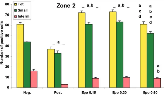
The CD31-positive cells count showed the occur-rence of 2 cell populations named small cells (22.3 ± 1.1 μm2; morphologically compatible with helper lympho-cytes) and intermediate cells (68.5 ± 6.1 μm2; morpho-logically compatible with cytotoxic and natural killer lymphocytes). The positive control group showed a reduction of the number of CD31-positive cells com-pared with the negative control group. Differently, EPO treatment showed an increase in the number of small cells and a light decrease of intermediate cell number in all the 3 zones (Fig. 2).
The CD68-positive cells count showed the occur-rence of 2 cell populations named very large cells (247.1 ± 15.2 μm2; morphologically compatible with macrophages) and large cells (129.2 ± 9.3 μm2; mor-phologically compatible with B-lymphocytes). In comparison to the negative control, the positive con-trol group exhibits a reduction of CD68-positive cell number in zone 2 and zone 3, whereas all EPO treat-ments showed reduction of very large cell number in all zones examined and a reduction of large cells in zone 3. Following higher EPO dose treatment, the Disclosure: The authors have no financial interest to
declare in relation to the content of this article. The Article Processing Charge was paid for by the Società Italiana di Chirurgia Plastica Ricostruttiva ed Estetica.
very large cells reduction was proportionally more evident (Figs. 3 and 4).
DISCUSSION
There is a wide presence of inflammatory cells in adipose mass extracted for refilling, suggesting the potential of the fat mass to produce an inflam-matory response.
EPO is a well-known factor able to induce tissue revascularization. EPO receptors have been found
in several human leukocytes; therefore, researchers supposed that the EPO receptors have a role in mod-ulating some signaling pathways.6
Our findings clearly remark the provascular properties of EPO. Specifically, we observed EPO to induce an increase in the number of CD31-positive microvessels. CD31 reactivity is a well-known marker, indicating the functional activation of microvessels in the tissue; therefore, several CD31-negative microves-sels observed in culture should be interpreted as
Fig. 1. Bar graph showing results of the morphometrical analysis performed on cd31-
positive or cd31-negative microvessels detected in zone 2 of the fat mass. aP < 0.05 vs neg; bP < 0.05 vs Pos; cP < 0.05 vs epo 0.15; dP < 0.05 vs epo 0.30. epo indicates erythropoietin
administration; neg, negative control with no treatment; Pos, positive control treated with the cocktail of trophic drugs (see Materials and Methods section).
Fig. 2. Bar graph showing results of the morphometrical analysis performed on
Fig. 3. Bar graph showing results of the morphometrical analysis performed on
cd68-posi-tive cells of zone 3, detected in the fat mass.
Fig. 4. Microphotograph panels showing the immunohistochemical positive signals occurring in the fat mass. cd68
quiescent microvessels. In addition, EPO presents a direct effect on tissue infiltrating leukocytes. The re-duction of macrophage number is an important find-ing that may evidence a reduced inflammatory state, with macrophages being the principal cells involved in the tissue inflammatory pathways. Other significant evidence is the increase of CD31-positive small lym-phocytes. In the absence of a more detailed analysis, the increase of the number of lymphocytes can be imputed only to a shift of cellular physiology from a nonactive state to an active state.
Small lymphocytes widely participate in the in-flammatory phenomena; on the other hand, they are also involved in the release of tissue trophic factors.7 These findings may indicate lymphocytes as crucial cells in supporting the tissue survival and they may also be at the origin of the observed reparative ef-fects detected on skin lesions and bone fractures.8,9
CONCLUSION
In conclusion, EPO treatment may be a useful strategy to reduce the inflammatory state of adipose grafted tissue and to sustain its revascularization.
Further studies are required to analyze the ex-tent of graft tissue damage related to inflammatory response and which pattern of cytokines is involved in modulating the inflammatory response. We think that a controlled inflammatory response may be use-ful to obtain a higher and more predictable survival rate in fat transplantation.
Optimizing the fat tissue graft take may lead to a reduction in the number of procedures required to obtain the desired outcome and thus improving the cost/benefit rate.
Liah Moalem, MD
Department of Plastic and Reconstructive Surgery Hospital “Maggiore della Carità,” Novara, Italy E-mail: [email protected] REFERENCES
1. Nishimura T, Hashimoto H, Nakanishi I, et al. Microvascular angiogenesis and apoptosis in the survival of free fat grafts.
Laryngoscope 2000;110:1333–1338.
2. Eto H, Kato H, Suga H, et al. The fate of adipocytes af-ter nonvascularized fat grafting: evidence of early death and replacement of adipocytes. Plast Reconstr Surg. 2012;129:1081–1092.
3. Nguyen A, Pasyk KA, Bouvier TN, et al. Comparative study of survival of autologous adipose tissue taken and transplanted by different techniques. Plast Reconstr Surg. 1990;85:378–386; discussion 387–389.
4. Chung S, Lapoint K, Martinez K, et al. Preadipocytes medi-ate lipopolysaccharide-induced inflammation and insulin resistance in primary cultures of newly differentiated hu-man adipocytes. Endocrinology 2006;147:5340–5351. 5. Lombardero M, Kovacs K, Scheithauer BW. Erythropoietin:
a hormone with multiple functions. Pathobiology 2011;78:41–53.
6. Lisowska KA, Debska-Slizień A, Bryl E, et al. Erythropoietin receptor is expressed on human peripheral blood T and B lymphocytes and monocytes and is modulated by re-combinant human erythropoietin treatment. Artif Organs 2010;34:654–662.
7. Beers DR, Henkel JS, Zhao W, et al. CD4+ T cells support glial neuroprotection, slow disease progression, and mod-ify glial morphology in an animal model of inherited ALS.
Proc Natl Acad Sci U S A 2008;105:15558–15563.
8. Klinger M, Marazzi M, Vigo D, et al. Fat injection for cases of severe burn outcomes: a new perspective of scar remodeling and reduction. Aesthetic Plast Surg. 2008;32:465–469.
9. De Ugarte DA, Ashjian PH, Elbarbary A, et al. Future of fat as raw material for tissue regeneration. Ann Plast Surg. 2003;50:215–219.