SCUOLA DI DOTTORATO DI RICERCA Scienze e Biotecnologie dei Sistemi Agrari e Forestali e delle Produzioni Alimentari
Indirizzo Scienze e Tecnologie Zootecniche Ciclo XXVIII
Influence of pathogens and pollution on Mugilidae health:
first evidence of mycobacteriosis and intersex condition in
extensively reared mullets from Sardinian lagoons
dr. Elisabetta AntuofermoDirettore della Scuola prof. Antonello Cannas
Referente di Indirizzo prof. Gianni Battacone
Docente Guida prof. Antonio Pais
INDEX
Abstract 1
Chapter 1 4
General Introduction 4
1.1 Global fish production 5
1.1.1 Production from capture fishery 5
1.1.2 Production from aquaculture 6
1.2 Aquaculture production systems and practices 7
1.2.1 Extensive aquaculture in the Mediterranean 8
1.2.2 Extensive aquaculture in Italy 8
1.2.3 Extensive aquaculture in Sardinia 10
1.3 Generalities of mullets 13
1.3.1 Mullet species in the Mediterrenean Sea 13
1.3.2 Morphology and morphometry 13
1.3.3 Food and feeding 16
1.3.4 Age and growth 17
1.4 Reproductive state of mullets 18
1.4.1 Sexuality 18 1.4.2 Oocyte development 19 1.4.3 Postovulatory follicles 20 1.4.4 Atresia 20 1.4.5 Ovarian maturity 20 1.5 Morphometric indices 21
1.5.1 Fulton’s condition factor 21
1.5.2 Epatosomatic index 22
1.5.3 Gonadosomatic Index 22
1.6 Current state of mullet culture 23
1.6.1 Global production of mullets 24
1.6.2 Products from mullets 25
1.7. Interaction between environment pathogens and fish health 25 1.7.1 Mugilids as biological indicator of water pollution 28
1.8 Pathogens in mullets 29
1.8.2 Mycobacteriosis in fish 31
1.8.3 Public health 37
1.9 Endocrine Disruptors Compounds (EDCs) 39
1.9.1 Intersex in mullets 42
1.9.2. Histological classifications of intersex 43
1.9.3 Distribution of intersex 44
1.10 References 48
Chapter 2 69
Sampling sites and mullet species.Processing techniques and morphometric
featuresof the fish examined 69
2.1 Study area 70
2.2 Lagoon characteristics 70
2.2.1 North western Sardinia 70
2.2.2 The Calich lagoon 71
2.2.3 North eastern Sardinia 72
2.2.4 The San Teodoro lagoon 72
2.2.5 Central western Sardinia 73
2.2.6 The Cabras lagoon 73
2.2.7 The Marceddì-San Giovanni lagoon 74
2.3 Mugilidae species of interest 75
2.3.1 Chelon labrosus 75 2.3.2 Liza aurata 76 2.3.3 Liza ramada 77 2.3.4 Mugil cephalus 78 2.4 Sampling techniques 79 2.5 Necropsy 80 2.6 Histopathology 81 2.7 Results 87 2.7.1 Cabras 89 2.7.2 Calich 91 2.7.3 Marceddì 94 2.7.4 San Teodoro 96
2.8 References 105
Chapter 3 107
Mycobacteriosis caused by atypical Mycobacteria in reared mullets: first evidence
from Sardinia 107
3.1 Introduction 108
3.2 Material and methods 110
3.2.1 Study area 110
3.2.2 Sampling 110
3.2.3 Anatomo-histopathological evaluation 110
3.2.4 Bacteriological and molecular analyses 110
3.3 Results 113
3.3.1 Macro and microscopical findings 113
3.3.2 Bacteriological and molecular findings 117
3.4 Discussion 122
3.5 Conclusion 124
3.6 References 126
Chapter 4 132
First evidence of intersex condition in extensively reared mullets from Sardinian
lagoons 132
4.1 Introduction 133
4.2 Material and methods 135
4.2.1 Study area 135 4.2.2 Sampling 135 4.2.3 Histology 135 4.3 Results 138 4.4 Discussion 143 4.5 Conclusion 146 4.6 References 147 Acknowledgments 152
Abstract
The multifactorial nature of fish health and diseases are generally linked to an imbalance of pathogen, resistance of the fish and environmental stress. Pollution is considered an anthropogenic factor that can affect fish life leading to immunosuppression that increases fish susceptibility to pathogens affecting their survival and growth rates.
Coastal waters and lagoons are typical environments devoted to extensive aquaculture. In Italy, which annual production of extensive reared fish is nearly 5.250 tons, mullets represent the most important cultured species with an average production of about 3.000 tons per year. The coastal environments in which they grow are constantly exposed to high levels of urbanization and, consequently, to the action of increasing amounts of contaminants discharged in waters. This fact can play a central role in some emerging issues like mycobacteriosis and gonadal abnormalities in cultured mullets, representing a real concern for fish health and reproduction.
The term “mycobacteriosis” or “fish tuberculosis” decribes a chronic systemic and progressive disease caused by mycobacteria belonging to the genus Mycobacterium. In particular, Mycobacterium marinum is a slow-growing non-tuberculous mycobacterium and it is considered the most common etiologic agent of mycobacteriosis in wild and cultured fish. This disease is considered a real risk for fishermen and aquarists that manipulate infected fish. The diagnosis of mycobacteriosis is principally made by histology when positive Ziehl-Neelsen stain granulomas are detected. The aim of the first part of this study was to investigate the occurrence of mycobacteriosis in extensively cultured Mugilidae (Chelon labrosus, Liza aurata, Liza ramada and Mugil
cephalus) of four lagoons from Sardinia by the use of histology, microbiology, PCR and
DNA sequencing. Twenty-five out of 495 mullets (148 specimens from Cabras, 120 from Calich, 89 from Marceddì, and 138 from San Teodoro), collected during summer and autumn of the years 2013, 2014 and 2015, were suspected of being infected with mycobacteriosis revealing granulomas containing acid-fast bacilli at histopathological examination. In particular, 10 out of 25 mullets were certainly affected by mycobacteriosis and Mycobacterium marinum was identified in 6 out of 10 as the primary cause, and the concordance obtained by histology, cultural evaluation and sequence analysis of the hsp65 gene was 100%. In the remaining 4 specimens,
Mycobacterium spp. were detected and the concordance obtained by histology and
granulomas with acid-fast bacilli were detected although culture confirmed the positivity for Mycobacterium spp. only in 6 cases, with an accordance of 43% with histology. In all of these cases, PCR-hsp65 and sequencing failed to identify atypical mycobacteria. Mullets affected by mycobacteriosis were mainly sampled in the Calich (10%) and San Teodoro (8%) lagoons. Only 2 subjects were positive for mycobacteriosis and no cases were observed in Cabras and Marceddì lagoons, respectively. This study confirms that histopathological examination is a very important diagnostic screening tool for the detection of mycobacteriosis in fish and PCR-hsp65 is a valid and easy method to identify atypical mycobacteria, expecially for M. marinum. Our finding are worthy of attention because mycobacteriosis in mullets has been evidenced for the first time in Sardinia, suggesting that this disease may be underestimated also in other cultured fish species. These results confirm our expectation that mullets living in San Teodoro and Calich lagoons, both characterized by critical environmental conditions, could play a central role in understanding the occurrence of fish mycobacteriosis which, if not properly monitored, could represent a serious concern for public health.
Fish are among the most studied organisms for the effects of chemical contaminants on the development and reproductive processes. In coastal and estuarine ecosystems, euryhaline fish living in polluted waters like Mugilidae can frequently show sexual anomalies like intersex. This term describes alterations in gonadal development with the simultaneous presence of male and female reproductive stages in the same gonad of a gonochoristic species. In the second part of this study, adult specimens of three species of euryhaline mullets (Chelon labrosus, Liza aurata, and Mugil cephalus), from two Sardinian lagoons (Marceddì and San Teodoro) devoted to extensive aquacultural practices, were analyzed in order to identify putative alterations in gonads and in gamete development. Overall, 13 of the 158 mullets examined (8.2%) were affected by gonadal disorders: four subjects (one C. labrosus, two L. aurata and one M. cephalus) exhibiting an intersex condition were found in the Marceddì lagoon and the other nine (five C. labrosus, two L. aurata and two M. cephalus) in the San Teodoro lagoon. Twelve of these gonads were classified as testis-ova (TOs) and one, belonging to a C.
labrosus specimen, was a mixed gonadal tissue (MGT). Intersex condition was
evaluated using an intersex index and all the recorded values showed a mild ovotestis severity index (OSI). However, our findings suggest that fish gonadal disorders may be underestimated in extensive reared fish species, particularly in coastal brackish
environments polluted by intensive agriculture and animal husbandry activities. In conclusion, further research on emerging fish disorders and diseases in Sardinian coastal lagoons could confirm the hypothesis that species like mullets have to be considered as biological sentinels to detect the degree of pollution in extensive aquaculture systems, with the purpose of reducing risks to animal and human health.
Chapter 1
1.1 Global fish production
Capture fisheries involve capture of wild organisms while aquaculture is based on the rearing of fish, crustaceans, molluscs and algae. The interplay between these two activities are numerous: many of the farmed species are also fished, and in many cases fishery and aquaculture, especially in lagoons and lakes are integrated and their boundaries are not always so well defined (Cataudella & Bronzi, 2001).
Global fish production has grown regularly in the last six decades and in 2012 fisheries and aquaculture activities supplied the world with about 158 million tons of product, of which 136 million were for human consumption (Fig. 1.1).
Fish consumption increased in the last 60 years from 9.9 kg to 19.2 kg per capita due to the world population growth, urbanization, and the improving of fish production technology (Table 1.1)
Fig. 1.1 Trend in world capture fisheries and aquaculture production from 1950 to 2012
(from FAO, 2014).
1.1.1 Production from capture fishery
Capture fishery production in 2012 was of 79.7 million tons in marine waters and of 11.6 million tons in inland waters respectively. Nevertheless recent data show that capture fisheries have reached the limits of growth and it is absolutely necessary that these activities do not exceed the limits imposed by the natural law (FAO, 2014).
Table 1.1 World fisheries and aquaculture production and utilization (from FAO,
2014).
1.1.2 Production from aquaculture
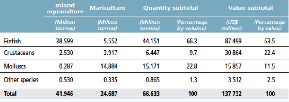
FAO introduced a definition of aquaculture, which reduces its confusion with capture fisheries: “aquaculture is the farming of aquatic organisms, including fish, molluscs, crustaceans and aquatic plants. Farming implies some form of intervention in the rearing process to enhance production, such as regular stocking, feeding, protection from predators, etc.” (FAO, 1988). In farming activities more than 600 aquatic species mainly fish, crustaceans and molluscs are cultured in different aquatic environments such as freshwater, brackish water and marine water. Aquaculture remains the productive activity showing the most rapid growth in the context of food production, with an average growth of 8.6% per year since 1980 (Costa-Pierce, 2002; FAO, 2014). Global aquaculture production of food fish has reached 66.6 million tons in 2012, where 44.2 were constituted of 38.6 million tons from inland aquaculture of finfish species and 5.6 million tons from mariculture (Table 1.2). In particular, growth of finfish is higher in inland than in mariculture and it is consequence of the easy-to-achieve type of aquaculture in developing countries.
Table 1.2 World production of farmed species groups from inland aquaculture and
mariculture in 2012 (from FAO, 2014).
1.2 Aquaculture production systems and practices
Aquaculture developed varieties of system that range from very extensive to hyper-intensive techniques. Intensive aquaculture is based on the rearing of fish in inland tanks or in marine floating cages where human intervention is necessary for feed and stocks management (MIPAAF, 2014). Extensive aquaculture cover large areas of water and characteristic environments are lagoons, delta river, estuaries, bays and ponds of inland areas generally near to the coasts. (Anras et al., 2010). This farming technique is practiced using freshwater or brackish water basins that cover extended areas. Generally cultured fish spend their earlier life in marine or freshwater and only the last part of rearing occur in brackish waters where the level of salinity varies from 0.5% to seawater (FAO, 2016). Extensive aquaculture is the closest farming methods to fishing and is regulated by minimal human intervention limited to simply catching aquatic organisms with artisanal fisheries as fixed capturing systems, nets or hand weapons (Cataudella & Bronzi, 2001).
More specialized methods are planned in different parts of productive cycle such as managing interventions, diet integration and control of juveniles (Bostock et al., 2010). However traditional practices may present differences moving from one country to another changing farming protocols and water management (Anras et al., 2010). Extensive aquaculture, that exploits mostly lagoon resources, constitutes a traditional model where general structures like hydraulic barriers as weirs and locks are used (Cataudella & Bronzi, 2001). Generally the management of the lagoon is organized in small farms where fishing license is owned by one or few cooperative that handle and sell fish stocks.
1.2.1 Extensive aquaculture in the Mediterranean
Several forms of aquaculture including extensive systems are now an important reality of fish production in the Mediterranean coastal lagoons and are part, from long time, of the cultural tradition of the regions, with around 400 coastal lagoons, covering a surface of over 641.000 ha. Productivity varies from a few to several hundred kilograms per hectare per year (kg/ha/year) based on the lagoon typology and ecology, although for many different reasons achieve consistent and updated fish production is still a challenge (FAO, 2015a).
Territory characteristics and several reared species (mainly seabream, seabass, mullet, sole and eel) are distinctive of different European regions. In a recent report, European Commission declares that aquaculture can still grow providing new job opportunities and gradually satisfy consumer internal request of safe and sustainable seafood products. Moreover in that report is observed that the increase of human activities (urbanization, tourism-related facilities, wastewaters from industrial and agricultural activities) have a harmful effect on this ecosystem and is becoming one of the major restraint to progress of the extensive aquaculture in Europe (European Commission, COM (2013) 229 final).
1.2.2 Extensive aquaculture in Italy
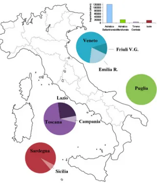
In Italy 198 lagoons, coastal lakes and ponds are present for a total surface of 167.575 ha, of which 40.000 are dedicated to extensive aquaculture. Lagoons are distributed mainly in four Italian geographic zones largely diverse in morphology and ecosystem: the northern Adriatic, the south Adriatic, the central Tyrrhenian and Sardinia and Sicily. (Fig. 1.2).
The extension of brackish lagoons that range from few ha to 1.660 ha, in the North of Italy (Veneto, Emilia-Romagna and Friuli-Venezia Giulia) are named "valli di pesca" and represents confinement of portion of coastal lagoon (vallicoltura) (Cataudella & Bronzi, 2001; MIPAAF, 2014).
Extensive aquaculture represents the 12,4% of national seafood production, with a production amounts ranging from 40 kg/ha of Lesina lagoon to 319 kg/ha of Sardinian lagoons (Cataudella & Bronzi, 2001).
Fig. 1.2 Lagoons in Italy: surface in the four areas mainly devote to extensive
aquaculture (histogram) and relative distribution in the nine regions (circles) (FAO, 2015a).
Italian production from lagoon environments is highly variable, and data began to be collected from 2008. FAO reports 2.787 tons of fish and 35.007 tons of shellfish for 2009 (FAO, 2015a and reference therein). Although lagoon system possesses a valuable biodiversity and richness in aquatic animals, only few species are of commercial interest. These generally include Mugilidae species, such as Chelon labrosus (thicklip mullet), Liza aurata (golden grey mullet), Liza ramada (thinlip mullet), Liza saliens (leaping mullet), and Mugil cephalus (grey mullet).
Other common fish species are Sparus aurata (seabream), Dicentrarchus labrax (European seabass), Anguilla anguilla (European eel), Atherina boyeri (Boyer’s sand smelt) (Cataudella & Bronzi, 2001).
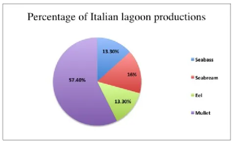
With a total national production of 5.250 tons of extensive reared fish, mullets represent the most important productive sector as they account for the 57% of total, with an annual production of 3.000 tons. Moreover seabream and seabass are largely diffuse and contribute for the 16% (850 tons) and 13% (700 tons) of national production, respectively; eels complete lagoons production with a percentage of 13% (700 tons) (MIPAAF, 2014) (Fig. 1.3).
Fig. 1.3 Extensive aquaculture productions in Italy: mainly fish species farmed and their
relative percentage (MIPAAF, 2014).
1.2.3 Extensive aquaculture in Sardinia
Sardinia has one of the most extended area of lagoons and ponds of Europe. According to Fenza et al. (2014), lagoons identify a coastal basin, characterized by brackish water, separated from the sea only by thin land boundaries that permit good water circulation, whereas in ponds exchanges with the sea are absent or modest and the water circulation is slower (Fenza et al., 2014).
Of the 77 coastal lagoons, covering a total area of around 15.000 ha, only 27 are now used for extensive aquaculture. These cover a total area of 5.700 ha, with 3.800 ha concentrated on the central western coast (Oristanese), where the most important lagoons are in Cabras (OR, 2.230 ha) and in S. Giovanni-Marceddi (OR, 1.600 ha). (Cataudella & Spagnolo, 2011; Fenza et al., 2014).
The other lagoons are present in the North-western area of Sardinia (Nurra), North East (Gallura -Baronia), South East (Ogliastra-Sarrabus-Gerrei), South (Cagliaritano) and South West (Sulcis-Iglesiente) as reported in Fig. 1.4.
Extensive aquaculture in Sardinia is practiced in lagoons and ponds with simpler technology compared to the extensive poly-culture method like “vallicoltura”. Generally in Sardinia production practices are based on traditional methods by using fixed capture systems generally V-shaped chambers called “lavorieri” built in wood, reeds, concrete or plastic (Fig. 1.5).
Fig. 1.4 Sardinian wetland areas used for extensive aquaculture (from Fenza et al.,
2014).
Fig.1.5. Typical plastic V-shaped “lavoriero”.
“Lavorieri” lead the fish moving from the sea towards the lagoon where they find abundance of food and a more protected environment than marine waters. These
systems permit to capture fish when they migrate from the lagoon back to the sea during their reproductive cycle (FAO, 2015a). In Sardinian wetlands are also diffuse different types of fishing with traps and nests to catch principally eels, gobies, crabs and cuttlefish (Fenza et al., 2014).
Among the many aquatic organisms that can find a proper habitat in wetlands, very few fish species of commercial interest are mainly extensively cultured in Sardinia: mullets and eels are the most represented, but also crabs and sand smelts. In waters where salinity is higher are captured seabreams and seabasses, the most valued species, but also gobies, red mullets and flounders can be found in these lagoons.
Sardinian ponds appear to be the most productive lagoons in Italy reaching a mean production of 50 kg/ha (a minimum of 25 and a maximum 320 kg/ha/year) (Cataudella & Bronzi, 2001). However Sardinian lagoons production have reached in the 1980s 600 kg/ha in the lagoon of Tortolì.
Many causes may play a role in determining these large differences in productivity. One can be attributed to the progressive changes of salinity and water temperature that may alter the ecological equilibria of wetlands as has been recorded in some ponds in Sardinia. Anyway pollution is becoming a serious problem having caused severe diseases in fish of many lagoons like in Cabras, Santa Giusta (OR) and San Teodoro (OT) (Cataudella & Spagnolo, 2011).
It is interesting to note how the property of wetlands is allocated in Sardinia. The “Regione Autonoma della Sardegna” (R.A.S.) is owner of twenty-four production ponds that give in concession to fishing cooperatives that can manage and sell fish stocks coming from those lagoons.
Only three ponds are privately owned (Fenza et al., 2014). However fish productions could be higher if implants could receive a relevant economic effort of modernization. This element is of great importance when you consider that the combination of fishing and aquaculture and their strong integration represent the future of this activity to safeguard the quality of products and for the safeguarding of resources. The combination of responsible fishing and sustainable aquaculture is considered to be the framework for the proper management of environmental resources.
1.3 Generalities of mullets
1.3.1 Mullet species in the Mediterrenean Sea
Fish of the family Mugilidae, class Actinopterygii, order Mugiliformes, includes 20 genera and 71 species (Eschmeyer & Fong, 2016; Eschmeyer et al., 2016). Four genera (Chelon, Liza, Mugil and Oedalechilus) and six species of mullets (thicklip mullet C.
labrosus, golden mullet L. aurata, thinlip mullet L. ramada, sharpnose mullet L. saliens, flathead mullet M. cephalus, boxlip mullet O. labeo) are endemic in the
Mediterranean Sea (Whitehead, 1984; FAO, 1995; Nelson, 2006; Turan, 2007).
1.3.2 Morphology and morphometry
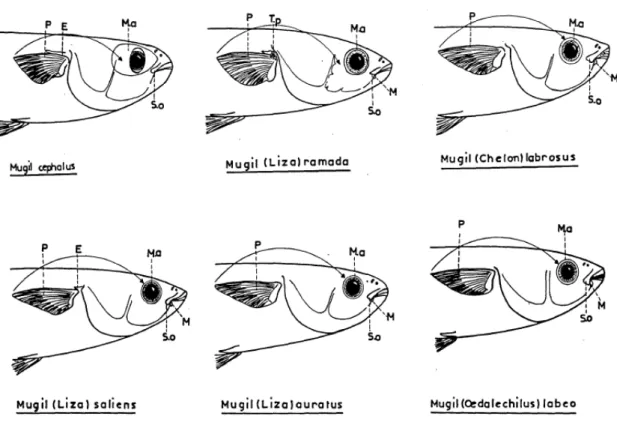
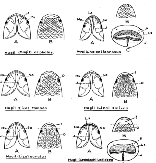
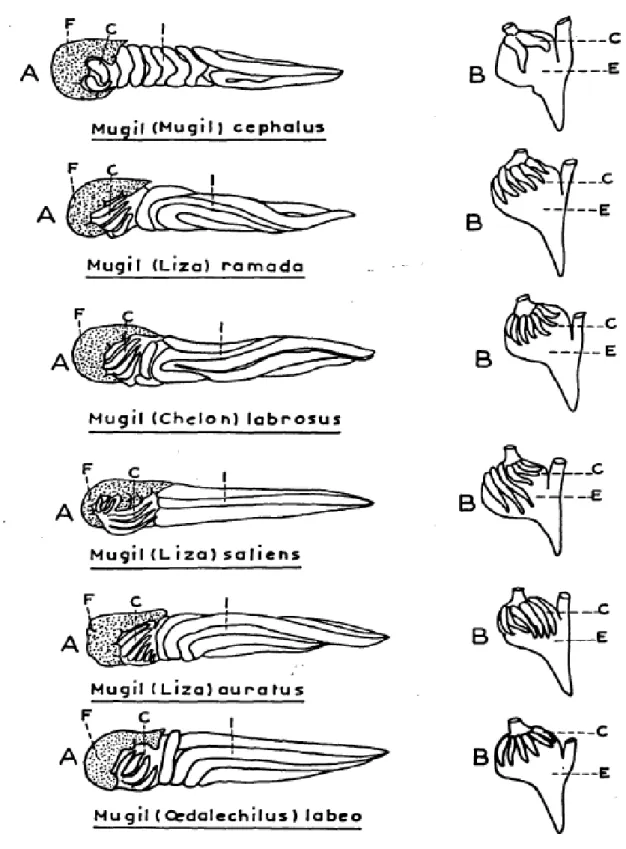
Although general features such as fusiform body, large scales, grey coloration on upper back and silver grey along flanks are common traits in Mugilidae, only some morphological characters are suitable to classify among different mullets species as reported by Farrugio (1977) and by Turan et al. (2011) see Figs. 1.5, 1.6 and 1.7, and Table 1.3 respectively.
Fig. 1.6 Profiles of mullet heads. E: axillary scale. M: maxillary. Ma: adipose
Fig. 1.7 Mullet heads. A: ventral face. B: dorsal face. C: frontal face. J: jugular space.
LS: upper lip. Mx: maxilla. O: scales. P: papillae. Pa: adipose eyelid. So: sub orbital (from Farrugio, 1977).
Fig. 1.7 Scheme of intestinal tract of mullets and profile of stomach. C: pyloric caeca.
Table 1.3 Descriptive taxonomic characters used to distinguish Mediterranean mullets.
First dorsal fin rays (DFR1), second dorsal fin rays (DFR2), ventral fin rays (VFR), anal fin rays (AFR), pectoral fin rays (PFR), pyloric caeca (PC) (from Turan et al., 2011).
However, in Mugilidae species the morphoanatomy is so similar and at the same time the evolutionary tool is not so straight in interpreting anatomical differences. Hence classifying species and genera in terms of phylogenetic inferences is problematic. Fortunately the extensive developments of Polymerase Chain Reaction (PCR) offer a valid support into the study of the phylogeny and diversity of the Mugilidae families and species (Durand et al., 2012).
1.3.3 Food and feeding
Mugilidae, generally known as mullets are distributed in all tropical and temperate seas, living in offshore and coastal waters, lagoons and estuaries (Boglione et al., 2006; Fenza et al., 2014). Mullets are euryhaline species able to adapt to a wide range of water salinities and to live both in fresh water (FW) than in sea water (SW) at different salinity levels. In particular some mullet species well tolerate FW and others inhabit waters more saline than normal SW. Euryhaline fishes need to maintain a particular homeostasis of their internal fluids and gills, alimentary tract, kidneys and neuroendocrine. This latter is the major organ system involved in osmoregulatory functions in mullets (McCormick, 2001).
Mullets are able to get feeding from water surface to mud bottom and have been named as mud or detritus feeders (Brusle, 1981). Diet consists mainly of organic matter suspended or present in sediment, benthic invertebrates, macroalgae and plankton. Benthic animals are generally present in the sediment and constitute the major source food of mullets. They are classified into meiofauna, macrofauna and epifauna (Cardona, 2016). For that reason mullets live in ecosystems where the sediment is plenty of
organic matter, like coastal lagoons and estuaries (Cardona et al., 2001).
The gastroenteric apparatus of young and adults mullets became similar when juvenile move from a zooplanktophagous to sedimentivorous diet (Ebeling, 1957; Albertini-Berhaut, 1987).
Mullets have a specialized feeding mode different than other species of fish. They orient their head down when eating by using their powerful jaw to scrape the bottom surface assuming sediment and associated food material (Odum, 1970; King, 1988). In particular they have toothed mouth to scrape microbial films, teeth and gills to remove large and small particles respectively and a stomach with a powerful gizzard (Ebeling, 1957; Capanna et al., 1974). Different oral and pharyngeal anatomical structures are also reported in mullets species: i.e. Chelon labrosus usually feeds organic debris and algae by using tubercles of its upper lip, Liza genus prefers small particles in soft mud while Mugil cephalus eat larger particles in sandy substrates (Boglione et al., 2006). However, the food quantity and quality vary considerably seasonally and during spawning migration some mullets species stop feeding (Odum, 1970; Cardona, 2001).
1.3.4 Age and growth
Growth in fish, the increase in length or weight directly related to age is a complex mechanism in which several factors and variables play a role. As is known, growth is considered as a process mostly driven by fish physiology strictly related to season, habitat, food, sex, water temperature, salinity, fish health etc. (Froese, 2006). Regarding mullets, authors report that a very rapid growth rate during the first two years of life is observed while reducing significantly in the rest of life (Brusle, 1981; Abbas, 2001). The greatest growth rate is reported in Mugil cephalus respect to the genus Liza and
Chelon labrosus. Low temperatures and scarce availability of food are considered
causes that may influences mullet growth. In addition reproductive period is known as a critical event in mullet life because storing of reserves for spawning migrations (Alvarez-Lajonchere, 1982). Mullet body structures, which proportionally increase with lifetime, i.e. natural rings on scales and otoliths are useful to calculate age (Morales-Nin, 1987). The first ring appears between one year and one and a half years of age (Hotos, 2003; Abdallah et al., 2012) and also environment factors such as food availability and temperature have been observed to influence the subsequent deposition rate of rings (Panfili et al., 2002). However the question to determine mullet age is still an open problem (Hsu & Tzeng, 2009).
1.4 Reproductive state of mullets 1.4.1 Sexuality
Reproduction in fish is regulated by neuroendocrine system. Gonadotropin-releasing hormone (GnRH) stimulates the synthesis and liberation of gonadotropins from the adenohypophysis. Some of them are related to the reproduction i.e. follicle-stimulating hormone (FSH) and luteinizing hormone (LH). The function of LH and FSH is still unknown even it is proved their action on the gonads to induce sex steroid productions involved in maturation of gametes (Devlin & Nagahama, 2002). FSH in female stimulates the production of 17β-estradiol by the ovary and induces vitellogenesis, while in males stimulates Sertoli cells proliferation and spermatogenesis. The value of LH is high in oocyte maturation and spermiation, whereas is undetectable at the first stages of gonadal development. The hypothalamus- pituitary-gonadal axis and the liver system regulating reproductive function in mullets are also influenced by environmental factors like temperature, photoperiod and water salinity (Cerdá-Reverter & Canosa, 2009). Mullets are species with sexes separate (gonochoristic) and with no sexual dimorphism so that distinguish between sexes are not feasible. Gonads are first undifferentiated and later develop into ovary or testis. Reproduction consists of gametes released into the coastal water and with an external fertilization (Kjesbu, 2009). The gonad shows two different types of cell named germ and somatic cells. In the ovary somatic cells develop into granulose and thecal cells, whereas in the testis differentiate into Sertoli and Leydig cells. Thecal and Leydig cells produce sex steroid hormone (Pandian, 2012). Testis grows in size and vascularization along with the increasing mullets length. The testis of mullets contains seminiferous tubules lined with a layer of germ cells that develop into spermatogonia, primary and secondary spermatocytes, spermatids and spermatozoa with four stages of sperm cell maturity (immature, maturing, functional maturation, and recovering stage) (Albieri & Araújo, 2010). The sperm move into the seminiferous tubules to the epididymis and then through the vas deferens to the urethral orifice (Guimaraes-Cruz et al., 2005). Folliculogenesis begin when oogonia differentiating into primary oocytes. In mullets, primary oocytes are small (80 to 100 μm) and relatively uniform in size (McDonough et al., 2005). At the beginning of reproductive development the oocytes increase in size and arrest at the first meiotic division (Stenger, 1959). Ovaries, when differentiated, increase vascularization and ovarian-cross section become more rounded in shape.
spawn gamete, synchronously developed, at the same time or over a very short interval period (Greeley et al., 1987; Render et al., 1995). Nature of spawning in sea mullet is still controversial and although differences between various Mugilidae species are present, they spawn once a year in autumn-winter, winter-spring or summer (Fenza et
al., 2014). Maturity stage of ovaries is a way to determine the duration of the
reproductive season of mullets that is defined as the time between the beginning and the end of spawning event.
This latter has been evaluated by using gonadosomatic indices (GSI) that indicate reproductive stage and the timing of spawning. However GSI is still questioned because reproductive activity may be not reliable in case of young female or individuals with anomalous ovarian development (Smith & Deguara, 2002).
Therefore additional evaluation of gonad maturity should include the classification of oocytes stage by histology that could permit a greater accuracy in determines the gonad maturity. Moreover the availability of histological data can be used to relate the GSI values with oocyte growth stages.
1.4.2 Oocyte development
Different classifications have been assessed to stage oocyte growth because immature ovarian germ cells (oogonia) develop into mature oocytes trough changes in their nuclear and cytoplasm structure based on two principally maturations referred as previtellogenic and vitellogenic stages (McMillan, 2007 and references therein).
• Primary growth (previtellogenic stage)
Nucleolar morphology characterizes two phases of primary growth of oocytes in fish: the chromatin-nucleolus phase and peri-nucleolus phase. In the first phase oocytes have scarce cytoplasm and a large nucleus, centrally located, which usually contains a single large basophilic nucleolus. Oocytes in this phase arrest their growth in diplotene of the first meiotic division. In the peri-nucleolus stage oocytes show basophilic cytoplasm and nucleus increases in size to form the germinal vesicle while nucleoli move to the periphery of the nucleus (McMillan, 2007 and references therein).
• Secondary growth (cortical alveolus stage)
The cortical alveolus stage is characterized by the presence of a clearly visible “zona radiata” while in the cytoplasm are observed empty spherical structures (lipid
vacuoles) called alveoli that increase in number and growth on the periphery of the oocytes. Proteins such as vitellogenin (VTG) synthesized endogenously are accumulated into the cortical alveoli and during fertilization the content is released within the vitelline membrane to preclude polyspermy and the ingress of pathogens. • Vitellogenesis
This stage is characterized by the deposition of VTG, the main precursor of yolk proteins, into oocytes (Tyler & Sumpter, 1996). VTG is an extraovarian plasmatic phosphoglycolipoprotein produced by the fish liver that plays a central role during the early embryo development (Babin et al., 1999). In vitellogenesis stage follicular cells increase in number and granulosa and theca cells as well as “zona radiata” are more evident.
• Maturation (germinal vesicle migration; hyalinization; hydration)
This phase is characterized by final oocyte maturation (hydration) and progression of meiosis. Although scarce reports are documented about final oocyte maturation and ovulation in mullet species, the ovulatory process includes numerous modifications in the oocytes and ovarian follicle (Lemos et al., 2014). Follicular rupture caused by proteolytic enzymes digestion and hydration processes determine the expulsion of the oocyte and is regulated partially by prostaglandins (Khan & Thomas, 1999).
1.4.3 Postovulatory follicles
After the oocyte is expelled the postovulatory follicles (POFs) are reabsorbed in 72-96 hours. Ovary at this stage can indicate a recent spawning as reported in few species of Mugilidae (Hsu et al., 2007; Lemos et al., 2014).
1.4.4 Atresia
The oocyte degeneration is called atresia and any stage of oocytes development can be affected. Atretic oocyte does not have the nucleus, show yolk granules and disruption of the vitelline membrane. Factors like environmental stress may increase the occurrence of follicular atresia negatively affecting fecundity (Sharma & Bhat, 2014).
1.4.5 Ovarian maturity
oocytes and small oocytes (previtellogenic), which are indicative of a conservative reproductive state of this species (González-Castro et al., 2011). To determine accurately the potential of fecundity, female mullets must be recognized as immature or mature, taking into account only maturity ovaries with no sign of recent spawning or atresia. Fecundity of mullets depends on various parameters like species, geographical area and season and generally increases with growth in size and age of the females (Brusle, 1981; Alvarez-Lajonchere, 1982; González-Castro et al., 2011). Ovarian stages maturity has been macro and microscopical studied and authors identified four to seven stages of ovarian maturity in Mugilidae (El-Halfawy et al., 2007; González- Castro et
al., 2011; Lemos et al., 2014). Ovarian stages are indicated as follow:
1. Virginal: translucent ovaries, weight less than 1 g with oogonia and few primary growth oocytes.
2. Immature: pink ovaries, weight between 1 and 3 g with primary growth oocytes. 3. Incipient maturity: pale to dark yellow ovaries; weight 10 to 20 g with primary growth and cortical alveoli oocytes. Females in this stage are considered as mature. 4. Advanced maturity: dark yellow to orange ovaries with prominent ovarian artery, weight between 30 and 280 g with rare primary growth oocytes and numerous yolked oocytes.
5. Spawning: translucent ovaries occupying the entire abdominal cavity and with oocytes ovulated.
6. Spent: reddish to flaccid ovaries occupying the 25% of the abdominal cavity with residual ovulated oocytes.
7. Resting: reddish or greyish ovaries, weight 4 to 10 g with primary growth oocytes and oogonia.
1.5 Morphometric indices
Several morphometric indices have been identified as potentially good indicators of the general well-being of wild and cultured fish. Among these, the following are the most used.
1.5.1 Fulton’s condition factor
K = (W/L
3)×100
where W is somatic weight (grams) and L is fish length (centimetres). Somatic weight is calculated as the total weight of fish less gonad and stomach content weights. Somatic weight is selected, as feeding intensity and gonad maturation can vary significantly and independently of condition between seasons and within and between stocks (Lambert & Dutil, 1997). Somatic weight is also a more precise reflection of condition, as available energy reserves of an individual will be located in somatic tissues (somatic cells as opposed to germ cells).
1.5.2 Epatosomatic index
Another important morphometric index is the hepatosomatic index (HSI), which is calculated by the formula
HSI = (LW/TW)×100
where LW and TW represent liver weight and total weight (grams), respectively.
1.5.3 Gonadosomatic Index
The gonadosomatic index (GSI) is an efficient tool for determining the reproductive condition of mullets. The physiological state of the gonads can be estimated through the ratio between the gonad weight (GW) in grams and the total body weight in grams (TW).
GSI = (GW/TW)×100
GSI has different average values between male and female and GSI has the highest values in the spawning season when mullet migrate to the sea. This is related in female to the remarkable growth in volume and weight of oocytes during the spawning season, whereas the male gonads do not have significant variation (Okumus & Bascinar, 1997; Hotos et al., 2000; Albieri & Araújo, 2010; Lemos et al., 2014). Highest
mean-gonadosomatic index (GSI) obtained for species of Mugilidae in different regions are reported in Table 1.4.
Table 1.4 Highest mean-gonadosomatic index (GSI) obtained for the species of
Mugilidae (from Crosetti & Blaber, 2016).
1.6 Current state of mullet culture
Mullets are species that at juvenile stage migrate from water sea to the costal lagoons and estuaries in order to grow and feed. Based on this characteristic the extensive aquaculture exploits the moving of young mullets towards coasts and the returning of adults to the sea during the breeding season leading to their capture. Mullet species have a different migration season and fry schooling together are easily caught at different times of the year. In specialized extensive culture they are collected and used for restocking purpose following specific rules (Koutrakis, 2015).
In some parts of Italy, these activities called “novellame per allevamento” are permitted in spring (from March to June) and in autumn (from September to December). The Ministry of Agriculture and Forestry Policies allows mullet fry capture in Italy through licence. Several licences were issued in 2014 in Italy, though in Sicily and in Sardinia mullet fry capture is no longer authorized. Fry capture can be considered as depletion in marine resources by a decrease of recruitment to wild stocks, at the same time wild fry and juveniles farmed in costal lagoons have a greater chance to survive with respect to wild (Ciccotti & Franzoi, 2001).
However these two remarks recommend an approach governed by sustainability criteria (IUCN, 2007). Mullet culture had great expectation during the 70s and 80s with a flourishing of interest and investments but the difficulties encountered to control their entire life cycle in restricted environments reduced the attention that was eventually pointed to other species. The above remarks and the low price of flesh mullets is still a
critical point leading many countries to abandon mullets culture to make more profit with different fish species in aquaculture activities. However culture mullets, because of their feeding at a low trophic level, could provide an alternative chance considering the progressive reduction of marine resources.
1.6.1 Global production of mullets
The ecological relevance of Mugilidae species cannot be underestimated and they constitute for human populations an important source of food in certain parts of the world (Whitfield et al., 2012). In some regions of the Mediterranean, mullets represent a diffuse commercial fishes both captured and extensive cultured (Turan, 2016). World mullet production has progressively increased in the last decade 2004-2013 (Fig. 1.8). Recent data from FAO reported that the production of Mugilidae from capture fisheries was 560.150 tons in 2013, whereas from aquaculture was 138.143 tons corresponding to 19.2% of world mullet production.
Fig. 1.8. World grey mullet production from captures and aquaculture (1950-2013)
(from FAO, 2015b).
Mullet are cultured both in inland (both freshwater and brackish water) and marine areas (both brackish water and marine) and Africa is the highest producer of mullet among continents reaching the 84% of the world aquaculture production as showed in Fig. 1.9. In 2013 in Italy, mullet production from aquaculture was 530 tons of which 380 tons were of Mugil cephalus and 180 tons was coming from ‘mullet nei’ (Mugilidae) (FAO, 2015b). Mugil cephalus and Chelon labrosus are the species with
the highest price and the larger commercial size (Cataudella & Monaco, 1983). However production data regard the different species of mullet are not available in official statistics (FAO, 2015b).
Fig.1.9 Mullet production from aquaculture in 2013 by continent (FAO, 2015b). 1.6.2 Products from mullets
The commercial interest of mullet is due to their flesh, which have different prices related to species, but in particular to egg roe considered traditional delicacies and sold at very high prize (Liao, 1981). Among mullet M. cephalus is the most commercially important species especially for egg roe that in Sardinian and in other Mediterranean regions are traditionally salted and dried (named bottarga) and represents an important income in the economy of aquaculture activities (Katselis et al., 2005). Bottarga can be sold as whole roe sac vacuum or grated dry and constitute a source rich in poly-unsaturated fatty acids (n-3 PUFA) (Rosa et al., 2009).
Factors such as size, shape, colour, salinity influence the quality and the price of bottarga, although mullet ovaries of different species of unknown origin and much cheaper than Mugil cephalus were mixed together as reported in Sardinia by Murgia (Murgia et al., 2002). However mullet roe are still a not recorded product sold trough local and traditional trades.
1.7. Interaction between environment pathogens and fish health
Fish are subject to pathogens and non-infectious agents (i.e. genetic anomalies, metabolic disorders and stress environments) that affect fish survival and growth rates
causing fish disease (Sparks, 1972). Fish are constantly exposed to infection in water containing potentially pathogenic agents but disease is generally linked to an imbalance of pathogen virulence, resistance of the fish, and environmental stresses (Snieszko, 1974). Disease state is strictly related to changes of these factors as reported in Fig. 1.10.
Fig. 1.10 Multifactorial nature of fish health issues. The interaction between
environment, pathogen and host causing fish disease
(www.cbf.org/Document.Doc?id=153).
Equilibrium between fish, pathogens and environments is generally observed in unpolluted waters where the possibility to develop diseases is certainly lower than in contaminated aquatic environment where the resistance of the fish to diseases is diminished. However, disease outbreaks are also related to fish overcrowding as well as the virulence of pathogens but is the synergy of pathogens and environmental stress that determine fish losses and mortality (FAO, 1993).
Pollution is considered an anthropogenic factor where human activities can affect fish life in sea and coastal waters. Intensive agriculture, industrial growth, urbanization and tourisms have severe impact in water pollution (Ziemann et al., 1992). Chemicals contaminants like pesticides, organochlorines (Dethlefsen et al., 1996), dioxin (Guiney
et al., 1996), DDT, toxins from algal blooms collapse (Noga et al., 1996) and heavy
metals can affect drastically fish health directly or trough their food even in low concentrations (Authman et al., 2015). Furthermore biological degradation of
discharged organic material usually modifies the aquatic ecosystem where untreated waste may spread pathogens causing concern in fish and human health (Dudley et al., 1980).
Also imbalances in water quality (temperature, pH, salinity, oxygen etc.) can cause dangerous growth of algae and plants that may determine an enhanced mortality in fish (FAO, 1993).
Several studies have shown that pollution can stress fish causing a reduction in the level of unspecific immunity possibly leading fish to immunosuppression that increases their susceptibility to pathogens (Snieszko, 1973). Specific contaminants (e.g. phenols, metals, pesticides etc.) are reported to determine a decrease in the number of leucocytes and antibodies often resulting in a wide array of immunosuppressive effects (McLeay & Gordon, 1977; Thuvander, 1989; Arkoosh et al., 1998).
In the aquatic ecosystem, pathogens live in water, sediment, plants and animals and are responsible for diseases in wild and farmed fish (Obasohan et al., 2010). Fish are reservoir of primary pathogens but they can also get opportunist invaders from environment. Virus bacteria and parasites use different way to determine infection in fish entering trough mouth, gills, ulcerated skin and gastrointestinal tract (Inglis et al., 1994).
Recently environmentalists are increasingly afraid about the growth of pathogenic microbes due to organic wastes discharged into water. Essentially coming from animal husbandry operations, the high concentrations of bacteria and nitrates are becoming a serious environmental and human health concerns. Materials deposited by waters form sediment that can harbour pathogens and toxic substances threatening the fish health through ingestion of contaminated food (Obasohan et al., 2010). Pathogens such as
Salmonella, Shigella, Aeromonas, Escherichia coli, Vibrio, Mycobacterium spp.,
viruses and hookworm larvae have been reported associated to pollution and to reduced water quality. Some examples of environmental changes predisposing disease are shown in Table 1.5.
Table 1.5 Environmental factors and fish susceptibility to certain diseases (Wedemeyer
& McLeay, 1981).
1.7.1 Mugilids as biological indicator of water pollution
Costal waters are natural environments for mullets as they function as the connection between brackish water and sea. Thus, mullets living in estuaries located in area with high level of urbanization are constantly exposed to the action of increased quantity of chemical and other contaminants discharged in water (Kennish, 2002). These pollutants (e.g. metals, pesticides, surfactants etc.), have multifarious effects on Mugilid health and the consequences affect mullets directly or via secondary outcomes like reduction of natural food that lead to a diminished resistance to disease (FAO, 1993). Through the
collection and analysis of samples mullets could be used as an indicator of the actual state of the environment. Thus, according to the National academy of sciences-national research council (NRC, 1991) definition, these species can be proposed as a biological sentinel to detect the degree of pollution of the marine ecosystems. The animals used to regulate and monitor the environment are called "animal sentinel system" (SSA) and identify a wide variety of environmental pollutants hazardous to human, animal health and ecosystems. The benthic feeding permit to incorporate heavy metals and other pollutants giving rise to a bioaccumulation of these contaminants in mullet tissues. Gills are always considered the primary target organ of the contaminants and toxins (Fernandes et al., 2003; Pereira et al., 2010). The different species of mugilids show a wide range in the sensitivity to different pollutants: juvenile Liza ramada are susceptible to herbicides but at low concentrations liver can still recovery its degenerative state (Biagianti-Risbourg & Bastide, 1995). Heavy metals are detected in
Liza ramada in the Tagus Estuary, Portugal, and are related to their feeding habits that
prefer finer sediment fractions (Pedro et al., 2008). Moreover the same mullet specie shows an increased polychlorinated biphenyls (PCBs) level with age (Baptista et al., 2013). Analogously, the increase of mercury in the tissues of Liza aurata measured in two different coastal ecosystems revealed a higher mercury concentration in the most polluted area (Tavares et al., 2011); moreover the gills has been used as a probe of a gradient of pollution in the Óbidos Lagoon, Portugal (Pereira et al., 2010).
From the above remarks Mugilides can act as a sentinel system and can be used to monitor the level of pollution and its distribution in the environment in order to reduce the risk assessment for the man and for the animal population and to check water quality in area where urban pressure near the coast can affect extensive aquaculture system. As a further remark in Italy tourism is one of the activities that have a major impact on these environments. It is the case of lagoons such as Lesina or Orbetello or of many Sardinian ponds (FAO, 2015a).
1.8 Pathogens in mullets
Diseases are caused by primary or opportunist pathogens in both wild and cultured fish. Many of these pathogens do not cause directly disease in fish but when encountering an immunocompromised host they could determine infections (Richards & Roberts, 1978). Infectious organisms are generally found as common component of the water flora mainly in eutrophic environments where stressful condition predisposes fish to bacterial
diseases (Snieszko, 1974).
A significantly role in culturing mullet is played by infectious diseases and parasites where disease-outbreaks have determined one of the main obstacles in the development of aquaculture industry. Several species of parasites can infect mullets: protozoa (Myxobolus episquamalis and Myxosporidium mugilis), trematodes (Microcotyle
mugilis, Tetraonchus vanbenedenii and Bedenia monticellii), crustaceans and copepods
(Ergasilus nanus, Caligus bonito, Lernanthropus mugilis, Lernaenicus neglectus and
Branchiella oblonga) (Kim et al., 2013). Viral and fungal diseases were also observed
in farmed mullets. Most of the known fungal pathogens are associated with genera
Aphanomyces, Achlya, Phialemonium, Ichthyophonus sp.
Virus disease are usually related to high mortality (lymphocystis; viral nervous necrosis; piscine Nodavirus infection; infectious pancreatic necrosis) (Ovcharenko, 2015). Farmed mullet are more susceptible to bacterial pathogens (Photobacterium
damselae subsp. piscicida, ex Pasteurella piscicida, Streptococcus aquamarinus, Achromobacter, Escherichia intermediate, Aeromonas hydrophila A. salmonicida, Vibrio anguillarum) that may persist for a long time on sediments containing decaying
organic matter. Polluted water can induce stress in fish inviting secondary infection. Mullets can act as vectors of dangerous bacteria transmissible to human posing a real problem for public health (Aeromonas hydrophila, Mycobacterium marinum, M.
fortuitum, Vibrio parahaemolyticus, Erysipelothrix rhusiopathiae and Leptospira icterohaemorhagiae) (Paperna & Overstreet, 1981).
1.8.1 Atypical mycobacteria
Several bacteria belonging to the genus Mycobacterium are characterized by a specific acid-alcohol resistance properties; this characteristic is due to the particular structure of their cell wall mainly composed of mycolic acids. Mycobacteria are nonmotile, aerobic, nonsporulating and rod shaped bacilli and considered as Gram-positive, even if, for the particular composition of their cell wall, this type of staining is not suitable for their identification. Their detection is best obtained using the Ziehl-Neelsen (ZN) method where mycobacteria are stained in red (Ziehl, 1882; Neelsen, 1883).
The classification of mycobacteria is very complex and this group include pathogenic mycobacteria causing tuberculosis (Mycobacterium tuberculosis, M. africanum, M.
bovis, M. microti). Atypical mycobacteria (AM), known also as mycobacteria other than
able to produce progressive chronic diseases in mammals, birds, reptiles and fishes. They are considered different from those of the Mycobacterium tuberculosis complex and M. leprae by the fact that they are not obligate pathogens but are opportunistic inhabitants of the environment (Primm et al., 2004).
In 1959 Runyon categorized atypical mycobacteria into 4 groups according to the pigmentation, the morphology and the growth speed of the colonies including both slow-growing (i.e., colony formation requires 7 days or more) and rapidly growing species (i.e., colony formation in less than 7 days).
In the past different classifications have been proposed to classify mycobacteria. (Bojalil et al., 1962; Woods & Washington, 1987) The current one distinguishes the mycobacteria species based on their clinical and pathological findings (Rastogi et al., 2001) and can be summarize as follow:
- Mycobacterium tuberculosis complex (MTC): M. tuberculosis, M. bovis, M.
africanum, M. microti, M. canettii, M. bovis subsp. caprae, M. pinnipedii
- Mycobacterium avium complex (MAC): M. avium subsp. avium, M. avium subsp. paratuberculosis, M. intracellulare, M. hominisuis
- Mycobacterium other than tuberculosis complex (MOTT): includes numerous species ubiquitous in nature, of which only M. leprae is an obligate pathogen.
1.8.2 Mycobacteriosis in fish
The term “mycobacteriosis” or “fish tuberculosis” is a chronic systemic and progressive disease caused by mycobacteria belonging to the genus Mycobacterium. About 150 species of fish are susceptible to infection of mycobacteria and have been found in ornamental and farmed fish also devoted to human’s consumption (Nigrelli & Vogel, 1963). Mycobacteriosis in fish is characterized by the presence of whitish gray nodules (granulomas) scattered both at cutaneous level and in visceral organs (Ghittino, 1985; Zanoni et al., 2008). The mycobacteria species most commonly found both in marine and fresh water as well as in wild, farmed and ornamental fish are mainly represented by M. marinum, M. fortuitum, M. chelonae (Adams et al., 1970; Wolke & Stroud, 1978; Noga, 1996) and less commonly by M. terrae, M. gordonae, M. scrofulaceum, M.
simiae, M. montefiorense, M. shottsii, M. chesapeaki, M. poriferae, M. neoaurum
(Lansdell et al., 1993; Tortoli et al., 1996; Herbst et al., 2001).
The mycobacteria were isolated from the environment and from cold-blooded animals in all parts of the world. Their distribution is due to their aptitude to grow at different
temperatures and to survive and proliferate in different environments (stagnant water, mud, rivers, lakes, drinking water, swimming pools). Soil and water are so considered as a reservoir of saprophytes mycobacteria where they can remain viable for months or years (Goslee & Wolinsky, 1976), able to survive in unfavourable environmental conditions (Kim & Kubica, 1973). The mycobacterial species most found in the environment are M. gordonae, M. kansasii, M. terrae, M. phlei, M. fortuitum, M.
chelonae, M. scrofulaceum (Biondi et al., 1982). The first cases of mycobacteriosis in
fish have been reported by several authors and are mainly caused by three species of the genus Mycobacterium (M. chelonae, M. fortuitum and M. marinum) (Gauthier & Rhodes, 2009). Mycobacterium marinum is the most commonly encountered bacteria in salt water fish (Jacobs et al., 2009). In contrast, M. chelonae was identified in cold-water fish, especially in salmonids (Brocklebank et al., 2003). Recently, cases of infection from other forms of mycobacteria as M. gordonae and M. terrae have been reported in fish even destined for human consumption (Mediel et al., 2000; Varello et
al., 2007).
The list of mycobacteria is constantly increasing due to the new molecular characterization systems used for their identification. Recently it has been described the first isolation of Mycobacterium salmoniphilum and Mycobacterium chelonae in sturgeons (Acipenser gueldenstaedtii) from a fish farms in Italy (Antuofermo et al., 2014; Righetti et al., 2014). Although various routes of infection are possible in fish, oral transmission through feces or carcasses of infected fish are recognized as the primary cause of mycobacteriosis (Ackleh et al., 2014). Furthemore, it has been demonstrated that amoebae or Paramecium caudatum are usually required for efficient transmission of M. marinum in some species (e.g. zebrafish: Harriff et al., 2007; Peterson et al., 2013). Other possibilities are equally plausible, including entrance through dermal wounds and transovarian transmission has been documented in fighting fish (Betta splendens) (Chinabut et al., 1994).
Most of experimental infections have used the intraperitoneal and intramuscular routes (Wolf & Smith, 1999; Gauthier et al., 2003). Moreover zebrafish embryos have been infected with M. marinum via bath exposure (Davis et al., 2002), as well as in adult zebrafish infected with M. marinum and M. peregrinum the gastrointestinal tract was indicated as the primary route of infection (Hariff et al., 2007). Nevertheless, mycobacteriosis seems to occur in association with seasonal factors including the availability of food, quality and water temperature as well as population density (Bragg
et al., 1990; Hennigan et al., 2013). Essentially the diagnosis of mycobacteriosis is
based on the detection of granulomas and acid-fast bacteria in tissue sections that are visible with Ziehl-Neelsen stain (Gauthier & Rhodes, 2009). However the diagnosis of fish mycobacteriosis is very complex and several investigations are necessary to identify mycobateria. Thus, histopathology remains the most common method for the diagnosis of fish mycobacteriosis, whereas culture and PCR-based methods are required for species identification (Toranzo et al., 2005). Normally symptoms are nonspecific (uncoordinated swimming, anorexia, apathy, weight loss, exophthalmos and ascites) and can be easily confused with other bacterial or fungal infections. The period of incubation of mycobacteriosis is not well established, and the first symptoms may arise from 6 weeks to 2 years based on the host’s immune system and water temperature (Ashburner, 1977).
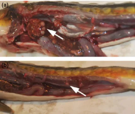
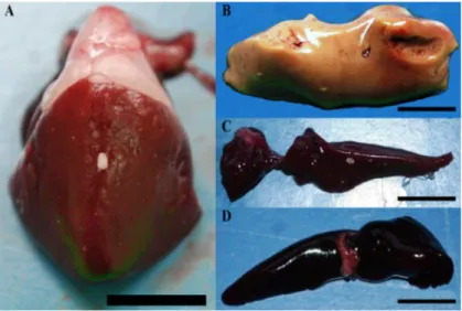
The disease may take several years to progress from the asymptomatic state to clinical illness (Snieszko, 1978; Bragg et al., 1990; Noga et al., 1990). Grossly, lesions caused by mycobacteria are gray-whitish nodules of varying diameter, disseminated in all visceral organs and in particular in the spleen, liver, kidney and gonads (Fig 1.11). Nodules can also be observed in the gills between lamellae (Gauthier & Rhodes, 2009).
Fig. 1.11 Visceral organs of a Russian sturgeon infected by Mycobacterium
salmoniphilum. (a) Multiple white roundish nodules are clearly visible in the liver
(white arrow). (b) Irregular nodules in shape are dispersed throughout kidney (white arrow) (photo by Righetti et al., 2014).
Microscopically atypical mycobacteria are pleomorphic rods of 1.5 X 0.25 micron in size. Typical granulomas are composed of epithelioid cells surrounded by a connective capsule of varying thickness, often associated with an area of central necrosis with a variable amount of acid-fast bacilli evident with Ziehl-Neelsen stain (Astrofsky et al., 2000; Decostere et al., 2004) (Fig 1.12).
Fig 1.12 Microscopic aspect of granuloma due to atypical mycobacteria in fish. (a)
Liver: granulomas are composed of epithelioid cells (arrow head) surrounded by a connective capsule often associated with an area of central necrosis (arrow). (b) Granuloma with fast rod shape bacilli positive to Ziehl-Neelsen.
Furthermore, as experimentally observed by Colorni et al. (1998) and recently by Ortega et al. (2014) and Antuofermo et al. (2016) granulomas can be also histologically classified in distinct evolutionary stages.
According to the histological pattern granulomas can be differentiated in relation to the disease development and they can be classified as subacute and chronic granulomas. Firstly granuloma is represented by an infiltration of flat epithelioid cells surrounding necrotic foci within it is possible to detect the presence of alcohol-acid resistant bacilli. Later evolutionary stage granulomas are characterized by the presence of a central portion of coagulative necrosis where there are colonies of mycobacteria, surrounded by epithelioid cells and by more concentric layers of fibroblasts. Chronic granulomas not always allow the identification of bacterial colonies. For a final diagnosis of mycobacteriosis, however, isolation and identification of the microorganism species are necessary since genus of mycobacteria is highly heterogeneous in terms of epidemiology and pathogenicity.
At this scope, specific media and phenotypical tests are commonly used. The classical method still used today is based on the phenotypic approach that unlikely takes a long
time (several weeks are required to mycobacteria for growth and achievement of their metabolic activity, which is essential to address the biochemical tests), and does not always produce a precise identification (Kent & Kubica, 1985). The cultural examination for the detection of mycobacteria, prepared on specific solid media (Lowenstein-Jensen and Stonebrink) are evaluated for morphology, temperature (28, 37, 43°C), speed of growth, and the ability to produce pigment (Fig. 1.13).
Fig. 1.13 Photo-induction show chromogenic colonies of M. marinum (right tube)
(photo by Florio et al., 2003).
These classical methods, however, are not always conclusive to identify all mycobacteria species. The culture condition can influence morphology and biosynthesis and sometimes the presence of strain varieties can alter the typicalbiochemical profile of mycobacteria (Ucko et al., 2002 and references therein). Mycobacterium marinum is one of the main fish pathogen described in association to zoonotic disease, thus the ability to distinguish among different strains remains of great importance (Haenen et al., 2013). So, different molecular techniques based on specific markers amplification and sequencing has been developed (Srinivasan et al., 2014).
In particular, the sequence comparison between stable parts of the bacterial genetic code allow at the same time bacterial identification and phylogenetic classification (Clarridge, 2004). For this reason, sequence analysis of the 16S rRNA gene has been widely used over the last two decades to establish phylogenetic relationships of bacteria, opening the way to molecular epidemiologic analysis. As previously reported PCR amplification and direct sequencing of 16S rRNA products allows proper taxonomic assignment in Mycobacterium species. (Ucko et al., 2002 and references therein). The 16S rRNA gene is a stable part of the bacterial genome composed by both
conserved and variable sequence regions (Srinivasan et al., 2015). The conserved regions sequences represent an useful tool to establish bacterial taxonomic levels, while the considerable sequence diversity in the 16S rRNA variable regions represents the most important current target for species classification (Chakravorty et al., 2007; Větrovský & Baldrian, 2013). However, the wealth of data available from public sequence databases highlighted that the comparison of the 16S rRNA sequence is often insufficient in closely related species (Ucko et al., 2002 and references therein).
For this reason, the protein-encoding genes analysis may be more discriminative than those encoding rRNA, and the highly conserved hsp65 gene of Mycobacterium species represents a useful candidate for this purpose, showing species-specific variations sequence (Ucko et al., 2002 and references therein). Nevertheless, hsp65 gene reveals higher sequence variability within the genus. However mycobacteria species are numerous and in order to identify species restriction enzyme digestion of PCR products by BstEII and HaeIII and separation of the restriction fragments on an agarose gel is required. Sequencing restriction pattern allow identifying different mycobacterial species (Telenti et al., 1993; Toranzo et al., 2005).
Prophylaxis is the best measure to prevent the transmission of atypical mycobacteria in fish due to the high resistance of these microorganisms in the environment. Formalin, sodium hypochlorite or phenolic compounds are necessary for the disinfection of the tanks in which there has been an infection with mycobacteria (Thoen & Schliesser, 1984). Additionally it is important to reduce overcrowding in fish farming or aquarium and immediately remove sick and dead fish. Also any importation of new individuals should be subjected to a quarantine period of 4-8 weeks (Belas et al., 1995). Treatments of affected individuals are not recommendable because atypical mycobacteria are resistant to the common chemotherapeutic agents used to treat human tuberculosis (Conroy & Solarolo, 1965).
All fish must be considered susceptible to mycobacteriosis, with a higher prevalence in farmed fish, where there may be significant losses (Gauthier & Rhodes, 2009). Anyway mortality in farmed fish is never on a large scale, but it is usually expressed in outbreaks (Gauthier et al., 2008). The spreading of the disease may be related to improper farm management that can induce a decrease in the resistance of the fish (Chinabut, 1999). Some fish species are still poorly studied and mycobacteriosis descriptions in mugilidi, are reported in the literature only sporadically. At present, acid-fast bacterial infections in wild Mugilidae are scarcely reported worldwide: Osman (1980) observed several
cases of mycobacteriosis in Liza aurata from Libya; Couch (1985) in an adult of Mugil
cephalus from the Gulf of Mexico. A recent study revealed in Venezuela the presence
of alcohol acid bacteria in 25% in Mugil curema (Aldeima et al., 2001). In Italy mycobacteriosis was detected in 2011 in mullets of the coastal lagoon in Lake Faro (Messina) and in 2014 in the eastern coast of the Ligurian Sea (Marino et al., 2012; Varello et al., 2014).
Furthermore, fish mycobacteriosis was also detected in cultured mullets (Aldeima et al., 2001; Salati et al., 2010). Among the emerging diseases of bacterial origin, fish mycobacteriosis are still poorly studied in Sardinia, one of the Italian regions most devoted to extensive aquaculture activities with more than 12.000 hectares of ponds and coastal lagoons.
1.8.3 Public health
Fish mycobacteriosis is a concern for public health because atypical mycobacteria act as zoonotic agents. In humans, skin infections by non-tuberculous mycobacteria are uncommon, but their importance has changed over the past years (Giavenni, 1980; Ghittino, 1985; Ghittino & Bozzetta, 1994; Zanoni et al., 2008) and has reached epidemic proportions (Nichols et al., 2004). Mycobacterium marinum has been recognized as the causative agent of “swimming pool granuloma” and it was isolated for the first time from skin lesions in swimmers of a thermal pool in Sweden (Norden & Linell, 1951).
In humans it causes skin granulomatous inflammation at the extremities (hands and fingers) in areas with pre-existing injuries (Philipott et al., 1963; Adams et al., 1970; Lim et al., 2000; Lahey, 2003). Skin lesions including dermal abscesses, fistulas, suppurative granulomas or multifocal nodules with sporotrichoid eruptions. Lesion can be firstly focal of red bluish colour with a diameter of 5-6 centimetres and evolving into skin ulceration. This disease in human is known as fish tuberculosis, piscine tuberculosis, fish tank granuloma, or possibly swimming pool nodules generally caused by Mycobacterium marinum (Fig. 1.14).
Mycobacterium marinum has an optimum growth temperature of 30°C, and does not
grow well at 37°C so is often missed in hospital laboratories. Two other species of mycobacteria M. fortuitum and M. chelonae are considered as zoonotic agents. These are considered as opportunistic pathogens causing generally skin infections but also lung abscess, endocarditis, meningitis and osteomyelitis (McCracken et al., 2000).