J Oral Maxillofac Surg 70:2433-2439, 2012
Proposal of a Presurgical Algorithm for
Patients Affected by Obstructive Sleep
Apnea Syndrome
Mariangela Giarda, MD,* Matteo Brucoli, MD,† Francesco Arcuri, MD,‡ Alberto Braghiroli, MD,§ Paolo Aluffi Valletti, PhMD,储 and Arnaldo Benech, PhMD¶Purpose: To propose an algorithm for the preoperative management of patients with obstructive sleep apnea syndrome (OSAS) and review the surgical outcomes in such patients.
Materials and Methods: This prospective cohort study involved 71 patients with OSAS who underwent presurgical upper airway endoscopy and cephalometry before being assigned to treat-ment categories based on the site(s) of obstruction, the pattern of collapse, the characteristics of the soft tissue, the air space between the base of the tongue and the posterior wall of the pharynx, and the severity of OSAS. Six months after surgery, they were followed up using polysomnography and the Epworth Sleepiness Scale. The pre- and postsurgical data were compared using a paired Student
ttest.
Results: The mean preoperative apnea/hypopnea index of the 71 patients (61 male and 10 female) was 40.98 events/hour (range, 14.7 to 87.6 events/hr), and the mean postoperative apnea/hypopnea index was 13.96 events/hour (range, 0 to 20 events/hr). The difference was statistically significant (P⬍ .001).
Conclusions: This algorithm was developed on the principle that every patient with OSAS should be considered individually. In the authors’ opinion, taking into account the number, site(s), pattern, and degree of the collapse/obstruction is a reasonable means of ensuring the correct diagnosis and treatment. ©2012 American Association of Oral and Maxillofacial Surgeons
J Oral Maxillofac Surg 70:2433-2439, 2012
Obstructive sleep apnea syndrome (OSAS) is character-ized by repetitive episodes of apnea/hypopnea during sleep, which are responsible for abrupt awakenings and a disrupted sleep microarchitecture. The obstructive episodes can also lead to major systemic complications
over time, such as pulmonary and systemic hyperten-sion, right heart failure, acute cerebrovascular accidents, ischemic heart disease, and arrhythmias. Furthermore, fragmented sleep can have major repercussions on day-time activities owing to sleepiness, fatigue, and mental disturbances and increase the risk of traffic and working accidents.1,2
During sleep, patients with OSAS develop a nega-tive pharyngeal transmural pressure that causes upper airway collapse. The caliber of the upper airway is smaller in patients with OSAS because of the effects of chronic snoring on the pharyngeal soft tissue (swell-ing and chronic inflammation), the deposition of ad-ipose tissue in the throat area, and skeletal abnormal-ities (micrognathia and mandibular hypoplasia). The obstruction can occur at any level: the most fre-quently affected sites are the retropalatal and retrolin-gual regions (isolated nasal narrowing is not respon-sible for the genesis of the disorder), and there is frequently more than 1 site of collapse.3-6
The treatment of choice is continuous positive airway pressure during sleep, which expands the Received from the Azienda Ospedaliera Maggiore della Carità,
Uni-versity of Piemonte Orientale, “Amedeo Avogadro”, Novara, Italy. *Resident, Department of Maxillofacial Surgery.
†Assistant, Department of Maxillofacial Surgery. ‡Resident, Department of Maxillofacial Surgery.
§Assistant, Department of Sleep Medicine, “Fondazione Salvatore Maugeri”, Veruno, Italy.
储Assistant, Department of Otorhinolaryngology.
¶Chairman and Professor, Department of Maxillofacial Surgery. Address correspondence and reprint requests to Dr Giarda: SCDU di Chirurgia Maxillo-Facciale, Ospedale Maggiore della Carità, Corso Mazzini 18, 28100 Novara, Italy; e-mail: mariangelagiarda@ yahoo.it
©2012 American Association of Oral and Maxillofacial Surgeons
0278-2391/12/7010-0$36.00/0 doi:10.1016/j.joms.2011.11.009
pharyngeal lumen in all directions and prevents upper airway collapse, thus leading to major bene-fits in morbidity and quality of life.7-10 The de-scribed side effects (a feeling of choking, dry
mouth, conjunctivitis, rhinitis, pressure ulcers, and bloating) are not serious and can be easily managed. However, the problem with ventilation therapy is poor compliance, because patients find the mask
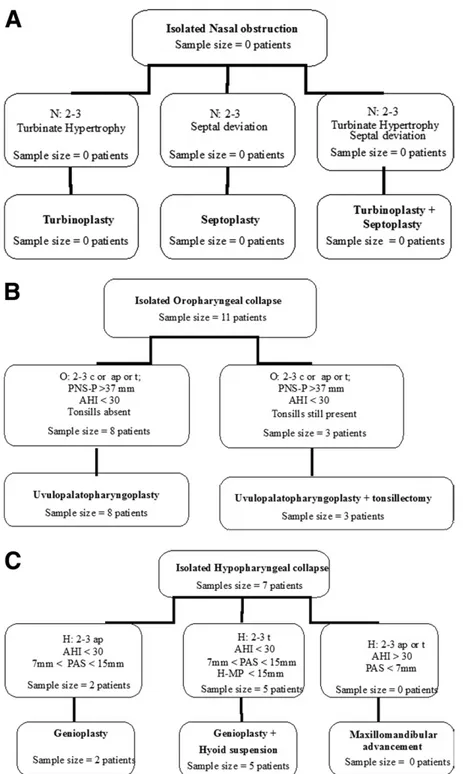
FIGURE 1. A to G, Preoperative algorithms, with 2 examples to facilitate reading. A, For an isolated grade 2 to 3 nasal obstruction, the authors
recommend turbinoplasty if the turbinates are hyperplastic, septoplasty for a septal deviation, and turbinoplasty plus septoplasty for a septal deviation and turbinate hypertrophy. B, For an isolated grade 3 to 4 circular, anteroposterior, or transversal oropharyngeal collapse identified by endoscopy, with an increased distance from the posterior nasal spine to point P (⬎37 ⫾ 3 mm), the authors recommend an isolated uvulopalatopharyngoplasty in the absence of tonsils or uvulopalatopharyngoplasty with tonsillectomy if the tonsils are present. AHI, apnea/hypopnea index; ap, anteroposterior; c, circular; H, hypopharynx; H-MP, distance from hyoid bone to mandibular plane; N, nose; O, oropharynx; PAS, posterior airway space; PNS-P, distance from posterior nasal spine to point P; SNA, sella-nasion-point A; SNB, sella-nasion-point B; t, transversal; UPPP, uvulopalatopharyngoplasty.
cumbersome and annoying to use every night for a lifetime.11 Consequently, surgery is emerging as a possible treatment option, with the goal of remov-ing the cause of the obstruction.12-15
The aim of this study was to formulate a preop-erative diagnostic algorithm for patients with OSAS based on the 3 potential sites of upper airway obstruction: the nose, oropharynx, and
hypophar-FIGURE 1. (continued)
Giarda et al. Presurgical Algorithm for OSAS. J Oral Maxillofac Surg 2012.
FIGURE 1. (continued)
ynx. The preoperative hypothesis was that the sur-gical treatment of the specific site of obstruction would lead to effective postoperative results and avoid useless treatments. The authors therefore re-viewed the surgical outcomes using the pre- and postoperative apnea/hypopnea index (AHI) and the Epworth Sleepiness Scale (ESS) as clinical parame-ters.
Materials and Methods
After creating a comprehensive flow chart based on the preoperative site of obstruction (Fig 1), the authors designed a prospective cohort study and enrolled patients with OSAS. The inclusion criteria were an age younger than 70 years, good general health, an intolerance to continuous positive air-way pressure, and a motivation to solve the
prob-lem; the exclusion criteria were cardiovascular dis-eases, diabetes, and psychiatric disorders.
The presurgical diagnostic algorithm involved upper airway endoscopy and cephalometry. The sites of collapse were evaluated using the endo-scopic nose, oropharynx, and hypopharynx classi-fication16 to identify the site(s) of collapse (nose, oropharynx, and/or hypopharynx), the pattern of collapse (circular, transversal, and/or anteroposte-rior) and the degree of collapse (grades 0 to 4;
Table 1). The cephalometric examination was per-formed using lateral-lateral teleradiography. Skele-tal relations were evaluated by angular measure-ments (sella-nasion-point A and sella-nasion-point B), and the soft tissue measurements included the air space between the base of the tongue and the posterior wall of the pharynx (posterior airway space), the length of the soft palate (distance from the posterior nasal spine to point P), and the dis-tance between the hyoid bone and the mandibular plane.13
Based on all the characteristics and the severity of the OSAS, the subjects were assigned to different treatments (Fig 1). The surgical procedures in-cluded septoplasty, turbinoplasty, genioplasty, maxillomandibular advancement, and uvulopalato-pharyngoplasty (UPPP), associated or not with ton-sillectomy and/or hyoid suspension, performed alone or in various combinations to ensure an op-timal outcome for each patient.
Six months after surgery, the patients were clin-ically followed by polysomnography and the ESS. A paired Student t test was used to compare the pre-with the postsurgical AHI (apnea/hypopnea events per hour) and the pre- with the postsurgical ESS scores.
The study was approved by the institutional review board.
Table 1. ENDOSCOPIC NOSE, OROPHARYNX, AND HYPOPHARYNX CLASSIFICATION Sites of collapse N Nose O Oropharynx H Hypopharynx Pattern of collapse c Circular t Transversal ap Anteroposterior Grade of collapse 0 No collapse/obstruction 1 Collapse⬍25% of pharyngeal lumen 2 Collapse 25%–50% 3 Collapse 50%–75% 4 Collapse⬎75%
Giarda et al. Presurgical Algorithm for OSAS. J Oral Maxillofac Surg 2012.
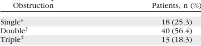
Table 2. SITES OF COLLAPSE/OBSTRUCTION SHOWN BY FIBROENDOSCOPY USING THE MUELLER
MANEUVER WITH THE PATIENT IN A SUPINE POSITION Site(s) of Collapse/ Obstruction Patients, n (%) Single* 18 (25.3) Double† 40 (56.4) Triple‡ 13 (18.3) *Isolated nasal obstruction, isolated oropharyngeal collapse, or isolated hypopharyngeal collapse.
†Nasal obstruction plus oropharyngeal collapse, nasal ob-struction plus hypopharyngeal collapse, or oropharyngeal plus hypopharyngeal collapse.
‡Nasal obstruction plus oropharyngeal collapse plus hy-popharyngeal collapse.
Giarda et al. Presurgical Algorithm for OSAS. J Oral Maxillofac Surg 2012.
Table 3. SITES OF COLLAPSE/OBSTRUCTION SHOWN BY FIBROENDOSCOPY USING THE MUELLER
MANEUVER WITH THE PATIENT IN A SUPINE POSITION
Site(s) of Obstruction/Collapse
Patients, n (%)
Isolated nasal site 0 (0)
Isolated oropharyngeal site 11 (15.5)
Isolated hypopharyngeal site 7 (9.8)
Oropharyngeal and hypopharyngeal sites 28 (39.5)
Oropharyngeal and nasal sites 8 (11.3)
Hypopharyngeal and nasal sites 4 (5.6)
Oropharyngeal, hypopharyngeal, and nasal sites
13 (18.3)
Giarda et al. Presurgical Algorithm for OSAS. J Oral Maxillofac Surg 2012.
Results
One hundred forty-two patients (132 male and 10 female; mean age, 50.5 yrs; age range, 35 to 70 yrs) underwent the multidisciplinary preoperative evalua-tion; 116 were considered suitable for surgical treat-ment, but 45 patients declined, so 71 patients (61 male and 10 female) were actually treated. The mean preoperative AHI was 40.98 events/hour (range, 14.7 to 87.6 events/hr), and the mean preoperative ESS score was 15 (range, 9 to 17).
Upper airway fibroendoscopy using the Mueller maneuver with the patient in a supine position iden-tified an isolated obstruction site in 18 patients (25.3%), a double obstruction in 40 (56.4%), and a triple obstruction in 13 (18.3%;Tables 2,3).
The mean postoperative AHI was 13.96 events/ hour (range, 0 to 20.5 events/hr), and the mean post-operative ESS score was 5 (range, 0 to 7). The data were analyzed using a paired Student t test, which showed that the differences were statistically signifi-cant (P ⬍ .001).
Discussion
The decision algorithm was developed on the prin-ciple that every patient with OSAS should be consid-ered individually and seems to have been validated by the significant decreases in the postoperative AHI and the ESS scores.
It is widely accepted that apneic events in patients with OSAS are related to the collapse of a narrow upper airway, which is not isolated in most cases and frequently involves multiple regions. The goal of sur-gery is to remove the cause of the obstruction by acting on the involved sites.
The diagnostic algorithm, which is based on an upper airway endoscopic examination and a cephalo-metric analysis, confirmed the frequent involvement of multiple sites in 67.5% of cases (52% with 2 sites and 15.4% with 3 sites;Table 2). Based on the results of the multidisciplinary examinations, the patients were offered 1 of many treatment options (Fig 1). Nasal obstruction indicates nasal surgery (septoplasty and/or turbinoplasty; Fig 1A). However, although 35.2% of patients showed nasal involvement, none of them had an isolated nasal obstruction (septal devia-tion, turbinate hypertrophy) capable of generating apnea (Table 3).
Oropharyngeal collapse identified by endoscopy (grades 3 to 4) and an increase in distance from the posterior nasal spine to point P (⬎37 ⫾ 3 mm) at cephalometry indicate UPPP (with tonsillectomy when the tonsils are present;Fig 1B).
Hypopharyngeal collapse identified by endoscopy (grades 3 to 4) and confirmed by a decrease in
pos-terior airway space (⬍11 ⫾ 1 mm) suggests genio-plasty. If the collapse has a transversal pattern and is associated with a decrease in distance between the hyoid bone and the mandibular plane (⬍15 ⫾ 3 mm), the authors propose genioplasty and hyoid suspen-sion in the same surgical sessuspen-sion (Fig 1C).
Maxillomandibular advancement with or without other procedures (septoplasty, UPPP, etc) is pro-posed for patients with moderate or severe disease, multiple obstruction sites, or alterations in the rela-tion of the jaws. It has positive effects for circular and transversal patterns of collapse (Fig 1C-G).
Even if it includes multiple procedures, the treat-ment plan is performed during the same surgical ses-sion because multiple sesses-sions discourage the pa-tients.
The palatal component was involved in the gen-esis of OSAS in 84.6% of patients, which is in line with published data indicating the pivotal role of the retropalatal region, but was isolated in only 15.5% of cases (Table 3). In patients with oropha-ryngeal collapse, the flaccidity of the palate causes its posterior displacement during sleep in a supine position. The hypopharyngeal components were solely responsible for OSAS in only 9.8% of cases, but were jointly involved in 73.2% (Table 3), which may explain why UPPP alone is often insufficient to control apnea. This large percentage of multiple collapse sites supports the need for therapeutic procedures selected based on an individual patient to ensure long-term success.
Upper airway fibroendoscopy during the Mueller maneuver is a simple means of morphologically analyzing the nasal cavity and the retropalatal and retrolingual regions. Although it is a subjective pro-cedure, the use of video recordings allows the find-ings to be assessed and discussed by different phy-sicians.16
The use of cephalometry to study OSAS in pa-tients was introduced by Riley et al.13 It is easy to perform, does not expose patients to a significant dose of radiation, and allows the analysis of the soft tissue and bony structures of the craniocervical region. However, it is limited because it provides only an anteroposterior view of the airway and does not show the presence of any lateral-lateral narrow-ing. Furthermore, because it is performed with the patient awake and standing, it does not allow any evaluation of the postural factors present during sleep.13
The published results of the surgical treatment of OSAS are divided into “surgical successes” and “sur-gical cures.” Sur“sur-gical success is defined as an AHI of fewer than 20 events/hour or a greater than 50% decrease in the AHI, and surgical cure is defined as an AHI of fewer than 5 events/hour.12 The authors
observed a considerable decrease in the AHI, which decreased from 40.98 to 13.96 events/hour. How-ever, because the authors considered the results of the different surgical procedures in the same statis-tical analysis, a more specific and comprehensive statistical evaluation is essential.
Although the present study has the weaknesses and limitations of a descriptive report and there was no control group, the authors believe that it provides a reasonable reflection of the usefulness of the present standard diagnostic flow chart for patients with OSAS, which was developed by a multispecialty team of otorhinolaryngologists, pulmonologists, maxillofacial surgeons, and anesthesiologists, allows an integrated approach, and allows the targeting of a specific sur-gical procedure to an individual case.
OSAS has a multifactorial etiology/pathogenesis and requires a multidisciplinary approach to reach a cor-rect diagnostic definition. The authors believe that the preoperative evaluation of patients with OSAS using a comprehensive diagnostic algorithm that takes into account the different sites of obstruction is a reasonable means of simplifying the complex pre-surgical approach to OSAS. However, further pro-spective clinical studies, possibly associated with more sophisticated clinical and radiologic parameters, are necessary to refine it.
References
1. Young T, Peppard PE, Gottlieb DJ: Epidemiology of obstructive sleep apnea: A population health perspective. Am J Respir Crit Care Med 165:1217, 2002
2. Schellenberg JB, Maislin G, Schwab RJ: Physical findings and the risk for obstructive sleep apnea. The importance of oropharyngeal structures. Am J Respir Crit Care Med 162:740, 2000
3. Remmers JE, deGroot WJ, Sauerland EK, et al: Pathogenesis of upper airway occlusion during sleep. J Appl Physiol 44:931, 1978
4. Hoffstein V, Mateika S: Differences in abdominal and neck circumferences in patients with and without obstructive sleep apnoea. Eur Respir J 5:377, 1992
5. Mortimore IL, Marshall I, Wraith PK, et al: Neck and total body fat deposition in nonobese and obese patients with sleep apnea compared with that in control subjects. Am J Respir Crit Care Med 157:280, 1998
6. Jamieson A, Guilleminault C, Partinen M, et al: Obstructive sleep apneic patients have craniomandibular abnormalities. Sleep 9:469, 1986
7. Engleman HM, Douglas NJ: Sleep. 4: Sleepiness, cognitive func-tion, and quality of life in obstructive sleep apnoea/hypopnoea syndrome. Thorax 59:618, 2004
8. Haentjens P, Van Meerhaeghe A, Moscariello A, et al: The impact of continuous positive airway pressure on blood pres-sure in patients with obstructive sleep apnea syndrome: Evi-dence from a meta-analysis of placebo-controlled randomized trials. Arch Intern Med 167:757, 2007
9. Barnes M, Houston D, Worsnop CJ, et al: A randomized controlled trial of continuous positive airway pressure in mild obstructive sleep apnea. Am J Respir Crit Care Med 165:773, 2002 10. Ballester E, Badia JR, Hernández L, et al: Evidence of the
effectiveness of continuous positive airway pressure in the treatment of sleep apnea/hypopnea syndrome. Am J Respir Crit Care Med 159:495, 1999
11. McArdle N, Devereux G, Heidarnejad H, et al: Long-term use of CPAP therapy for sleep apnea/hypopnea syndrome. Am J Respir Crit Care Med 159:1108, 1999
12. Bettega G, Pepin J, Veale D, et al: Obstructive sleep apnea syndrome. Fifty-one consecutive patients treated by maxillofa-cial surgery. Am J Respir Crit Care Med 162:641-9, 2000 13. Riley RW, Powell NB, Guilleminault C: Obstructive sleep apnea
syndrome: A surgical protocol for dynamic upper airway recon-struction. J Oral Maxillofac Surg 51:742, 1993
14. Borowiecki BD, Sassin JF: Surgical treatment of sleep apnea. Arch Otolaryngol 109:508, 1983
15. Elshaug AG, Moss JR, Southcott AM, et al: Redefining success in airway surgery for obstructive sleep apnea: A meta analysis and synthesis of the evidence. Sleep 30:461, 2007
16. Vicini C, Mira E: NOH Classification: A Proposal. Atti del XLVII Raduno Gruppo Alta Italia di Otorinolaringoiatria. Riccione, 2001, pp 155-163