Edited by: Marilena Valeria Iorio, Istituto Nazionale dei Tumori (IRCCS), Italy Reviewed by: Francesco Russo, Novo Nordisk Foundation Center for Protein Research, University of Copenhagen, Denmark Ajaikumar B. Kunnumakkara, Indian Institute of Technology Guwahati, India *Correspondence: Stefano Volinia [email protected]
Specialty section: This article was submitted to Cancer Molecular Targets and Therapeutics, a section of the journal Frontiers in Oncology Received: 01 June 2018 Accepted: 31 July 2018 Published: 29 August 2018 Citation: Corrà F, Agnoletto C, Minotti L, Baldassari F and Volinia S (2018) The Network of Non-coding RNAs in Cancer Drug Resistance. Front. Oncol. 8:327. doi: 10.3389/fonc.2018.00327
The Network of Non-coding RNAs in
Cancer Drug Resistance
Fabio Corrà, Chiara Agnoletto, Linda Minotti, Federica Baldassari and Stefano Volinia*
Department of Morphology, Surgery and Experimental Medicine, University of Ferrara, Ferrara, Italy
Non-coding RNAs (ncRNAs) have been implicated in most cellular functions.
The disruption of their function through somatic mutations, genomic imprinting,
transcriptional and post-transcriptional regulation, plays an ever-increasing role in cancer
development. ncRNAs, including notorious microRNAs, have been thus proposed to
function as tumor suppressors or oncogenes, often in a context-dependent fashion.
In parallel, ncRNAs with altered expression in cancer have been reported to exert a
key role in determining drug sensitivity or restoring drug responsiveness in resistant
cells. Acquisition of resistance to anti-cancer drugs is a major hindrance to effective
chemotherapy and is one of the most important causes of relapse and mortality in
cancer patients. For these reasons, non-coding RNAs have become recent focuses
as prognostic agents and modifiers of chemo-sensitivity. This review starts with a brief
outline of the role of most studied non-coding RNAs in cancer and then highlights
the modulation of cancer drug resistance via known ncRNAs based mechanisms. We
identified from literature 388 ncRNA-drugs interactions and analyzed them using an
unsupervised approach. Essentially, we performed a network analysis of the non-coding
RNAs with direct relations with cancer drugs. Within such a machine-learning framework
we detected the most representative ncRNAs-drug associations and groups. We finally
discussed the higher integration of the drug-ncRNA clusters with the goal of disentangling
effectors from downstream effects and further clarify the involvement of ncRNAs in the
cellular mechanisms underlying resistance to cancer treatments.
Keywords: non-coding RNAs, chemoresistance, drug sensitivity, miRNA, lncRNA, cancer, gene networks
miRNAs AND DRUG RESISTANCE IN CANCER
Chemotherapy represents the primary treatment for both early and advanced tumors. However,
drug resistance seriously limits the potency of conventional chemotherapeutics and novel biological
agents, this constitutes a major obstacle in the treatment of cancer (1). Then, a lot of effort is aimed
to identify new biomarkers, and to assess and predict the response of patients to drugs (2). Cancer
drug resistance is referred as intrinsic, if tumors demonstrate to be insensitive to therapeutic agents
before treatment, otherwise it is defined acquired if tumor becomes resistant during the treatment.
The acquisition of resistance to several types of anticancer drugs can be due to the expression
of transporters that eject drugs from cells, resulting in multidrug resistance (3). Nevertheless,
several other mechanisms are involved in resistance, including insensitivity to apoptosis induced
by drugs, increased repair of damaged DNA, decreased intracellular accumulation of therapeutics,
and induction of mechanisms capable of drug detoxification (1). Recent data showed that other
than by genetic and epigenetic changes, such as base mutations, amplifications, methylation and
other post-translational modifications, drug resistance might also
be due to non-coding RNA (ncRNAs) (4). The bulk of the human
transcriptome, excluding the ribosomal and mitochondrial RNA,
is represented by non-coding transcripts, including the most
studied miRNAs and the newly discovered long non-coding
RNAs (lncRNA) (5). MicroRNAs (miRNA) are small
non-coding RNA molecules (18–22 nt in length) that act as negative
regulators of gene expression through modulation of multiple
target mRNAs, by inhibition of translation (6–9). A number of
miRNA genes are located within intronic regions of genes, both
coding or non-coding for proteins and can be transcriptionally
regulated through their promoters (10). Other miRNAs are
found either within exons, including 3
′UTRs of mRNAs, or
clustered with other miRNA genes (11). Since their discovery
(12,
13), the number of annotated miRNAs in the human
genome has grown rapidly and they regulate a variety of cellular
processes, including apoptosis (14), differentiation (15) and cell
proliferation. miRNA deregulation has been demonstrated in
cancer (16–19). The role of miRNAs in controlling cellular
proliferation, differentiation and apoptosis, and their location
at sites of translocation breakpoints or deletions (20), suggests
that they might function as tumor suppressors or oncogenes
(21–23). Profiles of miRNA expression differ between normal
and tumor tissues, and among tumor types (18,
24–27). The
association of miRNAs with cancer was first revealed in chronic
lymphocytic leukemia (CLL), upon the discovery that
miR-15a and miR-16-1 were frequently deleted or down-regulated
(16,
28), and that their expression was inversely correlated to
that of BCL2 (29). Since then, numerous studies have provided
evidence for changes in microRNA expression in oncogenesis:
different cancer pathways can converge to affect the same
miRNAs and conversely a single miRNA can control an entire
transcriptional program, affecting a lot of target genes. The
deregulation of miRNAs is linked to cancer progression and
clinical outcome (30), and miRNAs have been proposed as
potential diagnostic markers, prognostics factors, and therapeutic
targets (27,
31–33). When aberrant microRNA expression is
directly involved in carcinogenesis (21), the inhibition of
selectd microRNAs may have therapeutic implications. Modified
antisense oligonucleotides have been designed ad-hoc and have
proven effective at inhibiting microRNA function in vivo in
mice (34,
35). The association of microRNA expression with
cancer prognosis, therapeutic outcome and response to therapy,
independently of other clinical covariates has been documented
(25,
26,
36,
37), and selected miRNAs may influence cancer
response to chemotherapy (38). The prognostic potential of
microRNAs has been demonstrated for CLL (37), lung cancer
(39), pancreatic cancer (25), and neuroblastoma (40) among
others. One of the firsts observation on a possible link between
miRNAs and drug resistance was reported in breast cancer (BC)
suggesting that increased sensitivity of patients to
anthracycline-based chemotherapy was related to deletion of chromosome 11q,
a region containing MIR125B1 (41). The effect of miRNAs on
chemotherapy was systematically studied by Blower et al. (42)
on NCI-60, a panel of 60 cancer cell lines, used by the National
Cancer Institute to screen >100,000 chemical compounds for
anticancer drug sensitivity (20,
38,
42). Overall, miRNAs can
mediate drug resistance through multiple pathways, including: (i)
cell cycle and proliferation control, (ii) survival and/or apoptosis
signaling pathways, (iii) DNA repair systems, (iv) specific
drug targets, (v) adenosine triphosphate–binding cassette (ABC)
transporter proteins, and/or drug metabolism, (vi) the epithelial–
mesenchymal transition (EMT) process (4,
6,
43,
44). For
example, miR-15b, miR-16 and miR-22 have been documented
as mechanisms in chemotherapy resistance (45,
46). Cell cycle
deregulation by miRNAs can induce resistance in cancer cells, as
confirmed for miR-224 (47). Also, miR-24 and miR-508-5p can
directly target enzymes involved in drug metabolism (48,
49).
In addition to the mechanisms described above, modulation of
epithelial-mesenchymal transition (EMT) can exert an effect on
cancer cell resistance. Importantly, once cancer cells undergo
EMT, chemo-resistance is increased and metastasis can occur
(50,
51). Normal stem cells are already more resistant to drug
treatment due to over-expression of drug efflux pumps and
anti-apoptotic proteins (52). In this context, miR-34, miR-125b,
miR-140, and miR-215 have an important role in conveying
drug resistance to cancer stem cells (2). Chemotherapy can
induce EMT and modulate the expression of miR-448 to promote
cancer cell progression (53); conversely miR-29c or miR-224 have
recently been shown to regulate the EMT process (54). miRNome
dysregulation in relation to chemotherapy has been described for
the most common tumor types: breast, ovarian, lung, prostate,
gastric and colon cancer, squamous and hepatocellular carcinoma
(HCC), cholangiocarcinoma, neuroblastoma and various types of
leukemia (55). Overall, these studies highlight the complexity of
adaptive/selective mechanisms in the establishment of resistance
to cancer therapies.
lncRNAs AND DRUG RESISTANCE IN
CANCER
lncRNAs have been linked to cancer progression and metastasis
(56), and recently intensive research has been devoted to
the molecular dissection of their roles, as well as to their
diagnostic and prognostic significance (57). lncRNAs are
mRNA-like transcripts 200 nt to ∼100 kb in length lacking significant
open reading frames. lncRNAs can be transcribed by RNA
polymerase II (RNA pol II), poly-adenylated and located within
nuclear or cytosolic fractions (58). lncRNAs can be divided
into different categories: if overlapping with any transcript
on sense or anti-sense strand lncRNAs will be classified as
(i) sense or (ii) antisense respectively. When its expression
is initiated along with a neighboring transcript, sense or
antisense, that is proximal, (iii) bidirectional. When deriving
from an intronic region, (iv) intronic or (v) intergenic if
placed between two genes (53). Generally, lncRNA expression
levels appear to be lower than those of protein-coding
genes (54), and lncRNAs might be preferentially expressed
in specific tissues (59). As to their functions, lncRNAs can
regulate the expression of genes in close proximity (cis-acting
regulation) or can target distant transcriptional activators or
repressors (trans-acting) (53,
60). Their mechanisms of action
are still diverse, and have been associated with a spectrum
of biological processes, for example, epigenetics, alternative
splicing, nuclear import, structural components, precursors to
small RNAs and regulators of mRNA decay (60–63). Thus
lncRNAs can regulate cellular functions such as chromosome
dosage compensation (64), imprinting (65), cell cycle progression
(66) and differentiation (67). Aberrant regulation of lncRNAs
is reported in a variety of diseases, including cancer (68–
71). Accumulating reports of misregulated lncRNA expression
across numerous cancer types suggest that also this class of
ncRNA can act in oncogenesis and tumor-suppression (72). A
number of useful databases providing molecular information on
lncRNAs are available (73). Loss of imprinting and redirecting
chromatin remodeling complexes (74), induction of metastasis
(75), depletion of miRNAs as “molecular decoy” or “miRNA
sponge” (76) and direct inactivation of tumor suppressor
genes (77) have been referred to specific lncRNAs. Preliminary
studies commenced to report the value of ncRNAs as potential
biomarkers in clinical settings (78,
79) and their roles in drug
resistance (80).
A NETWORK ANALYSIS: THE MOST
CENTRAL ncRNAs IN CHEMORESISTANCE
In recents years, an increasing number of studies have been
reported on ncRNAs, target gene modulation, and affected drug
functions, pharmacogenomics or chemoresistance. With the aim
to facilitate the classification of ncRNAs and drug targets, some
databases have been developed, such as NRDT (81) or
Pharmaco-miR (82), collecting all information about ncRNA-target
gene-drugs. There are large numbers, and growing, of both ncRNAs
and cancer drugs, thus the combinations between members of
the two groups are very difficult to manage in a traditional
review or interpretate in a database. Therefore we decided
to use machine-learning systems and to study the RNA-drug
interactions using a network-based approach. Basically, we took
from KEGG database all approved drugs used for cancer therapy.
Then, we searched in PubMed all recent studies (published from
2011 onwards) investigating ncRNAs in chemoresistance. This
selection was performed by batch analysis of PubMed-NCBI
(National Center for Biotechnology Information) using as major
topics the drugs from KEGG, ncRNA and chemoresistance.
The result of this screening was manually curated in order
to avoid and remove papers with generic statements and not
direct links between ncRNAs and drugs. Only the investigations
that proved (by in vitro/in vivo) experiments the existence
of a direct association between ncRNAs and chemoresistance
were then analyzed using a machine-learning tool. We thus
built a network of non-coding RNAs starting from a
human-curated selection of papers and applied an ad-hoc data mining
approach to dissect the network and identify the most important
ncRNA/cancer drugs interactions and cliques. We obtained
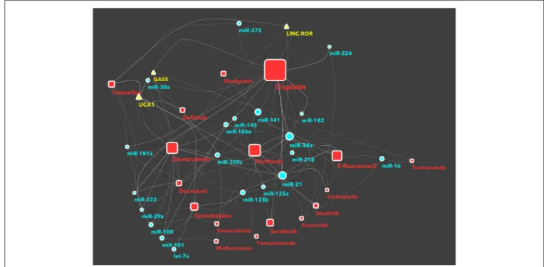
a fully connected network of 388 drug/ncRNA interactions
(edges) and 5 unconnected pairs (Supplementary Image 1).
We then went on with studying the network, which had
227 nodes: 150 miRNAs, 35 lncRNAs and 42 drugs. Three
graph theory measures were considered to define the most
relevant non-coding RNAs associated to therapeutics resistance:
(i) degree, indicating the number of links that an ncRNA had
with different nodes (here drugs) (ii) betweenness centrality, a
measure of centrality in the network based on shortest paths
(iii) closeness centrality, related to the distance between the
ncRNA and all the other nodes in the network. Then, we
ranked the nodes (drugs and ncRNAs) and edges (combinations)
in the network and collected the combinations from ncRNAs
with a degree >3 and a central position (closeness centrality
> 0.26 and betweenness centrality >0.003) (Figure 1 and
Supplementary Table 1
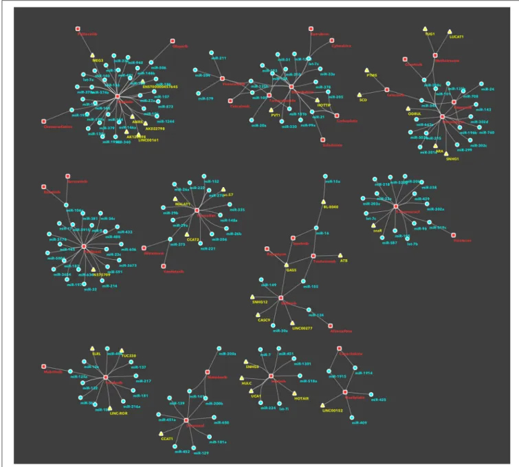
). Finally, we performed a community
structure analysis using Glay and Cytoscape (83) to identify
different clusters of ncRNAs and drugs. The clusters were
converted to subnetworks for convenient visualization. The
visual separation of clusters was improved by overlaying the
community structure on a graphic layout addressing specific
topology (Figure 2).
We wish to add a cautionary note to our reviewing effort. Even
in the genome-wide studies (a minority among those we included
in this review) for a number of conscious or unconscious reasons,
scientists often end up chasing the most “popular” ncRNAs
among others of “lesser pedigree.” Thus there is potentially a
positive bias toward well-known ncRNAs in the overall scheme,
and therefore in the final network. For this reason, we decided
to keep all associations and, although the “degree” (number of
associated drugs for an ncRNA) is important, we tried to avoid
biased selections and included in our review all ncRNAs/pairs.
Here we commence with describing the ncRNAs that are
most prominent in relation to chemoresistance, as detailed in
(Figure 1).
miR-21
has the highest scores (degree, betweenness centrality
and closeness centrality) as it was associated with several drugs.
The MIR21 gene is located at17q23.2, a region frequently
amplified in several tumors (84,
85). Its overexpression has
been observed in most cancer types and modulates the
resistance toward apoptosis-inducing drugs (86–91).
Down-regulation of miR-21 sensitizes cancer cells in vitro to
different chemotherapeutics, including cisplatin, etoposide and
doxorubicin (92–94). On the other hand, some drugs can
induce alterations in miR-21 levels: e.g. soladosine can inhibit
lung cancer cell invasion through miR-21 down-regulation,
via PI3K/Akt signaling pathway (95). Interestingly, exogenous
Epstein Barr virus modulates the PI3K/Akt pathway through
LMP1, thus enhancing miR-21 expression and contributing to
cisplatin reduced response in nasopharyngeal carcinoma (96).
Moreover, miR-21 delivered by exosomes augmented malignancy
in recipient cells and conferred paclitaxel resistance to ovarian
cancer cells (97). There was also a report for enhancement
of anticancer activity when Cao et al. reported that miR-21
induction sensitized gastrointestinal tumor cells to imatinib (98).
miR-34a
was reported to be downstream of p53 and to
function as a tumor suppressor (99). It is down-modulated in
colorectal cancer (CRC) (100). In 5-Fluorouracil (5-FU)-resistant
colon cancer cells ectopic expression of miR-34a inhibited cell
growth and attenuated the resistance to 5-FU through
down-regulation of SIRT1 and E2F3 (101), inhibition of LDHA (102)
and of c-Kit, thus reducing stem cell factor (SCF)-induced
FIGURE 1 | The network of non-coding RNAs and anti-cancer drugs. Each link between a drug and an ncRNA indicates a study in literature, investigating on the specific chemoresistance involvement of that ncRNA in cancer. The nodes (a ncRNA or a drug) shown in this figure have a degree >3, a central position in the network (expressed as betweenness centrality in the network description) >0.003, or a closer position relative to the companion drug (expressed as closeness centrality >0.26). The full network, with all nodes, is reported in supplemental information as Supplementary Image 1. Drugs are represented as red squares, miRNAs as light blue circles and lncRNAs as yellow triangles. The size of a node is proportional to its betweeenness centrality, while the size of a node name is proportional to its degree.
migration/invasion (103). Yang et al. demonstrated that
miR-34 targets BCL2 and sensitizes HCC cells to sorafenib
(104). In osteosarcoma cell lines miR-34a has been tested in
combination with celecoxib: the treatment showed decreased
cell viability, migration and invasion through regulation of the
Notch1/ROCK1-PTEN-Akt-GSK-3β axis (105). Moreover,
miR-34 could enhance the therapeutic efficacy of paclitaxel in resistant
prostate cancer (106). Its overexpression enhanced cisplatin
sensitivity, as confirmed in gastric cancer, by targeting MET
(107) and in lung cancer, through the p53/MICN axis (108).
Conversely, Pu et al. found that miR-34a overexpression in
osteosarcoma promoted resistance to several drugs (doxorubicin,
etoposide, carboplatin, cisplatin), via repression of AGTR1 (109).
The lncRNA Urothelial Cancer-Associated 1 UCA1 gene
is located at 19p13.12 (110). Different transcriptional isoforms
have been reported, UCA1 (1.4 kb), UCA1a (2.2 kb) and CUDR
(2.7 kb), generated by alternative splicing and poly-adenylated.
UCA1 is the most abundant isoform in various malignant
tumors such as bladder cancer, BC and HCC (110–113). UCA1
could promote drug resistance by directly binding to miR-204,
miR-18a and miR-16 (114). UCA1 emerged as a competitive
endogenous RNA (ceRNA) of multi-drug resistance associated
protein 1 (MDR1), inducing resistance to imatinib in CLL
cells by sequestering miR-16 (115). Overexpression of UCA1
up-regulated MDR1, resulting in imatinib resistance, whereas
its silencing had the opposite effect (116). In bladder cancer,
UCA1 enhanced chemoresistance to cisplatin by regulating Wnt
signaling (117) and to cisplatin/gemcitabine through modulation
of miR-195a (118). Recent studies reported that UCA1 regulates
tamoxifen resistance in BC (119). Liu et al. demonstrated
that the knockdown of this lncRNA could revert resistant
phenotype and increase tamoxifen sensitivity through inhibition
of the Wnt/β-Catenin pathway, thus further confirming the
oncogenic role of UCA1 in BC (120). Moreover, UCA1 was
shown to be released in exosomes by tamoxifen resistant
BC cells and increased tamoxifen resistance in ER-positive
recipient cells (121).
The members of miR-125 family (miR-125a,
miR-125b-1 and miR-miR-125b-125b-2) play an important role in tumorigenesis
and are potential biomarkers for cancer diagnosis, treatment
and prognosis in clinical settings (122). MIR125A gene is on
chromosome 19, while two separate loci on chromosomes 11
and 21 harbor MIR125B1 and MIR125B2, respectively (123).
miR-125b expression has been found negatively correlated
with 5-Fluorouracil resistance in HCC (124), while resistance
to pharmacological treatments with gentamicin, cetuximab,
doxorubicin and temozolomide by miR-125b still remains
controversial (88,
125–127). miR-125b regulates the resistance
to paclitaxel in colon cancer cells, in association with miR-125a
(128). Recent data strongly supports a relevant role for
miR-125b in conferring taxol resistance in BC, via suppression of
pro-apoptotic BCL2 antagonist killer 1 (Bak1) (129). In contrast,
in chondrosarcoma, overexpression of miR-125 enhanced the
sensitivity to doxorubicin by directly targeting ERBB2-mediated
glucose metabolism (130). miR-125a overexpression increased
the response to paclitaxel in cervical cancer, through STAT3
down-modulation (131). Sorafenib treatment in HCC showed
restoration of mir125 levels by sirtuin-7 and p21/p27 signaling
blockage inhibiting cell cycle progression (132). In AML cells,
via mubritinib, miR125a inhibited the ERBB pathway and cell
cycle proliferation and progression, suggesting that miR-125a
increased the sensitivity to the drug (133).
The MIR100 gene is at 11q24. Deregulation of
miR-100
has been reported in drug resistance; however, miR-100
expression can be either over-expressed or under-expressed
in diverse cancers (134). In ovarian cancer, miR-100 targets
mTOR therefore reverting the cell’s chemoresistance toward
cisplatin (135) and chondrosarcoma (136). In pancreatic cancer,
miR-100 mimics inhibit proliferation and increase sensitivity
to cisplatin by targeting FGFR3 (137). Recently, it has been
shown that down-modulation of miR-100 could increase
β-tubulin class V expression, promoting tumor cells proliferation,
with implications for paclitaxel resistance (138). Also,
miR-100 reduced ATM levels in a human glioma cell line (M059J)
and could sensitize tumor cells to ionizing radiation (139). In
vitro, miR-100 also induced the differentiation of BC stem cells
expressing a functional ER (140). Furthermore, in CRC cells
miR-100, together with miR-125b, negatively regulated Wnt/β-catenin
signaling, and restored responsiveness to cetuximab (125). On
the other hand, in mutant p53 pancreatic carcinoma, miR-100
up-regulation was related to gemcitabine resistance (88). In
accordance, the exosomes-mediated intercellular transfer of
miR-100, from drug resistant BC cells, could lead to resistance in
sensitive cells (141).
miR-200c
acts as a tumor suppressor, and could inhibit
the initiating steps of metastasis; a negative correlation
with ZEB factors has been reported, suggesting that this
miRNA-mediated regulatory pathway influences EMT (142–
147), potentially modulating drug resistance in advanced tumors.
miR-200c reverses resistance of lung cancer cells, both to
chemotherapeutics, like methotrexate (148), and to targeted
drugs, like crizotinib (149) and gefitinib (146,
150). In breast
and renal cancers, miR-200c could be involved in resistance or
re-sensitization to microtubule-targeting drug (151–153).
miR-141
is another member of the miR-200 family, also
involved in EMT, invasion, migration and drug resistance
(154).
miR-141
overexpression
contributes
to
acquired
chemoresistance, for both in vitro and in vivo models. The
initiation factor 4E (EIF4E) mRNA is a target of miR-141, that
is involved in drug-induced apoptosis, conferring resistance to
docetaxel-sensitive BC cells (155). miR-141 regulates cisplatin
sensitivity in non-small lung cancer cells via PDCP4 inhibition
and its inhibition increases cisplatin-induced apoptosis (156).
In oesophageal squamous cell carcinoma, miR-141 was highly
overexpressed in 5-Fluorouracil and oxaliplatin resistant cells
and contributed to acquired chemo-resistance via PTEN (157).
Moreover, in HCC cells, miR-141 was shown to confer resistance
to 5-Fluorouracil through the inhibition of KEAP1, thereby
reactivating the NRF2-dependent antioxidant pathway (158). Li
et al. discovered that miR-141 together with other miRNAs like
miR-16 contribute to prostate cancer chemoresistance via an
exosome network (159).
Two homologous microRNAs, miR-221 and miR-222, are
generally considered having an oncogenic activity (160). The
expression of miR-221 and miR-222 is highly up-regulated in
HER2/neu-positive human BCs resistant to endocrine therapy,
compared with HER2/neu-negative tissue samples (161); also,
in BC patients miR-222 is elevated in chemoresistant tissues
after surgery, compared with the pre-neoadjuvant samples
(162). miR-221/222 reduce the protein level of the cell cycle
inhibitor p27Kip1, conferring tamoxifen (161) and doxorubicin
resistance (162). Also, secreted miR-221/222 could serve as
signaling molecules and mediate communication of tamoxifen
resistance (163). Aberrant expression of miR-222 is tightly related
to poor overall survival (164) and affect oncogenic signaling
pathways associated with resistance to different drugs (165).
miR-222 also mediated BC cells resistance to adriamycin via
PTEN/Akt/FOXO1 (164). Furthermore, the exosome mediated
release of miR-222, miR-100 and miR-30a contributes to the same
effect on docetaxel and doxorubicin: loss of responsiveness in BC
cells (141). In oesophageal and prostate cancers, miR-221 could
modulate 5-Fluoruracil resistance via the Wnt/β-catenin-EMT
pathway (166) or RB1 (167), respectively.
miR-101
(168,
169) has a relevant role in autophagy.
Targeting the autophagy process is a promising therapeutic
strategy to improve chemotherapy efficiency. In BC cells
miR-101 inhibits basal autophagy, as well as etoposide- and
rapamycin-induced autophagy, thus sensitizing cancer cells
to 4-hydroxytamoxifen (4-OHT)-mediated cell death (170).
In HCC, miR-101 sensitizes cell lines to cisplatin-induced
apoptosis by targeting Mcl-1 (171). Likewise, miR-101 inhibits
autophagy and enhances chemo-sensitivity to doxorubicin of
osteosarcoma cells in vitro (172). In pancreatic cancer,
miR-101 up-regulation reverts gemcitabine resistance by inhibiting
the expression of ribonucleotide reductase M1 (RRM1) (173).
Moreover, recent studies demonstrate that miR-101 interacts
with lncRNA MALAT1 in regulatory networks that modulate
cisplatin and temozolomide resistance, in lung cancer (174) and
glioblastoma (80), respectively.
The miR-15/16 gene cluster in chromosome 13 (13q14) is
deleted or down-regulated in some cancer types (21). This
somatic alteration was reported to occur early in cancer
development and could represent a target for intervention (21).
miR-16 expression is affected by several drugs: in gastric cancer
cell lines etoposide and 5-Fluorouracil could increase the levels
of miR-16, both in vitro and in vivo, and the up-regulation of
miR-16 is modulated by p38 MAPK signaling pathway (175). In
BC, lapatinib and trastuzumab are reported to regulate miR-16
via PI3K/Akt (176). Noteworthy, the altered expression of both
miR-15a/16-1, due to the CXCR4 inhibitor BL-8040 induced the
apoptosis of AML blasts by down-regulating ERK, BCL2, MCL1
and cyclin-D1 (177).
The lncRNA GAS5, originating from the Growth
Arrest-Specific 5 gene, is down-regulated in multiple cancers. GAS5
inhibits proliferation and promotes apoptosis, thus playing a
tumor suppressor role (178). Several studies confirmed GAS5
as an mTOR effector, and its expression was directly correlated
with chemoresistance. Thus, enhancing GAS5 expression may
improve the effectiveness of rapalogues, as confirmed both in
prostate tumor cells and in mantle cell lymphoma cells (179,
180); also, the down-modulation of GAS5 caused resistance
to trastuzumab in BC (181). In lung adenocarcinoma cells
resistant to EGFR inhibitors, GAS5 enhance gefitinib-induced
cell death, via down-regulation of IGF1R (182). Lastly, in
bladder transitional cell carcinoma GAS5 inhibited malignant
proliferation and chemotherapy resistance to doxorubicin, partly
acting via BCL2 (183).
miR-106a
, a member of the miR-17 family, is associated with
poor prognosis, invasion and metastasis (184). In ovarian cancer
(OV), miR-106a inhibited cell survival and cisplatin resistance,
through downregulation of MCL1 (185); conversely expression
of miR-106a was higher in cisplatin-resistant OV. miR-106a may
be involved in the modulation of cisplatin-induced apoptosis by
regulating PDCD4 (186). In non-small cell lung cancer,
miR-106a also confers cisplatin resistance, by targeting adenosine
triphosphatase-binding cassette A1, an ABC transporter
(187). Otherwise, by targeting autophagy, miR-106a enhances
sensitivity of lung cancer cells to SRC inhibitors, including
saracatinib and dasatinib, expliciting once more the
context-dependent function of miRNAs (188). Further, dysregulation
of miR-106a conferred resistance to paclitaxel in OV; its
modulation resensitized resistant cells by targeting BCL10,
caspase-7, and ZEB1 (189). Down-modulation of miR-106a was
reported in gentamicin resistant hepatoma, participating to EMT
via the PDGF-D/miR-106a/Twist1 pathway; notably, in HCC
patients, miR-106a and Twist1 were associated with PDGF-D
expression (190).
miR-375
is involved in a positive feedback loop with ER in BC
(191) and its re-expression is sufficient to sensitize
tamoxifen-resistant cells. Furthermore, miR-375 partly reversed the EMT
process: metadherin (MTDH) was identified as a direct target of
miR-375 and tamoxifen-treated patients with higher MTDH had
a higher risk of relapse (192). Another miR-375 target is HOXB3;
miR-375 inhibited cancer stem cells (CSCs) phenotype and
tamoxifen resistance by regulating CSCs, through degradation of
HOXB3 (193). Epigenetically down-regulated miR-375 in
HER2-positive BC could induce trastuzumab resistance by targeting
IGF1R (194). 9-cis retinoic acid (Alitretinoin) modulated the
expression of 375 in BC depending on ER status: thus,
miR-375 was inhibited in ERα-positive cells while highly induced
in ERα-negative cells (195). The deregulation of miR-375 was
also observed in other malignancies: in medullary thyroid
carcinomas (MTC) miR-375 was the strongest up-regulated
miRNA (196). Vandetanib is a tyrosine kinase inhibitor for the
treatment of patients with recurrent or metastatic MTC that are
unresectable, and/or symptomatic (197). Interestingly, miR-375
over-expression associated with SEC23A down-regulation could
improve the efficacy of vandetanib (196). Thus, the expression
levels of miR-375 and SEC23A pointed to vandetanib sensitivity
and could be evaluated as predictive indicators for efficacy
of vandetanib in MTC. Analogously, up-regulation of
miR-375 increased the cisplatin-sensitivity of gastric cancer cells by
regulating ERBB2 and phospho-Akt (198).
A role in chemoresistance modulation has emerged for
putative tumor-suppressor miR-145 (199). miR-145 targeting of
MDR1 helps to restore drug efficacy in resistant cells and in vivo
models of bladder cancer and BC (200,
201). Moreover
miR-145 confirmed its role in reducing chemoresistance also with
paclitaxel (202) and doxorubicin (203), possibly via regulation of
EMT.
miR-218
has a physiological role in neuron development and
its loss of expression is involved in neurodegeneration (204). In
BC, it acts as a risk factor in ductal carcinoma in situ (DCIS)
(205). In association with platinum compounds, miR-218 and
miR-205 inhibit tumorigenesis and overcome chemoresistance
in lung cancer (206). In prostate cancer, miR-218 up-regulation
inhibited tumor growth and increased chemo-sensitivity to
cisplatin, by negatively regulating BCAT1 (207). Furthermore
miR-218 mediated autophagy and was associated with positive
response to paclitaxel in resistant endometrial carcinoma (208).
It also promoted apoptosis and caused cell cycle arrest in
CRC by targeting BIRC5, thus possibly enhancing first-line
5-FU treatment. Also, miR-218 through targeting the enzyme
thymidylate synthase (TS), enhanced 5-FU cytotoxicity in CRC
cells (209).
The let-7 family members are down-regulated in lung (210),
gastric (211), colon cancer (212) and in Burkitt’s lymphoma
(213). Loss of let-7 was associated with the shortened
post-operative survival of patients with lung cancer (210). The altered
expression of let-7a could increase chemoresistance to epirubicin
(214) and cytarabine (215). Furthermore, let-7a expression has
demonstrated to influence chemoresistance, due to maintained
treatment with gemcitabine, in pancreatic cancer patients (216,
217). Several studies have reported that let-7a acts as a tumor
suppressor in renal cell carcinoma (RCC), by targeting c-Myc
(218). let-7b and let-7e are down-regulated in glioblastoma and
ovarian cancer, respectively and promote resistance to cisplatin
by acting on the same target Cyclin D1 (219,
220). Reduced
levels of both let-7b and let-7c could determine the intrinsic
chemoresistance to 5-FU in RCC, possibly via AKT2 (221).
Clinically, 5-FU-based chemotherapy is considered moderately
effective in RCC due to rare response and severe toxicity (222);
transfection of let-7b or let-7c potentiated the efficacy of
5-FU in vitro at tolerable concentrations. Moreover, let-7c
up-regulation contributed to sensitize lung cancer cells with acquired
cisplatin resistance, by involving ABCC2 and Bcl-XL (223).
Interestingly, a combination of miR-224 and let-7i, reduced
imatinib resistance in CML, probably through targeting the
ST3GAL IV sialyltransferase (224).
miR-30a
was found to act as an oncosuppressor, but could
also promote tumor progression in several types of cancer (225).
The same dual activity was described for drug resistance. In
ovarian and lung carcinoma miR-30a interacted with cellular
receptors (EDNRA and EGFR) and played an important role
in overcoming the acquired resistance (226,
227), also via
exosomes (141). miR-181a is down-regulated in glioma and
lung cancer, while its up-regulation is involved in metastasis
and invasion in breast and oral squamous carcinomas (228).
Prostate cancer patients undergoing maintained treatment with
taxane develop resistance to the therapy. Recently, Armstrong
et al. discovered that miR-181a overexpression contributes to
docetaxel and cabazitaxel resistance in prostate cancer cells (229).
The role of miR-181a in cisplatin resistance is apparently dual:
in cervical squamous cancer, it could induce chemoresistance,
partly by down-regulating PRKCD (230), while it could reverse
cisplatin resistance in tongue squamous cell carcinoma, acting
through Twist1 (231).
miR-182
is overexpressed in a broad range of tumor types.
Clinical studies associated miR-182 with increased aggressiveness
and poor survival (232). miR-182 was also found to have a role
in chemoresistance. Acting as a negative regulator of PDCD4, it
determined a reduction of sensitivity to cisplatin and paclitaxel in
OV (233) and to cisplatin in lung cancer (234). Further, in HCC
miR-182 was directly correlated in vitro and in vivo with cisplatin
resistance, possibly by regulating TP53 (235).
In inflammatory bowel disease (IBD) and in cancer, miR-224
has an important function. By targeting p21, it participated in
cell cycle regulation at the G1/S checkpoint (236). miR-224 could
induce resistance to cisplatin in lung and ovarian cancer cell lines
(47,
237). In contrast, miR-224 promoted cisplatin sensitivity in
FIGURE 2 | Subnetworks/clusters of non-coding RNAs/drugs associations, according to community analysis. This figure depicts disjoint subnetworks corresponding to the different clusters in the whole network (Supplementary Image 1) and identified using the community analysis tool in Cytoscape. This is a simplification of the involvement of ncRNAs in drug resistance, as an ncRNA, or a drug, is represented only in a single cluster/subnetwork. For sake of completeness, all interactions are described in the main text and presented in the Supplementary Table 1.