Ultrasound Obstet Gynecol 2004; 23: 46–49
Published online 7 November 2003 in Wiley InterScience (www.interscience.wiley.com). DOI: 10.1002/uog.908
Effect of parity on second-trimester uterine artery Doppler
flow velocity and waveforms
F. PREFUMO, A. BHIDE, S. SAIRAM, L. PENNA, B. HOLLIS and B. THILAGANATHAN
Fetal Medicine Unit, Academic Department of Obstetrics and Gynaecology, St. George’s Hospital Medical School, London, UKK E Y W O R D S: Doppler; second-trimester; parity; uterine arteries
A B S T R A C T
Objectives To investigate the relationship between
second-trimester uterine artery Doppler findings and par-ity in a large pregnant population.
Methods Uterine artery Doppler studies were performed
in all singleton pregnancies at 18–23 weeks of gestation. The mean uterine artery resistance index and the presence or absence of protodiastolic notches were recorded. Two groups were identified: pregnancies not complicated by pre-eclampsia, and pregnancies with pre-eclampsia severe enough to require delivery at or before 32 weeks of gestation.
Results In the 4132 pregnancies uncomplicated by
pre-eclampsia, parity was shown to be an independent predictor for both mean uterine artery resistance index (beta= 0.073, P < 0.001) and the presence of bilateral protodiastolic notches (odds ratio= 0.67; 95% CI, 0.45–0.98). In the 17 pregnancies complicated with severe pre-eclampsia, uterine artery Doppler indices showed a trend towards being better predictors of disease in nulliparous compared with parous women.
Conclusion Parity has a significant effect on the resistance
index and the prevalence of protodiastolic notching in the uterine artery flow waveforms. This difference is clinically noticeable in its effect on notching. These findings suggest that some permanent modification may persist in the maternal vessels after a successful pregnancy, altering their impedance in subsequent pregnancies. Copyright 2003 ISUOG. Published by John Wiley & Sons, Ltd.
I N T R O D U C T I O N
Ultrasound assessment of uterine artery resistance in pregnancy has been used to screen for the subsequent
development of pre-eclampsia. Although numerous stud-ies have demonstrated the value of second-trimester uter-ine artery Doppler screening of high-risk populations, its role in low-risk populations is still far from defined1 – 7. The conflicting findings of these publications may be explained by variation in recruitment, methodology and definition of outcomes between the studies.
Nulliparity is a well known risk factor for pre-eclampsia8 and parity may have an effect on uterine artery blood flow in consecutive pregnancies. The aim of the current study was to investigate the relationship between second-trimester uterine artery Doppler indices and parity in a large series of pregnancies not complicated by pre-eclampsia.
M E T H O D S
All pregnancies booked and delivered at our hospital between September 1999 and December 2001 were identified from our computerized clinical database. We included in our study all singleton live births which had received ultrasound assessment of the uterine arteries between 18 and 23 weeks of gestation. Gestational age was calculated from the last menstrual period and confirmed by first-trimester ultrasound. All cases with chromosomal or structural abnormalities were excluded.
We identified pregnancies not complicated by pre-eclampsia and pregnancies in which severe pre-pre-eclampsia developed. Pre-eclampsia was defined as blood pressure
>140/90 mmHg, and proteinuria ≥ 300 mg in 24 h, or two readings of at least 1+ on dipstick analysis of midstream or catheter urine specimens if no 24-h urine collection was available. Pre-eclampsia was defined as severe when delivery at or before 32 weeks of gestation was necessary for maternal or fetal indications.
At ultrasound examination, the right and left uterine arteries were identified at the apparent crossover with
Correspondence to: Dr B. Thilaganathan, Fetal Medicine Unit, St. George’s Hospital Medical School, 4th Floor, Lanesborough Wing, Blackshaw Road, London SW17 0QT, UK (e-mail: [email protected])
Accepted: 17 July 2003
Parity and uterine Doppler 47 the external iliac artery using color Doppler. Pulsed-wave
Doppler was used to obtain uterine artery waveforms. When three similar consecutive waveforms were obtained, the presence of a protodiastolic notch was recorded, the resistance index (RI) measured, and the mean RI of the two vessels calculated9. The parity and smoking status of the mother were also recorded. All pregnancy outcomes were obtained from the delivery suite database.
Uterine artery RI required logarithmic transformation to give a better fit to the normal distribution. Therefore, standard deviations for RI are presented in terms of estimated coefficients of variation, i.e. as a proportion of the mean value rather than as an absolute value10. For inter-group comparisons, Student’s t-test, the Mann-Whitney U-test, the χ2 test or Fisher’s exact test were used as appropriate. Multiple regression analysis was performed to investigate the relationship between parity and uterine artery RI values. Correlation matrix, collinearity and residual analysis were performed to verify the validity of the model. Logistic regression analysis was performed to investigate the relationship between parity and the absence or presence of protodiastolic uterine artery notches. Correlation matrix and residual analysis were performed to verify the validity of the model. All calculations were performed using the SPSS software package (release 10.0.5, SPSS Inc., Chicago, IL, USA).
R E S U L T S
A total of 4132 singleton pregnancies not complicated by pre-eclampsia were identified. Of them, 3091 (74.8%) were nulliparous and 1041 (25.2%) parous. Demographic characteristics and uterine artery Doppler measurements of the whole study population and in the two groups are shown in Table 1. A multiple regression model including maternal age, smoking status, gestational age at scan and parity as independent variables with mean RI as the dependent variable was calculated (Table 2, adjusted R2= 0.008, P < 0.001). A logistic regression model including maternal age, smoking status, gestational age at scan and parity as independent variables was
calculated to predict the presence of bilateral uterine artery notches (Table 3, R2= 0.035, P < 0.005). In both these models, parity was shown to be significantly associated with variation in uterine artery RI and the prevalence of notches.
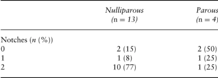
During the study period, severe pre-eclampsia was observed in 17 cases. Thirteen (76%) of these patients were nulliparous, and 4 (24%) parous. There was a trend for higher RI values in nulliparous women, but this did not reach statistical significance (Figure 1, P= 0.16). The dis-tribution of uterine artery notches in the cases with severe pre-eclampsia is displayed in Table 4. Despite a trend for a higher prevalence of notches in nulliparous women, the difference did not reach statistical significance (P= 0.44).
Table 2 Results of a multiple regression model with mean resistance index (RI) as the dependent variable (adjusted R2= 0.008,P < 0.001) in 4132 pregnancies not complicated by
pre-eclampsia Independent variable Beta* P Maternal age 0.020 0.214 Smoking 0.006 0.687 GA at scan −0.050 0.001 Parity≥ 1 0.073 <0.001
Uterine artery mean RI required logarithmic transformation. *Standardized regression coefficient. GA, gestational age.
Table 3 Results of a logistic regression model to predict the presence of bilateral uterine artery protodiastolic notches in 4132 pregnancies not complicated by pre-eclampsia
Independent
variable Odds ratio* 95% CI P
Maternal age 1.04 1.01–1.06 <0.01
Smoking 1.13 0.75–1.68 0.56
GA at scan 1.03 0.88–1.22 0.68
Parity≥ 1 0.67 0.45–0.98 0.04
GA, gestational age.
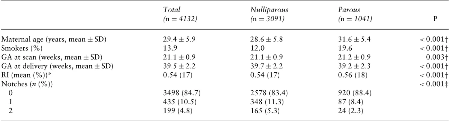
Table 1 Demographic characteristics and uterine artery Doppler measurements of 4132 women with pregnancy not complicated by pre-eclampsia Total (n= 4132) Nulliparous (n= 3091) Parous (n= 1041) P
Maternal age (years, mean±SD) 29.4± 5.9 28.6± 5.8 31.6± 5.4 <0.001†
Smokers (%) 13.9 12.0 19.6 <0.001‡
GA at scan (weeks, mean±SD) 21.1± 0.9 21.1± 0.9 21.2± 0.9 0.003†
GA at delivery (weeks, mean±SD) 39.5± 2.2 39.7± 2.2 39.2± 2.3 <0.001†
RI (mean (%))* 0.54 (17) 0.54 (17) 0.56 (18) <0.001†
Notches (n(%)) <0.001‡
0 3498 (84.7) 2578 (83.4) 920 (88.4)
1 435 (10.5) 348 (11.3) 87 (8.4)
2 199 (4.8) 165 (5.3) 24 (2.3)
*Since a logarithmic transformation was used for the mean resistance index, the SD is expressed as a percentage of magnitude;†Student’s
t-test;‡chi-square test; GA, gestational age; RI, resistance index.
48 Prefumo et al. Nulliparous Parous 0.2 0.3 0.4 0.5 0.6 0.7 0.8 0.9
Mean resistance index
Figure 1 Distribution of mean uterine artery resistance index (RI) values in nulliparous (n= 13) and parous (n= 4) women with pregnancies complicated by severe pre-eclampsia. Horizontal lines indicate means. The dashed line indicates the mean RI in pregnancies not complicated by pre-eclampsia (n= 4132).
Table 4 Distribution of uterine artery protodiastolic notches in nulliparous and parous women with pregnancies complicated by severe pre-eclampsia (n= 17) Nulliparous (n= 13) Parous (n= 4) Notches (n(%)) 0 2 (15) 2 (50) 1 1 (8) 1 (25) 2 10 (77) 1 (25) D I S C U S S I O N
The findings of this study indicate that parity has a significant effect on uterine artery Doppler indices measured in the second trimester of pregnancy. Although parity affects both uterine artery RI and the prevalence of notches, the effect on the latter is likely to be of greater clinical significance. In our series of uncomplicated pregnancies, parous women presented with slightly higher RI values when compared with nulliparous women. Although this difference was statistically significant, the multiple regression model using parity, maternal age, gestational age at ultrasound scan and smoking status was only able to explain less than 1% of the variance in mean uterine artery RI. Additionally, the difference in mean RI between nulliparae and multiparae was smaller than the established interobserver variability of uterine artery Doppler11,12.
In contrast, the prevalence of protodiastolic notches was significantly lower in parous women. After correcting for confounding factors in the logistic regression model, the odds ratio for bilateral notches in parous women was 0.67 (95% CI, 0.45–0.98). Hence, nulliparous women are 50% more likely to have bilateral notches than are parous women. These observations are supported by the findings described by Hafner et al.13who examined uter-ine artery perfusion in the first and second pregnancy in
1102 women. These authors observed that uterine pul-satility index (PI) in the same woman was similar in the first and second pregnancies, while notching appeared much more frequently in the first pregnancy.
Ideally the effect of parity should be demonstrated by comparing screening efficiency in nulliparous and parous women. However, a policy of two-stage screening2 with aspirin administration was followed in screen-positive pregnancies. This management policy, which is expected to decrease the risk of developing pre-eclampsia in the treated pregnancies, may have affected the two sub-groups to different extents14. Therefore, it is not appropriate to draw any conclusions from our data regarding the incidence of pre-eclampsia and the sensitivity, specificity or positive and negative predictive values of the test in the two groups. However, it is relevant to emphasize that almost 80% of nulliparae with pre-eclampsia severe enough to require delivery at 32 weeks or earlier presented with bilateral uterine notches, which were observed in only 25% of parous women with a similar disease severity. We are aware of the limits imposed by the small number of cases and the lack of statistical significance of this difference, but these findings suggest that the sensitivity of the test for the most severe cases is higher in the first pregnancy.
It is important to note that parity paradoxically served to increase the mean uterine artery RI and decrease the prevalence of notching. The literature is of little help in understanding why a previous pregnancy should have such contrasting effects on uterine artery Doppler indices and notching. The RI and PI are popular parameters used for characterizing arterial waveforms at Doppler ultrasound. Although their physiological meaning is still under dispute, mathematical models, in-vitro experiments and data from sheep all suggest that PI and RI are affected by both downstream resistance and vessel compliance15 – 19. However, clinically PI and RI are more directly dependent on factors affecting downstream vascular resistance, such as intervillous obstruction and failed spiral artery invasion15,17,18, whereas uterine artery notching may be an effect of abnormal compliance of the uterine/arcuate arteries15.
The findings of this study must be explained by the effect of pregnancy on the maternal vasculature. During placentation, trophoblastic cells infiltrate the spiral arteries. As a result, the thick-walled and muscular spiral arteries are transformed into thin-walled and floppy vessels that can dilate and accomodate the increased uteroplacental blood flow necessary for a successful pregnancy20,21. It is possible that some permanent modification persists in the maternal vessels as an effect of this process, altering their compliance in subsequent pregnancies. These changes may explain the lower prevalence of notches in parous women. Further studies are needed to investigate this physiological hypothesis and its effect on uterine artery Doppler screening programs.
Parity and uterine Doppler 49 A C K N O W L E D G M E N T
Dr. Prefumo was supported by a Marie Curie Fellowship of the European Community programme Quality of Life under contract number QLGA-CT-2000-52145.
R E F E R E N C E S
1. Bower S, Schuchter K, Campbell S. Doppler ultrasound screen-ing as part of routine antenatal scannscreen-ing: prediction of pre-eclampsia and intrauterine growth retardation. Br J Obstet Gynaecol 1993; 100: 989–994.
2. Bower S, Bewley S, Campbell S. Improved prediction of preeclampsia by two-stage screening of uterine arteries using the early diastolic notch and color Doppler imaging. Obstet Gynecol 1993; 82: 78–83.
3. North RA, Ferrier C, Long D, Townend K, Kincaid-Smith P. Uterine artery Doppler flow velocity waveforms in the second trimester for the prediction of preeclampsia and fetal growth retardation. Obstet Gynecol 1994; 83: 378–386.
4. Haddad B, Uzan M, Breart G, Uzan S. Uterine Doppler wave form and the prediction of the recurrence of pre-eclampsia and intra-uterine growth retardation in patients treated with low-dose aspirin. Eur J Obstet Gynecol Reprod Biol 1995; 62: 179–183.
5. Irion O, Mass´e J, Forest JC, Moutquin JM. Prediction of pre-eclampsia, low birthweight for gestation and prematurity by uterine artery blood flow velocity waveforms analysis in low risk nulliparous women. Br J Obstet Gynaecol 1998; 105: 422–429.
6. Papageorghiou AT, Yu CKH, Bindra R, Pandis G, Nico-laides KH for The Fetal Medicine Foundation Second Trimester Screening Group. Multicenter screening for pre-eclampsia and fetal growth restriction by transvaginal uterine artery Doppler at 23 weeks of gestation. Ultrasound Obstet Gynecol 2001; 18: 441–449.
7. Goffinet F, Aboulker D, Paris-Llado J, Bucourt M, Uzan M, Papiernik E, Br´eart G. Screening with uterine Doppler in low risk pregnant women followed by low dose aspirin in women with abnormal results: a multicentre randomised controlled trial. BJOG 2001; 108: 510–518.
8. Eskenazi B, Fenster L, Sidney S. A multivariate analysis of risk factors for preeclampsia. JAMA 1991; 266: 237–241.
9. Bower S, Kingdom J, Campbell S. Objective and subjective assessment of abnormal uterine artery Doppler flow waveforms. Ultrasound Obstet Gynecol 1998; 12: 260–264.
10. Bland JM, Altman DG. Statistics notes. Measurement error proportional to the mean. BMJ 1996; 313: 106.
11. Hollis B, Mavrides E, Campbell S, Tekay A, Thilaganathan B. Reproducibility and repeatability of transabdominal uterine artery Doppler velocimetry between 10 and 14 weeks of gestation. Ultrasound Obstet Gynecol 2001; 18: 593–597.
12. Papageorghiou AT, To MS, Yu CK, Nicolaides KH. Repeata-bility of measurement of uterine artery pulsatility index using transvaginal color Doppler. Ultrasound Obstet Gynecol 2001; 18: 456–459.
13. Hafner E, Schuchter K, Metzenbauer M, Philipp K. Uterine artery Doppler perfusion in the first and second pregnancies. Ultrasound Obstet Gynecol 2000; 16: 625–629.
14. Coomarasamy A, Papaioannou S, Gee H, Khan KS. Aspirin for the prevention of preeclampsia in women with abnormal uterine artery Doppler: a meta-analysis. Obstet Gynecol 2001; 98: 861–866.
15. Talbert DG. Uterine flow velocity waveform shape as an indicator of maternal and placental development failure mechanism: a model-based synthesizing approach. Ultrasound Obstet Gynecol 1995; 6: 261–271.
16. Michel E, Zernikow B. Gosling’s Doppler pulsatility index revisited. Ultrasound Med Biol 1998; 24: 597–599.
17. Saunders HM, Burns PN, Needleman L, Liu JB, Boston R, Wortman JA, Chan L. Hemodynamic factors affecting uterine artery Doppler waveform pulsatility in sheep. J Ultrasound Med 1998; 17: 357–368.
18. Bude RO, Rubin JM. Relationship between the resistive index and vascular compliance and resistance. Radiology 1999; 211: 411–417.
19. Adamson SL. Arterial pressure, vascular input impedance, and resistance as determinants of pulsatile blood flow in the umbilical artery. Eur J Obstet Gynecol Reprod Biol 1999; 84: 119–125.
20. Brosens IA, Robertson WB, Dixon HG. The physiological response of the vessels of the placental bed to normal pregnancy. J Pathol Bacteriol 1967; 93: 569–579.
21. Pijnenborg R, Dixon G, Robertson WB, Brosens I. Trophoblast invasion of human deciduas from 8 to 18 weeks of pregnancy. Placenta 1980; 1: 3–19.