I ffiw::
-wWffi
-*o-ff
:J
w7
L'NI\/ErcITA
[}ffi-L,q
CAIABRIA
Fucoltù di Farmucia
eScienze
della
Nutrizione
e
della
Sulute
Dip
urtimento
Farmaco-Biolo
gico
Dottoruto
di
Ricerca
in:
"Biochimicu Cellulure
ed
Attivitù
dei
Furmuci
in
Oncologiu"
Cumpo
di
Ricerca
MED/04-PATOLOGIA
GEIVERALE
XXII
Ciclo
Furnesoid
X
Receptor,
through
the
binding
wíth Steroidogenic
Fuctor
I
Responsive
Element,
inhibits
uromutase expression
in
tumor Leydig
cells.
Dottorando
Ch.mo
Prof
Diego
SISCI
w-Docente Tuto
Ch.mo
Prof.
Sebasti
Coordinatore
1
INDEX
Summary
Pag 1
Introduction
Pag 3
Materials and Methods
Pag 9
Results
FXR expression in normal and tumor testicular cells
Inhibitory effects of FXR agonists on aromatase
expression in R2C cells
SHP is not involved in the down-regulatory effects induced
by FXR ligand on aromatase
CDCAdown-regulates aromatase promoter activity
through SF-1 site
FXR protein binds to SF-1 RE in vitro and in vivo
CDCA inhibits R2C cell proliferation through FXR
activation
Pag 18
Pag 21
Pag 23
Pag 25
Pag 27
Pag 30
Discussion
Pag 33
References
Pag 38
Scientific Publications Performed during the Program
Pag 42
2
SUMMARY
Nuclear receptors (NRs) are ligand-activated transcription factors that have
central roles in nearly every aspect of development and adult physiology [1]. This
family contains 48 members in humans. Because of the importance of NR
functions in different metabolic pathways, they have become attractive targets for
drug discovery. The Farnesoid X Receptor (FXR) is a member of the nuclear
receptor superfamily that regulates bile acid homeostasis. It is expressed in the
liver and the gastrointestinal tract, but also in several non-enterohepatic tissues
including testis. Recently, FXR was identified as a negative modulator of the
androgen-estrogen-converting aromatase enzyme in human breast cancer cells.
In the present study we detected the expression of FXR in Leydig normal and
tumor cell lines and in rat testes tissue. We found, in rat Leydig tumor cells, R2C,
that FXR activation by the primary bile acid chenodeoxycholic acid (CDCA) or a
synthetic agonist GW4064, through a SHP-independent mechanism,
down-regulates aromatase expression in terms of mRNA, protein levels and its
enzymatic activity. Transient transfection experiment, using vector containing rat
aromatase promoter PII, evidenced that CDCA reduces basal aromatase promoter
activity. Mutagenesis studies, electrophoretic mobility shift and chromatin
immunoprecipitation analysis reveal that FXR is able to compete with SF-1 in
binding to a common sequence present in the aromatase promoter region
interfering negatively with its activity. Finally, the FXR activator CDCA exerts
anti-proliferative effects on tumor Leydig cells at least in part through an
3
In conclusion our findings demonstrate that FXR ligands as aromatase inhibitors
4
INTRODUCTION
In 1995, Forman et al. [1] and Seol et al. [2] isolated a novel cDNA that
encoded an ‘orphan’ nuclear receptor. At the time it was named the farnesoid X
receptor (FXR) on the basis of its weak activation by farnesol and juvenile
hormone III [1], and it has been subsequently classified as NR1H4. There are two
known FXR genes, which are commonly referred to as FXRα and FXRβ.
FXRα is conserved from humans to fish (teleost fish, Fugu rubripes) [3]. The
single FXRα gene in humans and mice encodes four FXRα isoforms (FXRα1,
FXRα2, FXRα3 and FXRα4) as a result of the use of different promoters and
alternative splicing of the RNA [4,5] (Figure 1A, B).
Figure 1. Genomic organization and protein isoforms of FXRα. (A) Structure of the FXRα gene. The 11 exons of the mouse and human FXRα genes are indicated, along with the two functional promoters that initiate transcription at exons 1 or 3. Alternative splicing of the initial RNAs produces four mRNAs. The alternative splicing of the 12 bp at the 3 end of exon 5, which encodes the MYTG motif, is indicated by the dark blue box. Asterisks indicate the translational start sites (ATG). (B) The four FXR protein isoforms with the different domains color-coded. Abbreviations: AF, activation function; DBD,
DNA-5
binding domain; LBD, ligand-binding domain. (Lee, F. Y.et al. (2006) Trends Biochem Sci 31,572-580)
FXRα3 and FXRα4 possess an estende N terminus, which encompasses the
poorly defined ‘Activation Function 1 domain’ (AF-1). In addition, FXRα1 and
FXRα3 have an insert of four amino acids (MYTG) immediately adjacent to the
DNA-binding domain in a region referred to as the ‘hinge domain’ (Figure 1 B).
The second FXR gene, FXRβ, encodes a functional member of the nuclear
receptor family in rodents, rabbits and dogs, but is a pseudogene in human and
primates [6]. FXRβ has been proposed to be a lanosterol sensor, although its
physiological function remains unclear.
FXRα is espresse mainly in the liver, intestine, kidney and adrenal gland, with
much lower levels in adipose tissue [1,4,5]. Like many other non-steroid hormone
nuclear receptors, regulates the expression of a wide variety of target genes
involved in bile acid, lipid and glucose metabolism by binding either as monomer
or as a heterodimer with the Retinoid X Receptor (RXR) to FXR response
6
Figure 2 FXR regulates a large number of target genes involved in bile acid, lipoprotein and
glucose metabolisms. FXR binds to DNA either as a heterodimer with RXR or as a monomer to regulate the expression of various genes. (Wang, Y. D. et al. (2008) Cell Research 18, 1087-1095)
Typical FXREs consist of an inverted repeat (IR) of the
canonical AGGTCA
hexanucleotide core motif spaced by 0 bp (IR-0) [10] or 1 bp (IR-1) [8, 9].
IR-1 is the primary binding sequence for FXR. FXR regulates human intestinal
bile acid binding protein (IBABP), small heterodimer partner (SHP), bile salt
export pump (BSEP), BA-CoA:amino acid N-acetyltransferase (BAT) [11] and
phospholipid transfer protein (PLTP) via IR-1 elements in the promoters of these
genes [12-15]. Besides IR-1, other FXREs include IR-0, direct repeat (DR),
everted repeat [16] of the core motif separated by eight nucleotides (ER-8) and
monomeric binding sites [10, 17-20] (Table 1). In summary, FXR can bind to a
7
(Wang, Y. D. et al. (2008) Cell Research 18, 1087-1095)
By binding to FXREs, FXR regulates many genes belonging to different
metabolic pathways (Figure 2). Activation of FXR alters the expression of
different groups of genes involved in BA homoeostasis, lipid metabolism,and
glucose balance. FXR is the primary sensor of BAs. FXR activates the expression
of short heterodimer partner (SHP) which interacts with other nuclear receptors
preventing their activation [13, 20, 21].
Recently, new functions of FXR beyond its roles in metabolism were discovered
in several nonenterohepatic tissues, including its control in regulating cell growth
and carcinogenesis [23-26]. For instance, it has been demonstrated that FXR
8
aromatase activity reducing local estrogen production which sustains tumor
growth and progression [25].
Estrogen dependency is also a feature of testicular tumor which is the most
frequent solid malignant tumour diagnosed in young men (20- 40 years old)
accounting for up to 20% of all malignancies diagnosed at this age. Ninety-five
percent of all human testicular neoplasms arise from germinal cells whereas
Leydig cell tumors are the most common tumors of the gonadal stroma [27]. The
molecular basis of testicular cell malignant transformation is poorly defined. It has
been reported that estrogen serum levels are elevated in patients with testicular
germ cell cancer as a consequence of increased local estrogen production
reflecting an higher aromatase activity present in Sertoli and Leydig cells [28].
Several studies on both rodents and humans indicate that prenatal, early post-natal
and adult exposure to an excess of estrogens might have a central role in the
mechanism leading to male reproductive tract malformations such as testicular
and prostatic tumors [29]. The biological significance of estrogen-induced
testicular tumorogenesis has been suggested by transgenic mice overexpressing
aromatase and exhibiting enhancement of 17β-estradiol (E2) circulating levels
[30]. About half of these male mice were infertile and/or had enlarged testis and
showed Leydig cell hyperplasia and Leydig cell tumors [30]. Recently, we
demonstrated aromatase and ERs expression in testis from patients affected by
Leydigioma in which high estrad
iol levels in the presence of ERα could
significantly contribute to tumor cell growth and progression [31]. Besides, we
9
tumorogenesis is an excessive estrogen production that stimulates a short
autocrine loop determining cell proliferation [32].
Aromatase activity is regulated primarly at the level of gene expression by
tissue-specific promoters and is present in testicular somatic cells and along the
maturative phases of male germ cells [33, 34]. A promoter proximal to the
translation start site, called promoter II (PII) regulates aromatase expression in
fetal and adult testis, R2C and H540 rat Leydig tumor cells, and in purified
preparations of rat Leydig, Sertoli, and germ cells [35, 36]. Specific sequences
seem to be mainly involved in aromatase expression: cyclic AMP
(cAMP)-responsive element (CRE)-like sequences binding CREB/ATF protein families
[37, 38] and a sequence containing an half-site binding nuclear receptors
(AGGTCA) in position -90 binding steroidogenic factor 1 (SF-1) [39] which is
essential for sex differentiation and development of gonads [40].
On the basis of all these observations, in this study we investigated in rat tumor
Leydig cells, R2C whether FXR activation by specific ligand chenodeoxycholic
acid (CDCA) or a synthetic agonist GW4064 may modulate aromatase expression
and antagonize estrogen signalling, inhibiting testicular tumor growth and
progression. We, for the first time, demonstrated that the molecular mechanism by
which FXR ligands inhibit aromatase gene expression in R2C cells is mediated by
a direct binding of FXR to SF-1 response element present in the aromatase
10
MATERIALS AND METHODS
Reagents
Nutrient Mixture F-10 Ham, Dulbecco’s Modified Eagle’s Medium/Nutrient
Mixture F-12 Ham (DMEM/F12), Dulbecco’s Modified Eagle’s Medium
(DMEM), L-glutamine, penicillin, streptomycin, fetal bovine serum (FBS), horse
serum (HS), phosphate-buffered saline, aprotinin, leupeptin,
phenylmethylsulfonyl fluoride (PMSF), bovine serum albumin (BSA) and sodium
orthovanadate were purchased by Sigma (Milan, Italy). TRIzol, Lipofectamine
2000 by Invitrogen (Carlsbad, CA, USA) and FuGENE 6 by Roche Applied
Science (Indianapolis, IN, USA). TaqDNA polymerase, RETROscript kit, 100-bp
DNA ladder, Dual Luciferase kit, TNT master mix, ImProm-II Reverse
transcription system kit and TK Renilla luciferase plasmid were provided by
Promega (Madison, WI, USA). SYBR Green Universa PCR Master Mix were
provided by Bio-Rad,
TaqMan rRNA Reagent kit by Applied Biosystems.
Antibodies against FXR, β-actin, GAPDH, Cyclin D1, Cyclin E and Lamin B by
Santa Cruz Biotechnology (Santa Cruz, CA, USA), antibody against Aromatase
by Serotec (Raleigh, NC, USA) and antibody against SF-1 kindly provided from
Dr. K. Morohashi (National Institute Basic Biology, Myodaiji-cho, Okazaki,
Japan). ECL system and Sephadex G-50 spin columns from Amersham
Biosciences
(Buckinghamshire,
UK).
[1β-
3H]androst-4-ene-3,17-dione,
[γ
32P]ATP, and [
3H]thymidine from PerkinElmer (Wellesley, MA, USA). Salmon
11
Plasmids
The plasmids containing different segments of the rat aromatase PII sequence
ligated to a luciferase reporter gene [1037/+94 (p1037), 688/+94 (p688),
-688/+94 mut (p-688m) (SF-1 site mutant),
-475/+94 475) and -183/+94
(p-183)] were previously described [39]. FXR responsive reporter gene (FXRE-IR1)
and FXR-DN (dominant negative) expression plasmids were provided from Dr.
T.A. Kocarek (Institute of Environmental Health Sciences, Wayne State
University, USA) [41]. FXR expression plasmid was provided from Dr. D.J.
Mangelsdorf (Southwestern Medical Center, TX, USA). SF-1 expression plasmid
and hCYP17 gene reporter were obtained from Dr. W. E. Rainey (Medical
College of Georgia, USA). XETL plasmid is a construct containing an
estrogen-responsive element from the Xenopus vitellogenin promoter, driving expression of
the luciferase gene.
Cell Cultures and animals
Rat Leydig tumor cells (R2C) were cultured in Ham/F-10 supplemented with 15%
HS, 2.5% FBS, and antibiotics. Mouse Leydig cells (TM3) were cultured in
DMEM/F-12 supplemented with 5% HS, 2.5% FBS, and antibiotics. Human
Cervix
tumor cells (HeLa) and Hepatoma cells (HepG2) were cultured in DMEM
supplemented with 10% FBS, 1% L-glutamine and antibiotics. The cells were
starved in serum free medium (SFM) 24 hours before treatments. Male Fisher 344
rats (a generous gift of Sgma-Tau), 6 (FRN) and 24 (FRT) months of age, were
used for studies. Twenty-four-month-old animals presented spontaneously
12
all animals were surgically removed by qualified, specialized animal care staff in
accordance with the Guide for Care and Use of Laboratory Animals (NIH) and
used for experiments.
Aromatase Activity Assay
The aromatase activity in subconfluent R2C cells culture medium was measured
by the tritiated water release a
ssay using 0.5 μM [1β-
3H]androst-4-ene-3,17-dione
as substrate [42]. The incubations were performed at 37°C for 2 h under an
air/CO2 (5%) atmosphere. The results obtained were expressed as pmol/h and
normalized to mg of protein (pmol/h/mg protein).
Total RNA extraction and reverse transcription PCR assay
Total RNA was extracted from R2C and TM3 cells using TRIzol reagent and the
evaluation of genes expression was performer by the reverse transcription-PCR
method using a RETROscript kit. The cDNAs obtained were amplified by PCR
using the following primers:
P450 aromatase
Forward 5’-AGCTATACTGAAGGAATCCACACTGT-3’
Reverse 5’-AATCGTTTCAAAAGTGTAACCAGGA-3’
FXR
Forward 5’-TTTCTACCCGCAACAACCGGAA-3’
Reverse 5’-GTGACAAAGAAGCCGCGAATGG-3’
rat-SHP
Forward 5’-CAGCCACCAGACCCACCACAA-3’
Reverse 5’-GAGGCACCGGACCCCATTCTA-3’
mouse-SHP
Forward 5’-CGTCCGACTATTCTGTATGC-3’
Reverse 5’-CTTCCTCTAGCAGGATCTTC-3’
L19
Forward 5’-GAAATCGCCAATGCCAACTC-3’
Reverse 5’-ACCTTCAGGTACAGGCTGTG -3’
13
The PCR was performed for 25 cycles for P450 aromatase (94°C 1 min, 58°C 1
min, 72°C 2 min), 35 cycles for FXR (94°C 1 min, 65°C 1 min, 72°C 2 min), 28
cycles for SHP (94°C 1 min, 65°C 1 min, 72°C 2 min) and 25 cycles for L19
(94°C 1 min, 60°C 1 min, and 7
2°C 2 min) in the presence of 1μl of first strand
cDNA, 1 μM each of the primers, 0.5 mM dNTP, Taq DNA polymerase (2
units/tube) and 2.2 mM magnesium chloride in a final volume of 25 μl. DNA
quantity in each lane was analyzed by scanning densitometry.
Immunoblot analysis
R2C, TM3, HepG2 cells or total tissue of FRNT and FRTT were lysed in 500 μl
of 50 mM Tris-HCl, 150 mM NaCl, 1% NP-40, 0.5% sodium deoxycholate, 2
mM sodium fluoride, 2 mM EDTA, 0.1% SDS, containing a mixture of protease
inhibitors (aprotinin, PMSF, sodium ortho-vanadate) for protein extraction.
Nuclear extracts were prepared as previously described [43]. Equal amount of
proteins were resolved on 11% SDSpolyacrylamide gel, transferred to a
nitrocellulose membrane and probed with FXR, Aromatase, Cyclin D1 and Cyclin
E antibodies. To ensure equal loading all membranes were stripped and incubated
with anti Lamin B antibody for nuclear extracts or anti-GADPH and anti-
β-actin
antibodies for total extracts. The antigen-antibody complex was detected by
incubation of the membranes with peroxidasecoupled goat mouse, goat
anti-rabbit, or donkey anti-goat IgG and revealed using the ECL System. The bands of
14
Immunofluorescence
R2C cells seeded on glass coverslips were treated with CDCA 50 and 100 μM for
24 h, washed with PBS and then fixed with 4% paraformaldehyde in PBS for 20
min at room temperature. Next, cells were permeabilized with 0.2% Triton X-100
in PBS for 5 min, blocked with 5% BSA for 30 min, and incubated overnight with
anti-aromatase antibody (1:100) in PBS overnight at 4°C. The day after the cells
were washed three times with PBS and incubated with the secondary antibody
anti-mouse IgG-FITC (1:200) for 1 h at room temperature. To check the
specificity of the immunolabelling the primary antibody was replaced by normal
mouse serum (negative control). Immunofluorescence analysis was carried out on
a OLYMPUS BX51 microscope using a 40x objective.
Transient transfection assay
R2C cells were transiently transfected using the FuGENE 6 reagent with FXR
reporter gene (FXRE-IR1)
in the presence or absence of FXR-DN or XETL
plasmid. A set of experiments was performed transfecting rat aromatase PII
constructs p-1037, p-688, p-475, p-183 and p-688m. HeLa cells were transiently
cotransfected with CYP17 gene promoter and FXR or SF-1 expression plasmids.
After transfection, R2C and Hela cells
were treated with CDCA 50 μM for 24 h.
Empty vectors were used to ensure that DNA concentrations were constant in
each transfection. TK Renilla luciferase plasmid was used to normalize the
efficiency of the transfection. Firefly and Renilla luciferase activities were
15
normalized based on the transfection efficiency measured by Renilla luciferase
activity.
RNA interference (RNAi)
R2C cells were transfected with RNA duplex of stealth RNAi targeted rat SHP
mRNA sequence 5’-ACUGAACUGCUUGAAGACAUGCUUU-3’ (Invitrogen,
Carlsbad, CA, USA), with RNA duplex of stealth RNAi targeted rat FXR
mRNAsequence 5-
UCUGCAAGAUCUACCAGCCCGAGAA-3 (Ambion), with
RNA duplex of validate RNAi targeted rat aromatase mRNA sequence 5
GCUCAUCUUCCAUAACCAGGtt-3 (Invitrogen, Carlsbad, CA, USA) or with a
stealth RNAi control to a final concentration of 50nM using Lipofectamine 2000
as recommended by the manufacturer. After 5 h the transfection medium was
changed with SFM and 24 after transfection the cells were exposed to CDCA
50µM and 100µM or GW 3 µM for further 24 h. These transfected cells were
used to examine the effects of silencing SHP and FXR gene expression on the
aromatase mRNA and protein content and the effects of FXR and aromatase
specific knock-down on cellular proliferation by [
3H]thymidine incorporation.
Electrophoretic mobility shift assay (EMSA)
Nuclear extracts from R2C cells were prepared as previously described [43]. The
probe was generated by annealing single-stranded oligonucleotides, labeled with
[γ
32P] ATP using T4 polynucleotide kinase, and purified using Sephadex G50 spin
columns. The DNA sequences used as probe or as cold competitors are the
16
lowercase letters):
SF-1
-AGGACCTGAGTCTCCCAAGGTCATCCTTGTTTGACTTGTA-mutated SF-1
-TCTCCCAAtaTCATCCTTGT-In vitro transcribed and translated SF-1 and FXR proteins were synthesized using
the T7 polymerase in the rabbit reticulocyte lysate system. The protein-binding
reactions were ca
rried out in 20μL of buffer [20 mmol/L HEPES (pH 8), 1
mmol/L EDTA, 50 mmol/L KCl, 10 mmol/L DTT, 10% glycerol, 1 mg/mL BSA,
50 μg/mL poly(dI/dC)] with 50,000 cpm of labeled probe, 20 μg of R2C nuclear
protein or an appropriate amount of SF-1 or FXR protei
ns and 5 μg of poly
(dI-dC). The misture were incubated at room temperature for 20 min in the presence
or absence of unlabeled competitor oligonucleotides. For experiments involving
anti-SF-1 and anti-FXR antibodies, the reaction mixture was incubated with these
antibodies at 4°C for 12 h before addition of
labeled probe. The entire reaction
mixture was electrophoresed through a 6% polyacrylamide gel in 0.25x Tris
borate-EDTA for 3 h at 150 V.
Chromatin immunoprecipitation (ChIP) and ReChIP assays
R2C cel
ls were treated with CDCA 50 μM for 1 h and then cross-linked with 1%
formaldehyde and sonicated. Supernatants were immunocleared with salmon
sperm DNA/protein A agarose for 1 h at 4°C. The precleared chromatin was
immunoprecipitated with specific anti FXR or anti polymerase II antibodies and
re-immunoprecipitated with anti SF-1 antibody. A normal mouse serum IgG was
used as negative control. Pellets were washed as reported, eluted with elution
17
obtained by phenol/chloroform/isoamyl alcohol (25:24:1) extractions and
precipitated with ethanol; 3 μl of each sample were used for PCR amplification
with the following primers flanking SF-1 sequence present in the P450arom PII
promoter region: ATGCACGTCACTCTACCCACTCAA -3’ and
5’-TAGCACGCAAAGCAGTAGTTTGGC -3’;
upstream of the SF-1 site
5-TGATAACGACTCCAGCGTCTTCA-3 and 5-CAGAGGAGAACAGGAAGAGTGC-3.
The amplification products were analyzed in a 2% agarose gel and visualized by
ethidium bromide stainin
g. Moreover, 5μl volume of each sample and input were
used for real time PCR. PCR reactions were performed in the iCycler iQ
Detection System (Biorad Hercules, CA, USA), using 0.1 μM of each primer, in a
total volume of 50 μL reaction mixture following the manufacturer’s
recommendations. SYBR Green Universal PCR Master Mix (Biorad Hercules,
CA, USA), with the dissociation protocol, was used for gene amplification.
Negative controls contained water instead of DNA. Final results were calculated
using the ∆∆Ct method as explained above, using input Ct values instead of the
18S. The basal sample was used as calibrator.
[
3H]thymidine in corporation
R2C cells were treated with CDCA 50 and 100 μM for 24h and 48 h. For the last
6 hours, [
3H]thymidine (1μCi/ml) was added to the culture medium. After rinsing
with PBS, the cells were washed once with 10% and three times with 5%
trichloroacetic acid, lysed by adding 0.1 N NaOH and then incubated for 30 min
18
set of experiment R2C cells were transiently transfected with FXR-DN expression
plasmid 24 h before starting with the same treatments mentioned above.
Anchorage-independent soft agar growth assays
R2C cells were plated in 4 ml of Ham/F-10 with 0.5% agarose and 5%
charcoal-stripped FBS, in 0.7% agarose base in six-well plates. Two days after plating,
media containing hormonal treatments (androst-4-ene-3,17-dione, CDCA), was
added to the top layer, and the appropriate media was replaced every two days.
After 14 days, 150 μl of MTT was added to each well and allowed to incubate at
37° for 4 h. Plates were then placed at 4°C overnight and colonies > 50 μm
diameter from triplicate assays were counted. Data are the mean colony number
of
three plates and representative of two independent experiments.
Statistical Analysis
Each datum point represents the mean ± S.D. of three different experiments.
Statistical analysis was performed using ANOVA followed by Newman-Keuls
testing to determine differences in means. P<0.05 was considered as statistically
19
RESULTS
FXR expression in normal and tumor testicular cells.
We first aimed to evaluate, by Western Blotting analysis, the expression of FXR
receptor in Leydig normal (TM3) and tumor (R2C) cell lines and in testes tissue
from younger (FRNT) and older (FRTT) Fisher rats.
The latter group have a high incidence of spontaneous Leydig cell neoplasma [44,
45], a phenomenon not observed in younger animals.
Immunoblot analysis revealed the presence of a FXR-immunoreactive protein
band at ~ 60kDa in all samples examined, particularly, FXR receptor seems to be
more expressed in R2C cells with respect to TM3 and in FRTT with respect to its
control FRNT (Figure 3).
Figure 3. FXR expression in different cells lines. Western blot analysis of FXR was done on 50 μg
of total proteins extracted from normal (TM3), tumor Leydig cells (R2C) and human hepatocytes cells (HepG2) or from tissues of normal (FRNT) and tumor (FRTT) Fisher rat testes. β-actin was used as a loading control.
Human hepatocyte cells (HepG2) were used as a positive control for FXR
expression. In R2C cells, incubation for 24 h with CDCA 50 and 100 μM, a
natural ligand of FXR, increased the level of the receptor at both mRNA and
protein levels. Because CDCA may also exert FXR-independent effects [46], the
influence of GW4064, a synthetic FXR agonist, was also investigated. We
observed that GW4064 (3µM) increased FXR mRNA and protein levels to a
similar order of magnitude as CDCA (Figs 4 A & B ).
20
Figure 4. FXR activation in R2C cells. A, Total RNA was extracted from R2C cells treated with
vehicle (-) or CDCA 50 and 100 μM or GW4064 3 μM for 24 h and reverse transcribed. cDNA was subjected to PCR using primers specific for FXR or L19 (ribosomal protein). NC: negative control, RNA sample without the addition of reverse transcriptase. The histograms represent the means ± S.D. of three separate experiments in which band intensities were evaluated in terms of optical density arbitrary units and expressed as percentages of the control, which was assumed to be 100%. *p<0.05, **p<0.01 compared to vehicle. B, Nuclear proteins were extracted from R2C cells treated with vehicle (-) or CDCA 50 and 100 μM or GW4064 3 μM for 24 h and then western blotting analysis was performed using anti-FXR antibody. Lamin B was used as loading control. The histograms represent the means ± S.D. of three separate experiments in which band intensities were evaluated in terms of optical density arbitrary units and expressed as percentages of the control, which was assumed to be 100%. *p<0.05 compared to vehicle.
Moreover, to assess the ability of CDCA and GW4064 to transactivate
endogenous FXR, we transiently transfected R2C cells with FXR responsive
A21
reporter gene (FXRE-IR1). As reported in Figure 5, CDCA and GW4064 induced
a significant enhancement in transcriptional activation of the reporter plasmid
even though to a higher extent under GW treatment,
these data were completely
reversed in the presence of FXR dominant negative plasmid FXR-DN.
Figure 5. CDCA and GW 4064 enhances FXR transactivation in R2C cells. R2C cells were
transiently transfected with FXR reporter gene (FXRE-IR1) and treated with vehicle (-) or CDCA 50 μM, 100 μM and GW4064 3μM for 24 h or co-trasfected with FXR dominant negative FXR-DN and treated with vehicle(-) or GW4064 3μM . The values represent the means ± S.D. of three different experiments performed in triplicate. * p<0.01 compared to vehicle.
22
Inhibitory effects of FXR agonists on aromatase expression in R2C cells.
Starting from previous findings showing that FXR activation represses aromatase
expression in breast cancer cells [25] we investigated the ability of FXR agonists
to modulate aromatase enzyme in R2C cells which have been shown to have high
aromatase expression and activity [39]
. Treatment with CDCA 50 and 100 μM for
24 h showed a down-regulation of aromatase mRNA and protein content in a dose
related manner. Similar results were observed upon treatment with GW4064
(3µM) for 24 h (Figs. 6 A & 2B).
Figure 6. Effects of CDCA on aromatase expression in R2C cells. A, Total RNA was extracted
from R2C cells treated with vehicle (-), CDCA 50 and 100 μM or GW4064 3 μM for 24 h and reverse transcribed. cDNA was subjected to PCR using primers specific for P450 aromatase or L19. NC: negative control, RNA sample without the addition of reverse transcriptase. The histograms represent the means ± S.D. of three separate experiments in which band intensities were evaluated in terms of optical density arbitrary units and expressed as percentages of the control which was assumed to be 100%. *p<0.05, **p<0.01 compared to vehicle. B, Total proteins extracted from R2C cells treated with vehicle (-), CDCA 50 and 100 μM or GW4064 3 μM for 24 h were used for immunoblot analysis of aromatase. GAPDH was used as a loading control. The histograms representthe means ± S.D. of three separate experiments in which band intensities were evaluated in terms of optical density arbitrary units and
23
expressed as percentages of the control which was assumed to be 100%. *p<0.01 compared to vehicle.
The down-regulatory effects of CDCA on the expression of aromatase was further
confirmed by immunofluorescence analysis. The strong P450 aromatase
immunoreactivity was detected in the cytoplasm as well as in the perinuclear
region of untreated R2C cells and it was drastically decreased upon CDCA at the
doses of
50 and 100 μM for 24 h (Figure 7).
Figure 7
Down-regulatory effects of CDCA on the expression of aromatase. R2C cells were treated with vehicle (-) or CDCA 50 and 100 μM for 24 h and aromatase expression was determined by immunofluorescence analysis. DAPI staining was used to visualized the cell nucleus. Each experiment is representative of at last 4 tests.
Next, we evaluated the effects of CDCA on aromatase enzymatic activity by
tritiated water release assay. As reported in Figure 8, exposure to CDCA 50 and
100 μM for 24 h reduced enzymatic activity in a dose dependent manner in R2C
cells.
Figure 8. Effects of CDCA on aromatase activity in R2C cells. R2C were cultured in the presence
of vehicle (-) or 50 and 100 μM of CDCA for 24 h. Aromatase activity was performed as described in Materials and Methods. The results obtained were expressed as pmol [3H]
24
H2O/h release and were normalized for mg protein (pmol/mg proteins/h). The values represent the means ± S.D. of three different experiments each performed with triplicate samples. *p<0.01 compared to vehicle.
SHP is not involved in the downregulatory effects induced by FXR
ligand on aromatase.
Induction of SHP expression is considered one of the canonical features of FXR
transactivation. SHP has been shown to be expressed in the interstitial
compartment of the adult testis, including steroidogenic Leydig cells [47].
We evidenced that SHP mRNA expression was significantly higher in R2C cells
compared with very low levels detected in TM3 cell line, but administration of
CDCA or GW4064 did not induce an increase of SHP mRNA in both cell lines.
However, to explore the role of SHP in CDCA-mediated repression of aromatase
gene, we knocked SHP by siRNA. SHP mRNA expression was effectively
silenced as revealed by RT-PCR after 24, 48 and 72 h of siRNA transfection (Fig.
9 A). As shown in Figure 9B and 9C, silencing of the SHP gene failed to reverse
the inhibition of aromatase expression induced by the specific FXR ligand in R2C
cells ruling out any SHP involvement in the inhibitory effects of CDCA on
25
Figure 9. SHP is not involved in CDCA-mediated down-regulation of aromatase. A, SHP mRNA expression in R2C cells which were not transfected (–) or transfected with RNA interference (RNAi) targeted rat SHP mRNA sequence as reported in Materials and Methods for 24, 48 and 72 h. L19 was used as loading control. NC: negative control, RNA sample without the addition of reverse transcriptase. The histograms represent the means ± S.D. of three separate experiments in which band intensities were evaluated in terms of optical density arbitrary units and expressed as percentages of the control, which was assumed to be 100%. *p<0.01 compared to vehicle. B, R2C cells were transfected with control siRNA or SHP siRNA for 24 h, and then treated with vehicle (-) or CDCA 50 and 100 μM for 24 h. Total RNA was extracted and RT-PCR analysis was performed to evaluate the expression of aromatase. L19 was used as loading control. NC: negative control, RNA sample without the addition of reverse transcriptase. The histograms represent the means ± S.D. of three separate experiments in which band intensities were evaluated in terms of optical density arbitrary units and expressed as percentages of the control, which was assumed to be 100%. *p<0.01 compared to vehicle. C, In the same experimental condition of B, total proteins were extracted and western blotting analysis was performed. GAPDH was used as loading control. The histograms represent the means ± S.D. of three separate experiments in which band intensities were evaluated in terms of optical density arbitrary units and expressed as percentages of the control which was assumed to be 100%. * p<0.05, ** p<0.01 compared to vehicle.
26
CDCA down-regulates aromatase promoter activity through SF-1 site.
The aforementioned observations led us to ascertain if the down-regulatory effects
of CDCA on aromatase expression were due to its direct inhibitory influence in
regulating aromatase gene transcriptional activity. Thus, we transiently transfected
in R2C cells plasmids containing different segments of rat PII aromatase (Fig.
10A). A significant reduction of promoter activity was observed in cells
transfected with p-1037 and p-688, p-475 and p-183 exposed to
CDCA 50 μM for
24 h. It is worth to note that construct p-688m bearing SF-1 mutated site displays
significantly lower basal activity compared with the p-688 plasmid while no
inhibitory effects were noticeable upon CDCA treatment (Fig. 10B).
Figure 10. Functional interaction between FXR and SF-1 site. A, Schematic map of the P450arom
proximal promoter PII constructs used in this study. All of the promoter constructs contain the same 3’ boundary (+94). The 5’ boundaries of the promoter fragments varied from 1037 to 183. Three putative CRE motifs (5’CRE at 335; 3’CRE at -231; XCRE at -169) are indicated as square. The AGGTCA site (SF-1 RE at-90) is indicated as rectangle. A mutated SF-1 binding site (SF-1 mut) is present in p-688m (black rectangle). B, Aromatase transcriptional activity of R2C cells transfected with promoter constructs are shown. After transfection, cells were treated in the presence of vehicle (-) or CDCA 50 μM for 24h. These results represent the means ± S.D. of three different experiments performed in triplicate. *p<0.01 with respect to the vehicle, **p<0.01 with respect to the the control of p688.
This latter result highlights the importance of the SF-1 binding site in the
regulation of aromatase expression in the R2C cells and suggests that the
inhibitory effect of CDCA requires AGGTCA sequence motif.
SF-1 is closely
27
related to Liver Receptor Homologue-1 (LRH-1) and both proteins recognize the
same canonical DNA motif [48]. However since LRH-1 is not expressed in R2C
cells (Figure 11) we focused our attention on SF-1.
LRH
Lamin B
TNT TM3 R2C
Figure 11. Western Blotting of LRH-1 in nuclear extracts of TM3 and R2C cells. Transcribed and
translated in vitro LRH- protein (lane 1) was used as positive control. Lamin B was used as loading control.
To further demonstrate the functional interaction of FXR with SF-1 binding site,
we transiently cotransfected Hela cells which do not express significant levels of
SF-1 [49] with hCYP17 promoter construct containing multiple SF-1 response
elements [50] with or without SF-1 plasmid in the presence of increasing amount
of FXR expression plasmid. SF-1 expression vector strongly increased the CYP17
promoter activity, which was progressively reduced by FXR overexpression
(Figure 12). We observed a similar results also in HeLa cells overexpressing FXR
and treated with CDCA. These data support the competitive role of FXR in
binding SF-1 site.
Figure 12.. HeLa cells were transiently cotransfected with CYP17 promoter and with SF-1
plasmid or empty vector (EV) in the presence of increasing amount of FXR expression plasmid. These results represent the means ± S.D. of three different experiments
28
performed in triplicate. In each experiment, the activities of the transfected plasmids were assayed in triplicate transfections., **p<0.01 with respect to the SF-1 alone.
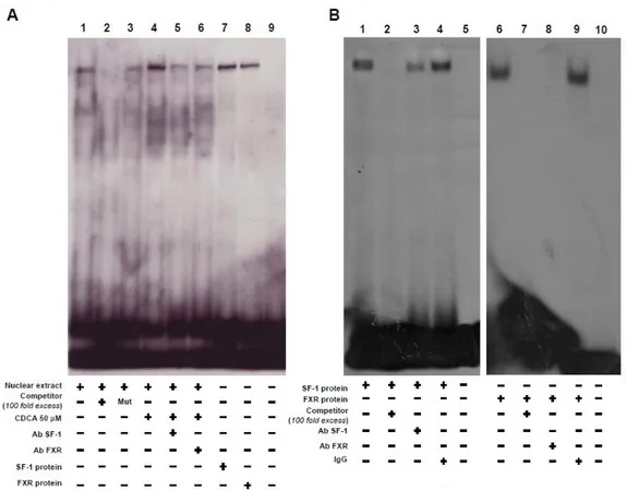
FXR protein binds to SF-1 RE in vitro and in vivo.
On the basis of the evidence that the inhibitory effect of CDCA on aromatase
requires the crucial presence of SF-1 RE, EMSA experiments were performed
using the SF-1 motif present in aromatase promoter as probe. We observed the
formation of a complex in nuclear extract from R2C cells (Figure 13A, lane 1),
which was abrogated by 100 fold molar excess of unlabeled probe (Figure 13A,
lane 2) demonstrating the specificity of the DNA binding complex. This inhibition
was not longer observed when mutated oligodeoxyribonucleotide was used as
competitor (Figure 13
A, lane 3). CDCA 50 μM for 6 h induced an increase in
DNA binding complex compared with control samples (Figure 13 A, lane 4). The
inclusion of anti-SF-1 and anti-FXR antibodies in the reactions attenuated the
specific bands suggesting the presence of SF-1 and FXR proteins in the complex
(Figure 13A, lane 5 and 6). Using SF-1 and FXR proteins transcribed and
translated in vitro, we obtained complexes migrating at the same level as that of
R2C nuclear extracts (Figure 13A, lane 7 and 8). Competition binding studies
revealed that both transcribed and translated SF-1 and FXR DNA binding
complex (Figure 13B, lane 1 and 6) were abrogated by 100-fold molar excess of
unlabeled probe (Figure 13B, lane 2 and 7). Finally the specificity of these bands
was proved by the drastically attenuation of the complex in the presence of
anti-SF-1 antibody, while the inclusion of anti-FXR antibody completely
immunodepleted the binding (Figure 13B, lane 3 and 8). IgG did not affect either
29
Figure 13. FXR binds, in vitro, to SF-1site within aromatase promoter region. A, Nuclear extract
from R2C cells were incubated with a double-stranded SF-1-specific sequence probe labeled with [γ32P]ATP and subjected to electrophoresis in a 6% polyacrylamide gel (lane 1). Competition experiments were performed adding as competitor a 100-fold molar excess of unlabeled probe (lane 2) or a 100-fold molar excess of unlabeled oligonucleotide containing a mutated SF-1 RE (lane 3). Lane 4, nuclear extracts from CDCA (50 μM) treated R2C cells. Lanes 5 and 6, CDCA-treated nuclear extracts were incubated with anti-SF-1 or anti-FXR antibodies respectively. We used as positive controls transcribed and translated in vitro SF-1 (lane 7) and FXR (lane 8) proteins. Lane 9 contains probe alone. B, SF-1 protein (1 μl) (lane 1)and FXR protein (1 μl) (lane 6) was incubated with a double-stranded SF-1 sequence probe labeled with [γ 32P] and subjected to electrophoresis in a 6% polyacrylamide gel. Competition experiments were performed adding as competitor a 100-fold molar excess of unlabeled probe (lane 2 & 7). SF-1 and for FXR proteins were incubated with anti-SF-1 antibody (lane 3) or anti-FXR antibody (lane 8) or IgG (lane 4 &9). Lanes 5 and 10 contain probe alone
The interaction of FXR with the aromatase gene promoter was further
investigated by ChIP assay. Using specific antibody against FXR and RNA-POL
II, formaldehyde cross-linked protein-chromatin complexes were
immunoprecipitated from R2C cells cultured with or without CDCA 50 and 100
30
reprecipitated with the anti SF-1 antibody. The results analyzed by PCR indicated
that FXR was weakly constitutively bound to the aromatase promoter in untreated
cells and this recruitment was increased upon CDCA treatment, which was
correlated with a reduced association of RNA polymerase II. Interestingly, by
Re-ChIP assay, we observed upon CDCA stimulation a significant reduction in SF-1
recruitment to the aromatase promoter.
Next, the anti-FXR antibody did not
immunoprecipitate a region upstream the SF-1 site located within the aromatase
promoter gene (Figure 14 A). ChIP assay was quantified by real-time PCR as shown in
Figure 14B.
Figure 14. FXR binds, in vivo, to SF-1 site within aromatase promoter region. A, R2C cells were
treated in the presence of vehicle (-) or CDCA 50 and 100 μM for 1 hour, then crosslinked with formaldehyde, and lysed. The precleared chromatin was immunoprecipitated with anti-FXR, anti-RNA Pol II antibodies and normal mouse serum (NC) as negative control. Chromatinimmunoprecipitated with the anti-FXR antibody was re-immunoprecipitated with anti-SF-1 antibody. The PII promoter (prom) sequence including the SF-1 site and that located upstream the SF-1 site were detected by PCR with specific primers, as described in Materials and Methods and B, 5μl volume of each sample and input were used for real time PCR.
To control input DNA, PII promoter was amplified from 30 μl initial preparations of soluble chromatin before immunoprecipitations. Similar results were obtained in multiple independent experiments.
31
CDCA inhibits R2C cell proliferation through FXR activation.
Finally, we evaluated the effect of CDCA on the growth of R2C cells by
measuring changes in the rate of DNA synthesis (
3H thymidine incorporation). As
shown in Figure 15 A, treatment with CDCA for 24 and 48 h reduced R2C cells
proliferation in a dose and time dependent manner. The specific involvement of
FXR in the antiproliferative response of R2C cells to CDCA was demonstrated by
the evidence that such inhibitory effects were completely reversed in the presence
of FXR dominant negative plasmid (Figure 15B) as well as after knocking down
FXR with a specific siRNA (Figure 15C).
Figure 15. CDCA effects on R2C cell proliferation. A, R2C cells were treated with vehicle(-) or
CDCA 50 and 100 μM for 24 and 48h or B, transiently transfected with FXR dominant negative (FXR-DN) for 24 h or C, transfected with control siRNA or FXR siRNA for 24 h, and then treated as above reported. Thymidine incorporation assay was performed. The results represent the means ± S.D. of three different experiments each performed with triplicate samples, and expressed as percentage of growth vs control which was assumed to be 100%.
It is well known that aromatase overexpression in tumor Leydig cells leads to a
consequent excess of in situ estradiol production that sustains tumor cell growth
and proliferation [30]. Since we demonstrated the ability of CDCA to
32
was able to antagonize the effect of an aromatizable androgen
androst-4-ene-3,17-dione (AD) on estradiol/ERα signaling in R2C cells. To this aim we performed
transient transfection experiment using XETL plasmid, which carries firefly
luciferase sequences under the control of an estrogen response element upstream
of the thymidine kinase promoter. As shown in Figure 16 we observed that the
exposure to CDCA (50
μM) per se did not elicit any changes in luciferase activity
but it completely reversed XETL activation induced by AD.
Figure 16. Transient transfection experment with XETL promoter plasmid in R2C cells. Cells
were treated with CDCA 50 μM in the presence or not of androst-4-ene-3,17-dione (AD) 100 nM for 24h. These results represent the means ± S.D. of three different experiments. In each experiment, the activities of the transfected plasmids were assayed in triplicate transfections. *p<0.01 with respect to the vehicle. **p<0.01 CDCA+AD treated vs AD alone.
Moreover, we examined if CDCA was able to inhibit the effect of AD on R2C cell
proliferation using two experimental approaches thymidine incorporation and
anchorage independent soft agar growth assay. As expected, treatment with 100
nM of AD, through its conversion into estradiol, increased thymidine
incorporation as well as the number of colonies present in soft agar (Figures 17A
& B) concomitantly with an increased levels of cell cycle regulators ciclin D1 and
33
exposure (Figures 17A, B & 17C). These data demonstrated that FXR ligand,
through an inhibition of aromatase activity, is able to reduce the estrogen
dependent tumor Leydig cells proliferation.
Figure 17. CDCA inhibits the effect of AD on R2C cells A, R2C cells were treated with
androst-4-ene-3,17-dione (AD) 100nM in the presence or not of CDCA 50 μM for 24h. Thymidine incorporation assay was performed. The results represent the means ± S.D. of three different experiments each performed with triplicate samples. *p<0.01 AD treated compared to vehicle. **p<0.01 CDCA+AD treated vs ADalone. B, R2C cells were seeded (10,000/well) in 0.5% agarose and the treated as described above. Cells were allowed to grow for 14 days and then the number of colonies >50μm were quantified and the results graphed. The results represent the means ± S.D. of three different experiments each performed with triplicate samples. *p<0.01 AD treated compared to vehicle. **p<0.01 CDCA+AD treated vs AD alone. C, Total proteins extracted from R2C cells treated with vehicle (-), Ad 100 nM, CDCA 50 μM and AD*CDCA for 24 h were used for immunoblot analysis of cyclin D1 and cyclin E. β-actin was used as a loading control. The histograms represent the means ± S.D. of three separate experiments in which band intensities were evaluated in terms of optical density arbitrary units and expressed as percentages of the control which was assumed to be 100%. *p<0.01 AD treated compared to vehicle. **p<0.01 CDCA+AD treated vs AD alone.
A B
34
DISCUSSION
FXR is highly expressed in the enterohepatic system where it drives bile acid
absorption and secretion, lipid, glucid metabolism, and immunological response to
intestinal bacterial overgrowth [51-55]. In hepatocytes, activation of FXR causes
both feedback inhibition of cholesterol 7ahydroxylase (CYP7A1), the
rate-limiting enzyme in bile acid biosynthesis from cholesterol, and activation of
intestinal bile acid binding protein [56]. In addition, several observations suggest
that FXR may also be involved in the control of steroid metabolism [25, 57].
Indeed, FXR activation results in the modulation of genes encoding androgen
precursor-synthesizing enzymes, namely dehydroepiandrosterone sulfotransferase
(SULT2A1), 5α-reductase and 3β-HSD (3β-hydroxysteroid dehydrogenase) in the
liver [58, 59]. Recently, FXR was shown to inhibit androgen glucuronidation in
prostatic cancer cell lines [60] and to suppress the activity of the aromatase in
human breast cancer cells [25]. The enzyme aromatase coded by the gene CYP19,
converts androgens in estrogens and is involved in the progression and growth of
various estrogen hormonal-induced neoplasms.
For instance, overexpression of aromatase plays a significant role in the excessive
estrogen production sustaining tumorogenesis in Leydig cells [30].
Here, we have documented that FXR is expressed in tissues of normal and tumor
Fisher rat testis and in Leydig normal and tumor cell lines. In R2C cells, the FXR
activators CDCA and GW4064, downregulate aromatase expression at both
mRNA and protein level, together with the inhibition of its enzymatic activity.
One of the well-characterized mechanism by which FXR down-regulates gene
35
both a DNA-binding domain and the NH2-terminal ligand independent activation
domain [21]. This receptor interacts with other nuclear receptors, including
Peroxisome Proliferator Activated Receptor (PPAR), RXR, Estrogen Receptor
(ER) and Liver Receptor Homolog-1 (LRH-1), preventing their activation of gene
transcription [13, 21]. In preadipocytes of cancerous breast tisuue, LRH-1 can
regulate via an alternate promoter (II) the expression of aromatase induced by
prostaglandin E2 [61, 62]. Moreover, SHP can inhibit LRH-1 induction of
aromatase [63].
LRH-1 is most homologous to SF1, which is essential for sex differentiation and
development of gonads [40], since they share a highly conserved DBD
(DBD>90% identity) and a moderately conserved LBD (LBD 56% identity).
SHP is detected in the interstitial cells of the adult testis and its expression has
been shown to be induced by FXR [47].
Our current study revealed that FXR activation does not induce SHP expression in
Leydig tumor cells in which the inhibition of aromatase protein by CDCA occurs
even when this nuclear receptor was knocked down. These results suggest that
SHP is not required for the effect of FXR ligand to down-regulate aromatase
expression, at least in R2C cells. On the basis of these observations, we focused
our attention on the direct effect of FXR on the transcriptional activity of
aromatase gene.
Distinctive tissues specific promoters are employed to direct the expression of
aromatse mRNA driving from a single aromatase gene.
The promoter located immediately upstream of the transcriptional initiation site
36
R2C Leydig tumor cells [35, 36]. A number of functional motifs have been
identified in the PII aromatase promoter: three motifs resembling cAMP response
elements (CRE) and an SF-1 binding site [39, 40].
We demonstrated by functional studies, using constructs containing different
5’-deleted regions of rat PII aromatase promoter, that CDCA treatment induces a
decreased transcriptional activity. The observed inhibitory effect of CDCA was
abrogated when a promoter fusion containing a mutated SF-1 element was
employed. These results clearly suggest that the integrity of SF-1 sequence is a
prerequisite for the down-regulatory effects of FXR ligand on aromatase promoter
activity. These findings raise the possibility that FXR and SF-1 are competing for
binding to a common site within this regulatory region. This assumption is further
supported by the observation that FXR expression vector is able to abrogate the
induction of SF-1 on human CYP17 promoter which contains multiple SF-1
response elements.
As a transcription factor, FXR binds to a specific consensus sequence (inverted
repeat of 2 AGGTCA half-sites) either as a monomer or as a heterodimer with a
common partner for NRs, as RXR to regulate the expression of various genes(4).
Location of an AGGTCA sequence at the -90 position supports a possible binding
of FXR to this promoter region, which we verified by EMSA experiments.
Nuclear extracts from R2C cells treated with CDCA revealed an increase in DNA
binding complex which was immunodepleted by both anti-SF-1 and anti-FXR
antibodies suggesting how the two proteins are able to bind the AGGTCA
sequence located in PII aromatase promoter.
The specificity of the binding was
37
of the DNA complex observed using SF-1 and FXR transcribed and translated in a
cell free system. In addition, the in vivo iinteraction between FXR and aromatase
promoter was further supported by ChIP assay, where upon CDCA treatment we
observed a reduced recruitment of RNA-POLII to this promoter addressing a
negative transcriptional regulation mediated by FXR. All together these data
suggest that FXR is able to compete with SF-1 in binding to a common sequence
within the PII promoter of aromatase interfering negatively with its activity.
Finally, in our study we demonstrated that FXR activator CDCA induces growth
inhibition in R2C cells which was reversed in the presence of FXR dominant
negative as well as after knocking down FXR with a specific siRNA addressing a
FXR dependency of this event.
However it is worth to mention, on the basis of our recent findings, that aromatase
overexpression, in Leydig tumor cells, determines an excessive local estradiol
production that is able to stimulate the expression of genes involved in cell cycle
regulation sustaining cell proliferation [32].
Here, we evidenced the ability of CDCA to reverse the stimulatory effects of an
aromatizable androgen androst-4-ene-3,17-dione(AD) at three different levels: 1)
E2/ERα signaling; 2) an anchorage dependent and independent R2C cell growth
proliferation; 3) expression of cell cycle regulators cyclin D1 and cyclin E.
The latter finding bring us to emphasize how the intrinsic property of FXR to
inhibit R2C cell proliferation sound to be not linked to any substantial effect on
cyclin D1 and cyclin E expression.
In conclusion, our results elucidate, for the first time, a new molecular mechanism
38
growth and progression. addressing FXR ligands as potential pharmacological
tools to be implemented in the novel strategies for testicular tumoral treatment.
The identification of this molecular mechanism will be helpful in defining new
39
REFERENCES
1. Forman BM, Goode E, Chen J, Oro AE, Bradley DJ, Perlmann T, Noonan DJ, Burka LT, McMorris T, Lamph WW, Evans RM, Weinberger C. (1995). Cell 81, 687–693
2. Seol W, Choi HS, Moore DD. (1995). Mol. Endocrinol. 9, 72–85
3. Maglich JM, Caravella JA, Lambert MH, Willson TM, Moore JT, Ramamurthy L. (2003). Nucleic Acids Res. 31, 4051–4058
4. Huber RM, Murphy K, Miao B, Link JR, Cunningham MR, Rupar MJ, Gunyuzlu PL, Haws TF, Kassam A, Powell F, Hollis GF, Young PR, Mukherjee R, Burn TC. (2002). Gene 290, 35–43
5. Zhang Y, Kast-Woelbern HR, Edwards PA. (2003). J. Biol. Chem. 278, 104–110 6. Otte K, Kranz H, Kober I, Thompson P, Hoefer M, Haubold B, Remmel B, Voss
H, Kaiser C, Albers M, Cheruvallath Z, Jackson D, Casari G, Koegl M, Pääbo S, Mous J, Kremoser C, Deuschle U. (2003). Mol. Cell. Biol. 23, 864–872
7. Song CS, Echchgadda I, Baek BS, Ahn SC, Oh T, Roy AK, Chatterjee B. (2001). J Biol Chem 276, 42549-42556
8. Li J, Pircher PC, Schulman IG, Westin SK. (2005). J Biol Chem; 280:7427-7434 9. Ananthanarayanan M, Balasubramanian N, Makishima M, Mangelsdorf DJ,
Suchy FJ. (2001). J Biol Chem; 276:28857-28865.
10. Song CS, Echchgadda I, Baek BS, Ahn SC, Oh T, Roy AK, Chatterjee B. (2001). J Biol Chem; 276:42549-42556.
11. Shibata M, Morizane T, Uchida T, Yamagami T, Onozuka Y, Nakano M, Mitamura K, Ueno Y. (1998). Lancet; 351:1773-1777.
12. Grober J, Zaghini I, Fujii H, Jones SA, Kliewer SA, Willson TM, Ono T, Besnard P. (1999). J Biol Chem; 274:29749-29754.
13. Goodwin B, Jones SA, Price RR, Watson MA, McKee DD, Moore LB, Galardi C, Wilson JG, Lewis MC, Roth ME, Maloney PR, Willson TM, Kliewer SA. (2000). Mol Cell; 6:517-526.
14. Pircher PC, Kitto JL, Petrowski ML, Tangirala RK, Bischoff ED, Schulman IG, Westin SK. (2003). J Biol Chem; 278:27703-27711.
15. Urizar NL, Dowhan DH, Moore DD. (2000). J Biol Chem; 275:39313-39317. 16. Huang W, Ma K, Zhang J, Qatanani M, Cuvillier J, Liu J, Dong B, Huang X,
40 17. Laffitte BA, Kast HR, Nguyen CM, Zavacki AM, Moore DD, Edwards PA.
(2000). J Biol Chem; 275:10638-10647.
18. Anisfeld AM, Kast-Woelbern HR, Meyer ME, Jones SA, Zhang Y, Williams KJ, Willson T, Edwards PA. (2003). J Biol Chem; 278:20420-20428.
19. Kast HR, Goodwin B, Tarr PT, Jones SA, Anisfeld AM, Stoltz CM, Tontonoz P, Kliewer S, Willson TM, Edwards PA. (2002). J Biol Chem; 277:2908-2915. 20. Claudel T, Sturm E, Duez H, Torra IP, Sirvent A, Kosykh V, Fruchart JC,
Dallongeville J, Hum DW, Kuipers F, Staels B. (2002). J Clin Invest; 109:961- 971.
21. Seol W, Choi HS, Moore DD. (1996). Science 272, 1336-1339
22. Seol W, Hanstein B, Brown M, Moore DD. (1998). Mol Endocrinol 12, 1551- 1557
23. Wang, Y. D., Chen, W. D., Moore, D. D. and Huang, W. D. (2008). Cell Research 18, 1087-1095
24. Journe, F., Laurent, G., Chaboteaux, C., Nonclercq, D., Durbecq, V., Larsimont, D., Body, J. J. (2008). Breast Cancer Res Treat 107, 49-61
25. Swales, K. E., Korbonits, M., Carpenter, R., Walsh, D. T., Warner, T. D., Bishop-Bailey, D. (2006). Cancer Res 66, 10120-10126
26. Modica S, Murzilli S, Salvatore L, Schmidt D R, Moschetta A. (2008). Cancer Res 68,9589-9594
27. Hawkins, C., Miaskowski, C. (1996). Oncol Nurs Forum 23, 1203-1211
28. Carroll, P. R., Whitmore, W. F. Jr., Herr, H. W., Morse, M. J., Sogani, P. C., Bajorunas, D., Fair, W. R., Chaganti, R. S. (1987). J Urol 137, 420-423
29. Bosland, M. C. (1996). Prog Clin Biol Res 394, 309-352
30. Fowler, K. A., Gill, K., Kirma, N., Dillehay, D. L., Tekmal, R. R. (2000). Am J Pathol 156, 347-353
31. Carpino, A., Rago, V., Pezzi, V., Carani, C., Andò, S. (2007). Eur. J. Endocrinol. 157, 239-44
32. Sirianni, R., Cimento, A., Malivindi, R., Mazzitelli, I., Andò, S., Pezzi, V. (2007). Cancer Res 67, 8368-8377
33. Aquila, S., Sisci, D., Gentile, M., Carpino, A., Middea, E., Catalano, S., Rago, V., Andò, S. (2003). Hum Reprod 18, 1650-1659
41 35. Young, M., Lephart, E. D., McPhaul, M. J. (1997). J Steroid Biochem Mol Biol
63, 37–44
36. Lanzino, M., Catalano, S., Genissel, C., Ando’, S., Carreau, S., Hamra, K., McPhaul, M. J. (2001). Biol Reprod 64, 1439-1443
37. Fitzpatrick, S. L., Richards, J. S. (1994). Mol Endocrinol 8, 1309–1319 38. Carlone, D. L., Richards, J. S. (1997). Mol Endocrinol 11, 292–304 39. Young, M., McPhaul, M. J. (1998). Endocrinology 139, 5082-5093 40. Parker, K. L., Shimmer, B. P. (1997). Endocr Rev 18, 361-377
41. Kocarek, T. A., Shenoy, S. D., Mercer-Haines, N. A., Runge-Morris, M. (2002). J Pharmacol Toxicol Methods 47, 177-187
42. Lephart, E. D. and Simpson, E .R. (1991). Methods Enzymol 206, 477–483 43. Anderws, N. C., and Faller, D. V. (1991). Nucleic Acids Res 19, 2499
44. Coleman, G. L., Barthold, W., Osbaldiston, G. W., Foster, S. J., Jonas, A. M. (1977). J Gerontol 32, 258–278
45. Jacobs, B.B., Huseby, R.A. (1967). J Natl Cancer Inst 39, 303–309 46. Nguyen, A., Bouscarel, B. (2008). Cellular Signalling 20, 2180-2197
47. Volle, D. H., Duggavathi, R., Magnier, B. C., Houten, S. M., Cummins, C. L., Lobaccaro, J.M., Verhoeven, G., Schoonjans, K., Auwerx, J. (2007). Genes Dev 21, 303-315
48. Pezzi V., Sirianni R., Chimento A., Maggiolini M., Bourguiba S., Delalande C., Carreau S., Andò S., Simpson E.R., Clyne C. (2004). Endocrinology 145, 2186-2169
49. Sugawara, T., Holt, J. A., Kiriakidou, M., Strauss, J. F. (1996). Biochemistry 35, 9052-9059
50. Hanley, N. A., Rainey, W. E., Wilson, D. I., Ball, S. G., Parker, K. L. (2001). Mol Endocrinol 15, 57-68
51. Makishima, M., Okamoto, A. Y., Repa, J. J., Tu, H., Learned, R. M., Luk, A., Hull, M.V., Lustig, K. D., Mangelsdorf, D. J., Shan, B. (1999). Science 284, 1362–3.
52. Kalaany, N.Y. and Mangelsdorf, D.J. (2006). Annu Rev Physiol 68, 159–191 53. Modica, S., Moschetta, A. (2006). FEBS Lett 580, 5492-5499
42 54. Jung, D., Inagaki, T., Gerard, R. D., Dawson, P. A., Kliewer, S. A., Mangelsdorf,
D. J., Moschetta, A. (2007). J Lipid Res 48, 2693-2700
55. Inagaki, T., Moschetta, A., Lee, Y. K., Peng, L., Zhao, G., Downes, M., Yu, R. T., Shelton, J. M., Richardson, J. A., Repa, J. J., Mangelsdorf, D. J., Kliewer, S. A. (2006). Proc Natl Acad Sci U S A 103, 3920-3925
56. Chiang, J. Y. (2002). Endocr Rev 23, 443-463
57. Lee, F. Y., Lee, H., Hubbert, M. L., Edwards, P. A., Zhang, Y. (2006). Trends Biochem Sci 31,572-580
58. Pircher, P. C., Kitto, J. L., Petrowski, M. L., Tangirala, R. K., Bischoff, E. D., Schulman, I. G., Westin, S. K. (2003). J Biol Chem 278, 27703-27711
59. Miyata, M., Matsuda, Y., Tsuchiya, H., Kitada, H., Akase, T., Shimada, M., Nagata, K., Gonzalez, F. J., Yamazoe, Y. (2006). Drug Metab Pharmacokinet 21, 315-323
60. Kaeding, J., Bouchaert, E., Bélanger, J., Caron, P., Chouinard, S., Verreault, M., Larouche,O., Pelletier, G., Staels, B., Bélanger, A., Barbier, O. (2008). Biochem J 410, 245-253
61. Clyne, C. D., Speed, C. J., Zhou, J., Simpson, E. R. (2002). J Biol Chem 277, 20591-20597
62. Zhou, J., Suzuki, T., Kovacic, A., Saito, R., Miki, Y., Ishida, T., Moriya, T., Simpson, E.R., Sasano, H., Clyne, C.D. (2005). Cancer Res 65, 657-663
63. Kovacic, A., Speed, C. J., Simpson, E. R., Clyne, C. D. (2004). Mol Endocrinol 18, 252-259
43