Performance of genotypic tropism testing
in clinical practice using the enhanced sensitivity
version of Trofile as reference assay:
results from the OSCAR Study Group
Valentina Svicher1, Roberta D’Arrigo2, Claudia Alteri1, Massimo Andreoni1, Gioacchino Angarano3,
Andrea Antinori2, Guido Antonelli4, Patrizia Bagnarelli5, Fausto Baldanti6, Ada Bertoli1,
Marco Borderi7, Enzo Boeri8, Isabella Bonn7, Bianca Bruzzone9, Anna Paola Callegaro10,
Roberta Cammarota1, Filippo Canducci8, Francesca Ceccherini-Silberstein1, Massimo Clementi8,
Antonella D’Arminio Monforte11, Andrea De Luca12, Antonio Di Biagio8, Simona Di Gianbenedetto12,
Giovanni Di Perri13, Massimo Di Pietro14, Lavinia Fabeni1, Giovanni Fadda24, Massimo Galli15,
William Gennari16, Valeria Ghisetti13, Andrea Giacometti5, Andrea Gori2, Francesco Leoncini17,
Franco Maggiolo9, Renato Maserati6, Francesco Mazzotta14, Valeria Micheli15, Genny Meini18,
Laura Monno19, Cristina Mussini16, Silvia Nozza20, Stefania Paolucci6, Saverio Parisi21,
Monica Pecorari16, Daniele Pizzi2, Tiziana Quirino22, Maria Carla Re7, Giuliano Rizzardini15,
Rosaria Santangelo24, Alessandro Soria23, Francesca Stazi1, Gaetana Sterrantino17,
Ombretta Turriziani4, Claudio Viscoli8, Vincenzo Vullo4, Adriano Lazzarin20,
Carlo Federico Perno1on behalf of OSCAR Group
1University of Rome “Tor Vergata”, Department of Experimental Medicine, Roma, Italy; 2INMI “L. Spallanzani”, Sequencing and Antiviral Drug Monitoring Unit, Roma, Italy; 3University of Foggia, Clinic of Infectious Diseases, Foggia, Italy;
4“Sapienza” University of Rome, Department of Experimental Medicine, Roma, Italy; 5Marche Politechnic University Medical School, Institute of Microbiology, Ancona, Italy; 6Molecular Virology Unit, Virology and Microbiology,
Fondazione IRCCS Policlinico San Matteo, Pavia; 7University of Bologna, Section of Microbiology of the Department of Hematology and Oncologic Science, Bologna, Italy; 8Vita-Salute San Raffaele University Laboratory of Microbiology
and Virology, Milan, Italy; 9San Martino Hospital, Microbiology and Virology Laboratory, Genova, Italy; 10Ospedali Riuniti, Department of Infectious Diseases, Bergamo, Italy; 11“S. Paolo” Hospital, Milano, Italy; 12Catholic University of Sacred Heart, Institute of Clinical Infectious Diseases, Roma, Italy; 13University of Turin,
Microbiology and Virology Laboratory, Torino, Italy; 14“S.M. Annunziata” Hospital, Firenze, Italy; 15“L. Sacco Hospital”, Institute of Infectious and Tropical Diseases, Milan, Italy; 16Modena University Hospital, Unit of Microbiology, Modena, Italy; 17“Careggi” Hospital, Firenze, Italy; 18University of Siena, Department of Molecular Biology,
Siena, Italy; 19University of Bari, Clinic of Infectious Diseases, Bari, Italy; 20“S. Raffaele” Scientific Institute, Department of Infectious Diseases, Milano, Italy; 21University of Padova, Department of Histology, Microbiology and Medical Biotechnology, Padova, Italy; 22Busto Arsizio Hospital, Busto Arsizio (MI), Italy; 23“S. Gerardo” Hospital, Monza (MI), Italy; 24Chatholic University of Sacred Hearth, Institute of Microbiology, Rome, Italy
Objective: The goal of the OSCAR programme is to evaluate the performances of genotypic HIV-1 tropism testing in clinical practice using the enhanced sensitivity version of Trofile (ESTA) as reference-assay.
Methods: HIV-1 coreceptor-usage was assessed using plasma samples from 406 HIV-1 infected patients by ESTA and by gp120 V3 population-sequencing followed by Geno2pheno (set at a False Positive Rate [FPR] of 10% and 5%). Results: ESTA was successful in 365 (89.9%) samples indicating R5 in 254 (69.6%), and DM/X4 in 111 (30.4% of sam-ples (104 [28.5%] DM and 7 [1.9%] X4). Genotypic-testing successfully assessed viral tropism for all 406 samsam-ples, in-cluding the 41 with undetermined result by ESTA. Genotypic-tropism testing at a FPR of 5% and 10% was 81.1% and 78.4% concordant with ESTA, respectively. Despite a sensitivity of 48.7% and 55.9% at a FPR of 5% and 10%, re-spectively, a high concordance (specificity: 95.3% for FPR of 5% and 88.2% for FPR of 10%) between genotypic-tro-pism testing and ESTA was reached in the detection of R5-tropic viruses.
Conclusion: Our results are in line with other European studies, and support the routine use of genotypic tropism testing in clinical-settings for monitoring of HIV-1 infected patients candidate to or failing CCR5-antagonists.
KEY WORDS: HIV, Tropism, V3 loop, Genotypic tropism testing, Trofile
SUMMARY
INTRODUCTION
Human immunodeficiency virus type 1 (HIV-1) entry into host cells is a multistep process that requires sequential interactions of the envelope glycoprotein gp120 first with the CD4 receptor and then with one of a family chemokine recep-tors, mainly CCR5 or CXCR4. The V3 loop in HIV-1 gp120 has been shown to be critical for co-receptor binding (Jensen et al., 2003). HIV-1 strains can be phenotypically classified accord-ing to virus ability to use the CCR5 (R5) and/or CXCR4 (X4) co-receptor. Thus, pure R5-tropic and pure X4-tropic virus can use only the CCR5 and CXCR4 co-receptors to enter the target cell, respectively (Berger et al., 1998), while dual-trop-ic virus can use both co-receptors. In a virus pop-ulation, the use of both co-receptors can be due either to the presence of dual-tropic clones or to a mixture of pure R5-tropic and X4-tropic clones or both. This is cumulatively defined as dual/mixed phenotype.
HIV-1 co-receptor usage is of central pathological and clinical importance. Indeed, it has been shown that R5-tropic viruses are generally re-sponsible for the establishment of the initial in-fection and predominate in the majority of new-ly HIV-1 infected patients, while the use of the CXCR4 co-receptor is generally seen in more ad-vanced stages of disease, and has been associat-ed with a more rapid CD4 decline and progres-sion to AIDS (Regoes et al., 2005; Berger et al., 1998). In addition, CCR5-antagonists are a new class of anti-HIV-1 drugs that specifically inhibit the entry of CCR5-tropic HIV-1 strains into the target cells by allosteric inhibition of the CCR5 co-receptor, (Dorr et al., 1995; Regoes et al., 2005; Princen et al., 2005). Maraviroc is the first ap-proved CCR5 antagonist, that entered clinical practice in 2007. The determination of HIV-1 tro-pism is mandatory before the prescription of CCR5 antagonists. In particular, the recent
guide-lines recommend to use a co-receptor tropism as-say whenever the use of a CCR5 antagonist is be-ing considered (DHHS guidelines). However, the definition of methodologies for a correct deter-mination of HIV-1 co-receptor usage in clinical practice is challenging. So far, several phenotyp-ic assays have been developed to determine HIV-1 tropism in clinical samples. Among them, the original Trofile assay (Monogram Biosciences), with a 10% sensitivity threshold for detection of minority X4 virus population, has been used to screen patients for inclusion in clinical trials of CCR5 antagonists (Whitcomb et al., 2007; Gulick
et al., 2007; Gulick et al., 2008; Fätkenheuer et al.,
2008; Saag et al., 2009; Soriano et al., 2009). In 2008, an enhanced sensitivity version of Trofile (ESTA) has been set up with a lower limit of sen-sitivity for detecting minority X4 virus of 0.3% in viral clone mixtures (Reeves et al., 2009). However, the sensitivity of ESTA with clinical samples at different levels of viremia is still poor-ly documented.
Phenotypic assays are complex, provided as a re-mote service by specialized companies and fur-ther made unpractical by high cost and long turn-around times. In addition, most of them cannot determine HIV-1 tropism in clinical samples with viremia below 1,000 copies/ml and thus cannot be used in the context of virological failure with low viremia.
For all these reasons, genotypic tropism assays are increasingly being used. They are based on the population-based sequencing of the patient-derived HIV-1 gp120 V3 domain, which is the ma-jor determinant for co-receptor binding. The ge-netic information contained in the V3 sequence is then used to infer HIV-1 tropism by using web-based bioinformatic interpretation tools. Genotypic tropism assays have the great potential for a routinely assessment of HIV-1 tropism, since they are more widely available, relatively inex-pensive and more rapid to perform than pheno-typic assays. In addition, they can be used either on plasma (through the amplification of viral RNA) or on peripheral blood mononuclear cells (PBMCs) (through the amplification of proviral DNA), thus representing a feasible option for tro-pism determination in the context of undetectable viremia, such as simplification and intensifica-tion therapeutic strategies.
Current data indicate that genotypic assays can Corresponding author
Corresponding author Carlo Federico Perno
Department of Experimental Medicine University of Rome “Tor Vergata” Via Montpellier, 1 - 00133 Rome Italy E-mail: [email protected]
be a valuable tool for assessing HIV-1 tropism to the point that they are already and routinely used in clinical practice in several European countries, such as United Kingdom, Germany, and France (http://www.viro.med.uni-erlangen.de/nrz/rec-ommendation080324.pdf; http://www.bhiva.org/ ClinicalGuidelines.aspx). In Italy, studies ad-dressing this point at a national level are still missing.
In this context, the OSCAR (Optimizing the Susceptibility to CCR5 Antagonists Response)
study was aimed at designing a new protocol for V3 sequencing and at defining the performances of genotypic tropism testing in clinical settings by using as ESTA as reference assay. To fulfill this goal, a pan-Italian network of clinicians and vi-rologists has been established throughout Italy that has allowed the collection of one of the largest cohort of patients with matched genotyp-ic and phenotypgenotyp-ic Tropism results.
METHODS
Study population
This study includes 406 plasma samples from HIV-1 infected patients, with plasma HIV-1 RNA >100 copies/ml and naïve to maraviroc, enrolled at 20 clinical centers throughout Italy in the framework of the OSCAR programme. For each patient, HIV-1 tropism was assessed both phe-notypically by ESTA and gephe-notypically by V3 se-quencing and tropism prediction. Plasma sam-ples for genotypic and phenotypic determination of HIV-1 tropism were collected at the same time-point. Demographic information, viral load, CD4 counts and antiretroviral treatment exposure were recorded at the time of the analysis.
Phenotypic characterization of HIV-1 co-receptor usage
ESTA was used for the phenotypic determination of HIV-1 co-receptor usage. Briefly, the patient’s derived env gene was amplified from plasma HIV-1 RNA and cloned into an expression vector, that is then co-transfected together with an env-defi-cient HIV genomic vector carrying a luciferase reporter gene into human embryonic kidney cells (HEK293). The resulting pseudo-typed HIV par-ticles were harvested and inoculated onto CX-CR4-expressing and CCR5-expressing U87 cells.
Samples that produced a signal (measured as rel-ative light units in a luciferase assay) only on CCexpressing cells were considered pure R5-tropic, and those that produced a signal only on CXCR4-expressing cells were considered pure X4-tropic; those that produced a signal on both cell types are considered dual or mixed tropic (D/M). Signal ablation with specific co-receptor in-hibitors is used to confirm the assigned tropism.
Sequencing of HIV-1 gp120 V3 domain
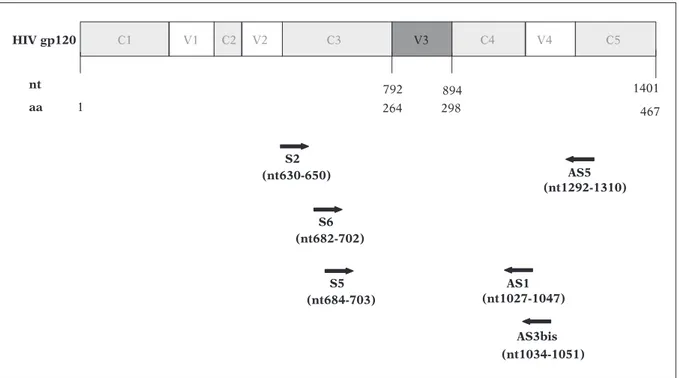
The OSCAR programme was specifically de-signed to set up a new protocol for population-based sequencing of the V3 loop. The protocol has been designed and optimized as follows. HIV-1 RNA was extracted on plasma samples by means of a commercially available kit (QIAamp RNA Viral Mini kit, Qiagen) according to the manufacturer’s instructions. The V3-containing region of the env gene was then reverse-tran-scribed and amplified using the forward primer V3S2 5’CAGCACAGTACAATGTACACA 3’ (nu-cleotide [nt]: 630-650 of HIV-1 HXB2 gp120 env gene) and the reverse primer V3AS5 5’ CTTCTC-CAATTGTCCCTCA 3’ (nt: 1292-1310) (Figure 1, Table 1). The conditions for reverse transcription and amplification were: one cycle at 50°C for 30 min, one cycle 94°C for 2 min, 40 cycles (94°C 30s, 52°C 30s, 72°C 40s), and a final step at 72°C for 10 min, using the following reaction mix: 25 ul of RNA template, 8 ul of 5 mM Mg++, 3 ul of Dnase Rnase free water, 0.75 ul of each primer at a concentration of 10 uM, 1 ul of Rnase out (40 U/ul), 1.5 ul of RT/Taq, 1 ul of dNTPs at a con-centration of 10 mM for a total of 40 ul.
When the RT-PCR product was not visible on agarose gel, a semi-nested PCR was used with the inner forward primer V3S6 5’ CTGTTAAATG-GCAGTCTAGC3’ (nt: 682-702) and the reverse primer V3AS5 (Figure 1, Table 1). Amplification conditions were: one cycle at 93°C for 12 min, 40 cycles (95°C 30s, 52°C 30s, 72°C 40s) and a final step at 72°C for 10 min, using the following re-action mix: 5 ul of buffer taq 10X, 4 ul of Mg++ at a concentration of 25 mM, 32.5 ul of Dnase Rnase free water, 0.9 ul of each primer at a con-centration of 10 uM, 1 ul of dNTPs at a concen-tration of 10 mM, 0.7 ul of Taq (5 u/ul) for a to-tal of 45 ul.
The PCR product was purified by Microcon PCR purification kit (Millipore). Negative and positive
control samples were included in each PCR run to exclude false-positive and false-negative reactions. PCR-products were then sequenced by using the BigDye terminator v.3.1 cycle sequencing kit (Applied-Biosystems), and an automated se-quencer (ABI-3100). Four different overlapping sequence-specific primers were used to ensure the coverage of the V3-sequence by at least two sequence segments (Figure 1). The sequencing
conditions were: one cycle 96°C 3 min, 25 cycles (96°C 30s, 50°C 10s, 60°C 4 min) and the follow-ing primers were used: V3S6 5’ CTGTTAAATG-GCAGTCTAGC 3’, V3S5 5’ GTTAAATGGCAGTC-TAGCAG 3’, V3AS1 5’ GAAAAATTCCCCTCCA-CAATT 3’ and V3AS3bis 5’ CAATTTCTGGGTCC-CCTC 3’ (Figure 1, Table 1).
For a subset of samples, some modifications of the above-mentioned protocol were followed. In S5 (nt684-703) nt 1401 aa 1 467 792 298 894 264 AS1 (nt1027-1047) S6 (nt682-702) AS5 (nt1292-1310) S2 (nt630-650) AS3bis (nt1034-1051) 4 V 2 V 1 V V3 C1 C2 C3 C4 C5 HIV gp120
FIGURE 1 - Location on HIV-1 gp120 coding region of the primers used for the amplification and sequencing of the
V3 loop. Primers S2 and AS5 were used for the RT-PCR reaction. Heminested PCR was directed by primers S6 and AS5. Primers used for sequencing reaction are S6, S5, AS1, and AS3bis.
TABLE 1 - Overview of the primer and cycling conditions for V3 PCR.
PCR Nested PCR
Sense primer 5’-CAGCACAGTACAATGTACACA-3’ 5’- CTGTTAAATGGCAGTCTAGC -3’
Antisense primer 5’-CTTCTCCAATTGTCCCTCA- 3’ 5’-CTTCTCCAATTGTCCCTCA-3’
PCR settings 30 mins 50°C, 1 cycle 12 mins
2 mins 94°C, 1 cycle 93°C, 1 cycle
30s 94°C 30s 95°C
30s 52°C, 40 cycles 30s 52°C 40 cycles
40s 72°C 40s 72°C
10 min 72°C 1 cycle 10 min 72°C 1 cycle
PCR, polymerase chain reaction.
particular, thirthy five plasma samples was se-quenced with an alternative method. Briefly RT-PCR was carried out with a 40 µl volume con-taining 10 µl sample RNA. 16 µl of MMix1, that contain 0.7 µl forward primer V3S2 (5’ CAGCACAGTACAATGTACACA 3’) and reverse primer V3AS5 (5’ CTTCTCCAATTGTCCCTCA 3‘) [15 µM], 1.8 µl dNTPs [10 mM], 1.2 µl dithio-threitol (DTT), 0.6 µl RNase Inhibitor [40 U/µl], 14.5 µl steril water, was mixed with the sample. The Second MMix (11.7 µl RT-PCR buffer, 06 µl Rnase-inhibitor, 1.2 µl RT Enzyme and 2.9 µl Dna Polymerase) was added after 10 min at 50°C. RT-PCR was run with the following temperature program: 20 min at 50°C and 2 min at 94°C, fol-lowed by 40 cycles with 30 s at 94°C, 30 s at 57°C, and 120 s at 68°C and a final extension for 7 min at 68°C.
CLIP sequencing reaction was performed by us-ing a 7-deaza-dGTP Cy5/Cy5.5 dye primer cycle sequencing kit (Siemens Healthcare Diagnostics, Milan Italy) according to the manufacturer’s in-structions. The four CLIP reaction mixtures con-tained 2.8 µl CLIP buffer, 8.8 µl molecular water, 2.8 µl forward primer V3S6 (5’ -Cy5.5-CTGT-TAAATGGCAGTCTAGC-3’) and reverse primer V3AS3bis (5’-Cy5-5’CAATTTCTGCCCCTC GGT-3’) [3 µM], 5 l sample cDNA, 3 µl of the four ter-minator nucleotides, and 4.4 µl Thermosequenasi Enzyme [32U/µl]. The CLIP-specific cycling pro-gram was 5 min at 94°C, followed by 30 cycles of 20 s at 94°C, 20 s at 55°C, and 60 s 70°C and fi-nal extension for 7 min at 70°C. Thereafter, Stop Loading Dye (6 µl) was added. Samples were heated to 94°C for 3 min and incubated at 4°C. Fragments were separated on a TruGene Tower (Siemens Healthcare Diagnostics, Milan Italy) with a 6% polyacrylamide gel. Sequence data were acquired and analyzed using the OpenGene DNA Sequencing System and read against a new-ly created HIV-1 V3 loop sequence-specific refer-ence library.
Genotypic prediction of viral tropism
HIV-1 co-receptor usage was inferred from the V3 nucleotide sequence by using the geno2pheno algorithm available at the following website http://coreceptor.bioinf.mpi-inf.mpg.de/. The sys-tem is based on a support vector machine methodology that has been trained with a set of V3 nucleotide sequences with known
phenotyp-ic tropism. The tool can also analyze amino-acid mixtures deduced from degenerate base calls. The result of the interpretation is given as a quanti-tative value, the false positive rate (FPR), that de-fines the probability of classifying an R5 virus falsely as X4.
HIV-1 co-receptor usage was inferred by using both the clonal and the clinical version of geno2pheno set at FPR of 10% and 5%. For clin-ical inference of viral tropism, viremia at the time of V3 sequencing and nadir of CD4 cell count were used.
Reproducibility of the test and validation of clinical center participating in the OSCAR programme
The OSCAR programme was designed to set up a new protocol for V3 sequencing to be shared with the majority of clinical and virological cen-ters in Italy. For this reason, a validation proce-dure for each virological center has been set up. In particular, for each center, 10 plasma samples were processed in duplicate, one by the center to be validated and the other by the reference cen-ter. In particular, 4 reference centers have been identified in Rome, Milan, Siena, and Padova; these centers have been previously validated us-ing the same above-mentioned procedure. The validation process was considered successful if the V3 nucleotide sequences obtained from the same plasma sample showed a degree of simi-larity of >95% and no change in HIV-1 tropism determination was observed. The validation was successful for all the 20 centers involved in the OSCAR programme.
Statistical analysis
To assess the performances of genotypic tropism testing, the sensitivity and specificity for the de-tection of X4 variants and their 95% confidence intervals (95% CIs) were calculated considering ESTA as reference assay. Sensitivity values were calculated as the proportion of samples that were considered as harboring X4-tropic viruses by genotypic test within the whole group of DM/X4 viruses detected by ESTA. Specificity values were reported as the proportion of patients with R5 viruses by genotype within all the R5 viruses by phenotypic test. Data were analyzed using the sta-tistical software package SPSS (SPSS Inc., Chicago, IL).
Phylogenetic analysis
For each sample, HIV-1 subtype was determined by using the geno2pheno algorithm and con-firmed by phylogenetic analysis of V3-containing nucleotide sequences. In particular, all the se-quences were aligned and compared with refer-ence sequrefer-ences for the Major HIV-1 subtypes, available at: http://hiv-web.lanl.gov/content/hiv-db/SUBTYPE_REF/align.html using CLUSTAL X (Thompson et al., 1994). The sequences were then manually edited by the Bioedit program (Hall et
al., 1999). Phylogenetic trees were generated
us-ing GTR Model of substitution with both NJ and Maximum Likelihood (ML) tree building meth-ods (Swafford KL, 1999). The best fitting nu-cleotide substitution model was tested with the Hierarchical Likelihood Ratio Test (HLRT) im-plemented in the Model Test V3.0 software (Posada et al., 1998). The statistical robustness within each phylogenetic tree was confirmed with a bootstrap analysis using 1000 replicates for the Neighbor-Joining (NJ) tree. All calculations were performed with PAUP* 4.0b10 (http://paup.csit. fsu.edu/about.html) software. For 39 patients, available pol sequences were used to confirm HIV-1 subtype by phylogenetic analysis (Alteri et
al., 2008). Phylogenetic analysis was also used to
identify potential cross-contaminations during the sequencing process.
RESULTS
Patient’s characteristics
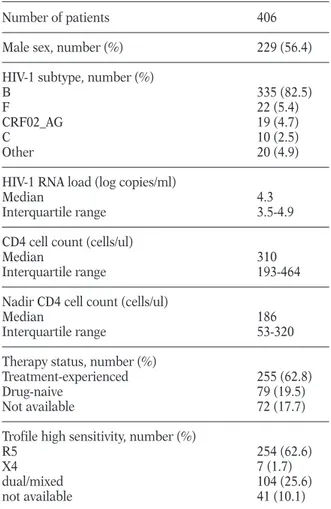
This study included 406 patients with HIV-1 RNA >100 copies/ml and naïve to maraviroc. Their clinical and viro-immunological characteristics are shown in Table 2. At the time of sample col-lection, the median viremia was 4.3 (IQR: 3.5-4.9) log10HIV-1 RNA copies/ml, and the median CD4 count was 310 (IQR:193-464) cells/ul. The large majority of patients harbored HIV-1 B subtype (N = 335, 82.5%). Other non-B subtypes or re-combinant forms observed at a frequency >2% were: F (N = 22, 5.4%), CRF02_AG (N = 19, 4.7%), C (N = 10, 2.5%).
Therapy status was known for 334 (82.3%) pa-tients. Among them, 255 (62.8%) were treatment-experienced patients. The proportion of anti-retroviral-treated patients was similar in B and non-B subtypes (61.3% versus 67.1%). No
statis-tically significant differences in viremia and CD4 cell count were observed between drug-naïve and treatment-experienced patients infected with HIV-1 B subtype (4.6 [3.8-5.0] log10copies/ml ver-sus 4.2 [3.4-4.9] log10copies/ml, and 325 [164-523] log10copies/ml versus 307 [193-450] log10 copies/ml, P>0.100).
Phenotypic characterization of HIV-1 co-receptor usage
Among the 406 plasma samples collected for the study, ESTA was obtained for 365 (89.9%) sam-ples indicating R5 in 254 (69.6%), and DM/X4 in 111 (30.4% of samples (104 [28.5%] DM and 7 [1.9%] X4) (Table 2). A significant lower preva-lence of pure R5-tropic viruses and a higher prevalence of DM-tropic viruses was observed in patients infected with HIV-1 B subtype than in those infected with non-B subtypes or recombi-nant forms (R5: 67.0% versus 82.3%, P=0.022;
TABLE 2 - Patients’ characteristics.
Number of patients 406
Male sex, number (%) 229 (56.4)
HIV-1 subtype, number (%)
B 335 (82.5)
F 22 (5.4)
CRF02_AG 19 (4.7)
C 10 (2.5)
Other 20 (4.9)
HIV-1 RNA load (log copies/ml)
Median 4.3
Interquartile range 3.5-4.9
CD4 cell count (cells/ul)
Median 310
Interquartile range 193-464
Nadir CD4 cell count (cells/ul)
Median 186
Interquartile range 53-320
Therapy status, number (%)
Treatment-experienced 255 (62.8)
Drug-naive 79 (19.5)
Not available 72 (17.7)
Trofile high sensitivity, number (%)
R5 254 (62.6)
X4 7 (1.7)
dual/mixed 104 (25.6)
DM: 30.7% versus 17.7%, P=0.045). All the 7 pure X4-tropic viruses were found in patients infect-ed with HIV-1 B subtype.
In the set of HIV-1 B subtype infected patients, we observed a higher prevalence of R5-tropic viruses and a lower prevalence of DM/X4-tropic viruses in drug-naïve than in treatment-experi-enced patients, even if not statistically significant (R5: 74.5% versus 62.6%, DM/X4: 25.5% versus 37.4%, P>0.100).
Evaluation of the performance of genotypic tropism testing
Efficiency of V3 sequencing
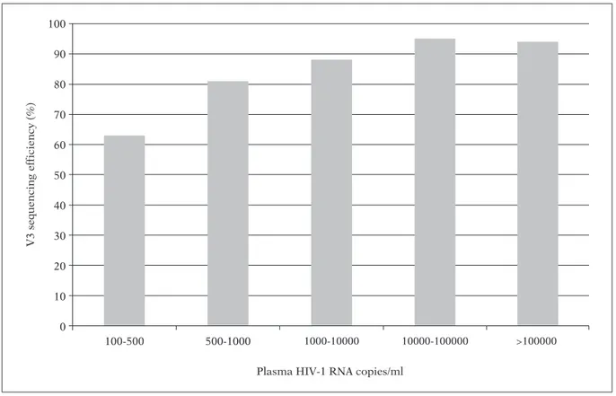
The protocol for V3 sequencing set up in the framework of the OSCAR programme showed an overall efficiency of 84.2%. This efficiency varied according to the level of viremia (Figure 2). In particular, for viremia ranging between 100-500 copies/ml, the rate of successful sequencing was 63%, it progressively increased to 81% and 88%
for viremia ranging between 500-1,000 copies/ml and 1,000-10,000 copies/ml, respectively, and reached ~95% for viremia >10,000 copies/ml (Figure 2). The efficiency of V3 sequencing was not affected by HIV-1 subtype.
Accuracy of genotypic tropism testing in detecting X4-tropic using ESTA as reference
We defined the accuracy of genotypic tropism testing in detecting X4-tropic viruses separately in the set of 303 matched genotypic and phenotyp-ic results from patients infected with HIV-1 B subtype and in the set of 62 matched genotypic and phenotypic results from patients infected with non-B subtypes or recombinant forms (Table 3). We used the geno2pheno algorithm (both the clonal and the clinical version) as interpretation tool since it is so far recognized as the most reli-able and robust algorithm for tropism determi-nation. The geno2pheno algorithm provides a quantitative value, the FPR, that is a measure of
Plasma HIV-1 RNA copies/ml 100 90 80 70 60 50 40 30 20 10 0 V 3 s e q u e n c in g e ff ic ie n c y ( % ) 100-500 500-1000 1000-10000 10000-100000 >100000
FIGURE 2 - V3 sequencing efficiency stratified by viremia levels (HIV-1 RNA copies/ml). In order to provide a
com-prehensive determination of the efficiency of V3 sequencing, the rate of successful sequencing has been determined in a set of 592 samples, including samples involved in the OSCAR programme, and also other plasma samples test-ed so far for both clinical and research purposes.
HIV-1 prediction to use the CCR5-coreceptor. We used as cut-off to discriminate between an R5-using or X4-R5-using virus a FPR of 10%, proposed by the German Recommendations of the National Reference Centre for Retrovirus for determining HIV-1 co-receptor usage, and a FPR of 5%, re-cently used to analyze V3 sequencing obtained from patients enrolled in the Phase III Clinical trial Motivate (Harrigan et al., 2009). If the FPR provided by geno2pheno for a specific V3 se-quence is higher than the established cut-off, the prediction of HIV-1 co-receptor usage is R5. The accuracy measures of the clonal and clinical ver-sion of geno2pheno algorithm are now reported: 1) Analysis of the clonal version of geno2pheno. By analyzing the 303 matched genotypic and phe-notypic results from patients infected with
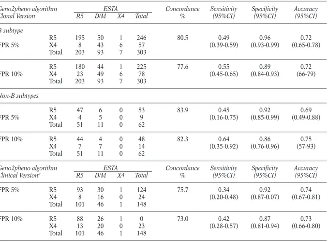
HIV-1 B subtype, we found that the overall concor-dance between ESTA and geno2pheno at a FPR of 5% and 10% was 80.5% and 77.6%, respec-tively (Table 3). The specificity (that measures the concordance between ESTA and genotypic tro-pism testing in detecting R5-viruses) was 96.0% and 89.0% and sensitivity (that measures the con-cordance in detecting DM/X4-viruses) was 49% and 55% when the FPR was set at 5% and 10%, respectively (Table 3). The performances of geno-typic tropism testing in the set of 62 matched genotypic and phenotypic results from patients infected with HIV-1 non B subtypes were not sig-nificantly different from those observed for B sub-type (Table 3).
2) Analysis of the clinical version of geno2pheno. In a subset of 148 HIV-1 B subtype infected patients,
TABLE 3 - Performances of genotypic tropism testing using the enhanced sensitivity version
of Trofile as reference assay
Geno2pheno algorithm ESTA Concordance Sensitivity Specificity Accuracy
Clonal Version R5 D/M X4 Total % (95%CI) (95%CI) (95%CI)
B subtype R5 195 50 1 246 80.5 0.49 0.96 0.72 FPR 5% X4 8 43 6 57 (0.39-0.59) (0.93-0.99) (0.65-0.78) Total 203 93 7 303 R5 180 44 1 225 77.6 0.55 0.89 0.72 FPR 10% X4 23 49 6 78 (0.45-0.65) (0.84-0.93) (66-79) Total 203 93 7 303 Non-B subtypes R5 47 6 0 53 83.9 0.45 0.92 0.69 FPR 5% X4 4 5 0 9 (0.16-0.75) (0.85-0.99) (0.49-0.88) Total 51 11 0 62 FPR 10% R5 44 4 0 48 82.3 0.64 0.86 0.75 X4 7 7 0 14 (0.35-0.92) (0.76-0.96) (57-93) Total 51 11 0 62
Geno2pheno algorithm ESTA Concordance Sensitivity Specificity Accuracy
Clinical Versiona R5 D/M X4 Total % (95%CI) (95%CI) (95%CI)
FPR 5% R5 93 30 1 124 75.7 0.34 0.92 0.74 X4 8 16 0 24 (0.20-0.48) (0.87-0.07) (0.67-0.81) Total 101 46 1 148 FPR 10% R5 88 26 1 0 73.0 0.42 0.87 0.73 X4 13 20 0 23 (0.28-0.57) (0.81-0.94) (0.66-0.80) Total 101 46 1 148
aThe performances of the clinical version of the geno2pheno algorithm have been evaluated in a subset of 148 HIV-1 B subtype infected patients. Abbreviation: ESTA, Enhanced Sensitivity Version of Trofile.
we also evaluated the concordance between the clinical version of geno2pheno algorithm and ES-TA (Table 3). Compared to the clonal version, the clinical version of geno2pheno takes into account the value of viremia at the time of V3 genotyping and the value of nadir CD4 cell count. The clini-cal version was 75.7% and 73.0% concordant with ESTA when the FPR was set at 5% and 10%, re-spectively, with a specificity ranging from 92% to 87% and a sensitivity ranging from 34% to 42%, respectively. Thus, in our dataset, the perform-ances of the clinical version were not significantly higher than that observed for the clonal one.
DISCUSSION
The OSCAR study has evaluated the perform-ances of a genotypic tropism testing in clinical settings using ESTA as reference assay in one of the largest set of matched genotypic and pheno-typic results collected so far. This has been feasi-ble by the establishment of an extensive network of clinicians and virologists throughout Italy that has allowed the collection and analysis of 406 matched genotypic and phenotypic results. In line with other European studies, this study supports the reliability and the use of genotypic tool for tropism determination in clinical practice. Several studies have evaluated the performances of genotypic tropism testing using the original version of Trofile or other phenotypic assays as reference (Low et al., 2007; Poveda et al., 2007; Raymond et al., 2008; Poveda et al., 2009; Chueca
et al., 2009; Harrigan et al., 2009; Seclen et al.,
2009). The performances of genotypic tropism testing in comparison to ESTA have been as-sessed only in a very limited number of studies using a limited number of patients (Strang et al., 2009, De Luca et al., 2009, Sanchez et al., 2009). Our and the above-mentioned studies are in agreement regarding the high concordance be-tween genotypic and phenotypic tropism testing (including ESTA) in the detection of R5-tropic viruses. For instance, the specificity observed in our dataset ranges from 86% to 96% when the clonal version of geno2pheno has been used. Conversely, a lower concordance is generally ob-served in the detection of DM/X4 viruses (sensi-tivity in our cohort ranging from 42% to 64%). This result can be explained either by the fact that
genotypic tropism testing cannot detect X4-trop-ic viruses representing less than ~10-20% of vi-ral population, or by the fact that the current al-gorithms for HIV-1 tropism prediction do not take into account additional regions in the env gene, outside the V3 loop. Even if the clinical rel-evance of including additional regions remains to be established, previous studies have demon-strated that mutations in the V1/V2 loops and the C4 region of the gp120 are involved in mecha-nisms underlying co-receptor binding and usage (Suphaphiphat et al., 2010; Pastore et al., 2006). A recent study has also reported an increase in the accuracy of interpretation when both V2 and V3 were used compared to V2 or V3 alone (Thielen et al., 2007).
Despite this lower sensitivity, recent studies have clearly shown that genotypic tropism testing is comparable with Trofile assay in predicting viro-logical response to a maraviroc containing HAART regimen. In particular, the virological re-sponse to a maraviroc-containing regimen has been retrospectively predicted using the original version of Trofile and population-based V3 se-quencing in a cohort of treatment-experienced patients enrolled in the framework of two phase III clinical trials MOTIVATE 1 and 2 and in the 1029 study (Harrigan et al., 2009). Both Trofile and population-based V3 sequencing were equal-ly successful in predicting virological response at week 12 and 24. Indeed, the percentage of viro-logical success (viremia <50 copies/ml) at week 24 was 46.4% and 46.1% when the baseline pres-ence of R5-tropic viruses was determined by Trofile or V3-sequencing, respectively (Harrigan
et al., 2009). Similar results have been recently
observed in drug-naive patients enrolled in the MERIT trials using ESTA and V3-sequencing (Mc Govern et al., 2010). Thus, these studies (although not randomized) support the use of genotyping in clinical settings to select candidates for mar-aviroc treatment. In addition, the 1029 study has shown that 45% of patients classified as R5 by the original Trofile and then reclassified as D/M by ESTA reached virological success (viremia <50 copies/ml) at week 48 of a maraviroc-containing regimen. In particular, by using the ultra-deep se-quencing methodology, a rapid decline of viremia (3 log) has been observed at week 12 in patients where X4-tropic viruses were present in less than 10% of the entire viral population (Swenson et
al., 2009). Another recent study have highlighted
the ability of a maraviroc-containing regimen to suppress HIV-1 replication in presence of DM-tropic viruses (Garcia et al., 2009; Garcia et al., 2010). Thus, overall results suggest that the use CCR5 antagonists could be enlarged to select pa-tients with DM-tropic viruses and underline the need of studies aimed at defining a more precise threshold of X4-tropic viruses below which a CCR5 antagonist together with other active drugs can be safely used in different clinical contexts. In this light, the use of highly sensitive assay for the determination of HIV-1 tropism (such as ESTA) could limit the number of patients that can ben-efit at least partially from treatment with a CCR5 antagonist.
Genotypic tropism testing has been also shown to be a valid and accurate assay in predicting the risk of clinical progression in both drug-naïve and treatment-experienced patients (Fouchier et al., 1992; De Jong et al., 1992; Brumme et al., 2004; Delobel et al., 1999; Lin et al., 2010). In particu-lar, in a prospective cohort study of patients ini-tiating antiretroviral regimen, patients with X4-tropic viruses predicted by genotyping had earli-er mortality aftearli-er starting thearli-erapy despite achiev-ing viral suppression to less than 500 copies/ml compared with patients with R5-viruses. Again, these results support the use of genotypic tropism testing can be a valuable tool in clinical practice not only to determine patients that can benefit CCR5 antagonist treatment but also for an opti-mized monitoring of HIV-1 infected patients. The methodology of V3 sequencing that has been designed in the framework of the OSCAR pro-gramme is characterized by a high rate of effi-ciency even in presence of viremia lower than 1,000 copies/ml. Indeed, the rate of successful se-quencing was 63% and 81% for viremia ranging from 100-500 copies/ml and 500-1,000 copies/ml, respectively, and reached ~95% for viremia >10,000 copies/ml. The availability of method-ologies that can assess HIV-1 co-receptor usage even when viremia is low, is crucial since they can be used even in those antiretroviral-treated patients with persistently low, but detectable, lev-els of HIV-1 RNA in the range of 50-1000 copies/ml.
In addition, the methodology of V3 sequencing used in this study is based on a single round of amplification of the V3 region. In other studies,
a triplicate round of amplification has been per-formed (Harrigan et al., 2009; Mc Govern et al., 2010). The use of single versus triplicate geno-typic analysis is still matter of debate. Indeed, preliminary data from clinical trials have sug-gested that genotypic analysis in triplicate may increase the detection of X4-tropic viruses. However, a recent study has evaluated the effect of triplicate testing on genotypic tropism predic-tion in routine clinical practice and has found that the triplicate testing resulted in an enhanced detection of X4-variants only in a small percent-age of patients (Wensing et al., 2010). Further studies are necessary to evaluate the clinical sig-nificance of triplicate versus single genotypic analysis for tropism prediction.
In conclusion, in line with other European stud-ies, our study supports the routine use of geno-typic tropism testing in clinical settings for mon-itoring HIV-1 infected patients and for a better tuning of in vivo efficacy of CCR5 antagonists.
Acknowledgements
We gratefully thank Federica Forbici, Fabio Continenza, Maria Santoro, Andrea Biddittu, Domenico Pinto and Marzia Romani for sequen-cing and data management, and all the other par-ticipants and members of the OSCAR study group. This work was financially supported by grants from from CHAIN, Collaborative HIV and Anti-HIV Drug Resistance Network, Integrated Project no.223131, funded by the European Commission Framework 7 Program.
OSCAR study group
The complete list of centers and members parti-cipating in the OSCAR programme is as follows:
“San Raffaele” Hospital (Milan): Adriano Lazzarin,
Massimo Clementi, Silvia Nozza, Filippo Canducci, Enzo Boeri; “L. Sacco” Hospital
(Milan): Giuliano Rizzardini, Massimo Galli
Valeria Micheli; “S. Paolo” Hospital (Milan): Antonella D’Arminio Monforte; Busto Arsizio
Hospital (Busto Arsizio [MI]): Tiziana Quirino; “S. Gerardo” Hospital, (Monza [MI]): Andrea Gori; Ospedali Riuniti (Bergamo): Franco Maggiolo,
Anna Paola Callegaro; IRCCS Policlinico S. Matteo
(Pavia): Renato Maserati, Fausto Baldanti,
Stefania Paolucci; University of Turin (Turin): Giovanni Di Perri, Valeria Ghisetti, Tiziano Allice;
Policlinico “S. Orsola-Malpighi” (Bologna): Marco
Martino” Hosptial (Genova): Claudio Viscoli,
Antonio Di Biagio, Bianca Bruzzone; Policlinico
of Modena (Modena): Cristina Mussini, William
Gennari, Monica Pecorari; Marche Politechnic
University Medical School (Ancona): Andrea
Giacometti, Alessia Monachetti, Patrizia Bagnarelli; “S.M. Annunziata” Hospital (Firenze): Francesco Mazzotta, Massimo Di Pietro; “Careggi”
Hospital (Firenze): Francesco Leoncini, Gaetana
Sterrantino; University of Siena (Siena): Maurizio
Zazzi; Policlinico“Tor Vergata” (Rome): Massimo
Andreoni; I.N.M.I. “L. Spallanzani” (Rome): Andrea Antinori, Carlo Federico Perno, Roberta D’Arrigo; University of Rome “La Sapienza”
(Rome): Vincenzo Vullo, Guido Antonelli,
Ombretta Turriziani; Catholic University “Sacro
Cuore” (Rome): Roberto Cauda, Andrea De Luca,
Giovanni Fadda, Maria Rosaria Santangelo;
University of Foggia and Bari: Gioacchino
Angarano, Laura Monno, Annalisa Saracino, Grazia Punzi.
REFERENCES
ALTERIC., SVICHERV., GORIC., D’ARRIGOR., CICCOZZI
M., ORCHIN., CECCHERINI-SILBERSTEINF., PONTIERI
S., SELLERIM., AVIANIBARDACCIS., GIULINIM., ELIA
P., SCOGNAMIGLIOP., BALZANOR., GIRARDIE., PERNO
CF. (2009). Characterization of the patterns of drug-resistance mutations in newly diagnosed HIV-1 in-fected patients naive to the antiretroviral drug. BMC
Infectious Diseases. 9, 111.
BERGERE.A., DOMSR.W., FENYOE.M., KORBERB.T.M.,
LITTMAN D.R., MOORE J.P., SATTENTAU Q.J.,
SCHUITEMAKERH., SODROSKIJ., WEISSR.A. (1998). A new classification for HIV-1. Nature. 391, 240.
BRUMME Z.L., DONG W.W., YIP B., WYNHOVEN B.,
HOFFMAN N.G., SWANSTROM R., JENSEN M.A.,
MULLINSJ.I., HOGGR.S., MONTANERJ.S., HARRIGAN
P.R. (2004). Clinical and immunological impact of HIV envelope V3 sequence variation after starting initial triple antiretroviral therapy. AIDS.18,
F1-F9.
CHUECAN., GARRIDOC., ALVAREZM. POVEDAE., LUNAJ. D., ZAHONERON., HERNANDEZ-QUEROJ., SORIANOV.,.
MAROTO C., MENDOZA C., GARCıA F. (2009).
Improvement in the determination of HIV-1 tro-pism using the V3 gene sequence and a combina-tion of bioinformatic tools. J. Med. Virol. 81,
763-767.
DEJONGJ.J., DERONDEA., KEULENW., TERSMETTEM., GOUDSMITJ. (1992). Minimal requirements for the human immunodeficiency virus type 1 V3 domain
to support the syncytium-inducing phenotype: Analysis by single amino acid substitution. J. Virol.
66, 6777-6780.
DELOBELP., SANDRES-SAUNEK., CAZABATM., PASQUIER
C., MARCHOUB., MASSIPP., IZOPETJ. (2005). R5 to
X4 switch of the predominant HIV-1 population in cellular reservoirs during effective highly active an-tiretroviral therapy. J. Acquir. Immune Defic. Syndr.
38, 382-392.
DORRP., WESTBYM., DOBBSS., GRIFFINP., IRVINEB.,
MACARTNEY M., MORI J., RICKETT G., SMITH
-BURCHNELLC., NAPIERC., WEBSTERR., ARMOURD., PRICED., STAMMENB., WOODA., ANDPERROSM.
(2005). Maraviroc (UK-427,857), A potent, orally bioavailable, and selective small-molecule inhibitor of chemokine receptor CCR5 with broad-spectrum anti-human immunodeficiency virus type 1 activi-ty. Antimicrob. Agents Chemother. 49, 4721-4732.
FOUCHIER R.A., GROENINK M., KOOTSTRA N.A.,
TERSMETTE M., HUISMAN H.G., MIEDEMA F. SCHUITEMAKERH. (1992) Phenotype-associated
se-quence variation in the third variable domain of the human immunodeficiency virus type 1 gp120 molecule. J. Virol.66, 3183-3187.
HALLT.A. (1999). BioEdit: a user-friendly biological
se-quence alignment editor and analysis program for Windows 95/98/NT. Nucleic acids symposium se-ries. 41, 95-98.
HARRIGANP.R., MCGOVERNR., DONGW. ., THIELENA.,
JENSENM., MOT., CHAPMAND., LEWISM., JAMESI.,
VALDEZH. (2009). Screening for HIV tropism using
population-based V3 genotypic analysis: a retro-spective virological outcome analysis using stored plasma screening samples from MOTIVATE-1.
Antivir Ther. 14 (Suppl. 1), A17.
HARRIGANP.R., MCGOVERNR., DONGW., THIELENA., JENSENM., MOT., CHAPMAND., LEWISM., JAMESI.,
ELLERYS., HEERAJ., VALDEZH. (2009). Screening
for HIV tropism using population-based V3 geno-typic analysis: a retrospective virologic outcome analysis using stored plasma screening samples from the MOTIVATE studies of maraviroc in treat-ment experienced patients. 5thIAS conference on HIV pathogenesis, treatment and prevention, 19-22 July 2009.
JENSENM.A., VAN’TWOUTA.B. (2003). Predicting
HIV-1 coreceptor usage with sequence analysis. AIDS
Rev. 5, 104-112.
JENSENM.A., LIF.S., VAN‘TWOUTA.B., NICKLED.C.,
SHRINERD., HEH.X., MCLAUGHLINS., SHANKARAPPA
R., MARGOLICKJ.B., MULLINSJ.I. (2003). Improved
coreceptor usage prediction and genotypic moni-toring of R5-to-X4 transition by motif analysis of human immunodeficiency virus type 1 env V3 loop sequences. J. Virol.77, 13376-13388.
LINN.H., KURITZKESD.R. (2009). Tropism testing in the clinical management of HIV-1 infection, Current Opinion in HIV and AIDS. 4, 481-487.
LOWA.J., DONGW., CHAND., SINGT., SWANSTROMR.,
JENSEN M., PILLAI S., GOOD B., HARRIGAN P.R.
(2007). Current V3 genotyping algorithms are in-adequate for predicting X4 co-receptor usage in clinical isolates, AIDS. 21, F17-F24.
MCGOVERNR., DONGW. ZHONGX. KNAPPD., THIELEN
A., CHAPMAN D., LEWIS M., JAMESI., VALDEZH.,
HARRIGANR. (2010). Population-based sequencing of the V3-loop is comparable to the enhanced sen-sitivity trofile assay in predicting virologic response to maraviroc ot treatment-naïve patients in the MERIT trial, 17thConference on retroviruses and opportunistic infections, February 16-19, 2010, San Francisco, CA, USA.
PASTOREC., NEDELLECR., RAMOSA., PONTOWS., RATNER
L., MOSIERD.E. (2006). Human immunodeficien-cy virus type 1 coreceptor switching: V1/V2 gain-of-fitness mutations compensate for V3 loss-of-fit-ness mutations. J. Virol.80, 750-758.
POSADAD., CRANDALLK.A. (1998). Modeltest: testing the model of DNA substitutionoxfordjournals.org - Bioinformatics - Oxford Univ Press.
POVEDAE., BRIZV., ROULETV., DELMARGONZALEZM.,
FAUDON J.L., SKRABAL K., SORIANO V. (2007). Correlation between a phenotypic assay and three bioinformatic tools for determining HIV co-re-ceptor use. AIDS.21, 1487-1490.
POVEDA E., SECLÉN E., GONZÁLEZ M.M., GARCÍA F., CHUECAN., AGUILERAA., RODRÍGUEZJ.J., GONZÁLEZ
-LAHOZJ., SORIANOV. (2009). Design and validation
of new genotypic tools for easy and reliable esti-mation of HIV tropism before using CCR5 antag-onists, JAC.
PRINCENK., SCHOLSD. (2005). HIV chemokine
recep-tor inhibirecep-tors as novel anti-HIV drugs. Cytokine
Growth. Factor Rev. 16, 659-677.
RAYMONDS., DELOBELAP., MAVIGNERAM., CAZABATAM.,
SOUYRISA C., SANDRES-SAUNE K., CUZIND L.,
MARCHOUB B., MASSIP P., IZOPET J. (2008).
Correlation between genotypic predictions based on V3 sequences and phenotypic determination of HIV-1 tropism. AIDS.22, F11-F16.
REGOESR.R., BONHOEFFERS. (2005). The HIV
core-ceptor switch: A population dynamical perspec-tive. Trends. Microbiol.13, 269-277.
SANCHEZV., ROBLEDANOC., CIPRIAND., MONTOLIOF.,
ESCOLANO C., PADILLAS., RAMOSJ.M., MASIAM.,
GUTIERREZF. (2009). Evaluation of genotypic al-gorithms to predict HIV-1 coreceptor tropism
as-say, 12th Europeans AIDS Conference/EACS, Cologne, Germany 11-14 Novembre.
SECLENE., GARRIDOC., GONZALEZM.M., GONZALEZ
-LAHOZ J., MENDOZA C., SORIANO V., POVEDA E.
(2009). High sensitivity of specific genotypic tools for detection of X4 variants in antiretroviral-expe-rienced patients suitable to be treated with CCR5 antagonists. J. Antimicrob. Chemother.
SYMONS J., VENDEKERCKHOVE L., PAREDES R., VERHOFSTEDEC., BELLIDOR., DEMECHELEERE., VAN
HAM M., VAN LELYVELD S.F.L., STAM A.J., VAN
VERSENDAALD., NIJHUISM., WENSINGA.M.J. (2010).
Effect of triplicate testing on genotypic tropism prediction in routine clinical practise. International HIV and hepatitis virus drug resistance workshop and curative strategies. June 8-10, Dubrovnik, Crotia. Antiviral. Therapy. 15, A101.
STRANGA.L., CAMERONJ., BOOTHC., GARCIAA., GERETTI
AM. (2009). Genotypic co-receptor tropism: corre-lation with enhanced Trofile. 7thEuropean HIV Drug Resistance Workshop. March 25-27; Stockholm, Sweden. Abstract 80.
SUPHAPHIPHATP., ESSEXM., LEET.H. (2007). Mutations
in the V3 stem versus the V3 crown and C4 region have different effects on the binding and fusion steps of HIV type 1 gp120 interaction with the CCR5 coreceptor. Virology.360, 182-190.
SWAFFORDK.L. (1999). PAUP 4.0: phylogenetic
analy-sis with parsimony (and other methods), version 4.0 b2a, Sunderland, Mass: Sinauer Associates Inc. SWENSONL., DONGW., MOT., WOODSC., THIELENA.,
JENSENM., GLASCOCKC., MONTANERJ., HARRIGAN
R. (2009). Quantification of HIV Tropism by « deep » sequencing shows a broad distribuition of prevalence of X4 variants in clinical samples that is associated with virological outcome, 16th Conference on retroviruses and opportunistic in-fections, February 8-11, 2009, Montreal, Canada. THIELEN ET AL. (2007). Improving HIV-1 co-receptor
usage prediction from genotype with regions out-side v3. 3rd International Workshop on Targeting HIV entry. 7-8 December 2007 Washington DC USA.
THOMPSON J.D., HIGGINS D.G., GIBSON T.J. (1994).
CLUSTAL W: improving the sensitivity of progres-sive multiple sequence alignment through se-quence weighting, position-specific gap penalties and weight matrix choice. Nucleic Acids Research.