Contents lists available atScienceDirect
Ticks and Tick-borne Diseases
journal homepage:www.elsevier.com/locate/ttbdisA retrospective study of the characterization of Rickettsia species in ticks
collected from humans
Valeria Blanda
a, Alessandra Torina
a,⁎, Francesco La Russa
a, Rosalia D
’Agostino
a, Kety Randazzo
a,
Salvatore Scimeca
a, Elisabetta Giudice
b, Santo Caracappa
a, Antonio Cascio
c, José de la Fuente
d,eaIstituto Zooprofilattico Sperimentale della Sicilia, Via Gino Marinuzzi 3, 90129 Palermo, Italy
bDepartment of Chemical, Biological, Pharmaceutical and Environmental Sciences, University of Messina, Viale Ferdinando Stagno d'Alcontres 31, 98166 S.
Agata-Messina, Italy
cDepartment of Health Promotion Sciences and Mother and Child Care“G. D'Alessandro”, University of Palermo, Via del Vespro, 129 - 90127 Palermo, Italy dSaBio. Instituto de Investigación en Recursos Cinegéticos, IREC (CSIC, UCLM, JCCM), Ronda de Toledo s/n, 13005 Ciudad Real, Spain
eDepartment of Veterinary Pathobiology, Center for Veterinary Health Sciences, Oklahoma State University, Stillwater, OK 74078, USA
A R T I C L E I N F O
Keywords: Ticks Rickettsia Spotted fever group Humans Zoonosis Molecular analysis
A B S T R A C T
Rickettsiae (family Rickettsiaceae, order Rickettsiales) are obligate intracellular bacteria transmitted by arthropod vectors. Several Rickettsia species causing vector-borne rickettsioses belong to the spotted fever group (SFG). Traditionally, Rickettsia conorii has been considered as the main etiologic agent of Mediterranean spotted fever. However, the molecular characterization of rickettsiae allowed identifying other species involved in spotted fever in the Mediterranean region.
In this study, 42 ticks collected from humans were subjected to morphological identification and molecular characterization of Rickettsia species potentially involved in human rickettsiosis in Sicily.
Fourteen ticks positive to at least two Rickettsia spp. molecular markers were used in the study. Identified Rickettsia spp. included R. conorii, found in Rhipicephalus sanguineus sensu lato and Rhipicephalus turanicus, Rickettsia aeschlimannii found in Hyalomma marginatum, Hyalomma lusitanicum, Dermacentor marginatus and Ixodes ricinus, Rickettsia massiliae found in R. turanicus and R. sanguineus s.l., and Rickettsia slovaca found in D. marginatus and R. sanguineus s.l.
Our results showed a great variety of zoonotic Rickettsia spp. in ticks collected from humans in Sicily. The Rickettsia spp. reported in this study were identified in previously recognized or new potential tick vectors in Europe, highlighting the risk of infection by different Rickettsia spp. for humans bitten by ticks in Sicily.
1. Introduction
In the last years, several bacterial, viral and parasitic diseases affecting humans and animals have emerged in Europe. Many of the etiological agents of these diseases are transmitted by arthropod vectors (de la Fuente et al., 2008). This phenomenon may be associated with social, ecological, environmental, and microbial risk factors, which act synergistically to facilitate emergence of these pathogens in Europe. Hard ticks (Acari: Ixodidae) are able to transmit pathogens such as viruses, bacteria and protozoa through their bite to humans and animals, and may serve as reservoirs and/or amplifiers for most of
these species (Estrada-Peña et al., 2014; Gortazar et al., 2014). The prokaryotic microorganisms of the order Rickettsiales (genera Rickettsia, Anaplasma, Ehrlichia) are agents of important diseases. These pathogens are transmitted by arthropod vectors and many of them can constitute a risk not only for animals but also for humans (de la Fuente et al., 2008). Rickettsiae (family Rickettsiaceae, order Rickettsiales) are obligate intracellular bacteria transmitted by ticks,fleas, lice and mites. Members of the genus Rickettsia may be classified into spotted fever group (SFG) rickettsiae, typhus group rickettsiae, the Rickettsia bellii group, and the Rickettsia canadensis group (Parola et al., 2013). Several Rickettsia spp. causing vector-borne rickettsiosis belong to the spotted
http://dx.doi.org/10.1016/j.ttbdis.2017.04.005
Received 5 December 2016; Received in revised form 10 April 2017; Accepted 10 April 2017
⁎Corresponding author.
E-mail addresses:[email protected](V. Blanda),[email protected](A. Torina),[email protected](F. La Russa),
[email protected](R. D’Agostino),[email protected](K. Randazzo),[email protected](S. Scimeca),[email protected](E. Giudice),
[email protected](S. Caracappa),[email protected](A. Cascio),[email protected](J. de la Fuente).
Abbreviations: DEBONEL, Dermacentor-borne Necrosis Erythema and Lymphadenopathy; gltA, citrate synthase; MSF, Mediterranean spotted fever; OmpA, outer membrane protein A; OmpB, outer membrane protein B; PCR, polymerase chain reaction; SENLAT, scalp escar neck lymphadenopathy; SFG, spotted fever group; s.l., sensu lato; TIBOLA, tick-borne lymphadenopathy
Available online 12 April 2017
1877-959X/ © 2017 The Authors. Published by Elsevier GmbH. This is an open access article under the CC BY-NC-ND license (http://creativecommons.org/licenses/BY-NC-ND/4.0/).
fever group (SFG), which represents one of the oldest-known vector-borne zoonosis (Parola et al., 2013).
In the Mediterranean area, Rickettsia conorii, comprising a variety of genospecies (Zhu et al., 2005), was traditionally considered as the main etiologic agent of the Mediterranean spotted fever (MSF). MSF is widely distributed through southern Europe, Africa and the Middle East, and it is an emerging or reemerging disease in some regions, while in some other countries of the Mediterranean basin incidence of MSF has increased in the past 10 years (Duque et al., 2012). However, in recent years, the amplification and sequencing of different molecular markers allowed the molecular characterization of strains, and the identification of many new Rickettsia spp. or subspecies within the SFG group involved in human rickettsiosis and considered as emerging pathogens (Kernif et al., 2016). They include R. slovaca, implicated in develop-ment of tick-borne lymphadenopathy (TIBOLA) or Dermacentor-borne necrosis erythema and lymphadenopathy (DEBONEL) in humans (Cazorla et al., 2003), R. helvetica (Fournier et al., 2004), R. aeschli-mannii (Raoult et al., 2002), R. massiliae (Beati and Raoult, 1993; Vitale et al., 2006), and R. monacensis (Jado et al., 2007; Simser et al., 2002). Other new Rickettsia spp. include R. sibirica sensu stricto (Shpynov et al., 2006), R. heilongjiangensis (Shpynov et al., 2006), R. mongoloti-monae (Psaroulaki et al., 2005), and R. akari (Radulovic et al., 1996). Recently, R. felis was also described as an emerging pathogen of medical importance (Perez-Osorio et al., 2008) also present in Sicily (Giudice et al., 2014).
The objective of this study was the molecular identification and characterization of Rickettsia spp. in ticks collected from humans through the use of a multi-gene assay for the amplification and sequencing of different molecular markers. These findings highlighted the importance of the molecular characterization of Rickettsia spp. in ticks collected from humans.
2. Material and methods 2.1. Tick collection and identification
During the years 2012 and 2013, 42 individuals have been bitten by ticks in the metropolitan city of Messina, in the Northeastern part of Sicily (Italy). They contacted the Policlinic Hospital of Messina, at the Complex Operative Unit of Infectious Diseases and the ticks were removed and collected from them. The area is characterized by a Mediterranean temperate climate and by a territory mostly mountai-nous, with some alluvial plains at the mouths of rivers. Only one of the individuals showed clinical manifestations of rickettsiosis. The rest of the individuals were asymptomatic. Collected ticks were stored in alcohol and identified using appropriate taxonomic keys (Apanaskevich et al., 2008; Manilla, 1998; Nava et al., 2015; Walker et al., 2000).
In case the collected ticks were in a state of preservation not suitable for morphological identification at the species level, they were sub-jected to molecular analysis for species identification. For this purpose, ticks were sectioned longitudinally and one half of each tick was used for DNA extraction. Tick halves were incubated overnight in 180μl of
Genomic Digestion Buffer and 20 μl of proteinase K to digest tick tissues. DNA was extracted using the PureLink Genomic DNA kit (Invitrogen, Carlsbad, CA, USA) following the manufacturer’s instruc-tions. The remaining half of each tick was preserved in alcohol. The extracted nucleic acids were analyzed by PCR amplifying a 360 bp fragment of the mitochondrial 12S rDNA (Beati and Keirans, 2001). 2.2. Rickettsia identification and analysis
The extracted nucleic acids were analyzed by polymerase chain reactions (PCR) targeting the outer membrane protein A (OmpA) (Oteo et al., 2006), outer membrane protein B (OmpB) (Choi et al., 2005) and the citrate synthase (gltA) (Roux et al., 1997) genes to detect the presence of DNA from Rickettsia spp. (Table 1). Each PCR reaction included a positive control, consisting of DNA from Rickettsia conorii Malish 7 strain cultured in VERO cells, and a negative control without DNA. PCRs were carried out in an MJ Research PTC-200 Peltier thermal cycler. PCR products were visualized after electrophoretic migration on a 1.5% agarose gel. PCR products were purified using commercial kits following the manufacturer’s procedures, quantified and sent for sequencing to Macrogen Inc. (Macrogen Europe, Amsterdam, The Netherlands).
Obtained sequences were analyzed using Bioedit (Ibis Biosciences, Carlsbad, CA, USA) and ClustalW version 2.0.10 (www.ebi.ac.uk/ clustalw) for nucleotide sequence identity to the reference strains reported in the GenBank. The Basic Local Alignment Search Tool (BLAST), DAMBE (http://dambe.bio.uottawa.ca/dambe.asp) and MEGA (www.megasoftware.net) software were used to obtain similar-ity percentages among the analyzed sequences. Neighbour-joining method was used for phylogenetic analysis using Clustal W. Obtained sequences were submitted to GenBank (accession numbers KT861865-KT861892).
3. Results
3.1. Eight tick species collected from humans in Sicily
Morphological tick identification allowed identifying the following tick species collected from humans in Sicily: Rhipicephalus turanicus (N = 13), Hyalomma lusitanicum (N = 11), Rhipicephalus sanguineus sensu lato (N = 7), Dermacentor marginatus (N = 4), Haemaphysalis punctata (N = 3), Hyalomma marginatum (N = 2), Ixodes ricinus (N = 1), and Rhipicephalus sp. (N = 1). For the Rhipicephalus sp. tick, morphological identification at species level was not possible due to its preservation state. This tick was identified as R. bursa by molecular techniques. Obtained sequence was submitted to GenBank (accession number KU512950).
3.2. Thirty three percent prevalence of SFG Rickettsia in ticks collected from humans in Sicily
Of the 42 tick samples, 14 (33%) resulted positive for at least two of
Table 1
PCR performed in this study for the amplification of different Rickettsia spp. molecular targets.
Target Primers Fragment length Reference
OmpA Rr190.70p ATGGCGAATATTTCTCCAAAA Rr190.701n GTTCCGTTAATGGCAGCATCT Rr190.602n AGTGCAGCATTCGCTCCCCCT 631 bp (first) 631 bp (semi-nested) Oteo et al. (2006)
OmpB rompB OF GTAACCGGAAGTAATCGTTTCGTAA rompB OR GCTTTATAACCAGCTAAACCACC rompB SFG IF GTTTAATACGTGCTGCTAACCAA rompB SFG IR GGTTTGGCCCATATACCATAAG 511 bp (first) 425 bp (nested) Choi et al. (2005) gltA RpCS.877p GGGGGCCTGCTCACGGCGG RpCS.1258n ATTGCAAAAAGTACAGTGAACA 381 bp Roux et al. (1997)
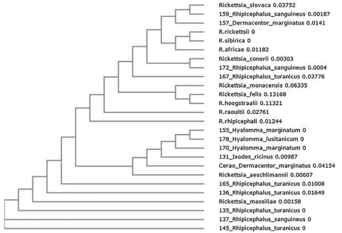
the Rickettsia spp. molecular markers used in the study (ompA, ompB and gltA). Positive PCR products were sequenced and the analysis of obtained sequences allowed identifying R. conorii, detected in R. sanguineus s.l. and R. turanicus, and other SFG Rickettsia (Table 2). R. aeschlimannii was found in five ticks belonging to the species H. marginatum, H. lusitanicum, D. marginatus and I. ricinus. R. massiliae was detected in four R. turanicus ticks and in R. sanguineus s.l., while R. slovaca was identified in D. marginatus and R. sanguineus s.l. Phyloge-netic multilocus analysis with ompA–ompB sequences (GenBank acces-sion numbers KT861865-KT861892;Table 2) confirmed the identity of the Rickettsia spp. identified in this study (Fig. 1).
4. Discussion
A great variety of zoonotic Rickettsia spp. were identified in this study in the ticks collected from humans in Sicily. The main agent of MSF, R. conorii, was detected in only two ticks belonging to the species R. sanguineus s.l. and R. turanicus. R. sanguineus s.l. is extensively recognized as the main vector of Rickettsia conorii. However, the role of R. turanicus as vector of R. conorii has not yet been proven (Parola et al., 2013), even iffindings of the pathogen in this tick species have been reported, also in Italy (Mancini et al., 2015). In our study, R. turanicus was collected from the only symptomatic patient. To the best of our knowledge, this is the first report of the possible vector role of R. turanicus in the transmission of R. conorii.
R. aeschlimannii was identified in known (H. marginatum and I. ricinus;Parola et al., 2013) and potentially new (D. marginatus and H. lusitanicum) tick vectors of this Rickettsia sp. R. aeschlimannii wasfirst described in 1997 in H. marginatum ticks from Morocco (Beati et al., 1997), and later detected in ticks from Niger, Zimbabwe, and Mali (Parola et al., 2001). In Europe, R. aeschlimannii is mainly associated with ticks belonging to the genus Hyalomma, and was identified in 2002 in a patient with MSF-like illness (Raoult et al., 2002). Since then, other reports of R. aeschlimannii infection were described in patients from South Africa (Pretorius and Birtles, 2002) and Greece (Germanakis et al., 2013). The pathogenicity of this bacterium to humans is not well understood, although MSF-like lesions were reported (Germanakis et al., 2013). Our study emphasizes the risk of rickettsiosis due to R. aeschlimannii in Sicily and other Mediterranean countries where H. marginatum is present.
R. massiliae is transmitted by tick vectors of the Rhipicephalus genus. In Europe, the vectors of this Rickettsia species are R. turanicus, R. sanguineus s.l., R. bursa, R. pusillus and I. ricinus (Parola et al., 2013). Ourfinding of R. massiliae in Rhipicephalus ticks collected from humans confirmed this pathogen-vector association and emphasized the possi-ble association between R. massiliae and human rickettsiosis in Sicily. In fact, until now, only three cases of human rickettsiosis due to R.
massiliae have been documented and confirmed by molecular methods in Europe, and two of these cases occurred in Sicily (Cascio et al., 2013; Parola et al., 2008; Vitale et al., 2006). Thefirst case was detected in a blood sample from a patient hospitalized with fever and skin rash in Sicily (Vitale et al., 2006). The second case was a patient who suffered from spotted fever and acute loss of vision in southern France (Parola et al., 2008), and the third case was a child showing scalp eschar and neck lymphadenopathy in Sicily (Cascio et al., 2013).
R. slovaca was originally isolated from D. marginatus in Czechoslovakia in 1968, and it is considered the main etiologic agent of TIBOLA (Lakos, 1997), DEBONEL (Oteo and Ibarra, 2002) and SENLAT (scalp escar neck lymphadenopathy) (Angelakis et al., 2010). In Europe, R. slovaca is usually associated with Dermacentor ticks. D. marginatus together with D. reticulatus are the recognized vectors for this Rickettsia sp. in Europe (Parola et al., 2013). In this study, evidences of R. slovaca infection in D. marginatus and R. sanguineus s.l. ticks collected from humans were reported. While a vector role of R. sanguineus s.l. has not yet been proven for R. slovaca, the association between R. slovaca and D. marginatus is widely documented, also in Sicilian ticks (Beninati et al., 2005). Moreover, ourfindings confirmed previous studies that reported a high risk of rickettsiosis due to R. slovaca in Sicily and other Mediterranean countries (Torina et al., 2012).
The results confirmed previous reports showing that several patho-genic Rickettsia spp. may be more prevalent than R. conorii ( Fernández-Soto et al., 2006), although in our study this pathogen was the only causing clinical signs in the individual from whom the tick was collected. All the other individuals did not show clinical manifestations of rickettsiosis. The absence of clinical signs in these individuals could be due to several factors. For example, for Rickettsia spp. found in their known vectors such as R. aeschlimannii in I. ricinus and H. marginatum, R. massiliae in R. turanicus and R. sanguineus s.l., R. conorii in R. sanguineus s.l., and R. slovaca in D. marginatum, the absence of clinical signs could be due to the fact that ticks were detected and removed soon after attachment and before pathogen transmission. Alternatively, other factors such as pathogen load and host immune response could also affect the clinical outcome. For Rickettsia spp. found in new tick spp., vector competence might affect pathogen transmission and disease.
5. Conclusions
The results reported in this study showed that, in addition to R. conorii, a variety of Rickettsia spp. are present in Sicily. Most of the Rickettsia spp. identified in this study were detected in ticks that are considered proven or potential vectors in Europe, suggesting a potential risk of infection by different Rickettsia spp. for humans bitten by ticks in
Table 2
Rickettsia spp. identified in ticks collected from humans in Sicily and GenBank accession numbers for the Rickettsia ompA and ompB sequences.
Identified Rickettsia spp. No. Tick species Sample ID GenBank ompA Accession Number GenBank ompB Accession Number Ricketttsia aeschlimannii D. marginatus Cerao_Dermacentor_marginatus KT861866 KT861880
H. marginatum 155_Hyalomma_marginatum KT861865 KT861879 5 H. marginatum 170_Hyalomma_marginatum KT861867 KT861881 H. lusitanicum 178_Hyalomma_lusitanicum KT861868 KT861882 I. ricinus 131_Ixodes_ricinus KT861869 KT861883 R. turanicus 135_Rhipicephalus_turanicus KT861870 KT861884 R. turanicus 136_Rhipicephalus_turanicus KT861873 KT861886 Rickettsia massiliae 5 R. turanicus 145_Rhipicephalus_turanicus KT861871 KT861887 R. turanicus 165_Rhipicephalus_turanicus KT861874 KT861888 R. sanguineus s.l. 137_Rhipicephalus_sanguineus KT861872 KT861885 Rickettsia conorii 2 R. turanicus 167_Rhipicephalus_turanicus KT861877 KT861891 R. sanguineus s.l. 172_Rhipicephalus_sanguineus KT861878 KT861892 Rickettsia slovaca 2 R. sanguineus s.l. 159_Rhipicephalus_sanguineus KT861876 KT861890 D. marginatus 157_Dermacentor_marginatus KT861875 KT861889
Sicily. The study reports the first evidence of R. conorii possible transmission by R. turanicus, suggesting that several still unrecognized tick spp. might be competent vectors for these pathogens. Consequently, it is necessary to use a multidisciplinary approach to characterize the potential tick vectors for SFG rickettsiae. Furthermore, since detected Rickettsia spp. may cause clinical rickettsiosis with signs different from those typically associated with MSF caused by R. conorii, physicians need to consider the occurrence of atypical signs of rickettsiosis in Sicily.
Declaration of interest
The authors declare that they have no any actual or potential conflict of interest including any financial, personal or other relation-ships with other people or organizations within three years of beginning the submitted work that could inappropriately influence, or be per-ceived to influence, their work.
Funding
This work was supported by Italian Ministry of Health (grant numbers IZS SI RC 02/13 and IZS SI RC 10/14).
Authors’ contributions
AT, SC and AC conceived and supervised the study. FLR, RDA, KR and SS performed the experiments. VB, EG and JF wrote the manu-script. All authors read and approved thefinal manuscript.
Acknowledgements
Authors thank Prof. Agustín Estrada-Peña (University of Zaragoza, Spain) for assistance with tick identification and Pippo Bono, Nicola Galati and Dr. Elda Marullo for their technical support.
References
Angelakis, E., Pulcini, C., Waton, J., Imbert, P., Socolovschi, C., Edouard, S., Dellamonica, P., Raoult, D., 2010. Scalp eschar and neck lymphadenopathy caused by Bartonella henselae after tick bite. Clin. Infect. Dis. 50, 549–551.
Apanaskevich, D.A., Santos-Silva, M.M., Horak, I.G., 2008. The genus Hyalomma Koch, 1844. IV. Redescription of all parasitic stages of H. (Euhyalomma) lusitanicum Koch, 1844 and the adults of H. (E.) franchinii Tonelli Rondelli, 1932 (Acari: Ixodidae) with afirst description of its immature stages. Folia Parasitol. (Praha) 55, 61–74.
Beati, L., Keirans, J.E., 2001. Analysis of the systematic relationships among ticks of the genera Rhipicephalus and Boophilus (Acari: Ixodidae) based on mitochondrial 12S ribosomal DNA gene sequences and morphological characters. J. Parasitol. 87, 32–48.
Beati, L., Raoult, D., 1993. Rickettsia massiliae sp. nov.: a new spotted fever group Rickettsia. Int. J. Syst. Bacteriol. 43, 839–840.
Beati, L., Meskini, M., Thiers, B., Raoult, D., 1997. Rickettsia aeschlimannii sp. nov.: a new spotted fever group rickettsia associated with Hyalomma marginatum ticks. Int. J. Syst. Bacteriol. 47, 548–554.
Beninati, T., Genchi, C., Torina, A., Caracappa, S., Bandi, C., Lo, N., 2005. Rickettsiae in ixodid ticks, Sicily. Emerg. Infect. Dis. 11, 509–511.
Cascio, A., Torina, A., Valenzise, M., Blanda, V., Camarda, N., Bombaci, S., Iaria, C., De Luca, F., Wasniewska, M., 2013. Scalp eschar and neck lymphadenopathy caused by Rickettsia massiliae. Emerg. Infect. Dis. 19, 836–837.
Cazorla, C., Enea, M., Lucht, F., Raoult, D., 2003. First isolation of Rickettsia slovaca from a patient, France. Emerg. Infect. Dis. 9 (135).
Choi, Y.J., Jang, W.J., Ryu, J.S., Lee, S.H., Park, K.H., Paik, H.S., Koh, Y.S., Choi, M.S., Kim, I.S., 2005. Spotted fever group and typhus group rickettsioses in humans, South Korea. Emerg. Infect. Dis. 11, 237–244.
Duque, V., Ventura, C., Seixas, D., Barai, A., Mendonça, N., Martins, J., da Cunha, S., Meliço-Silvestre, A., 2012. Mediterranean spotted fever and encephalitis: a case
Fig. 1. Phylogenetic analysis of Rickettsia spp. The evolutionary history was inferred by using the Neighbor-Joining method for ompA and ompB genes. Sequences of samples obtained in this study are shown using the identification number of the sample followed by the name of the tick species from which the DNA was isolated (Table 2). For each reference sequence present in GenBank, the name of the Rickettsia spp. is shown. Reference sequences included in the analysis are: Rickettsia aeschlimannii (ompA JF803906.1; ompB HQ335156.1), Rickettsia massiliae (JQ480842.1; AF123714.1), Rickettsia slovaca (EU622810.1; JN182796.1), Rickettsia conorii (HM050291.1; JN182801.1), Rickettsia monacensis (FJ919651.1; JX683117.1), Rickettsia felis (JN990593.1; GQ385243.1).
report and review of the literature. J. Infect. Chemother. 18, 105–108.
Estrada-Peña, A., Ostfeld, R.S., Peterson, A.T., Poulin, R., de la Fuente, J., 2014. Effects of environmental change on zoonotic disease risk: an ecological primer. Trends Parasitol. 30, 205–214.
Fernández-Soto, P., Pérez-Sánchez, R., Alamo-Sanz, R., Encinas-Grandes, A., 2006. Spotted fever group rickettsiae in ticks feeding on humans in northwestern Spain: is Rickettsia conorii vanishing? Ann. N. Y. Acad. Sci. 1078, 331–333.
Fournier, P.E., Allombert, C., Supputamongkol, Y., Caruso, G., Brouqui, P., Raoult, D., 2004. Aneruptive fever associated with antibodies to Rickettsia helvetica in Europe and Thailand. J. Clin. Microbiol. 42, 816–818.
Germanakis, A., Chochlakis, D., Angelakis, E., Tselentis, Y., Psaroulaki, A., 2013. Rickettsia aeschlimannii infection in a man, Greece. Emerg. Infect. Dis. 19, 1176–1177.
Giudice, E., Di Pietro, S., Alaimo, A., Blanda, V., Lelli, R., Francaviglia, F., Caracappa, S., Torina, A., 2014. A molecular survey of Rickettsia felis infleas from cats and dogs in Sicily (Southern Italy). PLoS One 9 (9), e106820.
Gortazar, C., Reperant, L.A., Kuiken, T., de la Fuente, J., Boadella, M., Martínez-Lopez, B., Ruiz-Fons, F., Estrada-Peña, A., Drosten, C., Medley, G., Ostfeld, R., Peterson, T., VerCauteren, K.C., Menge, C., Artois, M., Schultsz, C., Delahay, R., Serra-Cobo, J., Poulin, R., Keck, F., Aguirre, A.A., Henttonen, H., Dobson, A.P., Kutz, S., Lubroth, J., Mysterud, A., 2014. Crossing the interspecies barrier: opening the door to zoonotic pathogens. PLoS Pathog. 10, e1004129.
Jado, I., Oteo, J.A., Aldámiz, M., Gil, H., Escudero, R., Ibarra, V., Portu, J., Portillo, A., Lezaun, M.J., García-Amil, C., Rodríguez-Moreno, I., Anda, P., 2007. Rickettsia monacensis and human disease, Spain. Emerg. Infect. Dis. 13, 1405–1407.
Kernif, T., Leulmi, H., Raoult, D., Parola, P., 2016. Emerging tick-Borne bacterial pathogens. Microbiol. Spectr. 4 (3) EI10-0012-2016.
Lakos, A., 1997. Tick-borne lymphadenopathy-a new rickettsial disease? Lancet 350, 1006.
Mancini, F., Ciccozzi, M., Lo Presti, A., Cella, E., Giovanetti, M., Di Luca, M., Toma, L., Bianchi, R., Khoury, C., Rezza, G., Ciervo, A., 2015. Characterization of spotted fever group Rickettsiae in ticks from a city park of Rome, Italy. Ann. Ist. Super. Sanita 51, 284–2290.
Manilla, G., 1998. Fauna D’Italia, Acari Ixodida. Ed. Calderini, Bologna.
Nava, S., Estrada-Peña, A., Petney, T., Beati, L., Labruna, M.B., Szabó, M.P., Venzal, J.M., Mastropaolo, M., Mangold, A.J., Guglielmone, A.A., 2015. The taxonomic status of Rhipicephalus sanguineus (Latreille, 1806). Vet. Parasitol. 208, 2–8.
Oteo, J.A., Ibarra, V., 2002. DEBONEL (Dermacentor− borne − necrosis
−erythemalymphadenopathy). a new tick-borne disease? Enferm. Infecc. Microbiol. Clin. 202, 51–52.
Oteo, J.A., Portillo, A., Santibáñez, S., Blanco, J.R., Pérez-Martínez, L., Ibarra, V., 2006. Cluster of cases of human Rickettsia felis infection from Southern Europe (Spain) diagnosed by PCR. J. Clin. Microbiol. 44, 2669–2671.
Parola, P., Inokuma, H., Camicas, J.L., Brouqui, P., Raoult, D., 2001. Detection and identification of spotted fever group Rickettsiae and Ehrlichiae in African ticks. Emerg. Infect. Dis. 7, 1014–1017.
Parola, P., Socolovschi, C., Jeanjean, L., Bitam, I., Fournier, P.E., Sotto, A., Labauge, P., Raoult, D., 2008. Warmer weather linked to tick attack and emergence of severe rickettsioses. PLoS Negl Trop Dis. 2, e338.
Parola, P., Paddock, C.D., Socolovschi, C., Labruna, M.B., Mediannikov, O., Kernif, T., Abdad, M.Y., Stenos, J., Bitam, I., Fournier, P.E., Raoult, D., 2013. Update on tick-borne rickettsioses around the world: a geographic approach. Clin. Microbiol. Rev. 26, 657–702.
Perez-Osorio, C.E., Zavala-Velazquez, J.E., Arias Leon, J.J., Zavala-Castro, J.E., 2008. Rickettsia felis as emergent global threat for humans. Emerg. Infect. Dis. 14, 1019–1023.
Pretorius, A.M., Birtles, R.J., 2002. Rickettsia aeschlimannii: A new pathogenic spotted fever group rickettsia, South Africa. Emerg Infect Dis. 8, 874.
Psaroulaki, A., Germanakis, A., Gikas, A., Scoulica, E., Tselentis, Y., 2005. Simultaneous detection of Rickettsia mongolotimonae in a patient and in a tick in Greece. J. Clin. Microbiol. 43, 3558–3559.
Radulovic, S., Feng, H.M., Morovic, M., Djelalija, B., Popov, V., Crocquet-Valdes, P., Walker, D.H., 1996. Isolation of Rickettsia akari from a patient in a region where Mediterranean spotted fever is endemic. Clin. Infect. Dis. 22, 216–220.
Raoult, D., Fournier, P.E., Abboud, P., Caron, F., 2002. First documented human Rickettsia aeschlimannii infection. Emerg. Infect. Dis. 8, 748–749.
Roux, V., Rydkina, E., Eremeeva, M., Raoult, D., 1997. Citrate synthase gene comparison, a new tool for phylogenetic analysis, and its application for the rickettsiae. Int. J. Syst. Bacteriol. 47, 252–261.
Shpynov, S.N., Fournier, P.E., Rudakov, N.V., Samoilenko, I.E., Reshetnikova, T.A., Yastrebov, V.K., Schaiman, M.S., Tarasevich, I.V., Raoult, D., 2006. Molecular identification of a col- lection of spotted Fever group rickettsiae obtained from patients and ticks from Russia. Am. J. Trop. Med. Hyg. 74, 440–443.
Simser, J.A., Palmer, A.T., Fingerle, V., Wilske, B., Kurtti, T.J., Munderloh, U.G., 2002. Rickettsia monacensis sp. nov. a spotted fever group Rickettsia, from ticks (Ixodes ricinus) collected in a European city park. Appl. Environ. Microbiol. 68, 4559–4566.
Torina, A., Fernández de Mera, I.G., Alongi, A., Mangold, A.J., Blanda, V., Scarlata, F., Di Marco, V., de la Fuente, J., 2012. Rickettsia conorii Indian tick typhus strain and R. slovaca in humans. Sicily. Emerg Infect Dis. 18, 1008–1010.
Vitale, G., Mansuelo, S., Rolain, J.M., Raoult, D., 2006. Rickettsia massiliae human isolation. Emerg. Infect. Dis. 12, 174–175.
Walker, J.B., Keirans, J.E., Horak, I.G., 2000. The Genus Rhipicephalus (Acari: Ixodidae): a Guide to the Brown Ticks of the World. Cambridge University Press, Cambridge 643 pp.
Zhu, Y., Fournier, P.E., Eremeeva, M., Raoult, D., 2005. Proposal to create subspecies of Rickettsia conorii based on multi-locus sequence typing and an emended description of Rickettsia conorii. BMC Microbiol. 5, 11.
de la Fuente, J., Estrada-Peña, A., Venzal, J.M., Kocan, K.M., Sonenshine, D.E., 2008. Overview: ticks as vectors of pathogens that cause disease in humans and animals. Front. Biosci. 13, 6938–6946.