UNIVERSITÀ DEGLI STUDI DELLA TUSCIA DI VITERBO Dipartimento per l’agricoltura, le foreste, la natura e l’energia
DAFNE
Corso di Dottorato di Ricerca in Biotecnologie Vegetali - XXVII Ciclo.
Transcriptional regulation and dynamics of Arabidopsis nuclear
proteome in response to auxin and oligogalacturonides.
settore scientifico-disciplinare
BIO/04
Tesi di dottorato di:
Dott. Jacopo Ciarcianelli
Coordinatore del corso Tutore
Prof. Stefania Masci Prof. Benedetta M. Mattei
Firma ……….. Firma ………
Index
1 Introduction 6
1.1 Plant immunity 6
1.2 Basal Defense 10
1.3 Growth-Defense trade off
15
1.3.1Salicylic acid, jasmonates and ethylene are involved in defense
signaling 16
1.3.2 The growth-promoting hormone Auxin is also involved in defense
responses 18
1.3.3 Non-self recognition - Pathogen-Associated Molecular Patterns
(PAMPs) and Pattern Recognition Receptors (PRRs) 19
1.3.4 Damage-Associated Molecular Patterns (DAMPs) 22
1.3.4.1 Oligogalacturonides 22
1.3.4.2 Oligogalacturonide-induced responses involved in plant defense 23
1.3.5 PAMP-Triggered Immunity Crosstalk with Auxin 25
1.3.6 Salicylic Acid Crosstalk with Auxin 26
1.3.7 Oligogalacturonides Crosstalk with Auxin 26
2 Materials and Methods 28
2.1 Cloning of the promoter of IAA5 28
2.2 Plant growth and transformation with PIAA5-GUS 31
2.3 Induction of GUS expression driven by the promoter of IAA5 31
2.5 Analyses of GUS transcript level in response to IAA and IAA + OG
co-treatment 32
2.6 Plant material and growth conditions 33
2.7 Plant treatments 33
2.8 Analyses of the transcript levels of IAA5 and RetOX in response to the
treatments 34
2.9 Purification of Nuclei 35
2.10 SDS-PAGE denaturing electrophoresis and western blot analysis 36
2.11 Fluorescent microscopy 37
2.12 Protein extraction for the DNA Affinity Purification Experiments 37
2.13 Probes for the DNA Affinity Purification Experiments 38
2.14 DNA Affinity Purification of PIAA5 and DR5 binding proteins 39
2.15 Reduction, alkylation and in-solution digestion of proteins 39
2.16 Sample preparation for LC-MS/MS analysis 40
2.17 Identification and quantification of proteins with
mass spectrometry 40
2.18 LC-MS/MS analyses 42
2.19 Protein identification and quantification 43
2.20 Analysis of PIAA5 and DR5 binding proteins 44
2.21 Nuclear proteome analysis 44
2.22 Protein extraction for the nuclear proteome analysis 45
2.23 Isotopic labeling (dimethyl labeling) of peptide mixtures 46
2.25 Protein identification and quantification 48
2 .26 Statistical analysis 48
2.27 Subcellular localization of identified proteins and functional
annotation enrichment of regulated proteins 49
3 Results 50
3.1 Construction of a IAA5 promoter-GUS gene fusion 50
3.2 IAA-regulated activation of the PIAA5 promoter is inhibited by OG 53
3.3 Isolation and identification of proteins binding PIAA5 and DR5 57
3.4 Preparations of nuclear extracts for the DNA affinity purification 57
3.5 Probes for the DNA Affinity Purification Experiments 60
3.6 DNA Affinity Purification of PIAA5 and DR5 binding proteins 60
3.7 LC-MS/MS analysis of proteins isolated with DNA affinity
purification 62
3.8 Identification of DR5 and PIAA5 binding proteins 63
3.9 Label-free quantitative proteomics to find proteins involved in the OG
– auxin antagonism 64
3.10 TGA7 is a good candidate for a role in the OG – auxin antagonism 70
3.11 Dynamics of the nuclear proteome in response to IAA, OG, IAA +
OG 72
3.12 Label-based quantitative proteomics of nuclei 74
3.13 Label-free quantitative proteomics of nuclei 76
3.14 Differentially regulated proteins 79
3 .15 Biological Process over-representation of differentially regulated
proteins 79
3.16 IAA up-regulated processes 80
3.17 OGs and IAA shows antagonistic effect on the regulation of
proteins 82
3.18 Promoter analysis of IAA-induced proteins subjected to antagonism
by OG 82
4 Discussion 85
5.Appendix 108
6
1. Introduction
1.1.
Plant immunity
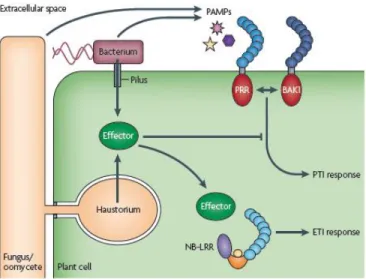
In an environment that is rich in harmful microbes, the survival of higher eukaryotic organisms depends on efficient pathogen sensing and rapidly mounted defence responses. Such protective mechanisms are found in all multicellular organisms and are collectively referred to as innate immunity (Medzhitov and Janeway, Jr., 1997; Akira et al., 2006). Because of their sessile lifestyle, plants cannot run away from invaders and need to defend themselves from threatening organisms by mounting a wide array of defense responses in a timely manner. Due to the absence of an adaptive immune system, plants rely on a so-called “innate immune system”, analogous to that found in animals (Nurnberger et al., 2004; Gomez-Gomez, 2004). The ability to detect and mount a defense response to potential pathogenic microorganisms has been paramount to the evolution and developmental success of modern-day plants. They are constantly exposed to microbes. To be pathogenic, most microbes must access the plant interior, either by penetrating the leaf or root surface directly or by entering through wounds or natural openings such as stomata, pores in the underside of the leaf used for gas exchange. Once the plant interior has been breached, microbes are faced with another obstacle: the plant cell wall, a rigid, cellulose-based support surrounding every cell. Penetration of the cell wall exposes the host plasma membrane to the microbe, where they encounter extracellular surface receptors that recognize pathogen- or microbe-associated molecular patterns (PAMPs or MAMPs) (Nurnberger and Kemmerling, 2006). Perception of a microorganism at the cell surface initiates PAMP-triggered immunity (PTI) (Dodds and Rathjen, 2010), which usually halts infection before the microbe gains a hold in the plant. Signals similar to PAMPs may arise from the plant itself because of the damage caused by microbes, which are now described as damage-associated molecular patterns (DAMPs) (Lotze et al., 2007) and can trigger PTI as well. Pathogenic microbes have evolved the means to suppress PTI by interfering with recognition at the plasma membrane or by secreting effector proteins into the plant cell cytosol that presumably alter resistance signalling or manifestation of resistance responses (Figure 1.1). Interestingly, the ability to deliver pathogen proteins directly into plant host cells to alter plant defence has become a unifying theme among plant pathogens (phytopathogens).
7
Once pathogens acquired the capacity to suppress primary defences, plants developed a more specialized mechanism to detect microbes, referred to as effector-triggered immunity (ETI) (Dodds and Rathjen, 2010). Effector-triggered immunity involves the direct or indirect recognition of the very microbial proteins used to subvert PTI by plant resistance (R) proteins. Activation of R protein-mediated resistance also suppresses microbial growth, but not before the invader has had an opportunity for limited proliferation. Not surprisingly, pathogens seem to have adapted effectors to interfere with ETI.
Figure 1.1: The plant immunity. Recognition of pathogen-associated molecular patterns (such as bacterial flagellin) by cell surface pattern recognition receptors (PRRs) promptly triggers PTI leading to basal immunity.
Many PRRs interact with the related protein BRASSINOSTEROID INSENSITIVE 1-ASSOCIATED KINASE 1 (BAK1) to initiate the PTI signalling pathway. Pathogenic bacteria use the type III secretion system to deliver effector proteins that target multiple host proteins to suppress basal immune responses. Plant resistance proteins (such as NB-LRR) recognize effector activity and restore resistance through effector-triggered immune responses (ETI). Adapted from (Dodds and Rathjen, 2010).
Disease is actually a relatively rare phenomenon in plants; the majority of plant species are resistant to infection by all isolates of any given microbial species (Dangl and Jones, 2001). The ability of an entire plant species to resist infection by all isolates of a pathogen species is termed non-host (or species) resistance. This is the commonest form of disease resistance in plants, and the infrequent change in the range of host species colonized by plant pathogens is indicative of its stability (Nurnberger and Lipka, 2005). Non-host resistance is thought to rely on both pre-formed barriers, such as the waxy cuticle and cell wall, which physically impede the growth and spread of the potential pathogen, and on the
8
induction of the basal defence system mounted in response to the recognition of non-self by the plant (Nurnberger and Lipka, 2005). An array of microbial-derived molecules termed pathogen-associated molecular patterns are recognized by pattern recognition receptors (PRRs) in the plant leading to signal transduction and the activation of a range of basal defence mechanisms including ethylene production, an oxidative burst, callose deposition, induction of defence related gene expression and, in some cases, hypersensitive response (HR)-like cell death (Nurnberger et al., 1994). The PAMP detection system present in plants corresponds conceptually to that of the innate immune system in animals; both recognize highly conserved microbial molecules and act as an early warning system for the presence of a potential pathogen (Ausubel, 2005). Plants also have a second system, cultivar-specific resistance, involving pairs of gene products—effector molecules from the pathogen and corresponding resistance (R) proteins in the plant. Recognition of an effector, or of its activity, by the appropriate R protein in the host leads to the HR and curtailment of pathogen growth, while loss of either of these proteins results in disease (Dangl and Jones, 2001). Since effectors are specific to particular pathogen strains, it has been proposed that cultivar-specific resistance fulfills an analogous role in plants to that of the adaptive immune system in vertebrates (Gomez-Gomez and Boller, 2002; Ausubel, 2005).
For many years view of the plant immune system was represented as a four phased 'zigzag' model (figure 1.2). In phase 1, PAMPs are recognized by PRRs, resulting in PTI that can halt further colonization. In phase 2, successful pathogens deploy effectors that contribute to pathogen virulence. Effectors can interfere with PTI. This results in effector-triggered susceptibility (ETS). In phase 3, a given effector is 'specifically recognized' by one of the NB-LRR proteins, resulting in effector-triggered immunity (ETI). Recognition is either indirect, or through direct NB-LRR recognition of an effector. ETI is an accelerated and amplified PTI response, resulting in disease resistance and, usually, a hypersensitive cell death response (HR) at the infection site. In phase 4, natural selection drives pathogens to avoid ETI either by shedding or diversifying the recognized effector gene, or by acquiring additional effectors that suppress ETI. Natural selection results in new R specificities so that ETI can be triggered again.
9 Figure 1.2: A zigzag model illustrates the quantitative output of the plant immune system. In this scheme, the ultimate amplitude of disease resistance or susceptibility is proportional to [PTI – ETS1ETI]. In phase 1, plants detect
microbial/pathogen-associated molecular patterns (MAMPs/ PAMPs, red diamonds) via PRRs to trigger PAMP-triggered immunity (PTI). In phase 2, successful pathogens deliver effectors that interfere with PTI, or otherwise enable pathogen nutrition and dispersal, resulting in effector-triggered susceptibility (ETS). In phase 3, one effector (indicated in red) is recognized by an NB-LRR protein, activating effector-triggered immunity (ETI), an amplified version of PTI that often passes a threshold for induction of hypersensitive cell death (HR). In phase 4, pathogen isolates are selected that have lost the red effector, and perhaps gained new effectors through horizontal gene flow (in blue)—these can help pathogens to suppress ETI. Selection favours new plant NB-LRR alleles that can recognize one of the newly acquired effectors, resulting again in ETI. Adapted from (Jones and Dangl, 2006).
In a recent work (Boller and Felix, 2009) it was proposed a new way to explain plant immunity in which effective innate immunity in plants, as in the case of innate immunity in vertebrates, is mediated through a single overarching principle, the perception of signals of danger. What may be categorized as PAMPs (or MAMPs), DAMPs, and effectors, might appear to the plant as one and the same type of signal that indicates a situation of danger (Figure 1.3).
10 Figure 1.3: Microbe-associated molecular patterns (MAMPs), damage-associated molecular patterns (DAMPs), and effectors are perceived as signals of danger. Extracellular MAMPs of prototypical microbes and DAMPs
released by their enzymes are recognized through pattern recognition receptors (PRRs). In the course of coevolution, pathogens gain effectors as virulence factors, and plants evolve new PRRs and resistance (R) proteins to perceive the effectors. When MAMPs, DAMPs, and effectors are recognized by PRRs and R proteins, a stereotypical defense syndrome is induced. RLK, receptor-like kinase; RLP, receptor-like protein; NB-LRR, nucleotide binding-site–leucine-rich repeat. Adapted from (Boller and Felix, 2009).
Indeed, gene expression data indicate that considerable overlap exists between the defense response induced by MAMPs, effectors, and endogenous elicitors. It remains to be seen, as an important challenge for future research, how signaling through MAMPs, endogenous DAMPs, and effectors converges into a stereotypical defense response.
1.2.
Basal defence
Induction of PTI in response to PAMPs or DAMPs occurs in both host and non-host plant species and is based on basal defense mechanisms. Studies of the effects of PAMPs and DAMPs point to a
11
stereotypical response, indicating that signaling converges to a common defense response. This is exerted trough a time course of events following PRR activation:
Very Early Responses (1–5 Minutes):
Ion fluxes. Among the earliest and most easily recordable physiological responses to MAMPs and DAMPs in plant cell cultures, starting after a lag phase of ∼0.5–2 min, is an alkalinization of the growth medium due to changes of ion fluxes across the plasma membrane (Boller, 1995; Nurnberger et al., 2004). These changes include increased influx of H+ and Ca2+ and a concomitant efflux of K+; an efflux of anions, in particular of nitrate, has also been observed (Wendehenne et al., 2002). The ion fluxes lead to membrane depolarization. PAMPs and DAMPs are known to stimulate an influx of Ca2+ from the apoplast and cause a rapid increase in cytoplasmic Ca2+ concentrations, which might serve as second messenger to promote the opening of other membrane channels (Blume et al., 2000; Lecourieux et al., 2002), or to activate calcium-dependent protein kinases (Boudsocq et al., 2010).
Oxidative burst. Another very early response to PAMPs and DAMPs, with a lag phase of ∼2 min, is the oxidative burst (Chinchilla et al., 2007). Reactive oxygen species can act as antibiotic agents directly or they may contribute indirectly to defense by causing cell wall crosslinking; in addition, reactive oxygen species may act as secondary stress signals to induce various defense responses (Apel and Hirt, 2004). The oxidative burst is an immediate and localized reaction that is believed to have several roles in plant defense (Low and Merida, 1996; Bolwell et al., 1999). The quantities of reactive oxygen species produced can be cytotoxic and thus are expected to be antimicrobial. Reactive oxygen species are thought to have direct (through cytotoxicity) and indirect (through signaling) roles in the plant cell death required for the HR. Reactive oxygen species induce the expression of defense related genes (Lamb and Dixon, 1997), and are implicated as second messengers that elicit other defense responses, including systemic acquired resistance (SAR) and the HR (Bolwell et al., 1999). SAR is the induction of defense mechanisms at locations remote from the original wound or infection site that serve to prepare the plant to defend itself against new attacks by pathogens (Sticher et al., 1997). In addition, reactive oxygen species drive the rapid peroxidase-mediated oxidative cross-linking of cell wall lignins, proteins, and carbohydrates, thereby reinforcing the wall against enzymatic maceration by the pathogen (Cote and Hahn, 1994).
12
O2- generating nicotinamide adenine dinucleotide phosphate (NADPH) oxidases are generally considered to be a major enzymatic source of ROS in the oxidative burst of plant cells challenged with pathogens or elicitors (Torres and Dangl, 2005; Torres et al., 2006). Two different NADPH oxidase genes in potato (Solanum tuberosum) are responsible for the elicitor induced biphasic oxidative burst(Yoshioka et al., 2001). In Arabidopsis, several genes encoding proteins with high similarity to the mammalian NADPH oxidase gp91phox subunit have been characterized. Among them, AtrbohD is required for the production of ROS during infection with different bacterial and fungal pathogens, including B. cinerea (Torres and Dangl, 2005) (Torres et al., 2006). Besides NADPH oxidases, other enzymes appear to be important in the elicitor-mediated oxidative burst, including apoplastic oxidases, such as oxalate oxidase (Dumas et al., 1993), amine oxidase (Allan and Fluhr, 1997), and pH-dependent apoplastic peroxidases (Frahry and Schopfer, 1998; Bolwell et al., 1995), which generate either O2- or H2O2.
Several studies report a PAMP-induced production of the reactive oxygen species nitric oxide (NO), a well-known second messenger in animals. However, contrary to animals, plants have no obvious NO synthase; furthermore, the indirect method used to measure NO may not be specific enough to discriminate it from other ROS products (Neill et al., 2008).
Activation of MAPKs. An early response to PAMP and DAMP signals is an activation of Mitogen-Activated Protein Kinase (MAPK) cascades (Pedley and Martin, 2005). The MAPK phosphorylation cascade is a highly conserved signal transduction mechanism that plays a key role in regulating many aspects of growth and development in eukaryotes. In plants, MAPK cascades have been associated with hormonal, abiotic stress, and disease defense responses and with the regulation of the cell cycle (Tena et al., 2001). A MAPK cascade consists of a core module of three kinases that act in sequence: a MAPK kinase kinase (MAPKKK) that activates, via phosphorylation, a MAPK kinase (MAPKK), which activates a MAPK (Figure 1.4). Activated MAPKs phosphorylate a number of different target proteins; the majority of targets appear to be transcription factors, but other targets include various protein kinases, phospholipases, and cytoskeletal proteins, all of which effect changes in gene expression and/or physiological responses appropriate to the stimulus in question (Widmann et al., 1999).
13 Figure 1.4. MAPK cascades and the cellular responses they influence following the recognition of microbial pathogens. Adapted from (Pedley and Martin, 2005).
Within the Arabidopsis genome sequence, 60 genes are predicted to encode MAPKKKs, 10 genes to encode MAPKKs, and 20 genes to encode MAPKs {2002 25518 /id}. Although there is likely to be some degree of functional redundancy, the high number of genes for MAPK cascade components indicates that plants rely heavily upon MAPK cascades for signal transduction. In particular, in Arabidopsis, a MAPK cascade, leading to AtMPK3 and AtMPK6 activation, is required for flg22-mediated responses (Asai et al., 2002). In Arabidopsis stimulated with flg22, a transient increase in AtMPK6 activity was observed, starting with a lag phase of ∼1–2 min and peaking after 5–10 min (Nuhse et al., 2000). A subsequent study made use of Arabidopsis leaf protoplasts transfected with various MAPK-related constructs to demonstrate the activation of two complete MAPK cascades by flg22, leading to the activation of AtMPK3 and AtMPK6 and culminating in the activation of WRKY-type transcription factors (Asai et al., 2002). DAMPs such as AtPep1 similarly induce a MAPK cascade (Huffaker et al., 2006).
Changes in protein phosphorylation. Activation of MAPK is accompanied by changes in protein phosphorylation. Pulse-labeling of Arabidopsis cells with radioactive phosphate, followed by two-dimensional gel electrophoresis, revealed dozens of proteins that showed increased phosphorylation within minutes of flg22 stimulation (Peck et al., 2001). With the advent of technologies that allow large-scale analysis of phosphopeptides, a number of proteins showing elicitor-responsive phosphorylation could be directly identified and their phosphorylation sites determined (Widmann et al., 1999).Using different technologies, two groups found a number of membrane proteins that display flg22-responsive phosphorylation in Arabidopsis cells;
14
intriguingly, both reported that RESPIRATORY BURST OXIDASE HOMOLOGUE D (RbohD), the NADPH oxidase that mediates the oxidative burst, is among these proteins (Nuhse et al., 2007; Benschop et al., 2007).
Early Responses (5–30 Minutes).
Ethylene biosynthesis. Among the earliest responses to MAMPs is an increased production of the stress hormone ethylene. Typically, an increased activity of l-aminocyclopropane-1-carboxylate (ACC) synthase activity can be detected within 10 min of treatment with MAMPs (Spanu et al., 1994).
Receptor endocytosis. Interestingly, FLS2, the PAMP flg22 receptor, undergoes ligand-induced endocytosis. A biologically functional FLS2-GFP fusion construct, stably expressed in Arabidopsis plants, disappears from its plasma membrane localization and appears in vesicles within ∼10–20 min of flg22 stimulation (Robatzek et al., 2006). FLS2 possibly has specific signaling functions after endocytosis, as described for certain receptors in animals, but endocytosis may also simply serve to remove and degrade the activated receptor (Robatzek et al., 2006).
Gene activation. Treatment of Arabidopsis plants with flg22 caused the induction of almost 1000 genes within 30 min and the downregulation of approximately 200 genes (Zipfel et al., 2004). The pattern of gene regulation in response to different PAMPs is almost identical, indicating that signaling through various PRR converges at an early step (Zipfel et al., 2006). In fact, fungal chitin and endogenous elicitors such as OGA seem to induce a similar set of genes (Ramonell et al., 2002; Ferrari et al., 2007), which suggests a stereotypical gene activation response to all PAMPs and DAMPs. Interestingly, among the induced genes, Receptor-like kinases (RLKs) are overrepresented. FLS2 and EFR are included in the induced genes, indicating that one role of early gene induction is a positive feedback to increase PRR perception capabilities (Zipfel et al., 2004).
Late Responses (Hours–Days).
Callose deposition. Arabidopsis leaves treated with flg22 and fixed and stained with aniline blue ∼16 h later display strong accumulation of fluorescent spots thought to represent callose deposits
15
(Gomez-Gomez et al., 1999). Although the biological foundation of this response is not clear, it has been used frequently, particularly to characterize pathogen effectors that interfere with MAMP signaling (Chisholm et al., 2006; Jones and Dangl, 2006).
Seedling growth inhibition. In Arabidopsis, a robust bioassay for PAMPs such as flg22 and elf18 is seedling growth inhibition. This response may reflect a physiological switch from a growth to a defense program, and it may be connected to the induction of amiRNA that negatively regulates the F-box auxin receptors TIR1 (transport inhibitor response 1), AFB2, and AFB3 (auxin signalling F-box proteins 2 and 3) and the consequent down regulation of auxin-responsive genes (Navarro et al., 2006).
1.3. Growth-Defense trade off
In their natural environments , plants are under continuous biotic stress caused by different attackers (e.g., bacteria, fungi, viruses, oomycetes, and insects) that compromise plant survival. Plants have thus evolved a variety of resistance mechanisms that can be induced after pathogen or pest attack (Glazebrook, 2005) but defense activation comes at the expense of plant growth. A fine regulation of the immune responses is necessary because the use of metabolites in plant resistance may be detrimental to other physiological processes impacting negatively in other economically interesting plant traits, such as biomass and seed production. This negative impact on growth could result from a diversion of resources away from growth and towards defense. Diversion of plant resources has been shown to occur at all levels, including machinery involved in transcription, translation, and protein secretion from cells as well as prioritization of carbon and nitrogen towards production of defense compounds. Transcriptomic and proteomic studies have demonstrated transcriptional reprogramming and altered protein profiles upon pathogen/herbivore detection to promote defense at the expense of growth (Jung et al., 2007; Denoux et al., 2008; Bilgin et al., 2010). The defense responses are regulated by phytohormones , that are small molecules which synergistically and/or antagonistically work in a complex network to regulate many aspects of plant growth , development , reproduction, and response to environmental stimuli. Then in adaptation to natural conditions, plants have evolved sophisticated mechanisms to regulate growth and defense responses, understanding the molecular mechanisms used by plants to balance growth and defense can enrich plant breeding and engineering strategies for
16
selection of elite genetic traits that will maximize plant fitness. Plant hormones play important roles in diverse growth and developmental processes as well as various biotic and abiotic stress responses in plants; for example, infection by diverse pathogens results in changes in the level of several of them (Robert-Seilaniantz et al., 2007; Adie et al., 2007). Plants hormones include auxins, gibberellins (GA), abscisic acid (ABA), cytokinins (CK), salicylic acid (SA), ethylene (ET), jasmonates (JA), brassinosteroids (BR) and peptide hormones. The identification and characterization of several mutants affected in the biosynthesis, perception and signal transduction of these hormones has been instrumental in understanding the role of individual components of each hormone signaling pathway in plant defense. Substantial progress has been made in the elucidation of individual aspects of phytohormone perception, signal transduction, homeostasis or influence on gene expression. However, the molecular mechanisms by which plants integrate stress induced changes in hormone levels and initiate adaptive responses are still poorly understood. Microbial pathogens have also developed the ability to manipulate the defence-related regulatory network of plants by producing phytohormones or their functional mimics; this results in hormonal imbalance and activation of inappropriate defence responses (Robert-Seilaniantz et al., 2007). For example, production of coronatine — a JA-Ile mimic by Pseudomonas syringae pv. tomato (Pst) bacteria, triggers the activation of JA-dependent defence responses leading to the suppression of SA-dependent defence responses and promotion of disease symptoms (Cui et al., 2005; Laurie-Berry et al., 2006). In addition, coronatine has been shown to prevent PAMP-induced stomatal closure which facilitates bacterial entry into the leaf (Melotto et al., 2006).
1.3.1.
Salicylic acid, jasmonates and ethylene are involved in
defense signaling
Three phytohormones—SA, JA and ET, are known to play major roles in regulating plant defence responses against various pathogens, pests and abiotic stresses such as wounding and exposure to ozone (Glazebrook, 2005; Lorenzo and Solano, 2005; Broekaert et al., 2006; Balbi and Devoto, 2008). SA plays a crucial role in plant defence and is generally involved in the activation of defence responses against biotrophic and hemi-biotrophic pathogens as well as the establishment of systemic acquired
17
resistance (SAR) (Grant and Lamb, 2006)). Mutants that are affected in the accumulation of SA or are insensitive to SA show enhanced susceptibility to biotrophic and hemi-biotrophic pathogens. Recently, it has been shown that methyl salicylate, which is induced upon pathogen infection, acts as a mobile inducer of SAR in tobacco (Park et al., 2007). SA levels increase in pathogen challenged tissues of plants and exogenous applications result in the induction of pathogenesis related (PR) genes and enhanced resistance to a broad range of pathogens. By contrast, JA and ET are usually associated with defence against necrotrophic pathogens and herbivorous insects. Although, SA and JA/ET defence pathways are mutually antagonistic, evidences of synergistic interactions have also been reported (Schenk et al., 2000; Beckers and Spoel, 2006; Mur et al., 2006). This suggests that the defence signaling network activated and utilized by the plant is dependent on the nature of the pathogen and its mode of pathogenicity. In addition, the lifestyles of different pathogens are not often readily classifiable as purely biotrophic or necrotrophic. Therefore, the positive or negative cross talk between SA and JA/ET pathways may be regulated depending on the specific pathogen (Adie et al., 2007).One of the important regulatory components of SA signaling is non-expressor of PR genes 1 (NPR1), which interacts with TGA transcription factors that are involved in the activation of SA-responsive PR genes. Arabidopsis npr1 plants are compromised in the SA-mediated suppression of JA responsive gene expression indicating that NPR1 plays an important role in SA-JA interaction (Spoel et al., 2007). Downstream of NPR1, several WRKY transcription factors play important roles in the regulation of SA-dependent defence responses in plants (Eulgem and Somssich, 2007). Several studies indicate that JA- and ET-signaling often operate synergistically to activate the expression of some defence related genes after pathogen inoculation (Penninckx et al., 1998; Thomma et al., 2001; Glazebrook, 2005). Microarray analysis of defence related genes revealed significant overlap in the number of genes induced by both JA and ET (Schenk et al., 2000). It has been shown that an Arabidopsis transcription factor, ethylene response factor 1 (ERF1) acts as a positive regulator of JA and ET signaling (Lorenzo et al., 2003). Recently, several members of ERF family have been shown to play important role in mediating defense responses in Arabidopsis (McGrath et al., 2005). However, how plants coordinate these complex interactions and what are the molecular mechanisms involved is not clear.
18
1.3.2.
The growth-promoting hormone Auxin is also involved
in defense responses
Auxins regulate many fundamental aspects of plant growth and development including stem and petiole elongation and root architecture in response to light, temperature, and gravity (Kazan, 2013). Auxin promotes the degradation of a family of transcriptional repressors called Auxin/Indole-3-acetic acid (Aux/IAA). Aux/IAA proteins bind to auxin response factors (ARFs) and inhibit the transcription of specific auxin response genes (Leyser, 2006). It has been shown that transport inhibitor response 1 (TIR1) is an auxin receptor that interacts with Aux/IAA proteins (Dharmasiri et al., 2005). TIR1 encodes an F-box protein that forms an Aux/IAA-SCFTIR1 (SKP1, Cullin and F-box proteins) complex and leads to the degradation of Aux/IAA proteins via ubiquitin/26S proteasome pathway (Parry and Estelle, 2006). To regulate plant growth and development, auxin can induce the expression of three groups of genes: Aux/IAA family, GH3 family and small auxin-up RNA (SAUR) family (Woodward and Bartel, 2005). GH3 genes encode IAA-amido synthetases that are involved in the regulation of auxin homeostasis by conjugating excess IAA to amino acids (Staswick et al., 2005). Most of the total auxin in plants is found in the conjugated form and the formation of auxin conjugates is one of the important regulatory mechanisms for the activation or inactivation of IAA. Auxin responsive GH3 genes have been shown to play roles in plant defence responses in Arabidopsis and rice. Recently, GH3.5 has been shown to acts as a bifunctional modulator in both SA and auxin signaling during pathogen infection (Zhang et al., 2007). Exogenous application of auxin has been shown to promote disease caused by Agrobacterium tumefaciens (Yamada, 1993), Pseudomonas savastanoi (Yamada, 1993) and Pst DC3000 (Navarro et al., 2006). Similarly, co-inoculation of P. syringae pv. maculicola (Psm) 4326 and auxin has been found to promote both disease symptom and pathogen growth in Arabidopsis (Wang et al., 2007). These results indicate that auxin is involved in the attenuation of defence responses in plants. In contrast, blocking auxin responses has been shown to increase resistance in plants. Auxin resistant axr2-1 mutants of Arabidopsis showed reduction in Psm 4326 growth compared to wild type plants (Wang et al., 2007). Several studies have shown that pathogen infection results in imbalances in auxin levels as well as changes in the expression of genes involved in auxin signaling. For example, infection with Pst DC3000 resulted in increased IAA levels in Arabidopsis (O'Donnell et al., 2003). Interestingly, the bacterial type III effector avrRpt2, which encodes a cysteine protease, has been shown to modulate host auxin physiology to promote pathogen
19
virulence and disease development in Arabidopsis (Chen et al., 2007). Global gene expression analysis using microarrays revealed that Pst DC3000 induces auxin biosynthetic genes and represses genes belonging to Aux/IAA family and auxin transporters. Thus, Pst DC3000 activates auxin production, alters auxin movement and derepresses auxin signaling thereby modulating auxin physiology in Arabidopsis (Thilmony et al., 2006). This suggests that auxin promotes disease susceptibility and repression of auxin signaling could potentially result in enhanced resistance in plants. Indeed, down regulation of auxin signaling has been shown to contribute to plant induced immune responses in Arabidopsis. Navarro et al. (2006) showed that down regulation of auxin receptor genes by over expression of a micro RNA (miR393), which targets auxin receptors, increased resistance against Pst DC3000 in Arabidopsis. In contrast, activation of auxin signaling through over expression of an auxin receptor that is partially refractory to miR393-mediated transcript cleavage, enhanced susceptibility to Pst DC3000 (Navarro et al., 2006). These results suggest that auxin promotes susceptibility to bacterial disease, and that down-regulation of auxin signaling is part of the plant induced immune response. Treatment of Arabidopsis plants with an SA analog, benzothiadiazole S-methyl ester (BTH) results in the repression of a number of auxin responsive genes, including an auxin importer AUX1, an auxin exporter PIN7, auxin receptors TIR1 and AFB1, and genes belonging to auxin inducible SAUR and Aux/IAA family (Wang et al., 2007). Similarly, it was found that majority of the above auxin-inducible genes were also repressed in systemic tissues after induction of SAR, indicating that SAR response involves down-regulation of auxin responsive genes. However, the level of free auxin did not change after SA treatment. In addition, SA has been shown to inhibit the expression of the auxin-inducible reporter DR5::GUS (Zhang et al., 2007), leading to the hypothesis that SA stabilizes Aux/IAA auxin repressors by limiting auxin receptors needed for the down-regulation of Aux/IAA proteins.
1.3.3. Non-self recognition - Pathogen-Associated Molecular
Patterns (PAMPs) and Pattern Recognition Receptors (PRRs)
The ability to determine self from non-self is critical for plants to mount an effective immune response against potential pathogens. PAMPs, also known as general elicitors, offer one such opportunity. PAMPs are highly conserved and ubiquitous molecules widely distributed amongst microbial species
20
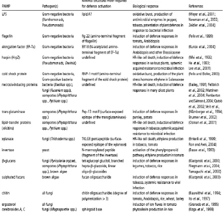
(pathogenic or not) where they carry out an essential function, but absent in the potential host species (Nurnberger and Lipka, 2005). A number of PAMPs that fulfill these criteria and elicit a defense response in plants have been identified from plant pathogens and reviewed in Nurnberger et al. (2004) (Table 1.1).
Table 1: Selected pathogen-associated molecular patterns (PAMPs) and their plant defence-inducing activities. Adapted
from (Nurnberger et al., 2004).
Different plant species respond to different PAMPs. For example tobacco responds to cold-shock protein while Arabidopsis does not, and only members of the Brassicaceae have so far been shown to respond to EF-Tu (Felix and Boller, 2003; Kunze et al., 2004). While this represents a diverse set of molecules, within the proteinaceous PAMPs two themes have emerged. These molecules typically contain a short (10–25) amino acid epitope that elicits a stronger defence response than the complete protein. For example, from Gram-negative bacteria flg22, a highly conserved stretch of 22 amino acids from the N terminus of flagellin, is a more potent elicitor than flagellin (Felix et al., 1999), and the same is true of a highly conserved 15 amino acid stretch including the RNA-binding motif RNP-1 from the cold shock protein (Felix and Boller, 2003) and an 18 amino acid stretch from the N terminus of the
21
elongation factor EF-Tu (Kunze et al., 2004). However, there are exceptions; the elicitor effect of NPP1 (necrosis-inducing Phytophthora protein 1) requires an intact protein and overlapping peptide fragments were inactive (Fellbrich et al., 2002), perhaps indicating that it is the activity of this protein that is detected by the plant rather than a specific amino acid sequence. Presumably, there would be a huge selective advantage for mutations within these epitopes that rendered them inactive as elicitors of plant defense systems. However, it would seem that, in many cases, such mutations also have a deleterious effect on the function of these proteins in the pathogen. For example, in Pep-13, a 13 amino acid internal peptide of a 42 kDa transglutaminase enzyme from the cell wall of Phytophthora sojae, substitution of Trp231 to Ala abolished elicitor activity in parsley but with a concurrent 98% reduction in transglutaminase activity (Brunner and et al., 2002). Thus, it appears that plants have evolved receptors that recognize short highly conserved amino acid stretches of certain microbial proteins that cannot easily be altered without loss of the protein function. That said, certain microbes may have evolved the capacity to avoid detection by specific PRRs. For example, Agrobacterium tumefaciens and Ralstonia solanacearum (pathogens) as well as Rhizobium meliloti (symbiont) possess functional flagellins that do not elicit a defence response in Arabidopsis and the N-terminal peptide from Pseudomonas syringae pv. tomato DC3000 (Pst) EF-Tu is not as potent an elicitor in Arabidopsis as those from other bacteria (Kunze et al., 2004; Sun et al., 2006). The evolution of non-eliciting PAMPs is one way in which pathogens can overcome non-host resistance in plants; however, the lack of a single eliciting PAMP has not yet been directly shown to affect the virulence of the pathogen. Some experiments have shown that deletion of a specific PRR in the host affects susceptibility; however, in other studies wild-type plants and plants lacking a PRR were equally susceptible (Sun et al., 2006) (Zipfel et al., 2004). This could be explained by the evolution in plants of recognition systems for multiple PAMPs from the same micro-organism. For example, Arabidopsis recognizes both flagellin and EF-Tu and these PAMPs activate the same signalling and defence responses in a nonsynergistic manner (Zipfel et al., 2006). A recent gene expression profiling study has also demonstrated that the lack of flagellin perception does not dramatically alter PAMP-induced gene expression during infection of Arabidopsis by Pst (Thilmony et al., 2006).
1.3.4.
Damage-Associated
22
In addition to sensing invading microbes by means of PAMPs (infectious non-self), plants and animals can also sense infectious-self or modified-self via damage-associated molecular patterns (DAMPs). Many plant pathogens produce lytic enzymes to breach the structural barriers of plant tissues. The products generated by these enzymes may function as endogenous elicitors. Such DAMPs typically appear in the apoplast and, as in the case of PAMPs, can serve as danger signals to induce innate immunity (Matzinger, 2002).
1.3.4.1. Oligogalacturonides
Oligogalacturonides (OGs) are linear molecules of two to about twenty α-1,4-d-galactopyranoslyuronic acid (GalA) residues. OGs were the first plant oligosaccharins, biologically active carbohydrates that act as signal molecules, to be discovered (Bishop et al., 1981; Hahn, 1981). OGs are released upon fragmentation of homogalacturonan (HG) from the plant primary cell wall (Cote et al., 1998) by wounding or by pathogen-secreted cell wall-degrading enzymes (for example polygalacturonases, PGs). Indeed, PGs are not elicitors per se, but are rather able to release elicitor-active molecules from the host cell wall. When the activity of a fungal PG is modulated by apoplastic PG-inhibiting proteins (PGIPs), long-chain oligogalacturonides are produced (De Lorenzo et al., 2001; De Lorenzo and Ferrari, 2002) (Figure 1.5). OGs cannot be considered true PAMPs, since they are not derived from the pathogen. However, they are considered the classic examples of DAMPs that are generated by the host cell during the infection process.
23 Figure 1.5 : Model for the OG accumulation during pathogen infection.
Chemically pure OGs can act as endogenous elicitors (Galletti et al., 2009). Biological responses to OGs occur in at least five of the six subclasses of dicotyledonous plants Magnoliidae, Hamamelidae, Asteridae, Rosidae, Dilleniidae (Reymond et al., 1996; Cote and Hahn, 1994) in a monocot (Moerschbacher et al., 1999) and a gymnosperm (Asiegbu et al., 1994). A number of different biological responses to OGs have been reported, and the particular response observed depends on the plant species, the bioassay, and the chemical structure of the OG used (Cote et al., 1998). A spectrum of modified and unmodified OGs of various lengths are active in different systems (reviewed by (Cote and Hahn, 1994).
The biological responses of plants to OGs can be divided into two broad categories: plant defense and plant growth and development (Cote and Hahn, 1994).
1.3.4.2. Oligogalacturonide-induced responses involved in plant
defense
Pathogens enter plant tissues in at least three ways: digesting cell walls, entering through wounds, and invading through natural openings such as stomata. Pectins are one of the first targets of digestion by invading pathogens (Pagel and Heitifuss, 1990). OGs are released when PGs and endopectate lyases (PLs) secreted from the pathogen degrade the homogalacturonan in the cell (Cote et al., 1998). The OGs released are a carbon source for the pathogens, but can also be detected by plants as signals to initiate defense responses. Exogenously added OGs inhibit the light-induced opening of stomata in tomato and Commelina communis L. leaves (Lee et al., 1999) and elicit a variety of defense responses, including accumulation of phytoalexins (Davis et al., 1986), glucanase and chitinase (Davis and Hahlbrock, 1987; Broekaert and Pneumas, 1988). Stomatal openings provide access to inner leaf tissues required by many plant pathogens (Agrios, 1997), suggesting that the constriction of stomatal apertures is beneficial for plant defense. One of the first responses observed after the addition of OGs that is clearly involved in plant defense is the production of active oxygen species, including H2O2, and O2- (Low and Merida, 1996). This oxidative burst occurs within a few minutes after the addition of OGs to suspension-cultured soybean (Legendre et al., 1993), tobacco (Rout-Mayer et al., 1997; Binet et
24
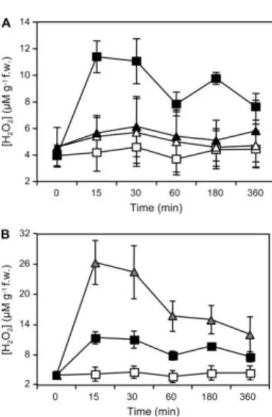
al., 1998) and tomato (Stennis et al., 1998) cells. Recently it was shown that, in Arabidopsis, production of H2O2 in response to OGs is mediated by AtrbohD (Figure1.6) (Galletti et al., 2008).
Figure 1.6 : Accumulation of extracellular H2O2 in response to OGs or G/GO in Arabidopsis seedlings. (a)
Arabidopsis wild-type and atrbohD seedlings were treated with water (H2O) or OGs for the indicated time (min).
Arabidopsis wild-type (squares) and atrbohD (triangles) seedlings were treated with water (white symbols) or OGs (black symbols). (b) Arabidopsis seedlings were treated with water (H2O, white squares), OGs (black squares), or G/GO (gray
triangles). H2O2 accumulation in the culture medium, expressed as mM g21 fresh weight, was measured at the indicated
times (min). Adapted from (Galletti et al., 2008).
OGs initiate signaling cascades that activate a plant defense. OGs rapidly activate AtMPK3 and AtMPK6 (Denoux et al., 2008), suggesting that, even though OGs and flg22 are perceived by distinct receptors, the signaling pathways mediated by these elicitors converge very early. Arabidopsis full-genome expression analysis reveals that OGs influence the expression of ~4000 genes (Ferrari et al., 2007). Some of these, such as AtWRKY40 (At1g80840), encoding a transcription factor that acts as a negative regulator of basal defense (Xu et al., 2006), CYP81F2 (At5g57220), encoding a cytochrome P450 and RetOx (At1g26380), encoding a protein with homology to reticuline oxidases, a class of enzymes involved in secondary metabolism and in defense against pathogens (Dittrich and Kutchan, 1991), are rapidly and strongly up-regulated upon exposure to elicitor. Early activation of genes in response to OGs is independent of SA, ET, and JA signaling pathways and of AtRbohD (Galletti et al., 2008). Exogenous treatment with OGs protects grapevine (Vitis vinifera) and Arabidopsis leaves against infection with the necrotrophic fungus Botrytis cinerea (Aziz et al., 2004; Ferrari et al., 2007), suggesting that production of this elicitor at the site of infection, where large amounts of PGs are
25
secreted by the fungus, may contribute to activate defenses responses. A variety of plant defense responses against microbial pathogens are regulated by the signaling molecules SA, JA and ET. Resistance to Botrytis cinerea induced in Arabidopsis by OGs is independent of SA, ET or JA signaling, but requires PHYTOALEXIN DEFICIENT3 (PAD3) (Ferrari et al., 2007), a gene involved in the metabolism of Trp-derived secondary compounds (Zhou et al., 1999).
1.3.5 PAMP-Triggered Immunity Crosstalk with Auxin
Auxin has long been implicated in suppressing plant defense due to the fact that many pathogens, including Pseudomonas syringae and Agrobacterium tumefaciens, can directly synthesize auxin or manipulate auxin synthesis and signaling in plants to promote disease (Chen et al., 2007). Analysis of plant transcriptional reprogramming following some pathogen infections has shown a general de-repression of the auxin pathway including promotion of auxin biosynthetic genes and de-repression of AUX/IAA genes resulting in enhanced plant susceptibility (Thilmony et al., 2006). To combat the effects of pathogen produced or induced auxin to promote disease, plants actively suppress auxin signaling during defense (Navarro et al., 2004). Following flg22-treatment, wild-type Arabidopsis plants show a reduction in both transcript and protein levels of the auxin F-box receptors, resulting in stabilization of AUX/IAA proteins and repression of auxin-responsive genes (Navarro et al., 2006). This suppression is partially due to the activity of the microRNA miR393 , which is induced by flg22 and directly targets and cleaves TIR1, AFB2, and AFB3 transcripts (Navarro et al., 2006). Suppression of auxin signaling has been shown to be biologically relevant to PTI, as overexpression of miR393 enhances resistance to virulent pathogens and overexpression of AFB1 increases susceptibility relative to that observed in wild-type plants, as measured by bacterial growth (Navarro et al., 2006).
26
One of the primary ways SA has been shown to inhibit growth is by suppression of auxin signaling. A microarray study revealed that a number of auxin responsive genes were affected by BTH treatment; 21 genes encoding proteins involved in auxin reception, import and export and signaling were down-regulated and two genes encoding GH3 enzymes were up-down-regulated (Wang et al., 2007). As GH3 enzymes are responsible for regulating auxin homeostasis by conjugating IAA with different amino acids (Staswick et al., 2005), the transcriptional profile indicates a general BTH-dependent repression of auxin homeostasis and signaling. A follow-up study confirmed this by investigating the effect of SA on auxin levels, uptake, sensitivity, and signaling (Wang et al., 2007). It was shown that SA does not affect auxin synthesis, but instead represses the expression of the TIR1/ABF F-box genes , resulting in stabilization of AUX/ IAA repressor proteins to decrease auxin signaling (Wang et al., 2007). One of the two GH3 genes identified in the microarray study encodes GH3.5 (Wang et al., 2007), which conjugates IAA with Asp (Staswick et al., 2005). The GH3.5 knockout mutants were shown to be compromised in SAR while overexpression lines exhibited a dwarf phenotype, accumulated higher levels of SA, had elevated expression of PR1, and increased resistance to Pto DC3000 (Park et al., 2007; Zhang et al., 2007).
1.3.7 Oligogalacturonides Crosstalk with Auxin
The biological responses triggered by OGs are well documented and similar in many aspects to those of MAMPs (Galletti et al., 2009). For example, OGs and flg22 activate defense responses effective against the microbial pathogens Botrytis cinerea and Pseudomonas syringae, respectively, independently of SA, ET, and JA (Zipfel et al., 2004; Ferrari et al., 2007). Both elicitors trigger a fast and transient response characterized by activation of early stages of multiple defense signaling pathways. However, the response to flg22 is stronger in both the number of genes differentially expressed and the amplitude of change. Even at very high concentrations, OGs do not induce a response that is as comprehensive as that seen with flg22. For example, SA-dependent secretory pathway genes and PR1 expression are substantially induced only by flg22 (Denoux et al., 2008). Exogenously added OGs influence the growth and development of plant tissues (Cote and Hahn, 1994). OGs inhibit auxin-induced pea stem elongation (Branca et al., 1988) and are also active in the tobacco thin-cell layer (TCL) (Tran Thanh Van et al., 1985) (Mohnen et al., 1990), and the tobacco leaf explant
27
bioassays (Bellincampi et al., 1993). When biologically active OGs are added to media containing specific auxin concentrations, TCLs that would normally form few or no organs form flowers, while TCLs that normally form roots form significantly fewer roots (Eberhard et al., 1989). Biologically active OGs inhibit root formation on tobacco (Bellincampi et al., 1993) and Arabidopsis (Savatin et al., 2011) leafs respectively and increase stomata formation (Altamura et al., 1998) on tobacco leaf explants incubated in media with specific auxin concentrations. OGs also inhibited the expression, induced by exogenous auxin, of GUS driven by the synthetic promoter DR5; and inhibited the accumulation of auxin early up-regulated transcripts (IAA5 [At1g15580], IAA19 [At3g15540], IAA20 [At2g46990], IAA22 [At1g19220], SAUR16 [At4g38860], SAUR-AC1 [At4g38850], and GH3.3 [At2g23170]). In every case reported to date where OGs regulate the growth and development of plant tissues, with the exception of fruit ripening, their effect is the opposite of the effect of added auxin (Branca et al., 1988; Eberhard et al., 1989) (Altamura et al., 1998; Savatin et al., 2011). The mechanism by which OGs act in opposition to the action of auxin is presently unknown; in Savatin D. V. et al. 2011 it was shown that OG - auxin antagonism does not involve any of the following mechanisms: (1) stabilization of auxin-response repressors; (2) decreased levels of auxin receptor transcripts through the action of microRNAs. These data suggest that OGs antagonize auxin responses independently of Aux/Indole-3-Acetic Acid repressor stabilization and of posttranscriptional gene silencing; It was therefore speculated that OG – auxin antagonism can be played at the level of transcriptional regulation on the promoter of auxin-inducible genes antagonized by OGs.
2 Materials and Methods
2.1 Cloning of the promoter of IAA5
The promoter of the auxin-responsive gene IAA5 (INDOLE-3-ACETIC ACID INDUCIBLE 5; TAIR accession: At1G15580) was cloned into the binary vector pCAMBIA 1391z (CambiaLab) using the “Cut & Paste” method . This involves preparing both a DNA fragment to be cloned (insert) and a
self-28
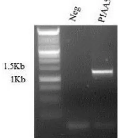
replicating DNA plasmid (vector) by cutting with two unique restriction enzymes that flank the DNA sequence and are present at the preferred site of insertion of the vector, often called the multiple cloning site (MCS). By using two different restriction enzymes, two non-compatible ends are generated, thus forcing the insert to be cloned directionally, and lowering the transformation background of re-ligated vector alone. The pCAMBIA 1391z vector contains the GUS reporter gene downstream of the MCS and two antibiotic resistance genes; the kanamycin resistance gene for selection of Escherichia coli and Agrobacterium tumefaciens transformants and the hygromycin resistance gene for the selection of plant transformants Figure 2.1. The promoter of IAA5 (PIAA5), the –1279 bp sequence upstream of the coding region of IAA5 gene, was amplified by PCR from genomic DNA of Arabidopsis thaliana. The primers used for the amplification contain at the 5’ extremity a restriction site for restriction enzymes; the forward (fw) primer contains the restriction site of pSTI restriction enzyme, while the reverse (rev) primer contains the restriction site of ECORI restriction enzyme. The PCR product was separated and visualized on 1% agarose gel stained with ethidium bromide (EtBr) and the fragment purified using QIAquick PCR Purification Kit (QIAGEN). Two hundred and fifty ng of the fragment containing the promoter of IAA5 and 100 ng of pCAMBIA 1391z were digested in parallel with pSTI and ECORI restriction enzymes (FastDigest—Thermo Scientific) according to the manufacturer instructions and purified using QIAquick PCR Purification Kit for the fragment and QIAprep Spin Miniprep Kit (QIAGEN) for the vector.
The digested fragment (18 ng) and vector (60 ng) were ligated with T4 ligase (Promega) according to the manufacturer instructions. The ligation between the fragment and the vector was confirmed by mixed primers PCR using the forward primer of PIAA5 that anneal on the fragment and the reverse primer that anneal on GUS reporter gene. The ligation product was subsequently used to transform E. coli DH5α electro-competent cells. E. coli DH5α electro-competent cells were transformed with 3 ng of ligation product between PIAA5 and pCAMBIA1391z by electroporation. Cell suspension was thawed on ice and 3 ng of ligation product were added. The cells were kept 2 min on ice, transferred in the electroporation cuvette and electroporated with BIO-RAD MicroPulser, electrical condition of 1.5 kV. After the electric pulse 1 mL of Luria Bertani (LB; tryptone 10 g ; yeast extract 5 g and NaCl 10 g in 1 L of deionized water) medium was quickly added to electroporated cells. Cells were recovered at 37 °C for 1 h with shaking, plated on LB containing 20 µg/ml of kanamycin and grown 16 hours at 37 °C to select the transformants. Transformants were screened by colony-PCR and positive colonies containing pCAMBIA1391z ligated with PIAA5 were inoculated in 5 ml of LB with 20 µg/ml of kanamycin and grown 16 hours at 37 °C to increase the copy number of the PIAA5 containing construct. The PIAA5
29
containing construct was purified with QIAprep Spin Miniprep Kit (QIAGEN), sequenced to verify the sequence of PIAA5 and digested with pSTI and ECORI restriction enzymes (FastDigest—Thermo Scientific) to confirm the presence of PIAA5. A. tumefaciens strain GV3101, containing rifampicin resistance on the genome and gentamycin resistance in the helper plasmid, was transformed by electroporation with the PIAA5 containing construct. Cell suspension was thawed on ice and 50 ng of PIAA5 containing construct were added. The cells were kept 2 min on ice, transferred in the electroporation cuvette and electroporated with BIO-RAD MicroPulser, electrical condition of 1.5 kV. After the electric pulse, 1 mL of Luria Bertani medium was quickly added to electroporated cells. Cells were recovered at 28 °C for 2 h with shaking, plated on LB containing 20 µg/ml of kanamycin, 20 µg/ml of rifampicin, 20 µg/ml gentamycin and grown 16 hours at 28 °C to select the transformants. The presence of PIAA5 containing construct was confirmed by colony-PCR on the trasformants. Positive colonies were inoculated in 5 ml of LB containing 20 µg/ml of kanamycin, 20 µg/ml of rifampicin, 20 µg/ml gentamycin and grown 16 hours at 28 °C, pelleted, re-suspended with LB containing 20% (v/v) glycerol and stored at -80 °C.
Primers used:
PIAA5 pSTI fw-5’AGCTCTGCAGAATTCGGTTGTATTTGCGGA-3’
PIAA5 ECORI rev-5’AAGCTGAATTCCTTTGATGTTTTTGATTGAAAAGTATT3’ GUSrev-5’ AGTTGCAACCACCTGTTGAT
30 Figure 2.1: the pCAMBIA 1391z vector is an Agrobacterium binary vector for plant transformation; it contains the GUS
reporter gene downstream of the MCS and two antibiotic resistance genes; the kanamycin resistance gene for selection of E.
coli and A. tumefaciens transformants and the hygromycin resistance gene for the selection of plant transformants.
31
The promoter (–1279 bp sequence upstream of the coding region) of the auxin-responsive gene IAA5, was cloned into the binary vector pCAMBIA 1391z (CambiaLabs), upstream of the GUS reporter gene (Jefferson et al., 1987). Arabidopsis thaliana plants (Columbia-0 ecotype) were grown on soil in a growth chamber for 30 days at 22°C, 70% relative humidity, with a photoperiod of 16 h light and light intensity of 100 µE m-2 s-1. Flowering plants were transformed by Agrobacterium tumefaciens mediated transformation using the floral dip method (Clough and Bent, 1998). The seeds collected after the floral-dip transformation were washed for 1 min in 1 mL of isopropanol and for 1 min in sterile ultrapure H2O (for two times). Seeds were surface sterilized for 10 min in 1 mL sterilization solution (1.6 % NaClO, 0.01% SDS) with shaking. To remove the sterilization solution, the seeds were washed under sterile flow hood with sterile ultrapure H2O (for five times). The sterilized seeds were wrapped in aluminum foil and placed at 4 ° C for 3 days to synchronize the germination (vernalization). In order to select the PIAA5:GUS transformant plants, seeds were plated on Petri dishes containing the selective solid medium, prepared by dissolving 2.2 g of Murashige and Skoog medium with vitamins (MS/2); 0.5% sucrose, 1% plant agar, and the antibiotic hygromycin (20 µg/ml) in 1 L of distilled water at pH 5.5.
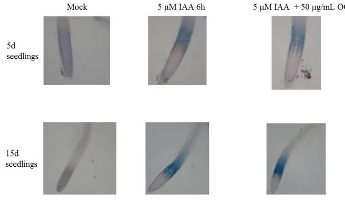
2.3 Induction of GUS expression driven by the promoter of IAA5
To induce the expression of GUS reporter gene in response to auxin (IAA), seeds from T2 generation of PIAA5:GUS transgenic lines and T3 generation of DR5:GUS transgenic lines (Ulmasov et al., 1997) were surface sterilized and vernalized as described above and grown 5 or 15 days in 6 well plates containing 5 mL of sterile MS/2 medium including vitamins and 1% sucrose in a growth chamber at 22°C, 70% relative humidity, with a photoperiod of 16 h light and light intensity of 100 µE m-2 s-1 ., The seedlings were treated for 6h with 2.5 µM IAA or 5µM IAA, to induce GUS expression under the control of PIAA5, or mock treated with H2O. DR5:GUS seedlings were treated for 6h with 2.5 µM IAA and used as positive control. To observe the effect of OG on the IAA-induced GUS expression the seedlings were treated for 6h with 5 µM IAA + 50 µg/mL OG or mock treated with H2O.
32
To reveal the auxin-induced GUS activity driven by PIAA5 or DR5; IAA-treated, IAA + OG co-treated and mock-co-treated seedlings, were placed in 6 well plates with 5 mL of GUS staining solution (50 mM Phospate Buffer pH 7 , 0.2% Triton-X 100 , 2 mM K3Fe(CN)6 , 2 mM K4Fe(CN)6 , 2 mM 5-bromo-4-chloro-3-indolyl-D-glucuronide); vacuum infiltrated for 5 min and placed at 37°C overnight (Jefferson et al., 1987). To reveal the GUS staining, seedlings were discolored with 5 washes of boiling ethanol. Seedlings were observed with a light microscopy (Nikon).
2.5 Analyses of GUS transcript level in response to IAA and IAA + OG
co-treatment
T2 generation of PIAA5:GUS transgenic lines were grown in 6 well plates containing 5 mL of sterile MS/2 medium including vitamins and 1% sucrose in a growth chamber at 22°C, 70% relative humidity, with a photoperiod of 16 h of light and light intensity of 100 µE m-2 s-1 . Seedlings were treated for 1 h with 1.5 µM IAA ; 1.5µM IAA + 100 μg/mL of OGs or mock treated with sterile ultrapure H2O. Treated seedlings were frozen in liquid nitrogen, homogenized with a MM301 ball mill (Retsch), and total RNA was extracted with Isol-RNA lysis reagent (5 prime) according to the manufacturer’s protocol. RNA was treated with RQ1 DNase (Promega) and first-strand cDNA was synthesized using ImProm-II reverse transcriptase (Promega) according to the manufacturer’s instructions. Real-time qPCR analysis was performed as previously described (Galletti et al., 2011) using a CFX96 real-time system (Bio-Rad). One microliter of cDNA (corresponding to 50 ng of total RNA) was amplified in a 30 mL reaction mix containing 1X GoTaq real-time PCR system (Promega) and 0.4 mM of each primer. Expression levels of GUS, relative to UBQ5, were determined using a modification of the Pfaffl method (Pfaffl, 2001) as previously described (Ferrari et al., 2006).
Primer sequences are:
-UBQ5fw-5’-GGAAGAAGAAGACTTACACC, -UBQ5rev-5’-AGTCCACACTTACCACAGTA; -GUSfw-5’- AATGGTGATTACCGACGAAA -GUSrev-5’- AGTTGCAACCACCTGTTGAT
33
2.6 Plant material and growth conditions
Arabidopsis thaliana Columbia-0 seeds (10 mg; approximatively 500 seeds) were surface sterilized and vernalized as described above. One liter of liquid medium was prepared by dissolving 2.2 g of Murashige and Skoog medium with vitamins (MS/2) and 1% sucrose, in distilled water at pH 5.5. The liquid medium was sterilized with filtration apparatus under a sterile flow hood. The seeds were placed in 500 mL Erlenmeyer flasks containing 100 mL of medium previously autoclaved at 120°C for 20 min, and grown for 15 days at 22°C, 70% relative humidity, with a photoperiod of 16 h light and light intensity of 100 µE m-2 s-1 .
2.7 Plant treatments
For the DNA Affinity Purification experiments 15-days-old Arabidopsis thaliana seedlings were treated in the Erlenmeyer flasks for 1 h with IAA and IAA+ OG co-treatment.
For the nuclear proteomic analyses 15-days-old Arabidopsis thaliana seedlings were treated in the Erlenmeyer flasks for 1 h with IAA, OG , IAA+ OG co-treatment and mock treated.
Auxin treatments were performed by adding 100 μL of 1.5mM IAA dissolved in sterile ultrapure H2O to the Erlenmeyer flasks to a final concentration of 1.5 μM IAA. Ten mg of oligogalacturonides (degree of polymerization 10-15) were dissolved in 1 mL of sterile ultrapure H2O (10 mg/mL) and added to the Erlenmeyer flasks to a final concentration of 100 μg/mL of OGs. Co-treatments were performed by adding 100 μL of 1.5mM IAA dissolved in sterile ultrapure H2O and 1 mL of 10mg/mL OG solution into the Erlenmeyer flasks to a final concentration of 1.5 μM IAA + 100 μg/mL of OGs. Mock treatments were performed by adding 1 mL of sterile ultrapure H2O to the Erlenmeyer flasks.
2.8 Analyses of the transcript levels of IAA5 and RetOX in response to
the treatments
To evaluate the effectiveness of the treatments, the transcript levels of IAA5 and RetOx were analysed by semi-q PCR. Seedlings were frozen in liquid nitrogen, homogenized with a MM301 ball mill
34
(Retsch), and total RNA was extracted with Isol-RNA lysis reagent (5 prime) according to the manufacturer’s protocol. RNA was treated with RQ1 DNase (Promega) and first-strand cDNA was synthesized using ImProm-II reverse transcriptase (Promega) according to the manufacturer’s instructions. IAA5 transcript levels were measured to verify the response to IAA treatment and IAA + OG co-treatment, while RetOX transcript levels were measured to verify the response to OG treatment. UBQ5 is not involved in the response to IAA or OG and was used as reference gene. The mixture of the reagents for the PCR were prepared in sterile tubes on ice, according to the manufacturer instruction (RBC Bioscencies):
1X Reaction Buffer 0.1 μM dNTP mix 0.2 μM Primer mix 1 μL cDNA
1.25U RBC Taq DNA polymerase (5U/μl) sterile ultrapure H2O.
Semi-qPCR were performed on Mycycler personal thermal cycler (Bio Rad) and the program used was:
- 94°C for 2 min
- 35 cycles at 94°C for 20 sec; 58°C for 20 sec; 72°C for 20 sec
UBQ5 and RetOx amplicons were taken at the step of primers annealing of the 29th cycle while IAA5 amplicons were taken at the step of primers annealing of the 35th cycle.
PCR products were separated and visualized onto 1% agarose gel stained with EtBr .
Primer sequences: -UBQ5fw-5’-GGAAGAAGAAGACTTACACC-3’ -UBQ5rev-5’-AGTCCACACTTACCACAGTA-3’ -IAA5fw-5’- ACCGAACTACGGCTAGGTCT-3’ -IAA5rev-5’- CTGTTCTTTCTCCGGTACGA-3’ -RetOxfw-5’- AGGTTCTCGAACCCTAACAACA-3’ -RetOxrev-5’- GCACAGACGACACGTAAGAAAG