R E S E A R C H
Open Access
Preterm birth: seven-year retrospective
study in a single centre population
Roberta Granese
1, Eloisa Gitto
2, Gabriella D
’Angelo
2,6*, Raffaele Falsaperla
3, Giovanni Corsello
4,
Donatella Amadore
1, Gloria Calagna
5, Ilaria Fazzolari
1, Roberta Grasso
1and Onofrio Triolo
1Abstract
Background: Preterm birth is a health and social problem, considered the leading cause of neonatal mortality worldwide. It is associated with higher rates of neurodevelopmental morbidity, sensorineural impairments and other complications. The aim of the study was to describe the incidence and the major risk factors associated with preterm birth.
Methods: We performed a single center, observational and retrospective Cohort study in the Division of Obstetrics and Gynaecology, University Hospital“G. Martino”, Messina. Clinical records of all pregnant women who delivered from 1st January 2010 to 31 of December 2016 were collected.
Results: In the 7 years considered, a total of 7954 pregnant women were included in our study. The majority of all preterm births were due to infants born late preterm (71.83%), 26.45% were due to preterm and 1.72% to extremely preterm. The preterm cohort had a higher proportion of history of preterm delivery (p < 0.0001), and unmarried (p = 0.003) and underweight or obese patients (p < 0.0001). In addition, prematurity was associated with presence of uterine anomalies (p < 0.0001), vaginal/urinary infections (p = 0.02), poli/oligohydramnios (p < 0.0001), maternal diabetes (p = 0.004),
hypertension (p < 0.0001), short cervical length (p < 0.0001).
Conclusions: We suggest prompt identification of all risk factors associated with preterm birth to apply immediate and appropriate specific interventions.
Keywords: Birth, Prematurity, Risk factors, Cervical length, Trans-vaginal cervical screening Background
Preterm birth (PTB) is a serious health and social prob-lem, considered the leading cause of neonatal mortality worldwide [1, 2]. It is associated with higher rates of neurodevelopmental morbidity, sensorineural impair-ments and other complications of the respiratory, gastrointestinal and renal systems. Epidemiologic evi-dence also suggests that preterm babies have a signifi-cantly increased risk of many chronic degenerative diseases in adulthood, including coronary heart disease, stroke, hypertension, and type II diabetes mellitus [3]. The incidence rates are higher in underdeveloped
countries (11.8%) compared to those most developed (9.3%) [4]. The high incidence of this condition even in most developed countries is due to the fact that even though the incidence of preterm delivery is decreasing for greater prevention of known risk factors, it is also in-creasing due to iatrogenic PTB [5], an improvement of reproductive technology with, consequently, multiple gestations and also an increased maternal age [6].
According to World Health Organization (WHO), pre-term birth is defined as any birth before 37 completed weeks of gestation [7]. A decreasing gestational age is as-sociated with increasing complications for premature ba-bies. The gestational age cut off used to distinguish a PTB from spontaneous abortion varies by geographic areas. In our study the cut off considered is 21 weeks and 5 days, as suggested by “Florence Card” in 2008 [8] and reported in the protocol of our Hospital. In the US, for example, 20 weeks is used as the lower gestational © The Author(s). 2019 Open Access This article is distributed under the terms of the Creative Commons Attribution 4.0
International License (http://creativecommons.org/licenses/by/4.0/), which permits unrestricted use, distribution, and reproduction in any medium, provided you give appropriate credit to the original author(s) and the source, provide a link to the Creative Commons license, and indicate if changes were made. The Creative Commons Public Domain Dedication waiver (http://creativecommons.org/publicdomain/zero/1.0/) applies to the data made available in this article, unless otherwise stated.
* Correspondence:[email protected]
2Neonatal and Pediatric Intensive Care Unit, Department of Human
Pathology of Adult and Childhood“G. Barresi”, University Hospital “G. Martino”, via Consolare Valeria 1, Gazzi, Messina, Italy
6Department of Clinical and Experimental Medicine, University of Messina,
Messina, Italy
age limit [9], while in other regions, especially lower and middle income countries, 28 weeks is often used as the lower limit [9]. We must therefore consider the lower gestational age cut off when we compare PTB rates between different geographic areas.
The research carried out and the preventive measures adopted in this field over the years have allowed a reduc-tion of the incidence of PTB demonstrating the import-ance of identifying the risk factors associated with this condition [10].
The aim of this study was to assess the trend of the in-cidence of preterm birth and the major risk factors, combining both maternal risk factors (RFs) and cervical length (CL) and with the inclusion in our cohort of women with a history of previous PTB.
Methods
We performed a single center, observational and retrospect-ive Cohort study in the Division of Obstetrics and Gynae-cology, University Hospital “G. Martino”, Messina, Italy. As a standard protocol, each patient, on admis-sion, signed an informed consent allowing data col-lection for research purposes. The study design was in accordance with the Helsinki Declaration and was approved by the Institutional Review Board of the University Hospital involved. All the design, analysis, interpretation of data, drafting and revisions followed the guidelines for reporting observational studies (STROBE) [11], available through the EQUATOR (Enhancing the QUAlity and Transparency Of health Research) network (http://www.equatornetwork.org/).
Clinical records of all pregnant women who delivered from 1st January 2010 to 31 of December 2016 were col-lected, while patients who underwent cerclage, pregnan-cies complicated by intrauterine fetal death or serious fetal malformation and multiple pregnancies have been excluded (Table1).
We defined preterm birth all the deliveries before 36 + 6/7 week/day of gestation according to the WHO [7]. We planned to look at subgroups of PTBs, such as ex-tremely early preterm (i.e. 21 + 5–23 + 6), preterm (i.e. 24–33 + 6) and late preterm (i.e. 34–36 + 6) [12].
Statistical analysis
Statistical analysis was conducted using SAS version 9.2 (SAS Institute Inc., Cary, NC, USA). Continuous vari-ables are presented as mean (SD) or median (interquar-tile range), where appropriate, and categorical variables as frequencies and percentages. Differences between control and preterm cases were assessed using unpaired Student’s t-tests or Mann–Whitney U test, as appropri-ate. The chi-square test was used to compare frequen-cies. Bivariate associations were tested by Spearman
correlation. A conservatively adjusted a priori alpha was set atp < .01 due to multiple comparisons.
Results
A total of 8179 pregnant women were admitted to de-liver in our Institution in the period considered [Fig.1a]. Seven thousand nine hundred fifty-four cases were in-cluded in our study and 225 were exin-cluded as they did not fall within the inclusion criteria. The preterm cases were 639 (7.8%). Demographic and clinical characteris-tics of the preterm cohort and the term controls are shown in Table1.
The rate of preterm birth in our Institution has shown a relatively stable or a slightly decreasing trend from 7.2 to 6.9% over the 7 year period (Fig.1b).
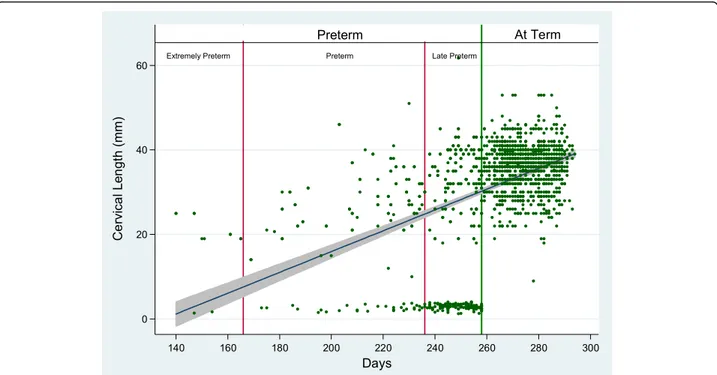
The majority of all preterm births were due to infants born late preterm, 71.83% (n = 459), 26,45% (n = 169) were due to preterm and 1.72% (n = 11) to extremely preterm (Fig.2).
The preterm cohort showed a higher proportion of history of a prior spontaneous PTB (p < 0.0001), unmarried women (p = 0.003) and underweight or obese patients (p < 0.0001). In addition, PTB was associated with presence of uterine anomalies (p < 0.0001), vaginal/urinary infections (p = 0.02), poli-oligodramnios (p < 0.0001), maternal diabetes (p = 0.004) and hypertension (p < 0.0001).
The median value of cervical length measured from 16th to 24th week of pregnancy was 31 mm in cases who delivered preterm and 38 mm in controls, demon-strating a significant difference between the two groups (p < 0.0001) (Table 1 e Fig. 2). The CL measurement, using a cut off of 25 mm according to the SIEOG guide-lines (28), showed a sensibility of 78%, demonstrating to be an adequate screening test for the women at risk of PTB, even if the specificity was only 3%.
Among PTB, cervical length positively correlated with gestational age (r = 0.42, p < 0.0001) (Fig.2). A sub-analysis of preterm birth categories based on gestational age showed a stronger correlation between gestational age and cervical length among preterm and extremely preterm (r = 0.50,p = 0.0002), while no correlation was found among late preterm (r = 0.01, p = 0.85). Among PTB, cervical length correlated with older mothers (r = 0.21,p = 0.025). No other correla-tions were found.
Discussion
In our observational and retrospective Cohort study of pregnant women admitted to an Obstetric Department in Southern Italy, the incidence of preterm birth was 7.8%. The rate of preterm birth decreased from 7.2 to 6.9% (Fig.1b), together with an increase of births in our Hospital (Fig.1a). However, the value remains high, con-sidering that preterm birth is associated with most of
a
b
Fig. 1 a Trends of annual births in our Institution. b Trends of preterm birth annual cases in our Institution
Table 1 Clinical and demographic characteristics of control and preterm cases
Characteristics Controls (n = 7315) Preterm Cases (n = 639) p-value
Maternal age (years), mean (SD) 31.87 (6.04) 31.68 (5.65) 0.025
Pre-pregnancy weight (kg), mean (SD) 63.44 (13.98) 63.73 (13.32) 0.2
Maternal BMI, n (%) Underweight 293 (4) 102 (16) < 0.0001*
Normal 4462 (61) 262 (41)
Overweight 1682 (23) 51 (8)
Obesity 878 (12) 224 (35)
Marital status, n (%) Married 5998 (82) 492 (77) 0.003*
Unmarried 1317 (18) 147 (23)
Parity Nulliparous 4096 (56) 367 (57.5) 0.5
Parous 3219 (44) 272 (42.5)
Prior history of PTB, Yes/No, n (%) 256/7059 (3.5/96.5) 40/428 (8.5/91.5) < 0.0001*
Treatment for cervical dysplasia
Yes/No, n (%) 110/ 7205 (1.5/98.5) 13/626 (2/98) 0.2
Cervical Length (mm), median (IQR) 38 (34–42) 31 (23–37) < 0.0001*
Uterine Anomalies, Yes/No, n (%) 66/7249 (0.9/99.1) 22/617 (3.4/96.6) < 0.0001*
Vaginal/Urinary Infections Yes/No, n (%) 1463/5852 (20/80) 115/524 (18/82) 0.02*
Poly/oligohydramnios
Yes/No, n (%) No 7096 (97) 518 (81) < 0.0001*
Polyhydramnios 37 (0.5) 13 (2)
Oligohydramnios 182 (2.5) 102 (9)
Hypertension, Yes/No, n (%) 366/6949 (5/95) 115/524 (18/82) < 0.0001*
Thyroid disease, Yes/No, n (%) 1024/6291 (14/86) 83/556 (13/87) 0.5
Diabetes, Yes/No, n (%) 732/6583 (10/90) 93/546 (14.5/85.5) 0.004*
Maternal Surgery, Yes/No, n (%) 2677/4638 (36.6/63.4) 252/387 (39.5/60.5) 0.2
Psychological Disorders, Yes/No, n (%) 37/7278 (0.5/99.5) 7/632 (1.1/98.9) 0.3
the pregnancy-related mortality and and short and long-term disability [2].
The slightly decreasing rate of PTB could be, partially, ex-plained with the utmost attention in the prevention of this condition based on the known risk factors. Many features are associated with PTB, such as maternal demographic characteristics [6], nutritional status/ physical activity, prior ob/gyn history, current pregnancy characteristics (i.e. vagi-nal bleeding, uterine contractions, poly/oligohydramnios, psychological problems, use of drugs or infection) [13–16]. Among these risk factors, history of PTB and short CL are considered the most important predictive factors [17–20]. Particularly, a short cervix in the second trimester (i.e. a cervix measure < 15 mm, in a single pregnancy) [21] is a powerful predictor of spontaneous PTB, regardless of ob-stetrical history [22].
In our analysis, we only included data on risk factors, which were clearly reported in all clinical records or sig-nificantly present in the folders. We report a significant association between PTB and risk factors such as vagi-nal/urinary infections, underweight or obese patients, unmarried status, the presence of uterine anomalies, poly/oligohydramnios, hypertension, diabetes, a history of PTB and, finally, a short cervical length. Many of these risk factors could be identified with a good medical history and also treated, leading to an improvement of the risk at the beginning of pregnancy.
In our study, particular attention has been given to CL measurement. While “late preterm” cases demonstrated a large variability of CL values, the “preterm” and “very
early” preterm cases were more correlated with short cervical length, independently to a history of previous preterm. This data, confirming previous studies [23], as-sumes that cervical length helps to identify better the case at risk for early and very early PTB. Probably, in late preterm cases, other risk factors should be also con-sidered, such as maternal disease (i.e.: severe hyperten-sion resistant to medical therapy) or fetal pathological conditions which prompt the gynaecologist to induce labor, determining an iatrogenic PTB.
Our findings suggest a universal cervical screening (i.e. not only for women at higher risk) should be performed in order to discover, at an early stage, asymptomatic women with a short cervix (2–5% of total) [24] and to ad-minister a preventive therapy [25, 26]. The CL measure-ment could be taken during the midtrimester ultrasound screening by all obstetric ultrasonographers who have re-ceived appropriate training. Transvaginal ultrasound is safe, and when performed by trained operators results are reproducible with a relatively low inter-observer variation rate of 5–10% [27]. Currently, the cervical measurement is performed only in women with a higher risk of preterm birth or with symptoms of preterm labour (to reduce hospitalization for tocolysis) [28, 29], in women who undergo cervical cerclage or in twin pregnancies [28]. Data on the utility of the cervical length measurement is reported in many papers [23,25,28–31] but, according to the guidelines of the Italian Society of Ultrasound in Obstetrics and Gynaecology (SIEOG), the use of ultra-sound for the evaluation of the uterine cervix in a low-risk
population is not supported by sufficient scientific evidence [28]. This statement has been confirmed in the NICE (Na-tional Institute for Health and Care Excellence) guideline [32] and in the SCOG (Society of Obstetricians and Gynae-cologists of Canada) guidelines, which do not consider the consequent therapeutic interventions valid [33]. Cervical screening is one tool that can be utilized to identify women at increased risk in order to allow for interventions to pre-vent, delay, or prepare for preterm births, without the need to undergo to biochemical testing for specific markers for preterm labour. Many authors have questioned if universal midtrimester transvaginal CL screening meets the criteria outlined by the WHO of an adequate screening test [34]. Some authors [35, 36] concluded that this screening for women with a singleton gestation, followed by treatment with vaginal progesterone for those with a short cervix, meets all 10 criteria outlined by the WHO for endorsing the implementation of a screening test in clinical medicine [34].
Also the recent Italian guidelines of the Confalonieri Ragonese Foundation recommend performing a cervical measurement during the second trimester ultrasound screening (19–23.6 weeks) of all pregnant women and to ad-minister 200 mg of Progesterone to those women with a cer-vix between 10 and 20 mm [21]. This suggestion is motivated by the results of a systematic review where it was shown that Progesterone administration to a sample of women without a history of PTB and with a cervix between 10 and 20 mm demonstrated a significant reduction of PTB and neonatal morbidity, while in the subgroup with a cer-vical length < 10 mm or between 21 and 25 mm did not give any benefits in prevention of PTB [37]. Another recent sys-tematic review and meta-analysis [26] from randomised controlled trials confirmed that vaginal progesterone was as-sociated with a significant reduction in the risk of preterm birth, respiratory distress syndrome, composite neonatal morbidity and mortality, birth weight < 1500 and < 2500 g and admission to the neonatal intensive care unit. The study reported a reduction of about 40–50% of neonatal death, re-spiratory distress syndrome, intraventricular haemorrhage and proven neonatal sepsis. Moreover, maternal adverse events, congenital anomalies and adverse neurodevelopmen-tal and health outcomes at 2 years of age did not report any difference between groups, confirming that vaginal proges-teron is, at the same time, efficacious and safe [26].
We also use to administrate vaginal progesterone in all the cases at risk for a short CL. This procedure has probably contributed, together with all other preventive measures, to the slight reduction of preterm birth over the years in our Hospital.
Conclusions
Based on our results, we suggest prompt identification of all risk factors associated with preterm birth to apply immediate and appropriate specific interventions.
We also recommend, confirming the evidence of other studies [26,38–40], a transvaginal CL measurement dur-ing the midtrimester ultrasound screendur-ing, in order to identify the women most at risk who could benefit of a progesterone therapy, without the need to undergo to fur-thers biochemical testing for specific markers for PTB.
All these procedures could reduce the rate of PTB and the associated neonatal morbidity and mortality.
Abbreviations
CL:Cervical length; NICE: National Institute for Health and Care Excellence; PTB: Preterm birth; RFs: Risk factors; SCOG: Society of Obstetricians and Gynaecologists of Canada; SIEOG: Italian Society of Ultrasound in Obstetrics and Gynaecology; WHO: World Health Organization
Acknowledgements
The authors gratefully acknowledge the support by families of all these preterm babies.
Funding
This research did not receive any specific grant from funding agencies in the public, commercial, or not-for-profit sectors.
Availability of data and materials
Data sharing not applicable to this article as no datasets were generated or analyzed during the current study.
Authors’ contributions
All authors made substantial contributions and approved the final version of the article. RG and EG made substantial contributions in the drafting the article, conception and design of the study, analysis and interpretation of data. RF and GC made substantial contributions in the revising manuscript critically for important intellectual content. DA made substantial contributions in the acquisition of data, analysis and interpretation of data. GD made substantial contributions in the acquisition of data and analysis. GC made substantial contributions in the acquisition of data and analysis. IF made substantial contributions in the acquisition of data and analysis. RG made substantial contributions in the acquisition and interpretation of data. OT made substantial contributions in final approval of the version to be submitted.
Ethics approval and consent to participate Yes
Consent for publication Yes
Competing interests
The authors declare that they have no competing interests.
Publisher’s Note
Springer Nature remains neutral with regard to jurisdictional claims in published maps and institutional affiliations.
Author details
1Obstetrics and Gynecology Unit, Department of Human Pathology of Adult
and Childhood“G. Barresi”, University Hospital “G. Martino”, via Consolare Valeria 1, Gazzi, Messina, Italy.2Neonatal and Pediatric Intensive Care Unit,
Department of Human Pathology of Adult and Childhood“G. Barresi”, University Hospital“G. Martino”, via Consolare Valeria 1, Gazzi, Messina, Italy.
3General Pediatrics and Pediatric Acute and Emergency Unit,
Policlinico-Vittorio-Emanuele University Hospital, University of Catania, Catania, Italy.4Department of Maternal and Child Health, University of Palermo, Palermo, Italy.5Obstetrics and Gynecology Unit,“Villa Sofia Cervello
Hospital”, University of Palermo, Piazza Salerno 1, Palermo, Italy.6Department
Received: 4 February 2019 Accepted: 29 March 2019
References
1. Saccone G, Perriera L, Berghella V. Prior uterine evacuation of pregnancy as independent risk factor for preterm birth: a systematic review and meta-analysis. Am J Obstet Gynecol. 2016;214:572–91.
2. Mathews TJ, MacDorman MF. Infant mortality statistics from the 2006 period linked birth/infant death data set. Natl Vital Stat Rep. 2010;58:1–31. 3. Saigal S, Doyle LW. An overview of mortality and sequelae of preterm birth
from infancy to adulthood. Lancet. 2008;371:261–9.
4. Martin JA, Kochanek KD, Strobino DM, Guyer B, MacDorman MF. Annual summary of vital statistics 2003. Pediatrics. 2005;115:619–34.
5. Granese R, Calagna G, Sollano A, et al. Data comparison between pharmacological induction of labour and spontaneous delivery. A single centre experience. Ginekol Pol. 2016;87:697–700.
6. Ciancimino L, Laganà AS, Chiofalo B, Granese R, Grasso R, Triolo O. Would it be too late? A retrospective case-control analysis to evaluate maternal-fetal outcomes in advanced maternal age. Arch Gynecol Obstet. 2014;290:1109–14. 7. World Health Organization WHO. Recommended definitions, terminology
and format for statistical tables related to the perinatal period and use of a new certificate for cause of perinatal deaths. Modifications recommended by FIGO as amended October 14, 1976. Acta Obstet Gynecol Scand. 1977; 56:247–53.
8. I grandi prematuri. Note bioetiche. Comitato Nazionale per la Bioetica. La “Carta di Firenze”, 2008. Presidenza del Consiglio dei Ministri.
9. Vincenzo Berghella. Preterm Birth: Prevention and Management. John Wiley & Sons, 19 gen 2010.
10. Suhag A, Berghella V. Short cervical length dilemma. Obstet Gynecol Clin N Am. 2015;42:241–54.
11. Von Elm E, Altman DG, Egger M, Pocock SJ, Gøtzsche PC, Vandenbroucke JP. STROBE initiative. The strengthening the reporting of observational studies in epidemiology (STROBE) statement: guidelines for reporting observational studies. Lancet. 2007;370:1453–7.
12. Berghella V. Preterm birth prevention. In: Berghella V, editor. Obstetric Evidence Based Guidelines. London- New York; 2012. p. 150–64. 13. Goldenberg RL, Culhane JF, Iams JD, Romero R. Ep¬idemiology and causes
of preterm birth. Lancet. 2008;371:75–84.
14. Mercer BM, Goldenberg RL, Das A, et al. The preterm predic¬tion study: a clinical risk assessment system. Am J Obstet Gynecol. 1996;174:1885–95. 15. American College of Obstetricians and Gynecologists. ACOG Practice
Bulletin. Assessment of risk fac¬tors for preterm birth. Clinical management guide¬lines for obstetrician-gynecologists. Number 31, October 2001. (Replaces Technical Bulletin num¬ber 206, June 1995; Committee Opinion num¬ber 172, May 1996; Committee Opinion number 187, September 1997; Committee Opinion num¬ber 198, February 1998; and Committee Opin¬ion number 251, January 2001). Obstet Gynecol. 2001;98:709–16. 16. Hoyme UB. Pragmatic prevention of preterm birth and evidence based
medicine. Arch Gynecol Obstet. 2016;294:1–3.
17. Adams MM, Elam-Evans LD, Wilson HG, Gilbertz DA. Rates of and factors associated with recur¬rence of preterm delivery. JAMA. 2000;283:1591–6. 18. Esplin MS, O’Brien E, Fraser A, et al. Estimating recurrence of spontaneous
preterm delivery. Obstet Gynecol. 2008;112:516–23.
19. Durnwald C, Walker H, Lundy J, Iams JD. Rates of recurrent preterm birth by obstetrical history and cervical length. Am J Obstet Gynecol. 2005;193:1170–4. 20. Jakobsson M, Gissler M, Sainio S, Paavonen J, Tapper AM. Preterm delivery
after surgical treatment for cervical intraepithelial neoplasia. Obstet Gynecol. 2007;109:309–13.
21. Gestione del parto pretermine. Fondazione Confalonieri Ragonese su mandato SIGO, AOGOI, AGUI 2016. Facilitatore A. Regalia (www.sigo.it). 22. Leung TN, Pang MW, Leung TY, Poon CF, Wong SM, Lau TK. Cervical length
at 18–22 weeks of gesta¬tion for prediction of spontaneous preterm deliv¬ery in Hong Kong Chinese women. Ultrasound Obstet Gynecol. 2005; 26:713–7.
23. Granese R, Mantegna S, Mondello S, et al. Preterm birth: incidence, risk factors and second trimester cervical length in a single center population. A two-year retrospective study. Eur Rev Med Pharmacol Sci. 2017;21:4270–7. 24. Martin JA, Hamilton BE, Sutton PD, et al. Centers for Disease Control and
Prevention National Center for Health Statistics National Vital Statistics sys¬tem. Births: final data for 2007. Natl Vital Stat Rep. 2010;58:1–85.
25. Romero R, Conde-Agudelo A, Da Fonseca E, et al. Vaginal progesterone for preventing preterm birth and adverse perinatal outcomes in singleton gestations with a short cervix: a meta-analysis of individual patient data. Am J Obstet Gynecol. 2017;218:161–80.
26. Saccone G, Simonetti B, Berghella V. Transvaginal ultrasound cervical length for prediction of spon¬taneous labour at term: a systematic review and meta-analysis. BJOG. 2016;123:16–22.
27. Owen J, Iams JD. What we have learned about cervical ultrasound. Semin Perinatol. 2003;27:194–203 National Institute of Child Health and Human Development Maternal-Fetal Medicine Units Network.
28. Linee Guida Societa’ Italiana di Ecografia Ostetrico Ginecologica, SIEOG 2015. Gruppo Editoriale EDITEAM.
29. Berghella V, Palacio M, Ness A, Alfirevic Z, Nicolaides KH, Saccone G. Cervical length screening for prevention of preterm birth in singleton pregnancy with threatened preterm labor: systematic review and meta-analysis of randomized controlled trials using individual patient-level data. Ultrasound Obstet Gynecol. 2017;49:322–9.
30. Navathe R, Saccone G, Villani M, et al. Decrease in the incidence of threatened preterm labor after implementation of transvaginal ultrasound cervical length universal screening. J Matern Fetal Neonatal Med. 2018;5:1–6.
31. Van der Ven J, van Os MA, Kazemier BM, et al. The capacity of mid-pregnancy cervical length to predict preterm birth in low-risk women: a national cohort study. Acta Obstet Gynecol Scand. 2015;94:1223–34. 32. NICE. Preterm labour and birth: methods, evidence and recommendations.
Guidelines 2015;25.
33. Lim K. SOGC. Clinical practice guideline. Ultrasonographic cervical length assessment in predicting preterm birth in singleton pregnancies. J Obstet Gynaecol Can. 2011;33:486–99.
34. World Health Organization Fact Sheet #363. November 2014. Available at:http:// www.who.int/mediacentre/factsheets/fs363/en/. Accessed 16 Mar 2016. 35. Combs CA. Vaginal progesterone for asymptomatic cervical shortening and
the case for universal screening of cervical length. Am J Obstet Gynecol. 2012;206:101–3.
36. Khalifeh A, Berghella V. Universal cervical length screening in singleton gestations without a previous preterm birth: ten reasons why it should be implemented. Am J Obstet Gynecol. 2016;214:603.
37. Romero R, Nicolaides K, Conde-Agudelo A, et al. Vaginal progesterone in women with an asymptomatic sonographic short cervix in the midtrimester decreases preterm delivery and neonatal morbidity: a systematic review and metaanalysis of individual patient data. Am J Obstet Gynecol. 2012;206:124 e1–19. 38. FIGO Working Group on Best Practice in Maternal-Fetal Medicine.
International Federation of Gynecology and Obstetrics. Best practice in maternal-fetal medicine. Int J Gynaecol Obstet. 2015;128:80–2. 39. Romero R, Nicolaides KH, Conde-Agudelo A, et al. Vaginal progesterone
decreases preterm birth < 34 weeks of gestation in women with a singleton pregnancy and a short cervix: an updated meta-analysis including data from the OPPTIMUM study. Ultrasound Obstet Gynecol. 2016;48:308–17. 40. Vintzileos AM, Visser GH. Interventions for women with mid-trimester short