UNIVERSITÀ DEGLI STUDI DI CATANIA
FACOLTÀ DI FARMACIA
DOTTORATO DI RICERCA IN BIOTECNOLOGIE
XXIII CICLO
Enhancement of efficacy and selectivity of
chemopreventive compounds in human breast
cancer cells by using Immunoliposomes
Coordinatore e Tutor:
Chiar.mo Prof. Federico Cicirata
Supervisor:
Chiar.mo Prof. Vicente Micol
Index
Introduction...1
1. The cancer burden………2
2. Distinctive features of cancer...6
3. Breast cancer...11
3.1 Detection and staging of breast cancer...11
3.2 Expression of estrogen receptor...15
3.3 The biology of HER2 and its importance in breast cancer...16
3.4 The monoclonal antibody Trastuzumab (Herceptin®)...19
3.4.1 The mechanisms of action……….19
3.4.1.1 Immune-mediated response...19
3.4.1.2 Inhibition of angiogenesis...20
3.4.1.3 Inhibition of HER2 extracellular cleavage...20
3.4.1.4 Inhibition of PI3K pathway...20
3.4.2 The resistance to Trastuzumab…….…...……..……….21
4. Cancer and bioactive compounds……….23
4.1 Curcumin………24
4.1.1 Origin of curcumin and its analogues...24
4.1.2 Molecular targets of curcumin………..26
4.2 Resveratrol……….29
4.2.1 Origin and chemestry……….29
4.2.2 Biological activity……….30
4.3 Limits in the use of bioactive natural compounds...32
5. Drug Delivery System...33
5.1 Features and advantages of DDSs...33
5.2 Liposomes...34
5.3 Immunoliposomes...38
5.3.1 Definition and advantages...38
5.3.2 Clearance of immunoliposomes from the circulation...39
5.3.3 Tumor cell binding: the importance to choise a target epitope...40
5.4 Therapeutic availability...41
Research’s Aim...43
Materials And Methods………44
1. Cell Cultures...45
1.1 Cell Lines...45
1.1.1 Thawing of JIMt1 and MCF7 cells...46
1.1.2 Passaging of JIMT1 and MCF7 cells...46
2. MTT assay...47
2.1 Experimental procedure……….48
3. Protein Extraction and analysis……….50
3.1 Preparation of cell lysate………..50
3.2 Determination of protein concentration………..50
3.3 Separation of proteins by Polyacrylamide Gel Electrophoresys (SDS-PAGE)...50
3.4 Western Blotting………51
3.5 Enhanced Chemioluminesciences detection………...51
4. Liposomes……….53
4.1 Chemicals...53
4.2 Liposomes preparation: thin film method...53
4.3 Size reduction of liposomes...53
4.4 Immunoliposomes preparation: antibody derivatization and conjugation to liposomes...55
4.4.1 Experimental protocol...56
4.5 Separation of no-encapsulated compounds and unbound antibody from liposomes...58
4.5.1 Separation of no-encapsulated compounds from liposomes by ultrafiltration...58
4.5.2 Separation of no-encapsulated compounds and unbound antibody from immunoliposomes by size exclusion chromatography...58
4.6 Quantitative analysis of lipid concentration...60
4.7 Size determination of liposomes...61
5. Quantification of encapsulated compounds by high performance liquid Chromatography...62
5.1 High performance liquid chromatography (HPLC): theory...62
5.2 Quantification of curcumin and resveratrol through HPLC...65
5.2.1 Experimental protocol...66
6. Cellular Uptake using flow citometry analysis...68
6.1 Principle of FACS analysis...68
6.2 Uptake studies...69
6.3 Analysis of HER2 expression...71
Results...72
1. Expression of HER2 on breast cancer cell lines...73
1.1 Detection of HER2 by Western Blot...73
1.2 Detection of membrane surface HER2 by Flow Cytometry...75
2. Cytotoxyc effects of free curcumin, free resveratrol and a combination of both on the viability of JIMT1 and MCF7...76
3. Quantification of drugs‟ content by HPLC analysis...81
4. Cytotoxic effect of curcumin and resveratrol incorporated into liposomes...84
5. Optimization of drugs‟ encapsulation into Immunoliposomes...89
6. Purification and characterization of liposomes and anti-HER2 Immunoliposomes...96
7. Comparative effect of the three delivery systems: free compounds, liposmoes and Immunoliposomes...99
8. Quantitative uptake and immunoliposomes‟ binding in JIMT1 and MCF7 cells using MICF technology...104
Discussion...120
List of Abbreviations...133 Acknowledgements...136 References...139
1. The cancer burden
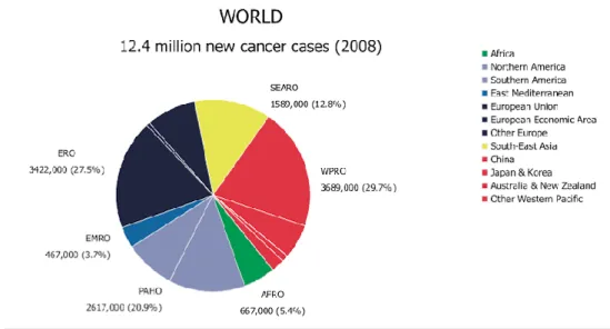
Cancer is the leading cause of death in economically developed countries and the second one in developing countries after cardiovascular diseases. Globally, there were an estimated 12.4 million incident cases of cancer in 2008 (6 672000 in men and 5 779000 in women) and 7.6 million deaths from cancer (4 293 000 in men and 3 300 000 in women) (Fig.1).
Figure 1 Distribution of Global Cancer cases by World Health Organization Region (2008)
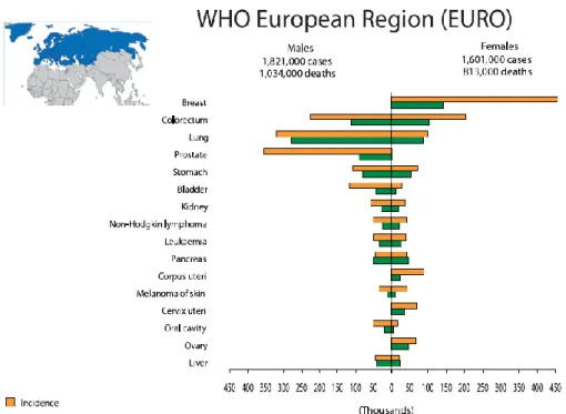
In the European Region (EURO), as in the rest of the world, the commonest incident cancer for men is lung cancer followed by prostate, colorectal, bladder and stomach cancer. They are also the commonest forms of cancer death in men (Fig. 2 and 3). Among women breast cancer is the commonest form of cancer and it is also the main cancer cause of death in women, followed by colorectal cancer, lung cancer and stomach cancer (Fig. 2 and
Figure 2 Cancer Incidence and Mortality in World Health Organization European Region (EURO) (Data from World Cancer Report 2008)
Figure 3 Incidence and mortality of the most common cancers in males and females in more developed and less developed countries. (Data
Globally, breast cancer comprises 23% of all female cancers that are newly diagnosed in more than 1.1 million women each year [1]. Breast cancer already is an urgent public health problem. Its incidence has grown rapidly during the last decades in many developing countries by up to 5% per year [2,3,4] and slowly in developed countries. Mortality rates have remained fairly stable between 1960 and 1990 in most of Europe and the Americas, then showed appreciable declines, which have reached 25-30% in northern Europe [5]. A plausible explanation could be that the screening programs in developed countries has increased the number of new detected cases and in parallel, the technical and pharmacological advances has reduced the mortality.
2. Distinctive features of cancer
The growth and development of breast cancer is similar to that of the other cancer types.
Cancer is a multi-step process through which cells undergo profound metabolic and behavioural changes, leading them to proliferate in an excessive and untimely way, to escape surveillance by the immune system, and at last to invade distant tissues to form metastases [6].
These changes arise through the accumulation of modifications in the genetic programmes that control cell proliferation and lifespan, the relationships with neighbouring cells and the capacity to escape the immune system. This process results in the formation of a mass of deregulated cells, which don't respect the rules that control normal cell growth and behaviour. A cancer cell is a cell that escapes the laws governing cell community life, attaining an independent survival advantage. In doing so, cancer cells try to adapt and fight off the defence systems of the organism and progressively adopt an aggressive and invasive behaviour. They become able to travel within the body and to home preferentially in hospitable organ environments as metastases. Metastatic cancer cells have become so good to adapt themselves to new conditions that they resist many attempts at killing them, including cytotoxic drugs or radiation treatments. This is the reason why most cancers are best treated at an early stage, i.e. at a time when cancer cells still have limited adaptive capacity and consequently are unable to bypass the effects of treatment. Recent experimental studies have identified the minimum number of steps needed to develop a fully cancerous cell [7] (Fig.
Figure 4 Acquired Capabilities of Cancer [7]
Physiologic changes and novel capabilities acquired during tumour development that represent the successful breach of an anticancer defence mechanism into cells and tissues.
Some fundamental rules must be violated:
- Self sufficiency in grow: cells should proceed to divide only when they receive appropriate signals; in the tumour this rule is broken and cells have permanently activated cell division by switching on the circuits that are normally activated when cells are stimulated by a hormone or a growth factor.
- Evasion from apoptosis: when confronted by stressful or improper conditions for DNA (Deoxyribonucleic acid) replication, cells activate self destruction programmes rather than allowing DNA replication to
proceed in conditions in which genes may become damaged. In cancer cells also these auto-destruction programmes are bypassed. To do that, cells have to get rid of its safety brakes, which normally should prevent aberrant cell divisions. These brakes are controlled by two master genes: RB (Retinoblastoma gene) and TP53 (Tumour Protein_53) which produces the p53 protein, a stress sensor that normally prevents cells from dividing when their environment is disturbed. When these two brakes are released by mutations, cells can not only divide themselves but also avoid entering programmed cell death, thus allowing the formation of a tumour mass.
- Insensitivity to antigrowth signals: another parameter eluded by cancer cells concerns cell division. Normal cells divide only a limited, fixed number of times, they are able to replicate their DNA and divide only for a finite number of times, thanks to particular structures at the end of each chromosome called telomeres. The telomeres are made of small DNA repeated sequences which become eroded each time the cells divide. When all repeats are gone, a cell cannot divide any longer and becomes a senescent cell. In a cancer cell, the activation of an enzyme called telomerase allows the addition of new repeats at the end of chromosomes, thus allowing the cell to divide an infinite number of times. Achieving these functional changes is enough for the cell to become cancerous.
- Angiogenesis: cancer cells to improve tumour supply in oxygen and nutrients promote angiogenesis, that is, the synthesis of new blood vessels dedicates to tumour vascularisation. They are able to do that by releasing of vascular endothelial growth factor (VEGF), which stimulate the growth and invasion of new blood vessels accelerating its proliferative capacity.
- Tissue invasion and metastasis: one of the main features that distinguish benign from malignant cancers is the ability of tumour cells to spread from their original location to invade and colonise distant organ sites. As long as the tumour remains confined to one specific
location, it remains curable and it can be removed surgically. Once tumour cells start to spread into the organism, they become more difficult to control. They may reach distant organ sites and form secondary tumours, called metastases; these tumour cells able to spread, have acquired special properties that make them more resistant to treatments and to destruction by the immune system. “Metastasis” is a term that origin from the combination of two Greek words, “meta”, meaning “beyond”, and “stasis”, that meaning “location”. A metastasis is a lesion that has changed position. It consists of a series of steps by which growing tumours disturb the architecture of the tissue where they arise, take the space and place of normal cells, infiltrate into healthy areas and cross vessel barriers to enter the lymphatic or blood circulation. They acquire the capacity to become independent from their organ of origin, to invade other organs, to travel in the body and to form colonies.
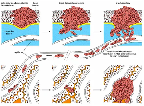
Expression of various Matrix Metallo proteinases (MMPs) has been found to be up regulated in every type of human cancer and correlates with advanced stages and metastatic properties [8, 9]. Up-regulation of MMP expression determinates degradation of basement membrane components, allowing the tumour cells to invade into the adjacent stroma and to break down the basement membranes associated with capillaries and lymphatic vessels allowing tumour cells to enter the circulation (Fig. 5). MMPs are also involved in cell migration by removing sites of adhesion, exposing new binding sites, cleaving cell-cell or cell-cell-matrix receptors and releasing chemoattractants from the extracellular matrix (ECM)[10].
Figure 5 Process of metastasis
Cells grow as benign tumor, break through the basement membrane, travel through the blood stream invading other tissues, adhere to capillary wall, escape from blood vessel and proliferate to form metastases.
3. Breast cancer
3.1 Detection and staging of breast cancer
Breast cancer (BC) is the most commonly diagnosed cancer among women. More than 1 in 4 cancers in women (about 28%) are breast cancers. The incidence is very low in females below the age of 15, and increases very steeply by the age of 45. After menopause, the production of estrogens and progesterone from the ovaries ceases, and the increase in breast cancer incidence rates with age slows down compared to premenopausal women. This suggests a significant implication of hormones in the etiology/development of breast cancer. In vitro experiments have shown that estrogen increases mammary cell proliferation, and also in vivo experiments in animals have demonstrated it [11]. In addition to estrogen exposure, there are other known risk factors for the development of the disease, they include: ethnicity, radiation exposure, family history, genetic predisposition such as mutations in Breast Cancer genes 1 and 2 (BRCA1 and BRCA2), and lifestyle factors, such as obesity, alcohol consumption, and lack of exercise [12].
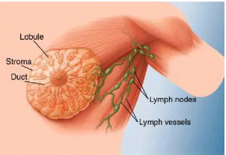
The mammary gland is the functional structure of the female breast. In adults, each mammary gland is composed of fifteen to twenty lobes, divided by adipose tissue. Each lobe is subdivided into lobules, which contain the glandular alveoli that secrete the milk into a series of secondary tubules. These tubules converge to form a series of mammary ducts, which in turn converge to form a lactiferous duct that drains at the tip of the nipple (Fig. 6). It is generally believed that most breast cancers (approximately 75%) occur within the terminal ductal-lobular unit (TDLU), consisting of the lobule and its adjacent ducts [12]. However, there is still not complete agreement as to whether breast cancer originates as a disease of a single cell, whose progeny spread through a single duct system and accumulate multiple genetic changes toward malignant transformation, or as a cluster of genetically unstable cells and ducts that are simultaneously involved [13].
Figure 6 Anatomy of the breast
Over time, cancer cells can invade nearby healthy breast tissue and make their way into the underarm lymph nodes, small organs that filter out foreign substances in the body. If cancer cells get into the lymph nodes, they then have a pathway into other parts of the body.
When cancer cells break away from the primary (original) tumor and travel through the lymph system or blood to other places in the body, another (secondary) tumor may form. The secondary tumor is the same type of cancer as the primary tumor. For example, if breast cancer spreads to the bones, the cancer cells in the bones are actually breast cancer cells; the disease is metastatic breast cancer, not bone cancer.
Tumour cells that are capable of spreading have acquired special properties that make them more resistant to treatments and to destruction by the immune system. Therefore, detection of distant spread and metastases is often an indicator of poor prognosis for the patient. This is reflected in the
TNM classification system, which provides a universal system to describe the anatomic extent of a cancer.
The staging system normally used to classify the stages in breast cancer is called TNM, which stands for „tumour, node, metastasis‟. So TNM staging takes into account the size of the tumour, whether the cancer has spread to nearby lymph nodes, and whether it has spread to other parts of the body (metastasis).
The T stages are numbered 1 to 4 and describe the size of the tumour (Fig.
7a, 7b, 7c and 7d). The N stages are numbered 0 to 3 and describe which
lymph nodes are affected, if any.
The M stages are M0 (no sign of cancer spread) and M1 (cancer has spread to another part of the body).
A B C
D
Figure 7 Tumor size stages
a), b) and c) illustrate T1, T2 and T3 stages of breast cancer; in d) pea, peanuts, walnut and lime show tumor size.
3.2 Expression of estrogen receptor
Many breast tumors have feature that distinguishes them from most other tumors: the estrogenic hormone-dependence for their development and growth [14-19]. Breast tissue requires input from estrogens to grow, consequently tumors that arise from this tissue, show estrogenic dependence for their proliferation [18].
Hormone receptors, such as the estrogen receptor (ER) and progesterone receptor (PR), are determinants of breast tumor behavior and may suggest etiologic pathways [19,20]. ERs bind estrogen and facilitate protein synthesis, cell division and breast cell proliferation [21-23]. PRs, in turn, are regulated by circulating levels of estrogen [19,20].
Expression of ER has long been known to be one of the most important prognostic factors in breast cancer [24,25]. The clinical investigation of tamoxifen, a competitive antagonist of the estrogen receptor in breast tissue used as an anti-cancer treatment in the 1960‟s, has had a big effect on survival of women with estrogen positive breast cancer. The survival benefit of ER positivity per se, coupled with the beneficial effects of tamoxifen, means that women who have cancer which expresses ER, have the best chance to survive their breast cancer in a shorter term. However, the risk of recurrence and death vary over time in patients with ER positive and ER negative disease. A data analysis of 2006 generated from three clinical trials of chemotherapy showed that the risk of recurrence in ER negative patients was highest in the first two to three years after treatment, which then decreased dramatically with increasing time from diagnosis. In patients with ER positive disease, the risk of an event in the first few years after diagnosis was low, but in a long term, the hazards were slightly higher for patient with ER positive disease when compared with ER negative disease [26]. This demonstrates that, with the development of modern chemotherapeutic regimens, patients with ER negative cancer, who survive the first 5 years after diagnosis, have at least as good long term survival as patients with ER positive cancer.
3.3 The biology of HER2 and its importance in breast cancer
The human epidermal growth factor receptor 2 (HER2) also known as HER-2/neu or ErbB2, is a member of the ErbB family of cell surface receptor tyrosine kinases (RTKs) involved in the transmission of signals controlling normal cell growth and differentiation [27]. The family consists of 4 receptors sharing a high degree of identity; HER1 (EGFR, ErbB1), HER2 (ErbB2), HER3 (ErbB3) and HER4 (ErbB4). The HER2 receptor, a 185 kDa transmembrane glycoprotein, is encoded by the HER2 gene, a proto-oncogene located on the long arm of chromosome 17q21 [28]. The erbB receptors share a similar structure comprised of a cysteine-rich extracellular region, a lipophilic transmembrane segment, an intracellular domain with tyrosine kinase activity and a carboxy terminal domain that is autophosphorylated upon receptor activation [29] (Fig. 11).
The receptors exist as monomers which upon ligand binding associate to form of homo- or hetero-dimers, resulting in the activation of intrinsic kinase activity, ultimately leading to stimulation of intracellular signaling cascasdes [30]. Notably, HER2 is the preferred dimerization partner of the other HER-family receptors [31] and HER2-containing heterodimers result in potent mitogenic signaling, especially HER2:HER3 heterodimers [30,32,33]. The preference for HER2-containing dimers is likely due to the open conformational state of HER2 compared with the other receptors. Crystallographic studies have revealed that HER1, HER3 and HER4 exist in a closed conformation with the dimerization domain (domain II) tethered to domain IV preventing the formation of dimers with other HER receptors [34-36]. Upon ligand binding to domains I and III the receptors undergo a significant conformational rearrangement exposing the previously concealed dimerization arm for interaction with other HER receptors also in the active state [34,36,37].
In contrast, HER2 is an “orphan receptor” with no known endogenous ligands, however, this receptor is constitutively in a open conformation with the dimerization arm exposed, resembling the ligand-bound state of EGFR [34,37]. This open-conformation facilitates dimerization with other HER family
receptors defining a key role for HER2 in the signal transduction of ligand-driven heterodimeric complexes.
HER2 is normally expressed at low levels in adult cell types including the breast epithelium, the central nervous system, bone, muscle, skin, heart, lungs and intestinal epithelium [28,38]. The function of HER2 in adult tissues is not fully understood, however the receptor appears to play a role in the proliferation and differentiation of epithelial cells [30] and in the protection of cardiomyocytes against apoptosis [39]. In fetal tissues, however, HER2 is widely expressed and is critical to normal development [38]. The clinical significance of HER2 in cancer was first discovered in the early 1980s following the identification of the neu onocogene, the mutationally active rat homologue of HER2 [40]. The human homologue was soon identified [28,41] and found to be overexpressed in a mammary carcinoma cell line [42]. On the basis of these findings, Slamon and co-workers examined HER2 expression in a series of primary human breast tumours and reported a significant association between HER2 overexpression, relapse and patient survival [43]. HER2 is now known to be overexpressed in approximately 20-30% of BCs [44], and overexpression is also common in ovarian, prostate, lung, gastric and oral cancers [45].
Overexpression of HER2 is a combined result of increased transcription and protein translation. Indeed, breast cancer cells may have as many as 100 copies of the gene per cell compared with two copies of the HER2-gene in normal cells [46]. Moreover, there are approximately 20 thousand receptors per cell on normal cells, but breast cancer cells may contain as many as 500 thousand to 2 million HER2 receptors per cell [47]. At this high level of HER2 receptor expression, the kinase activity of HER2 becomes constitutively activated which appears to exert potent mitogenic and transforming effects on cells [47].
This high density of HER2 promotes the formation of HER2 heterodimers as well as the formation of ligand-independent constitutively active HER2 homodimers [32,48,49]. HER2-containing dimers persist longer on the cell surface due to their slower rate of internalization, resulting in overactive cell
signaling and leading to dysregulated cell growth, proliferation and malignant transformation [32].
Pathologically, HER2 overexpression is associated with large tumor size, lack of ER/PR expression, presence of nodal metastasis, high nuclear grade and ductal histology [50]. Clinically, HER2 is associated with aggressive disease, increased risk of relapse and poor long-term survival [43,50].
Figure 8 Structure of HER2
The protein structure consists in two ligand-binding region (LD1 and LD2), two cysteine regions (CR1 and CR2), a short transmembrane domain (TM), a catalytic tyrosine kinase domain (TK) and a carboxy terminal tail (CT). Numerous sites of tyrosine phosphorylation within the TK and CT domains are indicated by circled P. Letters on the right (A-D) indicate areas that are altered or mutated in certain naturally occurring or experimentally induced cancer.
3.4 The monoclonal antibody trastuzumab (Herceptin®)
Given the critical importance of HER2 in some forms of breast cancer, extensive research has focused on HER2 inhibitors as potential anticancer agents. Trastuzumab (Herceptin®, Genentech, Inc., San Francisco, CA), is currently the only HER2-targeted therapeutic agent that has received marketing clearance from the U.S. Food and Drug Administration (FDA) for use in the treatment of patients with HER2- overexpressing breast cancer [47]. Trastuzumab is a humanized mAb that binds specifically to HER2 on the C-terminal portion of the extracellular domain (ECD) near the juxtamembrane region in domain IV of the receptor [51]. Trastuzumab was constructed by grafting the complementary-determining regions (CDRs) from the murine mAb 4D5 into a human kappa IgG1 to avoid eliciting a human anti-mouse antibody (HAMA) response in patients [52].
3.4.1 The mechanisms of action
The effectiveness of trastuzumab appears to be correlated with the level of HER2 expression in breast cancer, and with the accessibility of tumors to the drug [53]. However, the mechanism by which trastuzumab induces regression of HER2-overexpressing tumors is not completely understood, but several molecular and cellular effects have been observed in experimental in
vitro and in vivo models. Several of the proposed mechanisms are described
below.
3.4.1.1 Immune-mediated response
One of the proposed mechanisms of trastuzumab anti-tumor action is through antibody-dependent cellular cytotoxicity (ADCC) [54-56]. Specifically, the natural killer (NK) cells are important for the ADCC response to trastuzumab [47]. These cells express the Fcγ receptor that binds the Fc domain of the IgG1 trastuzumab antibody, and promotes lyses of trastuzumab-bound cancer cells. The importance of this immunological effect was revealed by Clynes et al. [57], who achieved a tumor regression rate of 96% in mice bearing HER2-overexpressing BT-474 xenografts treated with
trastuzumab, but only a 26% reduction in tumor growth in mice lacking the Fcγ receptor.
3.4.1.2 Inhibition of angiogenesis
Both primary and metastatic breast cancer are dependent on angiogenesis for tumor growth [54]. Trastuzumab can modulate different pro- and antiangiogenic factors to achieve angiogenesis suppression [55,56,58].
3.4.1.3 Inhibition of HER2 extracellular cleavage
The ECD of HER2 can be released by proteolytic cleavage from the full-length receptor, yielding a 110-kDa fragment that can be detected in vitro in cell culture medium, and a 95-kDa amino-terminally truncated membrane-associated fragment with increased kinase activity [51]. The HER2-ECD can also be detected in vivo in serum, and is currently measured in the clinic with an FDA approved ELISA-based test to follow-up and monitor patients with metastatic breast cancer [59]. Molina et al. [60] demonstrated in HER2- overexpressing SK-BR-3 and BT-474 human breast cancer cells that trastuzumab can block metalloprotease-mediated cleavage of the HER2-ECD. Moreover, several clinical studies have demonstrated that a decline in serum HER2-ECD during trastuzumab treatment correlates with improved tumor responsiveness and progression-free survival [61,62], which indirectly supports the hypothesis that trastuzumab may act by inhibiting HER2 cleavage [63].
3.4.1.4 Inhibition of PI3K pathway
Overexpression of HER2 receptor tyrosine kinases leads to ligand-independent homodimerization and autophosphorylation of tyrosine residues on the cytoplasmic domain of the receptors [64]. Phosphatidylinositol 3' kinase (PI3K) associates with these phosphorylated tyrosine residues and activates downstream effectors, which ultimately leads to enhanced protein synthesis, cell proliferation, survival and motility [64]. Trastuzumab can inhibit the PI3K pathway.
3.4.2 The resistance to trastuzumab
Despite the fact that trastuzumab-based treatment strategy has established a milestone in the therapy of HER-2 positive breast cancer with attractive clinical benefits, either as a single agent or in combination settings, one of the major drawbacks of the trastuzumab-therappy is trastuzumab resistance, even in highly selected HER-2 overexpressed patients. In fact, only about 30% of HER-2 positive metastatic breast cancer patients respond to trastuzumab and approximately 70% of patients with overexpressed HER-2 receptor may have primary resistance [65]. Additionally, the majority of those patients who achieve initial efficacy tend to develop secondary trastuzumab resistance within one or two years [66].
Several mechanisms have been postulated in an attempt to explain both intrinsic and acquired resistance to trastuzumab but it is not completely understood.
a) Cleavage of HER-2 extracellular domain to form the truncated HER-2 receptor and the overexpression of membrane associated mucin MUC4 to mask or block the trastuzumab binding site, which can interrupt the interaction between HER-2 receptor and this antibody [67-70].
b) Although trastuzumab reduces HER2-mediated signaling, it might not reduce signaling mediated through other HER receptors [47]. For example, heterodimerisation of HER2 with other receptors of the erbB2-family can be induced by ligands of HER1, HER3 and HER4, and in the presence of an excess of ligands, the resulting heterodimers may initiate mitogenic signaling even in the presence of trastuzumab [47]. Indeed, increased levels of the ErbB family ligands EGF and heregulin blocked trastuzumab-mediated growth inhibition of HER2- overexpresssing breast cancer cell lines, and this inhibition was associated with increased signaling from HER2/HER1(EGFR) and HER2/HER3 complexes [71,72]. Valabrega et al. compared tumor tissue from patients with advanced HER2-positive breast cancer before and after trastuzumab treatment, and observed a strong increase in TGF-α production upon disease progression, suggesting a possible role
of EGFR signaling by TGF-α as mediator of acquired resistance to trastuzumab. Indeed, these authors found that trastuzumab was less efficient at inhibiting the growth of HER2-positive SK-BR-3 cells engineered to overexpress Tumor Growth Factor alpha (TGF-α compared to the parental cells [73].
c) bypass signalling through the non-EGFR family growth factor receptor insulin-like growth factor-1 receptor (IGF-1R), enables activation of the downstream signal cascades without the participation of HER-2. Therefore, the further understanding of trastuzumab action and resistance mechanisms highlights the need of novel targeted drugs aiming at HER-2 overexpression [74,75].
4. Cancer and Bioactive compounds
Recently, attention has been focused on identifying bioactive compounds able to inhibit the process of carcinogenesis.
The interest of the medicine on natural compound rises at the time of
Hippocrates, the father of modern medicine, who said two and a half thousand years ago, “Let food be the medicine and medicine be the food.” Galen of Pegamon (129–199 A.D.), a Greek physician and follower of Hippocrates‟ teachings was said to have prescribed various foods, including peeled barley, milk, and various vegetables for the treatment of cancer [76]. Approximately 35 years ago, the National Cancer Institute (NCI) initiated a Diet and Cancer Program to elucidate the role of food and its nutrients in cancer prevention and therapy. These studies were among the first to show that consumption of fruits, vegetables, and whole grains are associated with a decreased risk of many types of cancers [77]. Subsequently, numerous case-control studies were conducted which yielded several potential dietary constituents as possible chemopreventive and therapeutic agents. In another study, the National Cancer Institute collected about 35,000 plant samples from 20 countries and has screened around 114,000 extracts for anticancer activity [78]. Of the 92 anticancer drugs commercially available prior to 1983 in the US and among worldwide approved anticancer drugs between 1983 and 1994, 60% are of natural origin [78]. Natural products discovered from medicinal plants have played an important role in the treatment of cancer. Natural products or their derivatives comprised 14 of the top 35 drugs in 2000 based on worldwide sales [79]. Two plant derived natural products, paclitaxel and camptothecin were estimated to account for nearly one-third of the global anticancer market or about $3 billion of $9 billion in total annually in 2002 [80].
The beneficial effects of fruits and vegetables have been attributed to, among other things, the high content of bioactive compounds that are non-nutrient constituents commonly present in food [81].
against cancer stand out curcumin and resveratrol that were the subject of our research [82-97].
4.1 Cucumin
4.1.1 Origin of curcumin and its analogues
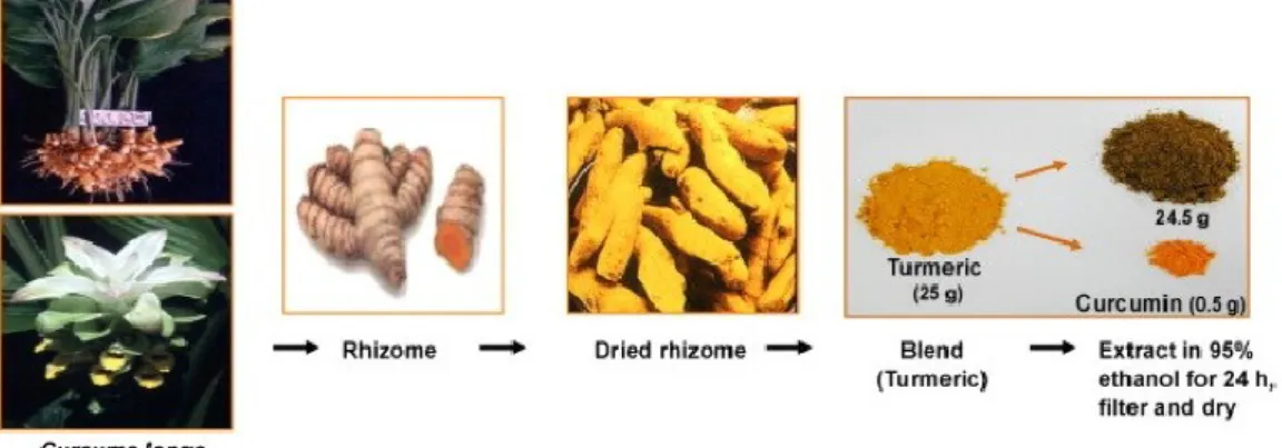
Curcumin (Curc) is the yellow spice derived from the roots, rhizome, of the plant Curcuma longa. It is native of India and Southeast Asia and it has been used to treat a broad range of ailments in Ayurvedic medicine for at least 4000 years. The powdered extracts of dried roots, often called turmeric, may contain volatile and nonvolatile oils, proteins, fat, minerals, carbohydrates, moisture and curcuminoids. As shown in Fig. 9 the curcuminoids constitute approximately 5% of most turmeric preparations and they are a mixture of three principal compounds: curcumin (sometimes referred to as curcumin I), demethoxycurcumin (curcumin II) and bisdemethoxycurcumin (curcumin III). The majority of commercially available curcumin contains the following composition: curcumin I (77%), curcumin II (17%) and curcumin III (3%) [98] (Fig. 10).
Curcumin was first isolated in 1815 by Vogel [99] and its chemical structure was confirmed by Lampe and Milobedezka in 1910 [100]. It is an oil-soluble coloring compound, readily soluble in alkali, ketone, acetic acid, and chloroform, while insoluble in water at acid or neutral pH.
Figure 9 The extraction of curcumin
4.1.2 Molecular targets of curcumin
As shown in Fig. 11 many molecular targets have been proposed for curcumin. It modulates many regulatory proteins including transcription factors, enzymes, cytokines, adhesion molecules, grow factors and this suggests that curcimin is able to act upon a wide number of biochemical and molecular cascades.
Curcumin shows anti-cancer activity both in vitro and in vivo through a wide variety of mechanisms. It acts over different cancer cell lines in vitro, such as breast, lung, prostate, pancreas, bladder, ovary, kidney, cervix, brain, bone marrow and skin cancer [101]. It has also been shown to potentiate the effect of chemotherapeutic agents [102-105] and that of gamma-radiation [106] in cell culture.
The most accepted curcumin activities are described below and resumed in
Fig 11:
Curcumin has been responsible for the induction of apoptosis, due to the production of reactive oxygen species (ROS) that are known as mediators of intracellular signaling pathways. Excessive production of ROS leads to oxidative stress, loss of cell function, and ultimately apoptosis [107]. ROS production leads to depolarization of the mitochondrial membrane and releases pro-apoptotic molecules from mitochondria into the cytosol, which may act to induce apoptosis [108]. In addition, cytochrome-c release from the mitochondrial membrane results in an increased level of cytochrome c at the cytoplasm and nucleus, which induces apoptosis through mitochondrial permeability transition [109]. ROS generation by curcumin also releases apoptosis inducing factor (AIF) and endonuclease G (EndoG) into the cytosol and nucleus where they induce chromatin condensation and DNA fragmentation [110]. The curcumin mechanism of ROS-triggered cell death also involves the p53 tumor suppressor gene. Many reports suggested that p53 is a potential mediator for ROS-dependent apoptosis [111]. The p53 is a tumor suppressor protein, which not only plays
a central role in the cellular stress response pathways but also promotes apoptosis through a variety of mechanisms. The p53 is involved in cell signal transduction, cellular response to DNA damage, genomic stability, cell cycle control, and apoptosis.
Moreover, curcumin has the ability to protect lipids, hemoglobin, and DNA against oxidative degradation.
Several studies demonstrated that curcumin also inhibits cyclooxygenase (COX) activity in rat peritoneal neutrophils and human platelets [112]. It is believed that over-expression of COX-2 is related with a wide variety of diseases including colon, lung, and breast cancers. COX is a key enzyme responsible for the conversion of arachidonic acid to prostaglandins and thromboxanes. COX-2 is the inducible form of COX, which is over-expressed at inflammatory sites, and research evidence has indicated the critical role of COX-2 in tumor promotion and carcinogenesis. Furthermore, curcumin inhibits the metabolism of arachidonic acid to prostaglandin D2 in mouse epidermis [113]. Curcumin has the ability to inhibit COX-2, but not COX-1 gene expression in colon carcinogenesis [114].
Curcumin showed also inhibitory effects on nuclear factor-kappa β (NF- β), mitogen-activated protein kinases (MAPK), and several cytokines, and was found to reduce the expression of TNF-α- induced Interleukin (IL)-1b, IL-6, IL-8, and TNF-α itself [115,116]. MAPK play a key role in inflammatory stimuli and environmental stresses. Three sub-groups of MAPK cascades (ERK, JNK, p38) have been identified in mammalian cells. The ability of curcumin to regulate the MAPKs signaling pathway might contribute to the suppression of inflammation in many cancer cell lines.
Curcumin also modulates MMPs; they are members of zinc-dependent endopeptidases, which are over-expressed in tumor infiltration. MMP2 and MMP9 are often involved in the tumor angiogenesis, mainly through their degradation of extracellular
matrix [117]. It has been reported that curcumin reduces the invasion and metastasis of cancer cells, suppressing the MMPs expression and inhibiting the TPA-induced activation of ERK and transcriptional activation of NF-kB in human breast cancer epithelial cells [118]. NF- β is a transcription factor that widely acts as a regulator of genes that control cell proliferation and cell survival. The NF- β proteins exist in the cytoplasm in an inactive state, but translocates to the nucleus by the activation of various kinases, phosphorylation, and degradation of the I- β, the NF-- β cytoplasmic inhibitor [119]. It was observed that NF-kB promotes or inhibits apoptosis depending on the phenotype, but activation of the NF- β is reported to protect cells against apoptotic stimuli in the majority of tumor cell lines via the initiation of cell survival genes [120]. Curcumin inhibited the I- β kinase (IKK) signaling complex responsible for the phosphorylation of IkB, thereby blocking improper activation of NF- β and induce apoptosis [121]. Bcl-2 and Bcl-XL are anti-apoptotic proteins that are regulated by NF- β. Several studies have shown that curcumin efficiently induces apoptosis in various cancer cell lines through the suppression of these anti-apoptotic proteins [122].
Figure 11 Multiple molecular targerts of curcumin [105]
4.2 Resveratrol
4.2.1 Origin and chemistry
Resveratrol (RESV) (3,5,49-trihydroxystilbene) was first discovered by Michio Takaoka more than 60 years ago, in the resin of Veratum grandiflorum (false hellebore). Then, RESV was also detected in grapevines (Vitis vinifera) in 1977 by Langcake, who found that the compound was synthesized by leaf tissues in response to fungal infection or exposure to ultraviolet light. This property classifies it as a phytoalexin, compounds produced by plants in
response to damage, stress or infection [123]. RESV is a polyphenol and a member of the stilbene family. It can be found in the cis- or trans-configuration and in a glycosylated form [123]. It has been found in many plants, including grapes, peanuts, berries, Polygonum roots, and other traditional oriental medicine plants [124]. RESV has been reported to be a phytoestrogen due to its structural similarity to the estrogenic agent diethylstilbestrol (Fig. 12).
Figure 12 Chemical structure of cis- and trans-resveratrol, diethylstilbestrol (synthetic estrogen), and 17ß-estradiol
4.2.2 Biological activity
In recent years, research on RESV has described several beneficial effects of this compound on human health. It has been reported to have both anticarcinogenic and cardioprotective activities, which could be attributed to its antioxidant and anticoagulant properties [125]. RESV has been reported
to be effective in inhibiting platelet aggregation and lipid peroxidation, altering eicosanoid synthesis, modulating lipoprotein metabolism [126,127], and exhibiting vaso-relaxing and anti-inflammatory activities [128,129]. For its anticarcinogenic activities, potential mechanisms of RESV action have been studied extensively, though there is no clear consensus on the matter. RESV has been reported to inhibit the three major stages of carcinogenesis: initiation, promotion, and progression (Fig13).
Anti-initiation activity was related to its antioxidant and antimutagenic effects and induction of phase II drug-metabolism enzymes.
Anti-promotion activity was indicated by anti-inflammatory effects and inhibition in vitro of cyclooxygenase and hydroperoxidase.
Anti-progression activity was related to its capability to induce human promyelocytic leukemia cell differentiation. It also inhibits the development of preneoplastic lesion in carcinogen-treated mouse mammary glands in culture and inhibits tumorogenesis in a mouse skin cancer model [130].
Many in vitro studies have addressed the RESV activities in breast cancer cells. RESV exhibits its action in both sensitive and hormone-resistant breast cancer cells. It has also been reported to exhibit anti-initiation, anti-promotion, and anti-progression activities in breast cancer cells, where these properties seem to be related to regulation of xenobiotic carcinogen metabolism and anti-inflammatory, antiproliferative, and proapoptotic effects [130-132].
Figure 13 Biochemicals mechanisms responsible for chemopreventive and chemotherapeutic potential of resveratrol
4.3 Limits in the use of bioactive natural compounds
Although, curcumin and resveratrol have shown significant efficacy in cell culture studies, they elicited limited efficacy in various clinical studies. Their introduction into the clinical setting is hindered largely by their poor absorption, rapid metabolism, or a combination of both, ultimately resulting in poor bioavailability upon oral administration [133,134].
Therefore, to circumvent these limitations and to ease their transition to clinics, alternate strategies should be explored. Drug delivery systems such as nanoparticles, liposomes, microemulsions, and polymeric implantable devices are emerging as one of the viable alternatives that have been shown to deliver therapeutic concentrations of various potent chemopreventives such as curcumin, resveratrol and other bioactive compound into the systemic circulation.
5. Drug Delivery Systems
5.1 Features and advantages of DDSs
Drug delivery systems (DDSs) can improve the pharmacological properties of traditional drugs by altering drug pharmacokinetics and biodistribution [135,136]. DDS can include liposomes, nanospheres, micelles, dendrimers, as well as various polymeric-based systems [137-139]. Among the many DDS available, liposomes became very popular (gained popularity) in recent years thanks to their clinical success.
Due to their small size (about 100 nm or less), they readily extravasate from circulation through vascular gaps or defects attributed to ongoing angiogenesis that is typical of tumour sites [140], which have been reported to be about 200 nm or greater [141]. DDS retention within these sites is generally high due to the poor lymphatic drainage observed within tumors [142,143]. Furthermore, their lower size limit of diameter ensures that these vehicles do not randomly penetrate normal vessel walls. In cancers, an imbalance in factors that regulate angiogenesis, such as overexpression of , results in both increased vascular permeability and chaotic tumour-vessel architecture. In combination, these effects cause enhanced permeation and retention (EPR), resulting in high local drug concentrations.
5.2 Liposomes
Liposomes are spherical, self-closed structures formed by one or more concentric lipid bilayers containing an aqueous phase inside and between the bilayers [144] (Fig. 14). The lipids used in the formation of liposomes are usually comprised of a hydrophilic headgroup and two hydrophobic fatty acyl chains. These amphiphilic molecules spontaneously assemble into aggregates in an aqueous environment. Water-soluble molecules occupy the aqueous compartment, whereas molecules of a more lipophilic character occupy the lipid bilayers. Liposomes can vary substantially in size and lamellarity.
Figure 14 Schematic illustration of a liposome
They are subdivided into multilamellar vesicles (MLVs) consisting of several concentric bilayers, large unilamellar vesicles (LUVs) and small unilamellar vesicles (SUVs) (Fig. 15).
Liposomes can be formed by a variety of methods. When they are prepared by hydration of the dried lipid mixture, they spontaneously form MLVs. Other procedures, such as prolonged exposure to ultrasound or pressure-driven filtration through small-pore filters, cause MLVs to form SUVs or LUVs.
Figure 15 Classification of liposomes
The clinical success of liposomes has also made them very popular drug carriers for various chemotherapeutics. For example, the clinically approved drugs DaunoXome and Doxil are liposomal formulations that encapsulate the commonly used chemotherapeutic agents daunorubicin and doxorubicin respectively.
The pharmacokinetics of liposomes depend on their physiochemical characteristics such as size, surface charge, membrane lipid packing, steric stabilization, dose and route of administration [145]. In general, larger liposomes are eliminated from the blood circulation more rapidly than smaller ones [146]. Binding of opsonins to liposomes depends on liposome size; consequently, the reticuloendothelial (RES) uptake of liposomes by the liver is size-dependent [147]. The action of the reticuloendothelial system results in rapid removal from the blood and accumulation in tissues such as liver and spleen. In general, for a given liposome composition, the larger the liposome, the faster the clearance by the RES [148–150].
Optimal liposome size depends on the tumor target. In tumor tissue, the vasculature is discontinuous, and pore sizes vary from 100 to 780 nm. By
comparison, normal vascular endothelium is < 2 nm in most tissues, 6 nm in postcapillary venules, 40–60 nm for the kidney glomerulus, and up to 150 nm for sinusoidal epithelium of the liver and spleen. Most liposomes are 100 nm [151].
Negatively charged liposomes were believed to be more rapidly removed from circulation than neutral or positively charged liposomes; later studies have indicated that the type of negatively charged lipid affects the rate of liposome uptake by the RES. For example, liposomes containing negatively charged lipids that are not sterically shielded (phosphatidylserine, phosphatidic acid, and phosphatidylglycerol) are cleared more rapidly than neutral liposomes of similar composition. However, liposomes containing sterically shielded lipids (for example ganglioside-GM1 and phosphatidylinositol) are cleared even more slowly than neutral liposomes [152].
Thus, one way to reduce liposomal uptake by the RES is by creating sterically stabilized liposomes. “Steric stabilization” refers to the colloidal stability resulting from attachment of hydrophilic polymers or glycolipids on the liposomes (Fig. 16). The best-studied stabilizers are polyethylene glycol and ganglioside GM1. Sterically stabilized liposomes (Stealth) showed prolonged lifetimes in the circulation as compared with nonstabilized liposomes [153–157]. Sterically stabilized liposomes are also less reactive toward serum proteins and less susceptible to RES uptake than nonstabilized liposomes [153]. The mechanism by which sterically stabilized liposomes are thought to decrease RES-mediated uptake is that the stabilizer occupies the space immediately adjacent to the liposomal surface, excluding other macromolecules from this space. Consequently, access to and binding of blood plasma opsonins to the liposome surface are hindered, preventing interactions with RES macrophages. However, although sterically stabilized liposomes prolong circulation time and decrease liposomal uptake by the RES, they do not actively target the liposome to the tumor.
Polyethylene glycol (PEG) is the most widely used polymeric steric stabilizer. PEG is a linear polyether diol with many useful properties. It is highly soluble
in aqueous and organic media and possesses very low immunogenicity and antigenicity [158] and is non-toxic. It can be attached to the liposome surface in various ways, but the most widely used method is to anchor the polymer in the liposome membrane via a cross-linked lipid (for example PEG-DSPE)
[19]. It was shown that steric stabilisation of liposomes with PEG increases their longevity in the circulation [160]. A supplementary method to ensure long circulation times involves adding cholesterol (Chol) to the lipid bilayer. Cholesterol acts as a spacer between phospholipids of the liposome membrane because of its inflexible structure, preventing demixing of the lipids and reduces PEG chain-chain interactions. This results in improved steric stabilization of the liposomes.
5.3 Immunoliposomes
5.3.1 Definition and advantages
A large variety of therapeutically active molecules (e.g. antitumor drugs, oligonucleotides, DNA, enzymes, peptides and hormones) have been successfully incorporated in liposomes. Especially in the field of cancer chemotherapy, much effort has been invested to successfully achieve site-specific drug delivery with liposomal systems. Active targeting of liposomes to tumor cells is generally attempted by conjugating ligands to the liposomal surface which allow a specific interaction with the tumor cells [161].
Several types of ligands have been used for this purpose, including antibodies, antibody fragments [159,162-164], vitamins [165], glycoproteins [166,167], peptides (RGD-sequences) [168,169], and oligonucleotide aptamers [170].
The first report on antibody-targeted liposomes came from Torchilin et al. about two decades ago [171]. These antibody-targeted liposomes (further referred to as immunoliposomes) were shown to be able to specifically bind to the antigen that is expressed on the target cells. Targeted delivery systems of this type have two basic advantages: because the drug is released at the tumor site instead of circulating widely through the body, it should be more effective for a given dosage; it should also have fewer harmful side effects because smaller amounts of the drug come into contact with healthy tissue. Several coupling techniques have been described for conjugating antibodies or their fragments to liposomes, each with their own advantages and drawbacks [163,172,173]. Many in vitro experiments have demonstrated highly specific binding of immunoliposomes to target cells. However, despite the excellent targeting properties in vitro, successful results on targeting of immunoliposomes in tumor models are scarce up to now.
5.3.2 Clearance of immunoliposomes from the circulation
The most important barrier limiting the usefulness of immunoliposomes for targeted drug delivery has been the rapid recognition and removal from the blood by cells of the mononuclear phagocyte system (MPS), particularly the macrophages in liver and spleen [174,175]. In addition, the presence of antibodies conjugated to the liposomal surface makes immunoliposomes highly susceptible to Fc-receptor-mediated phagocytosis and, as a result, even more prone to rapid clearance [174,176]. The Fc-receptor family, which is expressed by different cells of the MPS, binds the constant region (Fc) of antibodies resulting in internalization of antibody-opsonized complexes [177]. Similarly, immunoliposomes bearing whole antibodies are cleared rapidly due to exposed Fc parts [174,176,178].
The advent of so-called long-circulating liposomes produced by coating the liposome surface with the polymer polyethylene glycol, has revived interest in targeted drug delivery [179,180]. As previous mentioned, PEG-coating sterically stabilizes the liposomal membrane by decreasing interactions with destabilizing and opsonic factors in vivo. As a consequence, PEG-coated liposomes show longer circulation times and reduced uptake by the MPS. There are different methods available for coupling antibodies to PEG-liposomes [181]. For attaching antibodies to the surface of PEG-PEG-liposomes, two main strategies have been followed: those in which the ligand is coupled directly to the liposome bilayer (Figure 17 A and B) and those in which the ligand is attached to the terminal end of PEG (Figure 17 C) [159,164,182]. The latter strategy yields protein present at the surface of the PEG coating. Indeed, it has been shown that the clearance rate of PEG-immunoliposomes is dependent on the antibody density at the liposome surface [159,164,183]. At low antibody density (below 50 μg mAb/μmol PL), the PEG-immunoliposomes are cleared at rates only slightly more rapid than those few minutes [159]. Likely, clearance is mediated by the exposed Fc-region of the whole antibodies conjugated to the PEG-terminal ends.
Figure 17 Illustration of antibodies' conjugation to liposomes
Type A: „PEG-free‟ immunoliposome with antibody directly linked to the lipid; Type B: PEG-immunoliposome with antibody directly linked to the lipid; Type C: PEG- immunoliposomes with antibody conjugated to the distal end of the PEG chain.
5.3.3 Tumor cell binding: the importance to choice a target epitope
For the successful delivery of antineoplastic drugs by targeted liposomes, it is required that the drugs are delivered to every malignant cell. Therefore, liposomes should be targeted to surface molecules that are present on tumour target cells and, most importantly, that are not expressed at similar levels by normal cells. Since tumour cells are known for their heterogeneity with respect to phenotype, expression of the target epitope on all malignant cells is very unlikely. Targeted drug delivery will mainly affect those cells expressing the target epitopes at high densities whereas target epitope-negative cells or those with low-density expression are likely to become less susceptible to the treatment. For our research we have chosen HER2 receptor as target, which, as previously mentioned, is expressed in about 20-30% of breast cancer cells, while its expression is only at low level in normal
5.4 Therapeutic availability
After tumour cell binding, the encapsulated drug should become therapeutically available. In principle, the delivery of encapsulated compounds to tumor cells can take place via four different mechanisms:
release of encapsulated compounds from cell surface-bound immunoliposomes with subsequent uptake of free drug by the tumor cells [184],
transfer of lipophilic drugs from the immunoliposomal bilayers to the plasma membrane of tumor cells [185]
endocytosis of cell-surface receptor-bound immunoliposomes with subsequent intracellular release of encapsulated compounds [6or fusion of the immunoliposomal membrane with the target cell-membrane or endosomal cell-membrane [187].
The first mechanism aims for extracellular release of liposome-encapsulated compounds, whereas in the latter three mechanisms, the drug will be released onto or inside the cell. Intracellular release of antitumor drug has as main advantage that it may overcome multidrug resistance, which is a common mechanism for cancer cell adaptation or survival [188-190]. In general, tumor cells circulating in the bloodstream require intracellular delivery as extracellular delivery will result in fast diffusion and redistribution of the drug over the blood compartment [191]. In case of solid tumors, the extracellular release of drug at the tumor site seems preferable as this may lead to diffusion of drug within the tumor mass allowing the drug to reach also those tumor cells that do not express the targeted antigens or that are out of reach for the relatively large immunoliposome carriers. This effect may also occur after intracellular delivery of certain drugs which have physico-chemical characteristics that promote the leakage or active transport of a fraction of these drugs out of the target cells
Research's Aim
Curcumin and resveratrol are two of the most studied bioactive dietary compounds. They have therapeutic activities for a large spectrum of the most common diseases, including cancer, since they modulate multiple cellular pathways, but they show severe limitations in their bioavailability due to their hydrophobic character that prevent their success in chemotherapy. Most common problems are poor absorption, low half-life, due to rapid clearance and inactivation by metabolic enzymes. The development of suitable drug delivery systems and, in particular, of liposomes, may be an appropriate strategy for the effective administration and of these compounds.
Liposomes increase drugs' half life and reduce their clearance, but they don‟t discriminate among the different cellular types, then this is a factor limiting their effectiveness and suitability to be used in cancer therapy.
For this reason, in the last decade the research against cancer is oriented towards 'targeted therapy', which is based on the possibility to deliver drugs to specific tumor sites, narrowing the field of action compared to conventional chemotherapy and decreasing side effects. The specific aims were:
1. To develop liposomal formulations of curcumin, resveratrol, or curcumin+resveratrol in order to improve their bioavalability and their cytotoxic effects on human breast cancer cellular models.
2. To design and optimize immunoliposomes containing the anti-HER2 antibody to target breast cancer cells expressing that surface antigen; 3. To evaluate the cytotoxic effects of the compounds in three different
systems: free compounds, compounds incorporated into regular liposomes and incorporated into anti-HER2 immunoliposomes on two breast cancer cell lines showing different HER2 expression levels; 4. To demonstrate the specificity and selectivity of the immunoliposomes'
systems in the cytotoxicity studies compared to regular liposomes or free compounds;
5. To evaluate the cellular uptake of the compound and the liposome cell binding in the optimum system by a novel image-based flow cytometry approach.
1. CELL CULTURES
1.1 Cell lines
Two breast cancer cell lines, JIMT1 and MCF7, have been used for this study.
JIMT1 cell line was derived from a patient diagnosed with breast cancer at the age of 62 years. The tumor was a grade 3 invasive ductal breast cancer. It‟s a relatively novel cell line, commercially available since 2004. As a cell line, JIMT1 carry phenotypic hallmarks of HER-2-positive breast cancer that is, histologically, representing a high-grade invasive ductal carcinoma lacking expression of estrogen and progesterone receptors [192]. Although it is a HER2 positive cell line, it is resistant to the conventional therapy with trastuzumab [193].
MCF7 is one of the most used cell lines to study breast cancer. It is a hormone responsive breast cancer cell line with differential sensitivities to estrogens and anti-estrogens, positive expression of estrogen receptor, progesterone receptor and differences in tumorigenicity and proliferation rates with JIMT1 [194], but the main difference between both cell lines is that, differently to JIMT1, MCF7 cells present a very low HER2 expression.
Both cell lines were cultured in Dulbecco‟s modified Eagle medium (DMEM) with Glutamax (GIBCO), supplemented with 50 units/ml penicillin, 50mg/ml streptomycin (GIBCO), and 10% of heat-inactivated fetal bovine serum (FBS) (GIBCO). The cell were maintained in monolayer in T25 or T75 flasks (SARSTEDT, Spain), and incubated at 37°C in an atmosphere of 95% air and 5% CO2. All cell work was carried out in a tissue culture hood (equipped with a laminar flow cabin), using only sterile equipment in direct contact with the cells.
1.1.1. Thawing of JIMT1 and MCF7 cells
Cells were kept in liquid nitrogen in cryotubes. Tubes were removed from the liquid nitrogen tank and put into a 37°C water bath for about 30-60 sec. The cells were transferred to a 15 mL tube using 5 mL pre-heated DMEM and centrifuged at 1500 rpm for 5 min. Media was removed by aspiration and the cell pellet was “flipped” and resuspended in 5 mL DMEM before transferred to a T25 flask.
1.1.2 Passaging of JIMT1 and MCF7 cells
Cells were passaged every 3rd or 4th day, when they had reached 100% confluence. Media was removed by aspiration using sterile Pasteur pipette and a vacuum line. 5 mL phosphate buffered saline (PBS) was added twice to the opposite side of where the cells were attached. The flask was gently rocked about 5 times to wash away media residues from the cells. PBS was removed by aspiration. 1 mL Trypsin/EDTA (Ethylenediaminetetraacetic acid) (GIBCO) (in case of T25 flask use), or 2 mL Trypsin/EDTA (in case of T75 flask use) was added directly to the cells and the flask was incubated for 5 min in 37°C, 5% CO2. Cells were detached by gently tapping the flask. To neutralize trypsin's effect an amount of DMEM five times more of total trypsin volume was added to the flask; that is 5 mL of DMEM for 1 mL of trypsin/EDTA or 10 mL of DMEM for 2 mL of trypsin/EDTA. Cells were suspended in the media, transferred to a 15 mL tube and centrifuged at 1500 rpm for 5 min. Supernatant was removed by aspiration and the cell pellet was “flipped” to separate the cells. The cells were resuspended in 10 mL DMEM and passaged into T75 flasks containing 5 mL DMEM yet.
2. MTT Assay
Traditionally, the determination of cell growth is done by counting viable cells after staining with a vital dye. One of this approaches is trypan blue staining, which is a simple way to evaluate cell membrane integrity (and thus assume cell proliferation or death), but the method is not sensitive and cannot be adapted for high-throughput screening. Measuring the uptake of radioactive substances, usually tritium-labelled thymidine, is accurate but it is also time-consuming and involves handling of radioactive substances.
The MTT system is a means of measuring the activity of living cells via mitochondrial dehydrogenases. The MTT method is simple, accurate and yields reproducible results. The key component of this method is (3-[4,5- dimethylthiazol-2-yl]-2,5-diphenyl tetrazolium bromide) or MTT. Solutions of MTT, dissolved in medium or balanced salt solutions without phenol red, are yellowish in colour. Yellow MTT enters the cells and passes into the mitochondria where mitochondrial dehydrogenases of viable cells cleave the tetrazolium ring, yielding dark purple formazan crystals which are insoluble in aqueous solutions (Fig.18).
The crystals can be dissolved in a organic solvent such as Dimethylsulfoxide (DMSO) or isopropanol and the released, solubilised formazan reagent is spectrophotometrically measured at570 nm. The assumed advantage of this cell assay system is that this reaction can only take place in living cells with functional mitochondria. It is also assumed that the amount of formazan formed during a given exposure period is directly proportional
in the number of viable cells per well.
When the amount of purple formazan produced by cells treated with an agent is compared with the amount of formazan produced by untreated control cells, the effectiveness of the agent in causing death of cells can be deduced, through the production of a dose-response curve. An increase in cell number results in an increase in the amount of MTT formazan formed and an increase in absorbance.
The MTT method is useful in the measurement of cell growth in response to mitogens, antigenic stimuli, growth factors, cytotoxicity studies, so it's
possible derivate cell growth curves.
Figure 18 Cleavage of the yellow-colored tetrazolium salt, 3-[4,5- dimethylthiazol-2-yl]-2,5-diphenyl tetrazolium bromide (MTT) into a purple-colored formazan by the mitochondrial enzyme succinate-dehydrogenase
2.1 Experimental procedure
Cell suspensions were seeded into 96-well microplates. They were left one day in order to reach a confluence of 80%. Then medium were aspirated and the plates were incubated at 37°C for 72 hours with different drugs' concentrations resuspended in the medium, wells with only medium served as blanks.
After 3 days, medium and consequently drugs were aspirated and MTT was added (100 L of a solution of 5%MTT in DMEM). After 3-5 hours of incubation in 37°C in dark, MTT was removed by aspiration, plates were washed with PBS and 100 L of DMSO were added in each well; to allow the solubilisation of formazan's crystal, plates were mixing on an agitator for 10 minutes and cellular viability was quantified using an absorbance spectrophotometer microplate reader (SPECTROstar Omega, BMG LABTECH) measuring absorbance at 570nm with background correction at
The absorbance measured was proportional to the number of living cells in each well.
The cytotoxic IC50 values (inhibitory concentration 50%) for the drugs were
determined from log concentration-effect curves in GraphPad Prism (GraphPad Software Inc., La Jolla, CA, USA), using nonlinear regression analysis. Data are presented as the means ± standard error of the mean.
![Figure 4 Acquired Capabilities of Cancer [7]](https://thumb-eu.123doks.com/thumbv2/123dokorg/4483186.32402/14.892.246.671.122.633/figure-acquired-capabilities-of-cancer.webp)
![Figure 11 Multiple molecular targerts of curcumin [105]](https://thumb-eu.123doks.com/thumbv2/123dokorg/4483186.32402/36.892.171.713.145.612/figure-multiple-molecular-targerts-curcumin.webp)


