UNIVERSITÀ DEGLI STUDI DI CATANIA
DOTTORATO DI RICARCA IN BIOLOGIA, GENETICA UMANA
E BIOINFORMATICA: BASI CELLULARI E MOLECOLARI DEL
FENOTIPO - XXV CICLO
DIPARTIMENTO DI ANATOMIA, BIOLOGIA E GENETICA, MEDICINA
LEGALE, NEUROSCIENZE, PATOLOGIA DIAGNOSTICA,
IGIENE E SANITÀ PUBBLICA
G.F. INGRASSIA
BIOLOGIA, GENETICA, GENOMICA CELLULARE E MOLECOLARE G. SICHEL
Manuela Santonocito
__________
Gene expression and human oocyte maturation: transcriptional control and
post-transcriptional mechanisms based on intercellular signalling via exosomal
miRNAs in follicular microenvironment
Tesi di Dottorato
Coordinatore:
Chiar.mo Prof. MICHELE PURRELLO Tutor:
Chiar.ma Prof.ssa CINZIA DI PIETRO
TABLE OF CONTENTS
1 Abstract 1
2 Introduction 5
From oocytes to embryo: a journey full of hurdles 5
Oogenesis and Folliculogenesis: coordinated processes under hormonal regulation
7
The role of intracellular communication in ovarian follicle development 16
Storage and regulation of maternal RNAs 19
MicroRNAs 21
Genomic location of miRNA genes 21
miRNA biogenesis 23
miRNA mediated post-transcriptional repression 25
miRNAs: new candidates for the regulation of the human COC 28
miRNAs in body fluids: new powerful biomarkers 29
Circulating miRNA stability and possible release mechanisms 32
Defining exosomes 34
Exosomes in biological fluids 36
Women’s infertility: reasons and remedies 37
Reproductive ageing 39
Apoptosis: overview of cell death signalling pathways 39
Apoptosis in the ovary: molecular mechanisms 43
Aneuploidy 48
3 Materials and Methods 50
Women enrolled in the studies 50
Sample collection 51
Human metaphase II oocytes 51
Follicular fluid 52
Plasma 52
Sample preparation 52
Vitrification protocol 52
Exosome purification 54
Size determination of exosomes 54
Flow cytometry of exosomes 55
Samples used in our studies: classification 55
Primer design 57
mRNA profiling in oocytes 57
RNA isolation, reverse transcription and profiling by Real-Time PCR 57
miRNA isolation, reverse transcription and profiling by Microfluidic Cards
59
Expression data analysis 61
Paper I 61
Paper II 61
Paper III 61
Network analysis 62
Paper IV 62
miRNA target prediction, gene ontology and pathway analysis 63
4 Results 65
Molecular profiling of human oocytes after vitrification strongly suggests that they are biologically comparable with freshly isolated gametes
65
TAp73 is downregulated in oocytes from women of advanced reproductive age
67
The apoptotic transcriptome of the human MII oocyte: characterization and age-related changes
69
HT analysis of AM in human oocytes 69
Single cell analysis 72
Exosomal microRNAs in Human Follicular Fluid: new actors in the communication between oocyte and somatic follicular cells
74
Exosome characterization 74
Expression profile of microRNAs 76
Genomic Analysis 78
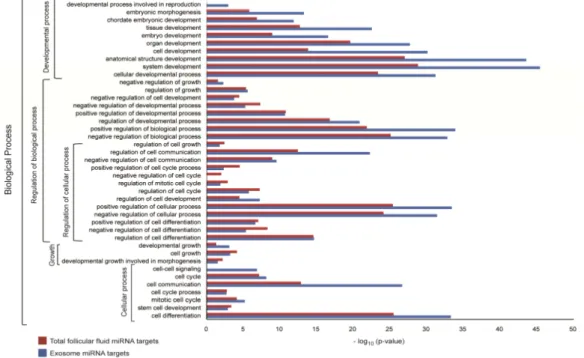
Gene Ontology and Pathway Analysis 80
Validated Targets 81
5 Discussion 84
6 Conclusions and future perspectives 91
1
1. ABSTRACT
Mammalian reproduction hinges upon the timely ovulation of a fully differentiated oocyte. Growth and development of human oocyte and somatic cell compartments of the ovarian follicle require a series of coordinated events that induce morphological, molecular and functional changes within the follicle, leading to cell differentiation and oocyte maturation. In particular, late oogenesis and early embryogenesis occur in the absence of transcription and rely entirely on maternal mRNAs stored in oocytes during its maturation. Moreover, mechanisms controlling both mRNA stability and their translation (e.g. cytoplasmic polyadenylation, and/or microRNAs) fix the ultimate molecular structure of the mature oocyte. Consequently, mRNA regulation at transcriptional and post-transcriptional level is a crucial step in germ cells and early embryo development. Given their nature of post-transcriptional regulators of gene expression, microRNAs (miRNAs) have recently been highlighted extensively for their possible involvement in translational programming of maternal mRNAs and therefore in the development of mammalian oocytes and embryos.
Studies on oocyte transcriptome are important to understand the biological pathways involved in oogenesis, totipotence and early embryonic development and genes regulating physiological pathways in gametes could represent potential candidate for reproductive disorders. Moreover, given that the bidirectional traffic between the oocyte and its surrounding somatic cells is very important for the acquisition of oocyte competence, the study of the transcriptomic profile of both granulosa/cumulus cells, follicular fluid (FF) and exosomes could improve our knowledge on the complex process of mammalian oocyte maturation as well as offer the opportunity to identify potential non-invasive markers of oocyte quality. These could be used in
2 Reproductive Medicine, in order to select the best gamete during medically assisted reproduction cycles. Accordingly, it is also useful to cryopreserve these gametes employing protocols that do not affect the biologic quality of oocytes.
This thesis aimed at characterizing mRNA and miRNA expression profiles in the female gamete and its microenvironment, respectively, under physiological and non-physiological conditions in order i) to assess the effect of vitrification on the biomolecular profile of the oocytes, ii) to understand the molecular basis of reproductive ageing and iii) to identify miRNAs in FF to be used as non-invasive biomarkers of oocyte quality.
We demonstrated that vitrification technique might be very helpful for preserving women’s fertility, since it keeps unaltered oocyte molecular profile and does not cause degradation of mRNAs essential for oocyte development (i.e. BMP15, FIGLA, GDF9, OCT4, TAF4B).
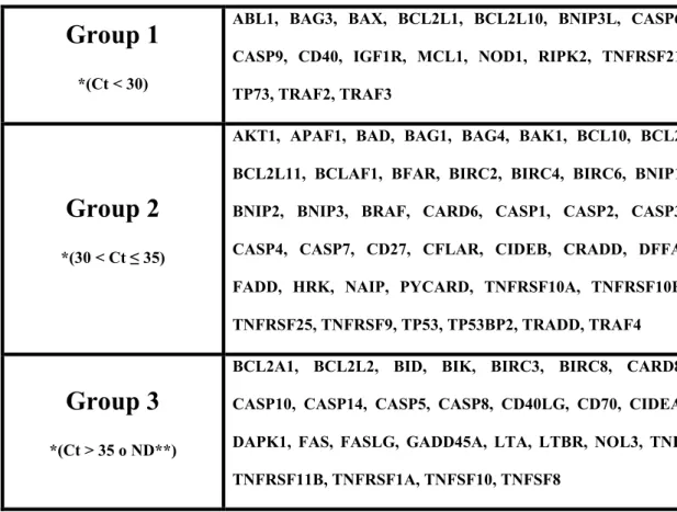
We determined Apoptotic Machinery (AM) transcriptome in mature MII human oocyte pools from women aged more than 38 years (old) and compared it to those of women aged up to 35 years (young). Subsequently, some AM candidate genes with a key role in apoptotic regulation were selected and analysed in single oocytes. These studies led us to identify AM transcripts never reported in human oocytes so far (BAG3, CD40, CFLAR, TNFRSF21, TRAF2, TRAF3) and to find out other differentially expressed genes in oocytes from older women. In fact, we found a significant upregulation of proapoptotic CD40, TNFRSF10A, TNFRSF21 and downregulation of antiapoptotic BCL2 and CFLAR. Our results demonstrate that during maturation old oocytes selectively accumulate mRNAs potentially able to trigger the extrinsic apoptotic pathway and express at low levels some survival factors: this condition could make old oocytes more inclined to apoptosis. Moreover,
3 we found TP73 among differentially expressed genes in human MII oocyte pools during reproductive ageing, a process closely related to the production of oocytes with a reduced developmental competence whose main hallmark is aneuploidy. In order to verify the potential involvement of TP73 isoforms in reproductive ageing, we determined their expression in single mature MII oocytes from women younger than 35 and older than 38 years. We found out that TAp73 isoforms are significantly downregulated in oocytes from women of advanced reproductive age. There is evidence that TAp73 interacts with some kinetochore proteins in order to stop the anaphase if chromosomes are not properly attached to the meiotic spindle. The absence of TAp73 removes this cell cycle brake, so causing genomic instability. Consequently, TAp73 downregulation in old oocytes could lead to aneuploidy in the developing embryos, explaining both the reduction of fertility and the increase of newborns with chromosomal abnormalities.
Finally, we profiled the expression of 384 miRNAs in human follicular fluid and its purified exosomal fraction with respect to plasma from the same women, providing the first molecular evidence of these bioactive vesicles inside ovarian follicle. Among the 37 miRNAs that we found upregulated in follicular microenvironment, the majority of them are carried by exosomes (exosome Follicular Fluid miRNAs – eFF-miRNAs) and are involved in signaling pathways critically important for follicle growth, oocyte maturation, and early embryo development (i.e. WNT, MAPK, ErbB and TGFβ signaling pathways). Moreover, eFF-miRNAs are able to negatively regulate genes encoding inhibitors of follicle maturation and meiosis resumption as PTEN, MTOR, P21 and RB1. These data could reveal new actors in the molecular communication among cells of ovarian follicle and eFF-miRNAs may represent valuable biomarkers of oocyte quality and reproductive disorders.
4 Data shown in this thesis were published in 2010 in Fertility and Sterility [Di Pietro C, Vento M, Guglielmino MR, Borzì P, Santonocito M et al. Molecular profiling of
human oocytes after vitrification strongly suggests that they are biologically comparable with freshly isolated gametes. Fertil Steril. 2010 Dec; 94(7):2804-7], in
2011 in Cell Cycle [Guglielmino MR, Santonocito M et al. TAp73 is downregulated
in oocytes from women of advanced reproductive age. Cell Cycle. 2011 Oct
1;10(19):3253-6], in 2013 in Apoptosis [Santonocito M, Guglielmino MR et al. The
apoptotic transcriptome of the human MII oocyte: characterization and age-related changes. Apoptosis. 2013 Feb;18(2):201-11]. The manuscript Exosomal microRNAs in Human Follicular Fluid: new actors in the communication between oocytes and somatic follicular cells by Santonocito M, Vento M et al. has been submitted for
publication in Molecular Human Reproduction.
For brevity, they will be referred in the text by Roman numerals: Paper I, II, III and IV respectively.
5
2. INTRODUCTION
From oocytes to embryo: a journey full of hurdles
Primordial germ cells (PGCs) are the most primitive undifferentiated sex cells, found initially outside the gonad. During the first month of gestation, these cells migrate to the gonadal ridge from the yolk sac endoderm. In the absence of the testicular determining factor, ovarian differentiation is established at 6–8 weeks of gestation, reflected in a rapid mitosis of the germ cells that peaks around 20 weeks of gestation to a total of about 7 millions of oogonia [Djahanbakhch et al., 2007] (Figure 1).
______________________________________________________________________ Figure 1. In human females, all germ cells are formed during fetal life. The total number of oocytes is at maximum at 5 months of gestation and drops to about 2 million (in both ovaries) by the time of birth (from Djahanbakhch et al., 2007).
After these multiple divisions oogonia stop proliferating, enter meiosis becoming
primary oocytes and their development is soon arrested at the diplotene stage of the
first meiotic prophase (prophase I). They remain at this stage until puberty, when a surge of luteinizing hormone (LH) induces the resumption of meiosis and ovulation of eggs arrested at metaphase II (MII). Almost all oogonia, which have entered the primary
6 oocyte stage, are individually surrounded by pregranulosa cells to form a primordial
follicle or they undergo atresia. Subsequently, the number of follicles decreases
dramatically to 1 000 000/ovary at birth and 300 000/ovary by puberty until complete depletion at menopause [Baker, 1963; Baker and Sum, 1976; Gougeon, 1986; Gougeon, 1996; Faddy, 2000] (Figure 1). The fate of each follicle is controlled by endocrine as well as paracrine factors [Richards et al., 1995]. After puberty, during each ovarian cycle, hundreds of resting primordial follicles enter the growing follicle pool developing through primordial, primary, and secondary stages before acquiring an antral cavity. At the antral stage, most follicles undergo atretic degeneration, whereas a few of them, under the cyclic gonadotropin stimulation that occurs after puberty, reach the preovulatory stage (Figure 2). Concomitantly, increases in local growth factors allow a positive selection of the dominant follicle from this cohort, thus ensuring its final growth and eventual ovulation, whereas the remaining cohort is led to its ultimate demise [Hirshfield, 1991; Gougeon 1996; MCGee and Hsueh, 2000] (Figure 2).
______________________________________________________________________ Figure 2. Follicle development and recruitment in human ovaries. Primordial follicles undergo initial recruitment to enter the growing pool of primary follicles. During cyclic recruitment, increases in circulating FSH allow a cohort of antral follicles to escape apoptotic demise. Among this cohort, a leading follicle emerges as dominant by secreting high levels of estrogens and inhibins to suppress pituitary FSH release. The result is a negative selection of the remaining cohort, leading to its ultimate demise. Concomitantly, increases in local growth factors allow a positive selection of the dominant follicle, thus ensuring its final growth and eventual ovulation (from MCGee and Hsueh, 2000).
7 The decline in oocyte number can be attributed to several mechanisms: germ cells in the cortical area migrating to the surface of the ovary and becoming incorporated within the surface epithelium or being eliminated in the peritoneal cavity, regression during meiosis, and failure to become encapsulated with granulosa cells to become primordial follicles. Once all the primordial follicles have been formed, continuous loss of oocytes occurs through the physiological process of follicular growth and atresia, which continues throughout the woman’s life [Djahanbakhch et al., 2007].
Since the pool of oocytes in the mammalian ovary is already fixed after birth, ovarian senescence is linked to the decreasing supply and eventual exhaustion of the pool of primordial follicles. It has been generally accepted that, in most mammalian species, oocytes cannot renew themselves in postnatal or adult life. However, a few recent studies challenge the idea that mammalian species have lost the capacity for oocyte production after birth, showing that female germline stem cells still exist in mammalian ovaries after birth, but this concept is still controversial [Johnson et al., 2004]. These hypotheses, which need further confirmations, suggest that the pool of primordial follicles in adult mammalian ovary may represent a dynamic rather than a static population, which is characterized by structures continuously subjected to degeneration and differentiation processes.
Oogenesis and Folliculogenesis: coordinated processes under hormonal
regulation
Oocyte development is the final outcome of a sophisticated biological process that is hormonally regulated and tightly connected to the differentiation of other highly specialized cells within the follicle, which provides for and influences the quality of the oocyte (Figure 3).
8 ______________________________________________________________________ Figura 3. Synchronization between oogenesis and folliculogenesis.
The oogenesis begin when PGCs migrate to the gonad and start to proliferate by mitosis giving rise to oogonia. Following this event and before follicle formation, mitotic divisions stop and germ cells initiate meiosis, becoming primary oocytes [Ginsburg et al., 1990] (Figure 3). Meiosis initiate with a prophase stage, a complex phase that is subdivided into five stages: leptotene, zygotene, pachytene, diplotene and diakinesis. Within the first period of the prophase, a series of crucial events occur, involving the pairing of homologous chromosomes, synapsis (close association between these chromosomes), and recombination or “crossing over” (exchange of genetic material). Subsequently, oocytes progress to the diplotene stage where they enter into a prolonged resting phase called “dictyate” [Hunt and Hassold, 2008]. Prophase events are vital for germ cell survival and meiotic progression, and errors occurring along this stage, as well as throughout the consecutive phases of meiosis, may originate and/or contribute to
9 female meiotic aneuploidies. Oocytes remain arrested at the dictyate stage of meiosis I throughout oogenesis until childhood when, each month from puberty to menopause, one primary oocyte resume and complete meiosis I, giving rise to a secondary oocyte arrested at metaphase II stage. These oocytes will complete meiosis II only after ovulation if sperm penetration occurs thus fertilizing the egg (Figure 3).
However, the oocyte cannot reach this point of development without the support of its follicle. Folliculogenesis is an intricate process by which one follicle per cycle is selected to develop fully and thereby create a mature oocyte. Soon after oocytes enter the prolonged diplotene stage of meiosis, the precursors to the follicular somatic cells encompass the oocyte in a single squamous layer to form primordial follicles. The large population of non-growing primordial follicles serves as the source of developing follicles and oocytes until the end of a female’s reproductive life. Until that time, there is a continuous recruitment of follicles from this pool, beginning with the formation of primary follicles. The oocyte in a primary follicle begins its extensive growth phase, and the surrounding follicular cells, now called granulosa cells (GC), become cuboidal and proliferative. When the growing oocytes are surrounded by more than one layer of granulosa cells, the follicle is called a secondary follicle. Early preantral follicles are independent of gonadotrophins for their initial growth, as evidenced by the fact that development to the primary and secondary stage can take place in the absence of hormones [Fortune and Eppig, 1979], although follicle stimulating hormone receptors (FSHR) are present on the GCs of these early follicles [O'Shaughnessy et al., 1996; Oktay et al., 1997]. Primary to secondary follicle transition is rather driven by local intraovarian paracrine factors produced by oocytes and their companion granulosa cells [Kol and Adashi, 1995]. The latter phase of folliculogenesis is termed antral
10 folliculogenesis, and its progress is entirely dependent on gonadotropic hormones, follicle stimulating hormone (FSH) and LH (Figure 4).
______________________________________________________________________ Figura 4. Classification of the major stages of mammalian folliculogenesis. In humans, preantral follicular development does not require stimulation by the pituitary gonadotropins whereas antral follicle growth is entirely dependent on them (Edson et al., 2009).
FSH induces luteinizing hormone receptor (LHR) expression in GCs, which will be required for follicles to respond to LH, the latter being crucial for triggering the ovulatory process [Sánchez and Smitz, 2012]. The antral stage is characterized by the appearance of a fluid-filled cavity, the antrum, which begins to form when follicles reach a critical size and a critical number of granulosa cells [Boland et al., 1994]. The follicular fluid (FF) consists of a complex mixture of proteins, metabolites, and ionic compounds, which are secreted from the granulosa and thecal cells and combined with plasma components that cross the blood–follicular barrier via the thecal capillaries [Angelucci et al., 2006; Hanrieder et al., 2008; Hanrieder et al., 2009; Rodgers and Irving-Rodgers, 2010]. This fluid provides a very important microenvironment and contains regulatory molecules that are important for the maturation and quality of the oocytes [Revelli et al., 2009]. Certain lipids, proteins, vitamins, and metabolites in the FF have been found to be associated with reproductive diseases [Kim YS et al., 2006], oocyte quality [Berker et al., 2009], embryo quality, and the outcome of in vitro fertilization (IVF) attempts [Wu et al., 2007; Wallace et al., 2012]. Additionally, the
11 appearance of the antral cavity establishes the morphological and functional separation of granulosa cells into mural granulosa cells (MGC), which line the follicle wall, and the cumulus cells (CC), which surround the oocyte. Each follicle is enveloped by a basal lamina, a specialized sheet of extracellular matrix that separates the internal follicle from the third somatic follicular cell type, the theca cells. Theca cells are a vascularized cell layer that defines the outer boundary of the antral follicle (Figure 5).
___________________________________________________________________ Figure 5. Schematic representation of an antral follicle (from Hennet and Combelles, 2012).
As mentioned earlier, the antral phase of follicular development is characterized by dependency on gonadotrophins, FSH and LH, which are cyclically secreted by the pituitary gland. Circulating levels of FSH support the growth of a group of antral follicles until the largest follicle begins producing estradiol (E2), an FSH suppressor, and switches to dependence on LH. This follicle is termed the dominant follicle, and this event is called follicle selection. As FSH levels decline, the rest of the antral follicles will regress in a process called atresia and the dominant follicle will be the only follicle capable of reaching ovulation. Atresia is the process by which all follicles regress if they do not reach ovulation, and as it progresses into its latest stages, the granulosa cell and cumulus cell populations die, followed by resorption of FF and
12 oocyte death. The underlying mechanism of this condition is apoptosis, or programmed cell death. Finally, preovulatory follicles containing fully-grown oocytes are ready to undergo ovulation, which is triggered by the preovulatory surge of LH (Figure 6). Ovulation is characterized by the rupture of the follicle wall and the release of the cumulus–oocyte complex; at this time the oocyte has resumed meiosis and has progressed to the metaphase II stage of meiosis (Figure 7).
___________________________________________________________________ Figure 6. The ovarian and uterine cycles regulated by gonadotropin hormones.
13 ___________________________________________________________________ Figure 7. Mature MII oocyte surrounded by cumulus cells ovulated from a mature follicle.
After ovulation, granulosa and theca cells become luteal cells and are responsible for the production of E2 and progesterone (P), the latter is a female hormone predominantly expressed in the corpus luteum (Figure 8), which plays a central role in the reproductive events associated with pregnancy establishment and maintenance [Eppig, 2001; Kwintkiewicz and Giudice, 2009]. In particular, high levels of progesterone causes a subsequent decrease in FSH levels due to its negative feedback effects on the hypothalamic-pituitary axis, thus inhibiting the development of other follicles. If egg fertilization does not occur, the corpus luteum breaks down causing a decrease in estrogen production with a subsequent increment of FSH secretion due to a positive feedback of these hormones on hypothalamus and pituitary gland, so the reproductive cycle begins again.
14 ___________________________________________________________________ Figure 8. Folliculogenesis and ovulation.
Therefore, the mechanisms underlying oogenesis, folliculogenesis and pregnancy maintenance take place on different scales and rely on a subtle interplay between regulatory signals derived from multiple organs, including the hypothalamus, the pituitary gland and the ovaries that form a functional endocrine axis, known as the hypothalamic-pituitary-ovary (HPO) axis, with hormonal regulations and feedback loops [Bacchus, 1975] (Figure 9). Neurons in the hypothalamus secrete Gonadotropin-releasing Hormone (GnRH) to regulate anterior pituitary gland gonadotrope cells. Gonadotrope cells produce FSH and LH, which control ovarian follicle development and ovulation by stimulating the granulosa cells to synthesize and secrete steroid hormones as well as peptidic hormones, such as estradiol and inhibin (INH) respectively, whose cumulated ovarian release impacts FSH secretion (Figure 9). During their development, antral follicles become more and more sensitive to FSH, then to LH, and they secrete increasing amounts of E2 and INH. The large estradiol amounts
15 secreted by the preovulatory follicle affect the secretion of GnRH from the hypothalamus and end up by triggering the GnRH ovulatory surge. As a result, the pituitary LH surge occurs and brings about the ovulation event [Clément and Monniaux, 2012], the end-point of follicular phase of the menstrual cycle during which the oocyte is released (Figure 8, 9).
______________________________________________________________________ Figure 9. Endocrine feedback loops between the ovary and the hypothalamic-pituitary axis. Left panel: The terminal development of follicles from the small antral growing stage (stage 1) is under the control of FSH secreted by the pituitary gland. In turn, the granulosa cells secrete steroid hormones, such as E2, as well as peptidic hormones, such as INH, whose cumulated ovarian release impacts FSH secretion. During their development, antral follicles become more and more sensitive to FSH, then to LH, and they secrete increasing amounts of E2 and INH (stages 2 and 3). Right panel: The large estradiol amounts secreted by the preovulatory follicle(s) (stage 3) impact the secretion of GnRH from the hypothalamus and end up by triggering the GnRH ovulatory surge. As a result, the pituitary LH surge occurs and results in the ovulation event during which the oocyte is released (from Clément and Monniaux, 2012).
Following ovulation, somatic cells of the ruptured follicle form the corpus luteum giving rise to the luteal phase of the menstrual cycle (Figure 8). During this phase, it secretes high levels of progesterone and moderate levels of estradiol to inhibit further release of GnRH and thus secretion of LH and FSH through negative feedback mechanisms, and transforms proliferating endometrium into secretory endometrium. If
16 pregnancy does not occur, corpus luteum degenerates in corpus albican and stops producing progesterone and estrogen, while the secretory endometrium breaks down and sheds during the ensuing menstrual period, thus the cycle starts again.
The role of intracellular communication in ovarian follicle
development
Growth and development of the somatic and germ cell compartments of the ovarian follicle occur in a highly coordinated and mutually dependent manner, which means that all these processes rely on continuing cross talk between the oocyte and its somatic cells. Historically, the oocyte was relegated to a passive role in the regulation of folliculogenesis; the key drivers being endocrine and ovarian somatic cell derived hormones and growth factors. However, over the past 20 years, it has become increasingly clear that the oocyte is a pivotal regulator of folliculogenesis, and there exists an important bi-directional communication axis between the oocyte and somatic cells [Albertini and Barret, 2003]. This kind of relationship is mainly based on two means of intercellular communication mediated by gap junctions, also known as connexins, and paracrine factors (Figure 10).
______________________________________________________________________ Figure 10. Oocyte-granulosa cell communication is essential for normal growth and development of both the oocyte and the follicle. Communication occurs via paracrine signalling (curved arrows) and
gap-17
junctional exchange of small regulatory molecules (straight arrow). This is a bi-directional communication axis (from Gilchrist et al., 2004).
The highly specialized cumulus cells have trans-zonal cytoplasmic processes, which penetrate through the zona pellucida (a glycoprotein membrane surrounding the plasma membrane of the oocytethat issecreted by both the oocyte and the follicular cells) and abut the oocyte membrane, forming the cumulus-oocyte complex (COC) [Albertini et al., 2001]. Gap junctions at the ends of these processes allow the transfer of small molecular weight molecules between oocyte and cumulus cell, and also between cumulus cells, whereas larger molecules are transported by receptor-mediated endocytosis [Gilchrist et al., 2004]. Molecules that pass via gap junctions include ions, metabolites, and amino acids that are necessary for oocyte growth, as well as small regulatory molecules that control oocyte development. In particular, connexins 43 (Cx43) and 37 (Cx37), play an important role in the maintenance of this communication: Cx43 is expressed by granulosa cells and required to form gap junctions between granulosa cells whereas Cx37, expressed in oocytes at all stages of follicle development, is crucial for an oocyte–granulosa cell gap junctional communication. In the absence of Cx43, gap junctions between somatic cells do not form and folliculogenesis arrests at the unilaminar stage [Juneja et al., 1999; Gittens et al., 2003; Gittens and Kidder, 2005], whereas mutation of Cx37 abolishes the production of mature Graafian follicles and fully grown oocytes and its meiotic competence is compromised [Simon et al., 1997; Carabatsos et al., 2000]. These experimental evidences demonstrate that this mode of communication in the ovary is essential for development and fertility, and is thought to play a key role in disseminating local and endocrine signals to the oocyte via the cumulus cells [Simon et al., 1997; Buccione et al., 1990].
18 A second means of communication between oocytes and granulosa cells is via paracrine signalling (Figure 10). Now there is widespread interest in paracrine factors secreted by oocytes and their role in the regulation of key granulosa cell processes and vice versa. For example, before the LH surge, oocytes do not only influence granulosa cell proliferation [Vanderhyden et al., 1992; Gilchrist, 2006] and differentiation [Diaz et al., 2007; Diaz et al., 2007], but very importantly, oocytes regulate metabolic activity of cumulus cells within the COC (aminoacid uptake, glycolysis and cholesterol biosynthesis) [Eppig, 2005; Sugiura et al., 2005; Su et al., 2008]. After the LH surge, oocytes regulate the expression of cumulus genes responsible for the mucification/expansion process [Sánchez and Smitz, 2012] (Figure 11).
______________________________________________________________________ Figure 11. Oocytes secrete soluble growth factors that regulate fundamental aspects of granulosa cell functions, including growth, differentiation, cumulus cell expansion and ovulation. The direct actions and secondary consequences of oocyte-secreted factors impact on fertility (from Hawkins and Matzuk, 2010).
Much of scientific interest has focused on oocyte-secreted TGF-β superfamily members, in particular growth differentiation factor-9 (GDF-9), GDF-9B (also called bone morphogenetic protein-15; BMP-15) (Figure 10, 11). Interest in these oocyte-secreted
19 factors has been fostered not only by the need to improve our understanding of fundamental mechanisms regulating folliculogenesis, but also because altered expression of some of these oocyte paracrine factors has profound effects on ovarian function and fertility, and perhaps ovarian disease [Buccione et al., 1990; Simon et al., 1997; Gilchrist et al., 2004; Yeo et al., 2009]. Targeted deletion to produce gdƒ9−/− mice causes an infertile phenotype in which follicles halt at the one-layer stage with enlarged oocytes thus demonstrating that GDF-9 is needed for granulosa cells to proliferate [Dong et al., 1996; Gosden, 2002]. This feedback communication between oocytes and somatic cells is also necessary to coordinate meiotic resumption and ovulation. Moreover, it has been demonstrated that immature or incompetent oocytes which fail to interact appropriately in this communication loop are not released [Albertini, 1992; Albertini et al., 2003; Plancha et al., 2005]. The ovulation process may therefore be considered a checkpoint event whereby only healthy and meiotically competent oocytes engineer their own release, thus ensuring a sort of natural selection mechanism of the best gamete among all those belonging to the reproductive pool [Russell and Robker, 2007].
Although many studies have contributed in pointing out to the significance of the interaction between oocyte and its surrounding somatic cells, there is still much to be learned about molecular pathways controlling early stages of oogenesis, folliculogenesis and oocyte maturation, which are determined by complex activation and interplay of many factors acting in a stage-specific manner. Since the follicle-oocyte dialogue seems to be a prerequisite to ensure a proper development and to preserve fertility, it is extremely important to elucidate the molecular basis of these events.
20 During oogenesis, oocytes increase in size (approximately 35-120µm in human) and in volume (~100-fold) [Eppig and O'Brien, 1996; Picton et al., 1998]. Fully-grown and meiotically competent murine oocytes have been estimated to contain ~6 ng of total RNA which is almost ~200 times the amount of RNA found in a typical somatic cell [Sánchez and Smitz, 2012]. Throughout this period, oocytes synthesize and accumulate RNAs (mRNAs, rRNAs, tRNAs, small RNAs) and proteins that are vital for their appropriate growth and maturation, and indispensable for the development into a viable embryo [Moore and Lintern-Moore, 1978; Bachvarova, 1985; Eichenlaub-Ritter and Peschke, 2002]. Synthesis of transcripts is highest in the earliest phases of development, which coincides with active proliferation of follicular cells; however, by the end of growth (antral stages) and at the time of oocyte maturation, silencing of transcriptional activity and a selective degradation of some mRNAs will be the predominant processes [Bachvarova et al., 1985; De La Fuente et al., 2004; Su et al., 2007]. In mouse, a decrease of ~30% in total RNA has been described to occur during maturation [Paynton et al., 1988]. Additionally, during the maternal-zygotic transition, embryonic transcription is initiated and other maternal mRNAs are inactivated or degraded through co-ordinated post-transcriptional regulation. Accumulating evidence indicates that a class of small silencing RNAs, particularly microRNAs (miRNAs), is implicated in the elimination of maternal transcript by their binding to the 3' untranslated region (UTR) of target RNAs. So far, very few studies have analysed the expression of and the role played by miRNAs during oocyte growth and preimplantation development [Tang et al., 2007]. Thus, it is imperative to examine transcription not only in the oocyte but also in somatic cells as well as FF when investigating what is needed to make a developmentally competent egg. The identification of oocyte-quality molecular markers that could be used to predict the developmental competence of oocytes more precisely
21 could be of help in establishing more objective criteria for the selection of the best gamete during medically assisted reproduction cycles.
MicroRNAs
miRNAs are short 20–22 nucleotides (nts) RNA molecules that function as negative regulators of gene expression in eukaryotic organisms. These highly conserved RNAs regulating gene expression constitute about 1-5% of predicted genes in animals genomes, and 10-30% of protein-coding genes are probably regulated by miRNAs. Recently, many small non-coding RNAs have been also identified in prokaryotic organisms and viruses. Since they were recognized in 2001, the biological significance of these newly identified small RNAs was elucidated, because more and more evidences suggest that miRNAs play an essential role in multiple biological processes through negative regulation of gene expression at a post-transcriptional level. They perform their functions by partial pairing with one or more 3’UTR of mRNA targets to promote their degradation or transcriptional repression [Bartel, 2009]. The majority of known miRNAs are evolutionarily conserved among species, demonstrating that miRNA mediated gene silencing pathways have essential roles in development, cell differentiation, cell proliferation, cell death, chromosome structure and virus resistance, signalling transduction, disease and cancer [Lee and Ambros, 2001; Zhang et al., 2007]. From a functional point of view, it has been demonstrated that most of miRNAs are able to recognize several mRNA targets; on the other hand, one specific mRNA can be regulated by more than one miRNA [Kim and Nam, 2006]. Moreover, studies from the last years have demonstrated that there is altered expression of miRNA genes in many human malignancies.
22 It was estimated that the human genome contains 1600 miRNA loci, encoding for 2042 mature miRNAs, distributed in all chromosomes except for Y chromosome, according to the latest release of miRBase database, the primary online repository for all miRNA sequences and annotation, where all known and newly identified miRNAs are deposited (www.mirbase.org). The identification of miRNA genes is the result of a combination of directional cloning together with computational approaches, based on the examination of genomic sequences to identify phylogenetic conservation of known miRNA genes, taking in account the high level of 5’ region conservation in all miRNA sequences. An integrative approach leading to the identification of a significantly higher number of miRNAs than previously expected in humans combined bioinformatic predictions with microarray analysis and sequence-directed cloning, allowing the identification of a huge amount of non-conserved miRNAs [Bentwich et al., 2005]. Around 50% of the known miRNAs are organized in genomic clusters and are transcribed as polycistronic primary transcripts. Generally, miRNAs belonging to the same cluster are structurally and functionally related to each other, demonstrating their origin as a result of duplication events during evolution; on the other hand, some miRNAs lying in close genomic regions can be unrelated to each other. One possible explanation for the existence of clustered miRNAs with related functions or structure is the possibility that they may play a synergic role in regulating the same genes, or different genes involved in the same pathway [Kim and Nam, 2006]. Despite it was originally inferred that most of miRNAs are located in intergenic regions, more recent analysis have shown that a vast majority of mammalian miRNA genes is located in well-defined transcription units, many of them within introns of coding genes in the sense orientation. Based on these findings, miRNAs can be categorized as follows: a) intronic miRNAs in coding regions;
23 b) intronic miRNAs in non-coding regions;
c) exonic miRNAs in coding regions;
d) exonic miRNAs in non-coding regions (Figure 12).
Based on the splicing pattern, some miRNAs have mixed features; moreover, for miRNAs comprised within coding regions, as expected for genes sharing the same promoters, miRNAs usually have similar expression profiles to their host transcript [Kim and Nam, 2006].
___________________________________________________________________ Figure 12. Genomic organization of miRNA genes (from Wahid et al., 2010).
miRNA biogenesis
miRNA biogenesis is a multistep process where a mature miRNA is generated from a miRNA gene, with several enzymes playing critical roles in the process (Figure 13). As it was already discussed, intronic miRNAs share their regulatory elements and primary transcript with their pre-mRNA host genes, and are undoubtedly transcribed by RNA
24 polymerase II (pol II). This gives a possible mechanism for coordinated miRNA and protein coding gene expression [Rodriguez et al., 2004].
______________________________________________________________________ Figure 13. miRNA biogenesis pathways and their regulation (from Winter et al., 2009).
All the characterized promoters for the other miRNA genes contain general RNA polymerase II transcriptional regulatory elements previously found in protein-coding genes: this, together with other experimental evidences, strongly suggests that all miRNAs are pol II products [Lee et al., 2004]. miRNA gene transcription by pol II produces a pri-miRNA of several hundreds of nts in length, with a 5’ cap and a polyadenylated 3’, which forms a hairpin stem-loop secondary structure within the
25 nucleus and enters a large complex called microprocessor complex (500–650 kDa). The main components of this complex are Drosha (an RNase III endonuclease) and the essential cofactor DGCR8/Pasha (containing two doublestranded RNA binding domains). Drosha asymmetrically cleaves both strands of the hairpin stem at sites near the base of the primary stem loop thus releasing a 60- to 70-nucleotides pre-miRNA with a 5′ phosphate and a 2-nucleotide 3′ overhang (Figure 13). This process is highly specific and predetermines the sequence of mature miRNA [Lee et al., 2004]. The pre-miRNAs are then transported to the cytoplasm by Exportin-5 (Exp5) (a member of the Ran transport receptor family): this transport process requires energy, a specific hairpin secondary structure, and is Ran-GTP dependent. When the pre-miRNA reaches the cytoplasm, one of its ends has already been predetermined by Drosha cleavage site selection. Once in the cytoplasm, the pre-miRNAs enter the RISC loading complex (RLC), made by Dicer (a second RNase endonuclease), TRBP (able to bind double stranded RNAs), PACT (protein activator of PKR) and Ago-2 [Gregory et al., 2005; Haase et al., 2005; Lee et al., 2006]. Dicer recognizes the 3’ overhangs of the pre miRNA through the PAZ domain, and its cleavage 22-nt from the end of the substrate releases a mature double-stranded miRNA (miRNA:miRNA* duplex) with 5’ phosphates and a 2-nt 3’ overhang [Gregory and Shiekhattar, 2005]. Finally, miRNA:miRNA* duplex is unwound by helicase into two single strands: in the majority of cases miRNA*, which is the less thermodynamically stable strand at 5’ end, is degraded, while the leading strand is incorporated into a ribonucleoprotein effector complex known as RNA-induced silencing complex (RISC) which induces gene silence at a post-transcriptional level [Zhang et al., 2007].
miRNA mediated post-transcriptional repression
26 RISC (RNA-induced silencing complexes), which identify miRNA targets by the perfect or nearly perfect complementarity between the miRNA and the mRNA. The better characterized components of RISC are Argonaute (AGO) proteins, with PAZ and PIWI domains: in mammals, four AGO proteins (AGO1 to AGO4) function in the miRNA repression but only AGO2 functions in RNA interference (RNAi), which is the process guiding the endonucleolytic cleavage of the target mRNA. Beyond Ago proteins, the complex contains other proteins playing critical roles as regulators of the main effectors of RISC inhibitory functions [Tolia and Joshua-Tor, 2007]. The complementary sites for miRNAs associated with RISC reside in the 3‘UTRs of target mRNAs and are generally present in multiple copies as a necessary condition to reach an efficient translational inhibition [Easow et al., 2007]. The specific site of mRNA recognition in miRNA molecules is called seed region, and is comprised between nucleotides 2-8; generally, in metazoan miRNAs bind the target mRNA with mismatches and bulges, in contrast, most plant miRNAs bind with near-perfect complementarity to sites within the coding sequence of their targets [Carthew and Sontheimer, 2009] (Figure 14).
______________________________________________________________________ Figure 14. Principles of microRNA–mRNA interactions in metazoan. The base pairing is perfect at the seed region and at miRNA 3’ half, while bulges and mismatches can be present in the central region (from Filipowicz et al., 2008).
Once associated to the target mRNA, RISC inhibitory functions can take place through several possible mechanisms:
27 - translational repression, in presence of mismatches in the core part of the seed region; - mRNA deadenylation and decay in case of perfect complementarity with the target mRNA (Figure 15).
______________________________________________________________________ Figure 15. Mechanisms of miRNA mediated post-transcriptional repression: a) repression of transcription initiation; b) inhibition of protein chain elongation; c) lysis of nascent peptide; d) mRNA deadenilation (from Pillai et al., 2007).
Translational repression can occur at the initiation or at post-initiation steps. Initiation of translation of most cellular mRNAs starts with the recognition of the mRNA 5′-terminal-7-methylguanosine (m7G) cap by the eukaryotic translation initiation factors (eIF) 4E, 4F and 4G, which together with PAPB1 are responsible for the association between the mRNA ends, triggering the translation process [Filipowicz et al., 2008]. Ago2 and related proteins can compete with eIF4E for m7G binding, preventing translation. An alternative mechanism of miRNA action was recently proposed at a post-initiation step, since studies with reporter mRNAs targeted by either synthetic or endogenous miRNAs have shown that repressed mRNAs were associated with active polysomes; nevertheless, how miRNAs could modulate the elongation or termination
28 process remains unclear [Petersen et al., 2006]. In eukaryotes, mRNA degradation can follow two pathways, both of them initiated by a gradual shortening of the mRNA poly(A) tail. The mRNA body can then be degraded by progressive 3′→5′ decay, which is catalysed by the exosome, or by the removal of the cap followed by 5′→3′ degradation, catalysed by the exonuclease XRN1. The final steps of mRNA degradation occur in P-bodies, cytoplasmic structures enriched in proteins participating in miRNA repression functions such as AGO proteins and GW182, and miRNAs themselves [Filipowicz et al., 2008]. Recent findings demonstrated that lowered mRNA levels account for most (≥84%) of the decreased target’s protein production: by using ribosome profiling to measure the overall effects on protein production and compare these to simultaneously measured effects on mRNA levels, Guo et al. showed that changes in mRNA levels closely reflect the impact of miRNAs on gene expression and indicate that destabilization of target mRNAs is the predominant reason for reduced protein output [Guo et al., 2010].
miRNAs: new candidates for the regulation of the human COC
As previously described, in the ovarian follicle, the maturing oocyte is nurtured and supported by CCs, the surrounding somatic cells. Disruption or deregulation of the CC interactions with the oocyte can affect oocyte quality and consequently embryo development and pregnancy outcome. Much knowledge on human oocytes and CCs has been generated over recent years mainly owing to technological advances in gene expression analysis [Assou et al., 2011]. Although such techniques have allowed entire profiling of the transcriptional activity in single human MII oocytes and the surrounding CCs [Assou et al., 2006; Kocabas et al., 2006; Grondahl et al., 2010], leading to the identification of several transcripts that are crucial for oogenesis and folliculogenesis,
29 however, the post-transcriptional regulation of these transcripts needs to be elucidated. This is particularly important also because the stability and translation of the maternal mRNAs, that are accumulated during oocyte maturation [Niakan et al., 2012] and that drive human preimplantation development, are controlled by post-transcriptional regulatory mechanisms [Bettegowda and Smith, 2007]. The miRNA repertoires are cell type specific and change markedly during development [Carthew and Sontheimer, 2009]. Changes in miRNA expression profiles have been linked to pathologies, such as cancer [Ventura and Jacks, 2009]. Moreover, miRNAs have been associated with infertility as shown in female mice in which Dicer, an essential factor in miRNA biogenesis, was genetically ablated [Murchison et al., 2007; Nagaraja et al., 2008]. Futhermore, the expression of several miRNAs, such as miR132 and miR212, has been found to be regulated by gonadotropins (FSH and LH) [Fiedler et al., 2008; Yao et al., 2009], even if this mechanism has not been fully investigated so far. Finally, analysis of mRNA expression during mouse and bovine oogenesis shows that a large proportion of maternal genes are regulated by miRNAs [Tang et al., 2007]. Therefore, regulation of gene expression in the oocyte throughout oogenesis at the transcriptional and post-transcriptional level is a crucial process that is tightly controlled in a stage-dependent manner, and this process ultimately ensures that the oocyte will mature and acquire full developmental competence. Thus, miRNA profiling might help us to better understand the regulation of transcripts involved in human reproduction.
miRNAs in body fluids: new powerful biomarkers
MiRNAs are involved in virtually all biologic processes and, because a single miRNA can target hundreds of mRNAs, aberrant miRNA expression is involved in the initiation of many diseases, including cancer. Distinguishable abnormalities in miRNA genes and
30 expression patterns were identified in almost all types of cancer, thus providing a strong rationale for the application of miRNAs as diagnostic/prognostic biomarkers. Expression profiles of miRNAs have been found significantly altered in numerous types of human cancers when compared with their corresponding normal tissues, among different subtypes within the same type of cancers, or among individual patients suffering from a same type of cancer but having different prognoses [Yan et al., 2008; Lebanony et al., 2009]. The analyses of miRNA signatures are in general limited to tissue biopsies; however, in the last few years several studies have shown the diagnostic and prognostic usefulness of circulating miRNAs, released by some cell types both under normal and pathological conditions [Chen et al., 2008] (Table 1). One of the first studies measuring miRNA levels in serum demonstrated that levels of miR-21 were associated with relapse-free survival in patients with diffuse large B-cell lymphoma [Lawrie et al., 2008]. Currently, one of the most complete studies highlighting the role of circulating miRNAs was carried out on a cohort of 303 non-small-cell lung cancer (NSCLC) patients through Solexa sequencing, and led to the identification of eleven serum miRNAs significantly altered between longer-survival and shorter-survival groups, while some of them were associated to overall survive [Hu et al., 2010].
31 Table 1. Serum miRNAs as biomarkers for cancer (from Kosaka et al., 2010).
So far, miRNAs have been found in human serum, plasma, and other body fluids [Takamizawa et al., 2004; Peter, 2009; Wurz et al., 2010] and specific compositions and concentrations were found for each body fluid analysed. The miRNA profiles of these fluids have been found to be associated not only with cancer and other diseases [Calin and Croce, 2006; Chun-Zhi et al., 2010; Pineau et al., 2010; Zhang et al., 2010] but also with healthy conditions. In fact, in healthy individuals the levels of cell-free miRNAs present in sera are stable [Chen et al., 2008; Gilad et al., 2008]. Mitchell and colleagues showed that under healthy conditions, the serum miRNA profile is similar to that of circulating blood cells [Mitchell et al., 2008]. Thus, alterations of serum miRNA levels may be indicative of physiological or pathological changes and may possibly be
32 used as surrogate biomarkers [Cortez and Calin, 2009]. For example, circulating miRNAs were found in the sera of pregnant women [Gilad et al., 2008]: miR-526a, miR-527 and miR-520d-5p showed a considerably high fold-change and could be used to distinguish pregnant from non-pregnant women with high accuracy. Moreover, placenta derived miRNAs (e.g. miR-141, miR-149, miR-299-5p and miR-517a) are detectable in the maternal plasma, and their concentrations decrease directly after childbirth [Chim et al., 2008; Luo et al., 2009]. Therefore, miRNAs have been discussed as novel non-invasive markers for prenatal diagnosis [Chim et al., 2008]. miRNAs have also been identified in the ovarian tissues of other species, such as mouse [Ahn et al., 2010], goat [Zhang et al., 2013], ruminant [McBride et al., 2012], etc. Moreover, miRNAs has been found in equine and human FF [Da et al., 2012; Sang et al., 2013] whose composition is tightly correlated with the developmental competence of the human oocyte [Wallace et al., 2012]. It contains essential substances involved in follicle growth, oocyte fertilization, glucose and lipoprotein metabolism which have been found to be associated with reproductive disorders such as premature ovarian failure (POF) and polycystic ovary syndrome (PCOS) [Dai and Lu, 2012]. Accordingly, the discovery of miRNAs in body fluids, particularly in FF, opens up the possibility of using them as non-invasive biomarkers in human reproductive diseases; moreover, given that FF is easily available during oocyte pick up, these miRNAs can be used as non-invasive predictors of oocyte quality in order to select the best gamete during medical assisted reproductive cyles.
Circulating miRNA stability and possible release mechanisms
To be able to use circulating miRNAs as a diagnostic marker, we need to gain a better understanding of the mechanisms by which miRNAs are released in the bloodstream.
33 Cell-free miRNAs in body fluids are stable under harsh conditions that would normally degrade most RNAs, including boiling, low/high pH, extended storage and multiple freeze-thaw cycles [Chen et al., 2008]. Moreover, there is evidence that serum miRNAs are particularly resistant to RNases digestion, since they can be detected in this body fluid containing high levels of these enzymes; this implies that they should be released in the bloodstream in a form that protects them from degradation. Some hypotheses have proposed that they are secreted in protected protein–miRNA complexes (AGO2, nucleophosmin 1-NPM1, and other RNA-binding proteins); another hypothesis is that they are produced as by-products of dead cells [Vickers et al., 2011; Xu et al., 2012]. Recent studies have demonstrated a novel mechanism of cell communication using miRNAs released in microvesicles (up to 1 µm), or in small membrane vesicles called exosomes (10-100 nm) [Kosaka et al., 2010] (Figure 16).
______________________________________________________________________ Figure 16. Origin of circulating miRNAs (from Xu et al., 2012).
These small vesicles of endocytic origin, which can also contain mRNAs and proteins in addition to miRNAs, can be formed through inward budding of endosomal
34 membranes, giving rise to intracellular multivesicular bodies (MVB) that later fuse with the plasma membrane, releasing the exosomes into the extracellular environment. In any case, circulating miRNAs enter the bloodstream and are taken up by the recipient cells by endocytosis or, hypothetically, by binding to receptors present at the recipient cellular membrane capable of recognizing RNA-binding proteins (Figure 17). However, more studies are necessary to elucidate how miRNAs are loaded into exosomes and how they can be internalized by recipient cells.
______________________________________________________________________ Figure 17. Biogenesis and mechanism of action of circulating miRNAs (from Cortez et al., 2011).
Defining exosomes
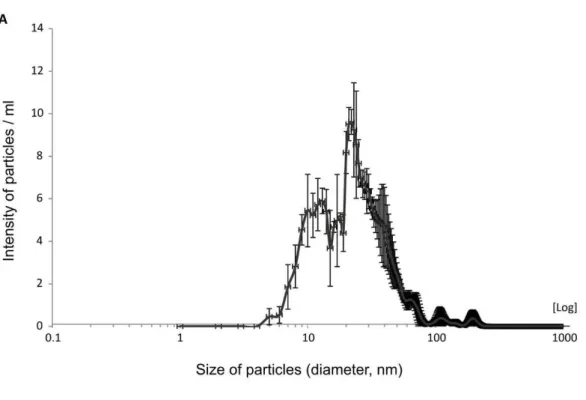
Although potentially any vesicle released by a cell carries cell type specific membrane and cytosolic components, there are a number of features that should be taken into consideration for the characterization of exosomes. Exosomes are between 40 and 100
35 nm in diameter, appear with a characteristic cup-shaped morphology (after negative staining) or as round well delimited vesicles as observed by transmission and cryo-electron microscopy, respectively [Conde-Vancells et al., 2008]. Exosomes float on sucrose gradient to a density that ranges from 1.13 to 1.19 g/ml [Thery et al., 2006]. Apart from their morphology, their unique protein and lipid composition enable their identification. Notably, these include cytoplasmic proteins such as tubulin, actin, actin-binding proteins, integrins, annexins and Rab proteins as well as molecules responsible for signal transduction (protein kinases, heterotrimeric G-proteins) [Thery et al., 2001; Skokos et al., 2001; Skokos et al., 2001]. Most exosomes also contain MHC class I molecules [Wolfers et al., 2001; Blanchard et al., 2002] and heat-shock proteins such as Hsp70 and Hsp90 [Thery et al., 2001; Thery et al., 2002]. The protein family most commonly associated with exosomes is the tetraspanins including CD9, CD63, CD81 and CD82 [Escola et al., 1998; Bard et al., 2004; Chaput et al., 2005] (Figure 18). While some of the proteins that are found in the proteome of many exosomal membrane preparations may merely reflect the cellular abundance of the protein, others are specifically enriched in exosomes and can therefore be defined as exosomal marker proteins (e.g. Alix, flotillin, TSG101, CD63). Another feature of exosomes is their enrichment in raft-lipids such as cholesterol, sphingolipids, ceramide and glycerophospolipids with long and saturated fatty-acyl chains [Wubbolts et al., 2003; Subra et al., 2007; Trajkovic et al., 2008]. Conversely, other exosomal proteins directly represent the proteome of the source cells. For example, analysis of urinary vesicles showed a link between exosomes containing aquaporin-2 and their origin from the urogenital tract [Pisitkun et al., 2004].
36 ______________________________________________________________________ Figure 18. Biogenesis, characteristics and functions of secreted extracellular vesicles.
Exosomes in biological fluids
Exosomes have been detected in many biological fluids, where they are secreted both under physiological and pathological conditions (e.g. blood, plasma, urine and cancerous pleural effusions); besides they are actively produced by many cultured cell types [Mathivanan et al., 2010]. Among the possible biological functions exerted by exosomes, they have been demonstrated to play a significant role in signalling, immune response, and tumor development [Denzer et al., 2000; Anand, 2010; Zhang and Grizzle, 2011]. The increased levels of tumor-derived exosomes in plasma and malignant effusions of patients with cancer suggest that exosomes can be a rich source for the discovery of blood-based diagnostic biomarkers of disease. One of the first reports showing the existence of miRNAs in exosomes was published by Valadi et al., who reported that exosomes released from human and murine mast cell lines contain mRNAs and miRNAs [Valadi et al., 2007]. Since then, many studies started to focus on the expression profiling of miRNAs from exosomes isolated from plasma of diseased patients, in order to identify new miRNA markers that are easily measurable in blood
37 samples. Two pivotal studies led to the identification of eight putative miRNA markers for the diagnosis of ovarian cancer (miR-21, miR-141, miR-200a, miR-200c, miR-200b, miR-203, miR-205, and miR-214) and twelve specific miRNAs (miR-17-3p, miR-21, 106a, 146, 155, 191, 192, 203, 205, 210, miR-212, and miR-214) for lung cancer, respectively: in both cases it was demonstrated that miRNA profiling could be performed in the absence of tissue and accurately reflects the tumor’s profile [Taylor and Gercel-Taylor, 2008; Rabinowits et al., 2009]. However, up to date very little is known about the presence of exosomes in FF since the only paper published on this subject is limited to equines. da Silveira and collegues reported the presence of cell-secreted vesicles containing miRNAs and proteins within equine ovarian FF and demonstrated that several exosomal miRNAs, being expressed exclusively or at higher levels in FF from old mares with respect to the young counterpart, could represent novel biomarkers of the age-related decline in oocyte quality and competence [da Silveira et al., 2012].
It is well known that a proper cell communication within the ovarian follicle is critical for the growth and maturation of a healthy oocyte that can be fertilized and develop into an embryo. Thus, the detection of exosomes even in human FF could reveal a new mechanism of communication between oocyte and follicular cells, as well as elucidate the critical role played by their miRNA cargo in the signalling pathways required for follicle development.
Women’s infertility: reasons and remedies
On average, optimal female fertility is maintained until 30 years of age and then decreases sharply as women towards the menopause, a process referred to us female reproductive ageing. It is dictated by a gradual decrease in both the quantity and the
38 quality of the oocytes and, therefore, tightly correlated with the depletion of the pool of ovarian follicles and aneuploidy increase. Accordingly, the modern trend to delay childbearing in the last 30 years has notably increased the risk of infertility. Nowadays infertility affects approximately 15% of reproductive-age couples [Uyar et al., 2013] and in addition to medical consequences, has significant social and financial implications. Other than reproductive ageing, the main causes responsible for altering fertility are environmental factors (i.e. smoke, diet, environmental pollution), life-style related risks (sports, jobs), diseases (i.e. endometriosis, POF, PCOS) and medical treatments (i.e. chemotherapy, radiotherapy, surgery). Among the treatment modalities available to infertile couples, IVF offers the highest success rates of pregnancy and live-birth outcomes. However, as experienced by many couples with infertility problems, an ovulated MII oocyte is not always a good egg, as it may resist fertilization or, when fertilized, may not be competent to sustain development. Since the quality of the female gamete has an impact on rates of preimplantation, implantation and pregnancy, a critical step in IVF treatment is assessment of oocyte competence to determine the most viable gamete to be transferred. To this aim, eggs have been studied so far to identify non-invasive markers that would help the selection of the gametes to fertilize.
Another challenge in IVF is the choice of the best option to preserve the oocytes of those women at risk of losing their fertility (i.e. after disease, surgery or chemotherapy), thus safeguarding their potential to become pregnant in the future. To date oocyte cryopreservation is considered the best helpful technique for fertility preservation and, particularly, vitrification is the most suitable protocol to be used for this purpose. Unlike the slow-cooling method, ultrarapid freezing rates applied for vitrification allow the formation of a glass-like egg, free of ice crystals that normally destroy the integrity of the cell.
39
Reproductive ageing
Female reproductive ageing is the process dictated by a gradual decrease in both the quantity and the quality of the oocytes. The pool of ovarian follicles is progressively reduced during female reproductive lifespan, which ends with menopause at a mean age of 50–51 years. With the decay in follicle numbers, oocyte quality also diminishes: in fact, fertilization rates decrease while the frequency of miscarriages increases [Broekmans et al., 2007]. Moreover, during in vitro fertilization cycles older women tend to produce less oocytes after hormonal stimulation and derived embryos have a lower implantation potential [Jones, 2008]. The two mechanisms mainly involved in the decrease of oocyte number and miscarriage increase with ageing are apoptosis and aneuploidies.
Apoptosis: overview of cell death signalling pathways
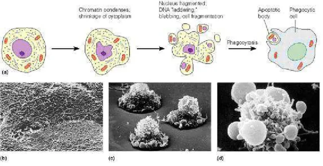
Programmed cell death (apoptosis) is a common type of cell death associated with morphological features that had been repeatedly observed in various tissues and cell types. Characteristic apoptotic features include cell membrane blebbing, cell shrinkage, chromatin condensation and DNA fragmentation, finally ending with the packaging of dead cell content into apoptotic bodies, which are recognized by neighboring cells or macrophages and cleared by phagocytosis, thereby avoiding an inflammatory response in surrounding tissues. Phagocytosis reaction is triggered by phosphatidylserine signal: it is normally present on the cytoplasmic side of the cell membrane, but exposure to the external surface of the cell membrane occurs when a cell undergoes apoptosis [Savill and Fadok, 2000]. Apoptosis is distinct from necrosis in which the cells suffer a major insult, leading to a loss of membrane integrity, swelling and disruption of the cells. During necrosis, the cellular contents are released uncontrolled into the environment which results in damage of surrounding cells and a strong inflammatory response in the
40 corresponding tissue [Leist and Jaattela, 2001] (Figure 19).
_________________________________________________________________________________________________________________________
Figure 19. Morphological features of apoptotic cells.
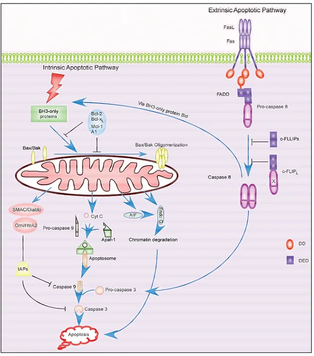
Apoptosis is an essential part of life for multicellular organisms that plays an important role in development and maintenance of tissue homeostasis [Prindull, 1995]. During development many cells are produced in excess which eventually undergo programmed cell death and thereby contribute to sculpturing organs and tissues [Meier et al., 2000]. Cellular apoptosis is tightly controlled by complex regulatory networks and balanced in a physiological context. Pro-survival signals enhance the expression and/or activity of anti-apoptotic regulatory molecules thereby keeping in check the activation of pro-apoptotic factors. A set of various anti-pro-apoptotic molecules and mechanisms has been identified such as NF-κB, AKT, BCL2, CFLAR and the IAP family of proteins. Every step in the apoptotic cascade is monitored and controlled by certain pro-survival signals [Vogelstein and Kinzler, 2004]. Pro-apoptotic factors such as BAX, BID and caspases can counteract those inhibitory molecules when apoptotic demise of a cell is timely and imperative. Caspases activation is a hallmark of apoptosis since this family of proteins are the main effectors of programmed cell death [Jin and El-Deiry, 2005]. Caspases are