UNIVERSITÀ DEGLI STUDI DEL PIEMONTE ORIENTALE
Dipartimento di Scienze della Salute
Corso di Dottorato di Ricerca in MEDICINA MOLECOLARE
Ciclo XXVIII
PhD course in Molecular Medicine
ISCHEMIA/REPERFUSION INJURY ON MICE STEATOTIC
HEPATOCYTES and DIFFERENTIAL EFFECTS OF
ADENOSINE A2A AND A1 RECEPTORS STIMULATION
SSD: MED-04
CO-ORDINATOR
TUTOR
Prof. Emanuele Albano Prof. Rita Carini
DOCTORATE
1
Contents
1. Introduction………...
.. .
031.1
Ischemia Reperfusion Injury……….. .
031.2
Non-alcoholic fatty liver disease (NAFLD)………....
041.2.1 Pathogenesis & Epidemiology of NAFLD……….... 04
1.2.2 Mechanism of Steatosis………. 06
1.3
Liver transplantation and marginal liver availment…………....
071.3.1 Steatotic Grafts……….. . 08
1.4
Ischemia-Reperfusion (I/R) injury
in steatotic liver……….
101.5
Strategies of protection………
131.5.1 Ischemic preconditioning……….……….….. 13
1.5.2 Adenosine and Adenosine receptors………... 15
1.6
Cytotoxicity signal mediators………. .
191.6.1 Endoplasmic reticulum (ER) and ER stress………... 19
1.6.2 Tumour necrosis factor receptor (TNF-R) adaptor factor 2 TRAF2…………. .21
1.6.3 Apoptosis signal-regulating kinase 1 (ASK1)……… 22
1.6.4 C-Jun N-terminal kinases JNK………... 25
1.7
Cell survival signal mediators……….
261.7.1 Phosphoinositide 3-Kinase (PI3K) and Protein Kinase B (PKB)-AKT axis…. 26
2. Aims of the project ……….
283. Paper I (published)………..
294. Paper II (to be submitted)...………
.
645. General Discussion………..
916. References………....
987. Acknowledgements………
..
1122
ABBREVATIONS
I/R Ischemia -ReperfusionNAFLD Non Alcoholic Fatty Liver Disease FFA Free Fatty Acid
ECD Extended Criteria Donor
OPTN Organ Procurement and Transplantation Network IP Ischemic Preconditioning
PI3K Phosphatidyl-inositole-3-kinase HIF-1 Hypoxia-inducible factor 1 ER Endoplasmic Reticulum
TRAF2 Tumor Necrosis Factor Receptor (TNF-R) Adaptor Factor 2 ASK1 Apoptosis Signal-Regulating Kinase 1
ROS Reactive Oxygen Species JNK C-Jun N-terminal kinase PI3K Phosphoinositide 3-Kinase PKB Protein Kinase B
HFD High Fat Diet Hp Hepatocytes
SHp Steatotic Hepatocytes A2aR Adenosine 2a Receptor A1R Adenosine 1 Receptor
CGS21680 2p-(2-carboxyethyl)-phenyl-amino-50-Nethylcarboxyamido- Adenosine
CCPA 2-Chloro-N6-cyclopenty–l adenosine,
NQDI-1 2,7-Dihydro-2,7-dioxo-3H-naphth-o[1,2,3dequin- oline-1- carboxylic acid ethyl ester.
DCFH-DA 2,7-dichlorofluorescin diacetate SiRNA Small interference RNA ATP Adenosine Triphosphate WRT Wortmannin
3
1. INTRODUCTION
1.1 ISCHEMIA -REPERFUSION (I/R) INJURY OF THE LIVER
Hepatic ischemia/reperfusion (I/R) injury is defined as the phenomenon during which cellular damage in an organ, caused by hypoxia, is paradoxically exacerbated after the restoration of oxygen delivery (Peralta et al., 2010). It is a dynamic process which involves the two interrelated phases of local ischemic insult and inflammation-mediated reperfusion injury (Zhai et al., 2013). This concept occurs in several organ systems such as the central nervous system, liver, heart, lung, intestine, skeletal muscle and kidney (Eltzschig et al., 2004).
Hepatic I/R injury is a frequent and major complication in clinical practice, which compromise liver function and increases postoperative morbidity, mortality, recovery and overall outcome (Serracino-Inglott et al., 2001). Liver, being an organ with high energy requirements, is highly dependent on oxygen supply and susceptible to hypoxic or anoxic conditions (Teoh et al., 2011). Extensive researches have investigated the mechanisms responsible for liver damage by I/R. I/R affect liver tissue for the combined alterations occurring during the ischemic period as well as during the reperfusion phase. The lack of oxygen during the ischemic period causes mitochondrial de-energization, ATP depletion and impairment of H+, Na+ and Ca2+ homeostasis (Selzner et al., 2007).
Upon oxygen re-admission, the formation of reactive oxygen species (ROS) by uncoupled mitochondria promotes oxidative stress and mitochondrial permeability transition, and results in a decreased capacity to synthesize ATP. These events are responsible for caspase activation, necrosis and apoptosis. Concomitantly, the activation of Kupffer cells releases ROS, nitric oxide (NO) and pro-inflammatory cytokines. The pro-inflammatory cytokines, in concert with the increased expression of adhesion molecules by sinusoidal endothelial cells, promote liver neutrophil infiltration that contributes to the progression of parenchymal injury (Jaeschke et al., 2003; Urakami et al., 2007).
I/R can induce liver dysfunction or failure that is still a significant clinical problem after tissue resection and transplantation surgery. In Europe over 1853 patients are waiting for a liver graft, while only about 1591 liver donors become available per year (European Liver Transplant Registry. 2014: http://www.eltr.org; Euro transplant Annual Report 2013-2014). Such dramatic organ shortage for transplantation, forces consideration of steatotic grafts, however meta-analysis
4
on total of 1000 patients shows that patients with steatosis have an up to two fold increased risk of postoperative complications, and those with excessive steatosis had an almost three fold increased risk of death (de Meijer et al., 2010).
1.2 NON ALCOHOLIC FATTY LIVER DISEASE (NAFLD)
Non-alcoholic fatty liver disease (NAFLD) is defined when lipids exceed 5% of the total liver weight (Reid et al., 2001). In a subset of the patients, NAFLD further evolves in non-alcoholic steatohepatitis (NASH) characterized by cell death by either apoptosis and necrosis and lobular inflammation with alteration or failure of hepatic functions.
At present, NAFLD/NASH represents the hepatic manifestation of the so called Metabolic Syndrome which is a complex of clinical manifestations associated with obesity and over-weights that includes diabetes, hypertension and hyper triglyceridemia.
1.2.1 PATHOGENESIS AND EPIDEMIOLOGY OF NAFLD
Based on the clinical and experimental data available at the time, the so called „two hit‟ model of progressive NAFLD was proposed in 1998. This model considered the development of steatosis to be the „first hit‟ increasing the sensitivity of the liver to the putative „second hits‟ leading to hepatocyte injury, inflammation and fibrosis. The best candidates for these second hits were considered to be oxidative stress and associated lipid peroxidation and cytokines; principally TNFα. Studies published over the subsequent seven years have led to revisions in this model of pathogenesis, although oxidative stress and cytokines retain a central role. The most important modifications to the model have come from an increased understanding of the sources of oxidative stress and cytokines, in particular the prominent role of insulin resistance, free fatty acids (FFA) and adipose tissue inflammation (Cortez-Pinto et al., 2006). Oxidative stress inhibits the replication of mature hepatocytes which results in expansion of the hepatic progenitor cell (oval cell) population. These cells can differentiate into hepatocyte-like cells, and both oval cell and intermediate hepatocyte-like cell numbers are strongly correlated with fibrosis stage, suggesting that cumulative hepatocyte loss promotes both accumulation of progenitor cells and their differentiation towards hepatocytes (Roskams et al., 2003). In chronic liver injury, the development of fibrosis/cirrhosis is dependent on the efficacy of hepatocyte regeneration, and therefore cell death with impaired proliferation of hepatocyte progenitors represents the proposed third hit in NAFLD pathogenesis (Jou et al., 2008).
5
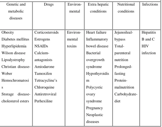
The numerous clinical situations that have been associated with NAFLD can be grouped into 6 etiological groups (Bellentani et al., 2010) (Table 1).
TABLE 1: CLASSIFICATION OF DIFFERENT CAUSES ASSOCIATED WITH NAFLD
Genetic and metabolic diseases Drugs Environ-mental Extra hepatic conditions Nutritional conditions Infections Obesity Diabetes mellitus Hyperlipidemia Wilson disease Lipodystrophy Christian disease- Weber Hemochromatosi s Storage disease- cholesterol esters Corticosteroids Estrogens NSAIDs Calcium- antagonists Amiodarone Tamoxifen Tetracycline‟s Chloroquine Antiretroviral Perhexiline Environ-mental toxins Heart failure Inflammatory bowel disease Bacterial overgrowth syndrome Hypothyroidis m Polycystic ovary syndrome Pregnancy Neoplastic diseases Jejunoileal- bypass Total-parenteral nutrition Prolonged- fasting Protein- malnutrition Carbohydrate- diet Hepatitis B and C HIV infection
NAFLD is common in Europe and now is the most frequent hepatic lesion in Western countries with prevalence rates reported to be anywhere between 2-44% in the general population and 42.6-69.5% in people with type 2 diabetes and rising up to 90% in morbidly obese individuals (Machado et al., 2006). About 15-20 % patients accounts for the pathological evolution form of NASH from the NAFLD with possible progression to cirrhosis or hepatocellular carcinoma. The more severe and clinically significant form of NASH is less common, affecting an estimated 2–3% of the general population and up to 37% of the morbidly obese (Neuschwander-Tetri et al., 2003). The particular concern with significant implications for future disease burden is the increasing prevalence of NAFLD in children and young adults. Studies have reported a 3% prevalence of
6
NAFLD in the general pediatric population, rising to 53% in obese children (Tominaga et al., 1995; Franzese et al., 1997). Recent surveys show that the prevalence of NAFLD across the Asia-Pacific region is at least 10%, and in some regions as many as one-third of individuals could be affected (Liu et al., 2012; Amarapurkar et al., 2007). Likewise, the reported prevalence of NAFLD ranges from 16% in Mexico, 23% in Italy, 30% in Israel, and 9.3% in Japan, respectively (Lazo et al., 2008). The epidemiological significance of NAFLD streams from the data published by the United States Centre for Disease Control and Prevention that estimates that about 66% of US adults in are overweight and half of those are obese. The prevalence of obesity is projected to increase in the United States up to 45% by 2025. Similarly, by 2030 the projected percent increase in type 2 diabetes mellitus is 32% in Europe, 72% in the United States, and 150% or greater in sub-Saharan Africa, India, and the Middle East.
As obesity and diabetes are important risk factors for NAFLD, it is likely that the prevalence of NAFLD will rise in the near future to epidemic proportions. A recent prospective cohort study using ultrasound and liver biopsy determined the prevalence of NAFLD in asymptomatic middle-aged patients to be 46.0% (Williams et al., 2011). Although hospital-based studies are flawed because of ascertainment bias, population-based studies using non-invasive imaging studies (e.g., sonography) suffer the poor specificity of sonography for the diagnosis of NAFLD. Recently, magnetic resonance imaging has been used to quantify the extension of hepatic steatosis. And using this technique, it is estimated that 31% of the U.S. population has NAFLD. In contrast, depending on the definition used, between 2.8% and 24% of U.S. adults have NAFLD according to a comprehensive National Health and Nutrition Examination Survey III (NHANES III) data set–based analysis.
1.2.2 MECHANISMS OF STEATOSIS
The accumulation of triglycerides (TG) originating from the esterification of free fatty acids (FFAs) and glycerol within the hepatocyte is a key point for NAFLD onset.
The contributing factors for the accumulation of FFAs within the liver include dietary sources, enhanced lipolysis in the adipose tissue, insulin resistance and “de novo” lipogenesis in the liver (Postic et al., 2008).
Available evidence suggests that fatty liver results from derangements in fatty acid metabolism in both the liver and the adipose tissue consequent to insulin resistance (Fabbrini et al., 2010; Sanyal et al., 2005; Tilg et al., 2008). In fact, insulin resistance promotes lipolysis in the adipose tissue
7
increasing circulating free fatty acid (FFA) levels and affects hepatocyte FFA metabolism (Tilg et al., 2008). Liver FFA influx through the portal circulation along with decreased FFA oxidation and enhanced “de novo” lipogenesis promote triglyceride accumulation within the hepatocytes (Sanyal et al., 2005).
Figure 1:Schematic mechanisms of metabolic defects leading to the development of hepatic steatosis
1.3 LIVER TRANSPLANTATION AND MARGINAL LIVER AVAILMENT
The lack of available organs for liver transplantation (LT) associated with the increased death rates among patients on most waiting lists for LT has triggered the use of so-called extended criteria donor (ECD) grafts, previously called „„suboptimal grafts‟‟. Among the wide range of these ECD livers, hepatic steatosis is one of the most frequent disorders (Nocito et al., 2006), which is mostly related to an increasing prevalence of NAFLD. The decision to implant or reject a steatotic liver for LT, however, is difficult, due to a risk of impaired graft function or even failure after implantation. How much and what types of fat represent a significant risk for primary non function (PNF) of the graft remains under debate. Ploeg et al originally suggested a classification of fatty change as mild (<30% of visualized hepatocytes involved), moderate (30% to 60 %), and severe (>60%), a system approximately applied by most transplantation Centre‟s (Ploeg et al, 1993).
8
The reserves of transplant steatotic livers are based on the strong association with primary non function (PNF) after a period of cold preservation, initially described by Todo (Todo et al., 1989). However, if a valid and standard method of assessment could be developed, it may be possible to maximize the use of fatty livers while simultaneously minimizing their risk to the recipient. In the early 1990s, four studies examined the relationship of fatty change to PNF. (D‟Alessandro et al., 1991; Markin et al., 1993; Ploeg et al., 1993). The largest of these assessed 390 frozen section biopsy specimens and found that 13% of grafts showing greater than 30% steatosis showed PNF compared with 2.5% of non steatotic grafts. Progressive deterioration in graft survival was observed from mild to massive steatosis. Thus, it was concluded that grafts with severe steatosis should be discarded, and those with moderate change should be evaluated in conjunction with other criteria, such as the condition of the recipient and availability of organs at that time. The institution involved, in line with most others worldwide, found no contraindication to transplanting livers with minimal change. This concurs with the findings of Ploeg et al, who found PNF rates as high as 80% in severely steatotic organs, but more worryingly, initial poor function rates as high as 30% in moderately steatotic livers.
1.3.1 STEATOTIC GRAFTS
Steatotic grafts are considered a risk factor for dysfunction or even primary non function of liver transplants; grafts with more than 50% fatty infiltration are routinely discarded. Steatosis is typically characterized quantitatively and qualitatively. The quantitative evaluation is based on the percentage of hepatocytes containing cytoplasmic fat inclusions. In the clinical setting, steatosis is usually reported as mild, moderate or severe if, respectively less than 30%, between 30% and 60%, or more than 60% of hepatocytes contain fat vacuoles within the cytoplasm (Nocito et al., 2006; McCormack et al., 2005;Selzner et al., 2001). In addition, fatty infiltration is divided quantitatively into two categories: microsteatosis and macrosteatosis. In microsteatosis (MiS), the cytoplasm of the hepatocytes contains tiny lipid vesicles without nuclear dislocation. MiS are usually encountered in mitochondrial disruption following acute viral, toxin- or drug-induced injury, sepsis and in some metabolic disorders (Silva et al., 2009). Macrosteatosis (MaS) is characterized by a single, bulky fat vacuole in hepatocytes, displacing the nucleus to the edge of the cell. This type is most commonly associated with obesity, diabetes, and hyperlipidemia and alcohol abuse. The underlying pathogenesis is related to an excessive triglyceride accumulation in the liver, mainly due to an increased uptake of fatty acids released from adipose tissue or
9
augmented by “de novo” synthesis (Nocito et al., 2006; McCormack et al., 2005; Selzner et al., 2001). Additionally, a defective hepatic export caused by reduced lipoprotein synthesis or impaired β-oxidation of fatty acids, further increases hepatic triglyceride content. Hence the use of grafts with MaS has been associated with increased rates of initial poor function (IPF), primary non function (PNF), and poorer outcome (Marsman et al., 1996). Estimation of steatosis using haematoxylin and eosin (H&E)-stained frozen section liver biopsy has been reported to be difficult and subjective (Franzen et al., 2005; Urena et al., 1999). Even Organ Procurement and Transplantation Network (OPTN) data regarding steatosis are recorded in broad ranges and until recently, did not differentiate between macro vesicular and micro vesicular steatosis (Feng et al., 2006). Therefore the reported variability in both the numbers and grading of steatotic donor livers may reflect differences in both qualitative and quantitative evaluations between different Centers (Selzner et al., 2001; Urena et al., 1998). Some experts believe that physical inspection of an expert in assessing the fat content is equivalent to biopsy (Cameron et al., 2006). However, this has not yet been validated and remains largely subjective. Body mass index (BMI) per se correlates weakly with presence and severity of steatosis (Ryan et al., 2002). Imaging studies alone are not proper tools for the accurate quantification of hepatic fat in all donor candidates (Kim et al., 2006). It has been suggested that differential quantification of color pixels in Oil Red O (ORO) stained liver biopsies using a computer methodology yields more objective and consistent estimation of liver fat content compared with visual interpretation of H&E stained sections (Fiorini et al., 2004), although these computer methods determine the total amount of fat and not the size of the fat droplet (i.e., micro vesicular vs. macro vesicular steatosis). Similarly, the additional negative influence of older donor age and hepatic steatosis has been underlined (De Carlis et al., 1999). A large retrospective single-center study has suggested that recurrent hepatitis C is more common in recipients of moderate and severe steatotic donor livers (Verran et al., 2003). Currently, a macro vesicular fat content of 30% in liver graft, a value with a historical basis resting on early nineties‟ observations is widely accepted for transplantation (D‟Alessandro et al., 1991). Grafts with moderate MaS (30–60%) may be utilized in the absence of additional risk factors in the donor or recipient livers with more than 60%, MaS should probably be excluded (Burke et al., 2004). There are recommendations to allocate livers of different degrees of steatosis based on the Model for End-Stage Liver Disease (MELD) scores of the candidates; these recommendations are however yet to be verified by multivariate analysis (Briceno et al., 2005).
10
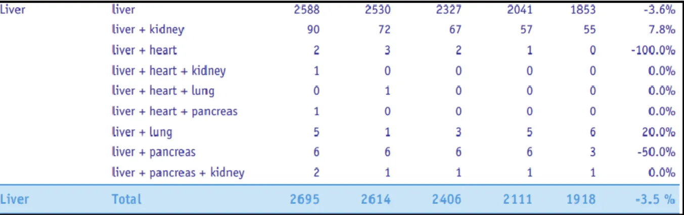
The Organ Transplant Registry update as of 2014 gives information that in the last five years the number of liver donors has decreased at the same time the waiting list for liver transplantation also increased and this disparity between available organs for transplantation and 12 to 24 months waiting list mortality have forced the clinicians to use marginal livers (Steatotic grafts) and even cadaveric grafts for liver transplantation.
TABLE 2: LIVER DONORS
TABLE 3 WAITING LISTS
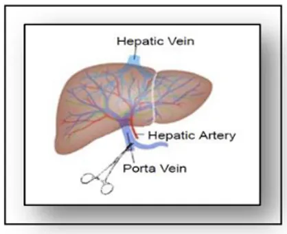
1.4 ISCHEMIA-REPERFUSION (I/R) INJURY IN STEATOTIC LIVER
Hepatic I/R injury can be categorized into warm and cold ischemia. Warm ischemia occurs in the setting of transplantation, trauma, shock and selective liver surgery, in which hepatic blood supply is temporarily interrupted. It may also occur in some types of toxic liver injury, sinusoidal obstruction and Budd-Chiari syndrome (Fernandez et al., 2012). Cold storage ischemia occurs during organ preservation before transplantation (Nickkholgh et al., 2008; Kupiec-Weglinski et
11
al., 2005). Mitochondrial dysfunction has been reported after prolonged cold ischemia in steatotic livers (Caraceni et al., 2004) and also low ATP levels in steatotic livers after transplantation (Jimenez-Castro et al., 2011).
Steatotic livers have been reported to be more susceptible to cold ischemia injury (Schemmer et al., 1999; Fukumori et al.,1999) and moderate to severe MaS steatosis has been observed as the leading cause of severe liver preservation injury (Briceno et al., 2005). In one experience with MaS steatotic livers, every additional hour of total ischemia time longer than 10 hours significantly increased the relative risk of graft and patient loss (Salizzoni et al., 2003). This highlights the difficult issue of acceptance steatotic livers previously evaluated and refused by other Centers, as in these cases ischemia times were always much longer.
Steatotic livers have been shown to be more susceptible to IR injury also after transplantation. During cold ischemia, structural changes attributable to the disruption of hepatic microcirculation caused by fat droplets and hepatocellular swelling, results in occlusion of the sinusoids. After reperfusion, loss of viable endothelial cells and activation of Kupffer cells are accentuated over the non steatotic graft. The migration of leukocytes and adherence to the vascular endothelium is an early and key step in I/R injury and is mediated by three classes of adhesion molecules: selectins, integrin, and immune globulins.
The major event of re-perfusion injury in steatotic livers are due to the abnormal accumulation of fat within the cytoplasm of hepatocytes, resulting in increased hepatocellular volume and narrowing of sinusoid, compromising the suitable graft revascularization and viability after transplantation. Moreover, several evidences indicated that an increased sensitivity of fatty hepatocytes to the harmful effects of reactive oxygen species (ROS) plays a pathogenic role in this event (Domenicali et al., 2005). Where Selzner et al showed the increased susceptibility of fatty livers to reperfusion injury is associated with a change in hepatocytes death form, where the lean rat liver had a prevalence of apoptosis death, while steatotic liver had more massive necrosis present after an ischemic insult (Selzner et al., 2000). After major liver resection, steatosis is associated with mortality higher than 14% respect to the 2% using normal liver. Several hypotheses have been suggested to explain the decreased tolerance of steatotic liver to I/R injury compared with normal livers. These include increased lipid peroxidation, neutrophil infiltration, and release of pro-inflammatory mediators and the alteration of micro circulation (Serafin et al., 2002). The end result is multiple alterations in the steatotic liver finally rendering it more susceptible to I/R. The donor medical history factors may enhance pre preservation injury. They
12
include a history of donor alcohol or drug abuse, presence of a fatty liver, cardiovascular instability after brain death, hypotension during the donor operation, and surgical trauma at the time of harvest.
IR injury is the underpinning of graft dysfunction that is seen in the marginal organ. On restoring the blood supply, the liver is subjected to insult, aggravating injury already caused by the initial ischemia (Clavien et al., 1992; Serracino et al., 2001). I/R injury to endothelial cells disrupts the sinusoidal microcirculation by up-regulating the attraction, activation, adhesion, and migration of neutrophils (polymorph nuclear cells [PMN]) causing local tissue destruction by release of proteases and oxygen-free radicals. I/R in liver transplantation lead to PNF/IPF and increased rejection, and contribute to high morbidity. Preservation injury in liver allografts occurs at four stages: (1) pre preservation injury, (2) cold preservation, (3) rewarming, and (4) reperfusion injury. Cold preservation is also associated with injurious effects. The Kupffer cells, endothelial cells, and Ito cells are more susceptible to cold IR injury as compared with hepatocytes. Sinusoidal endothelial cells undergo apoptosis and coagulated necrosis after cold storage followed by reperfusion of liver grafts (Gao et al., 1998). The sinusoidal cell lining, which is most sensitive to cold ischemia, becomes deficient, exposing the hepatocyte microvilli (Clavien et al., 1998). White blood cells attach where the sinusoidal cells vacated and obstruct the sinusoids and liver blood flow. Additionally, PMNs release numerous mediators, amplifying the inflammatory response (Engler et al., 1983; Varani et al., 1989, Cywes et al., 1993). Platelets, which adhere to the sinusoids, almost immediately on reperfusion aggravate the degree of preservation injury via a mechanism of procoagulant activity and cytokine release, which results in hepatocyte hypoxia (Cywes et al., 1993). Additionally, on reperfusion, Kupffer cells become activated, generating inflammatory mediators such as cytokines and oxygen-derived free radicals, which are injurious to endothelial cells and hepatocytes. The energy stores of the liver (e.g., ATP, glycogen) are depleted, severely compromising hepatocyte function (Lemasters et al., 1995; Kukan et al., 2001). Furthermore, morphologic changes to the endothelial cells are observed, resulting in an endothelin/ nitric oxide imbalance during the reperfusion period, which has been correlated with decreased liver blood flow (Serracino et al., 2001; Clavien et al., 1998; Chazouilleres et al., 1993).
13
1.5 STRATEGIES OF PROTECTION
1.5.1 ISCHEMIC PRECONDITIONING (IP)
The term ischemic preconditioning (IP) refers to the increase in tissue tolerance to ischemia/reperfusion (I/R) damage that can be induced by the pre-exposure to brief periods of ischemia followed by re-oxygenation (Yellon et al., 2005). This phenomenon was first described by Murry in the myocardium (Murry et al., 1986), but was subsequently observed in many other tissues (Yellon et al, 2005). In liver, studies in rodents have shown that 10 min interruption of blood supply followed by 10 min reperfusion reduces hepatic injury and inflammation during a subsequent extended period of I/R. Recent studies have shown that the effects of IP can be observed also by the application of a brief ischemia during the reperfusion period. This phenomenon, known as post-conditioning, demonstrates that the mechanisms of hepatoprotection induced by ischemic preconditioning may also act when hepatic damage has already started and suggests the possibility to activate these mechanisms also in case of liver damage different from I/R when surgical application of IP is not practicable.
The protection induced by IP takes place in two different phases. The first phase known as early preconditioning immediately follows the preconditioning stimulus and modulates different cellular functions. The second phase is known as delayed or late preconditioning; it starts 12-24 hours after the pre-conditioning stimulus, can last up to 3-4 days, and is characterized by gene transcription and “de novo” protein synthesis (Peralta et al., 1997). Despite these differences, both phases of preconditioning can be initiated by the same stimuli and partially share the same intracellular signal pathways.
Figure 2: Liver preconditioning with a brief cycle of ischemia-reperfusion (I/R) (10 minutes of ischemia +
14
“In vivo” and “in vitro” studies have clearly established that the onset of IP is triggered by the production of adenosine and by the subsequent stimulation of adenosine A2a receptor (Peralta et al., 1997; Nakayama et al., 1999; Hart et al., 2008; Carini et al., 2000; Carini et al., 2001). This was confirmed in our Laboratory with experiments using primary rat hepatocytes pre-conditioned with 10 minutes of hypoxia plus 10 minutes of re-oxygenation. In this model, the released adenosine to extra-cellular space induced hepatocyte protection by the autocrine stimulation of A2a receptors.
Surgical ischemic preconditioning raised hopes that it could be applied to patients to prevent the side-effect of major liver surgery, but the first application of IP in clinical trials have given conflicting results and in some cases IP did not afford protection and in some cases its protective effects were extremely variable. These contrasting outcomes of the clinical studies, the different protocols of IP application in humans, and the variable clinical settings have not allowed a definitive demonstration of the benefit of the clinical application of IP (Amador et al., 2007; Azoulay et al., 2005; Cescon et al., 2006; Koneru et al., 2007; Franchello et al., 2009; Jassem et al., 2006). Hence this observation has inhibited now, the routine use of IP in human liver surgery and has indicated the need of more efficient approaches to activate IP in patients.
In this regard, the pharmacological induction of liver preconditioning by targeted activation of one or more of the critical molecular mediators of IP may represent a more reliable technique to activate the intrinsic system of hepatoprotection in patients.
15
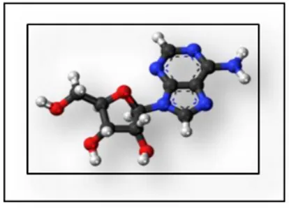
1.5.2 ADENOSINE AND ADENOSINE RECEPTORS
Adenosine is an endogenous purine nucleoside that modulates many physiological processes. Extracellular adenosine concentrations in normal cells are approximately 300 nM but these concentrations are elevated quickly during tissue damage and inflammatory reactions.
Figure 3: Adenosine molecular structure.
The released adenosine interacts with different subtype of adenosine receptors that modulate cell protection, inflammation and immunological responses. There are four kinds of adenosine receptors (ARs) A1, A2A, A2B and A3 that are of purinergic class and G protein coupled receptor. ARs have traditionally been classified based on their differential coupling to adenylyl cyclase to regulate cyclic AMP levels. The A1 and A3aRs are coupled to Gi proteins, while A2aAR and A2b AR are coupled to Gs proteins (Fredholm et al., 2011). Therefore, activation of the A2A and A2B ARs increase cyclic AMP production, resulting in activation of protein kinase A (PKA) and phosphorylation of the cyclic AMP response element binding protein (CREB). In contrast, activation of the A1 and A3AR inhibits cyclic AMP production and decreases PKA activity and CREB phosphorylation (Cunha et al., 2001; Fredholm et al., 2011; Paes-De-Carvalho et al., 2002).
The animal studies have, all together, shown that a brief ischemic stress induces a profound phenotypic modification that makes liver cells resistant to damage, and inhibits hepatic inflammatory reactions. Despite the nearly 25 year‟s research on liver IP, the knowledge of the proteomic modifications responsible for its production is still poor. To date a well-established notion is the role of the adenosine A2a receptor (A2aR) as a first inductor of rodent liver preconditioning. Since from the early study of Peralta et al (Peralta et al., 1997), a number of
16
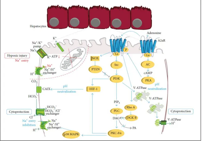
studies “in vivo” and “in vitro” have in fact clearly established that the brief ischemic stress triggers the onset of IP by inducing the release of adenosine in the extracellular space with subsequent stimulation of the A2aR of liver cells. By using primary rat hepatocytes preconditioned with a brief hypoxia-reoxygenation in the last years we have begun to analyse the signal network that following A2aR stimulation induces hepatocyte resistance hypoxic damage (Figure 4). This network involves Gs protein, adenylate cyclase and protein kinase A (PKA) that phosphorylates ADR2A and shifts its coupling to Gi protein and Src kinase and activates phosphatidyl-inositole-3-kinase (PI3K) and its downstream effector Akt. This allows the stimulation of phospholipase C, the recruitment of the specific isoforms and of protein kinase C (PKC) and the activation of p38 MAPK. Full activation of preconditioning responses also needs an A2R-induced down modulation of inhibitory enzymes of PKC and PI3K. Hypoxic preconditioning as well as A2aR stimulation, in fact, induces a RhoA-GTPase-dependent inhibition of the diacylglycerol kinases thus increasing diacylglycerol (DAG) and sustaining activation of the DAG-dependent PKC and . A2aR also induces the degradation of the PI3K inhibitor, phosphatase and tensin homologue deleted from chromosome 10 (PTEN) by an NADPH oxidase-dependent mechanism, allowing the maintenance of the PI3K-dependent signals (Alchera et al., 2010). These observations represent the first data on the modulation of constitutive hepatocyte signal proteins upon hypoxic preconditioning and their role in hepatocyte resistance to hypoxia. The knowledge on the induction of gene expression and protein synthesis in preconditioned liver is even lower. “In vivo” studies have shown that liver IP is associated with a stimulation of nuclear factor-κB (NF-κB) activity in the ischemic phase and its inhibition during hepatic reperfusion. The pro regenerative and protective effect of liver preconditioning are instead associated to the activation of the signal transducer and activator of transcription (STAT)/IL6 axis. Hypoxia-inducible factor 1 (HIF-1) is the main regulator of tissue adaptation to oxygen deprivation and it is found increased in human transplanted livers exposed to IP. Consistently the delayed protective effect of hepatocyte preconditioning is related to an A2aR/PKC/PI3K-dependent non hypoxic HIF-1 activation and to the consequent production of its target protein, carbonic anhydrase IX (Alchera et al., 2015).
17
Figure 4: Molecular mechanisms involved in hypoxic injury of primary rat hepatocytes and their
protection upon A2aR stimulation. Hypoxic damage: ATP depletion causes intracellular acidosis, inhibition of the Na+/K+ ATPase, and activation of the Na+/H+ exchanger with an increase in intracellular Na+ content and activation of the K+ channel. For A2aR protection: A2aR stimulation induces the sequential activation of PKA, Gs and Gi protein, Src, PI3K, PLC, PKC𝛿, and 𝜀 and p38 MAPK. A2aR also inhibits the negative regulators of PKC and PI3K, DGK, and PTEN. PI3K activates V-ATPase that maintains intracellular pH avoiding the activation of the Na+/H+ exchanger and Na+ overload. PI3K and PKC 𝛿 and 𝜀 activate HIF with production of CAIX. CAIX converts CO2 into bicarbonate that enters into
hepatocyte through the Cl−/HCO3− exchanger. This neutralizes intracellular pH without activation of the Na+/H+ exchanger and the consequent Na+ increase. (Alchera et al. Biomed Res Int. 2015)
18
The cytoprotective effects of adenosine during preconditioning are only part of the effects of adenosine. A separate bulk of researches have indeed demonstrated that adenosine dramatically increases at extracellular levels during tissue damage, ischemia and inflammation and by interacting with one or more of the four adenosine receptors (A1, A2a, A2b, A3), elicits autocrine and paracrine modulation not only of cell survival but also of inflammatory and immunological reactions (Fredholm et al., 2007; Hasko et al., 2008). Several studies show that adenosine may play pro-inflammatory or anti-inflammatory role depending on the type of adenosine receptor is engaged. Interestingly, some findings indicate that the different adenosine receptors might have dissimilar or even opposite effects. Well characterized is the pro-inflammatory activity of A1 and A2b receptor and in contrast, the immune suppressive action of the A2a receptor. A1R exerts a pro-inflammatory response by enhancing phagocytosis (Salmon et al., 1993), promoting chemotaxis (Schnurr et al., 2004; Rose et al., 1988) and enhancing neutrophils adherence to endothelium during inflammatory process (Cronstein et al., 1992). By contrast A2aR have a major role in suppressing immune response. Engagement of A2aR inhibits neutrophils adherence to endothelium during inflammation (McColl et al., 2006) and inhibits the activation of neutrophils, monocytes platelets and T-cells (Sullivan et al., 2001; Cooper et al., 1995; Koshiba et al., 1997). In animal models, A2aR-agonists can prevent lethal response to bacterial LPS and sepsis (Sullivan et al., 2004; Mazar et al., 2005). In macrophages, A2aR mediate inhibition of TNF-alpha and augment IL-10 production (Haskò et al., 2000; Ryzhov et al., 2008; Nemeth et al., 2005).
The adenosine receptors have contradictory effects on liver steatosis and lipotoxicity. In A1 KO mice ethanol-induced hepatic steatosis is reduced compared to WT mice, indicating a pro-steatotic action of A1 (Peng et al., 2009). On the other hand, recent studies in our laboratory have shown the protective effect of A2aR stimulation in lipoapoptotic liver presence (Imarisio et al., 2012). As described above, adenosine and ARs play a dynamic role in regulating normal cell physiology and also act as modulators in disease processes. A better understanding of the functions of these receptors, especially the newly identified receptor homomers and heteromers, could stimulate development of new therapies for the treatment of diseases.
19
1.6 CYTOTOXICITY SIGNAL MEDIATORS
1.6.1 ENDOPLASMIC RETICULUM (ER) AND ER STRESS
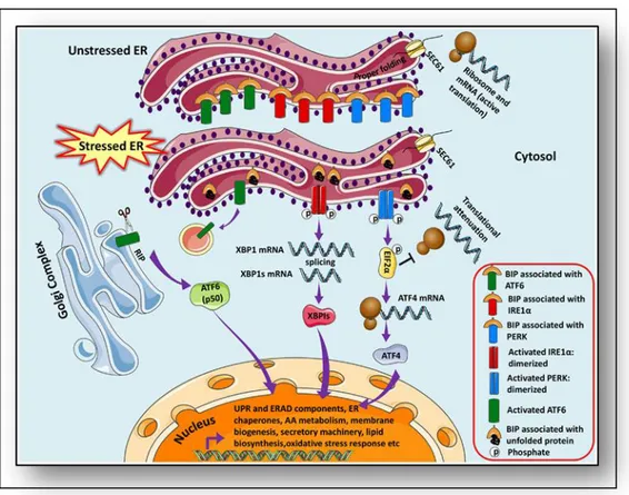
The endoplasmic reticulum (ER) is a central organelle of each eukaryotic cell responsible for protein folding, maturation, quality control and trafficking. Proteins of the plasma membrane, secreted proteins as well as proteins of the Golgi apparatus and lysosomes fold into their tertiary and quaternary structure in the ER. The ER is the major signal transducing organelle that senses and responds to changes of the homeostasis (Voeltz et al., 2002). Conditions interfering with the function of ER are collectively called ER stress. ER stress is induced by accumulation of unfolded protein aggregates (unfolded protein response, UPR) or by excessive protein traffic usually caused by viral infection (ER overload response, EOR). In eukaryotic cells, UPR -caused by formation of unfolded protein aggregates -uses an evolutionarily conserved signaling pathway during which the signal of unfolded proteins activates a set of ER-located sensors (Zhang et al., 2011).
The adaptive UPR comprises signal transduction pathways initiated by ER proximal UPR transmembrane proteins: inositol-requiring kinase 1 (IRE1α), activating transcription factor 6 (ATF6), and double-stranded RNA-activated protein kinase (PKR)-like endoplasmic reticulum kinase (PERK) in an attempt to restore homeostasis and normal ER functions (Schroder et al., 2005). These UPR transducer proteins are negatively regulated by the chaperone GRP78/BIP (immunoglobulin heavy chain binding protein) in unstressed or healthy ER at their luminal ends (amino terminal), however, increase in unfolded proteins causes dissociation of 78-kD glucose-regulated/binding immunoglobulin protein Grp78/BIP thereby releasing the inhibition and thus eliciting the response to stimuli that divert ER chaperones to misfolded proteins, IRE1, PERK and ATF6 initiate signal transduction processes. These events promote the expression of genes required to fold newly synthesized proteins and to degrade the unfolded proteins. Moreover, homeostasis and normal ER function are restored. However, when injury is excessive, the same ER stress signal in response transduction pathways can also induce cell death (Bertolotti et al., 2000; Pfaffenbach et al., 2011) (Figure 5).
20
Figure 5: Unfolded protein response pathways.
It has been demonstrated that the UPR is a fundamental intracellular signal transduction response that is critical for health and disease. ER stress and other cellular stress responses, such as inflammation and oxidative stress, are integrated in many pathophysiological processes. The ER stress response has recently been recognized in a wide spectrum of experimental models of liver injury, which is an emerging field of interest in the pathogenesis of nearly all types of human liver disease. In the liver, hepatocytes, similar to other secretory cells, are rich in ER. Due to its high capacity for protein synthesis, the UPR/ER stress response plays important roles in both preventing and mediating pathological changes in various liver diseases (Zhang et al., 2014). NAFLD implies that the ability to resolve ER stress has been compromised. Particularly, recent research demonstrated that ER stress and the UPR signaling are critically involved in the initiation and progression of nonalcoholic fatty liver disease (NAFLD). Under metabolic stress conditions, the UPR regulates transcriptional and translational programs that are associated with hepatic steatosis and inflammation; the major characteristics of NAFLD (Pagliassotti et al., 2012).Toxic lipids such as free fatty acids, diacylglyceride, phospholipids and free cholesterol activate several cellular stress pathways. The maintenance of ER function requires high concentrations of intra-ER
21
Ca2+, which is actively controlled by sarcoplasmic (endo) reticulum Ca2+-ATPase (SERCA). Free cholesterol accumulation triggers ER stress by altering the critical free cholesterol-to-phospholipid ratio of the ER membrane, which is needed to maintain its fluidity. Among the ER enzymes, SERCA ATPase is particularly sensitive to ER membrane cholesterol contents that can inhibit SERCA conformational changes and activity. Such changes induce a decrease in physiologically high intra-ER Ca2+.
The presence of ER stress and activation of the UPR in chronic diseases such as obesity is one of the most important factors for disease progression in NASH along with hepatocyte apoptosis and hepatic stellate cell (HSC) or Kupffer cell activation.
Moreover, in liver cells, ER response is involved in hepatic ischemia-reperfusion injury (I/R) that promotes protein unfolding and hence triggering as documented by the activation of XBP1 and ATF6 in the parenchyma of livers (Brenner et al., 2013). Accumulating evidence suggests perturbations at the ER as a novel sub cellular effector, possibly involved in promotion of cell death in various pathologies including the pathophysiology of organ preservation.
1.6.2 TUMOR NECROSIS FACTOR RECEPTOR (TNFR) ADAPTOR FACTOR 2 -TRAF2
The tumor necrosis factor (TNF) receptor adaptor factor (TRAF) family of proteins plays a pivotal role in different biological processes, including immunity, inflammation and apoptosis. The mammalian TRAF family comprises seven members: TRAF1 though TRAF7. Among these, TRAF2 and TRAF6 have been most extensively studied. In particular, TRAF2 associates, directly or indirectly, with members of the TNF receptor (TNFR) super family, including TNFR1 and TNFR2, RANK (a receptor that mediates differentiation and maturation of osteoclasts), and CD40 (a receptor important for the proliferation and activation of B cells). TRAF2 play a central role in the cellular response to stress and cytokines via their regulation of stress kinases, resulting in the activation of key transcription factors, including NF-κB, c-Jun and ATF2 (Xia et al., 2005). The function of TRAF2 and TRAF5 is best characterized in TNFR1 signaling, whereas TRAF6 and TRAF3 have been extensively studied in IL-1R or TLR signaling, TRAF3 has been demonstrated to be critical for virus-induced activation of IRF3-IRF7 and interferon production.
It has been known that ER stress can lead to altered lipid metabolism and hepatic steatosis. In particular, the IRE1α-XBP1 pathway has been reported to be required for the maintenance of
22
hepatic lipid homeostasis under ER stress conditions (Zhang et al., 2014). The association of ER stresses signaling and hepatic steatosis has been proven through the IRE1α/XBP1 pathway and the ER protein translocation pathway. When IRE1α is activated induces the unconventional splicing of the mRNA encoding X-box-binding protein 1 (XBP-1). The cytosolic domain of activated IRE1 binds TRAF2 and triggers the activation of the c-Jun N-terminal kinase (JNK), MAPK p38 and caspase-12. Fumihiko et al demonstrated that stress-induced oligomerization and activation of IRE1 could lead to clustering of TRAF2 that is bound to the COOH-terminal cytoplasmic portion of the IRE1, one of the ER transmembrane proteins involved in initiating signals from the ER. (Urano et al., 2000).
Mediating cellular response to ER stress has been proposed based upon the observation that ectopic expression of a domain negative mutant of TRAF2 lacking the N terminus Ring finger domain blocks ER stress-induced NF-κB and JNK/SAPK activation, and the mouse embryonic fibroblast derived from TRAF2 knock-out mice failed to activate NF-κB following ER stress (Mauro et al., 2006). TRAF2 is not only investigated in a contest of steatotic liver injury. In literature is not well know the exact relationship of ER stress-mediated cell death and cold I/R injury in liver transplantation, but results presented implicate ER stress on a broad scale as an important factor in this injury (Mosbah et al., 2012), shown that activation of the IRE-1 pathway regulates pro-apoptotic responses by activation of stress kinase JNK and mitogen-activated protein kinases. Finally, it has been demonstrated that the use of specific inhibitors of ER stress could represent effective strategies to reduce hepatic I/R injury (Peralta et al., 2010).
1.6.3 APOPTOSIS SIGNAL-REGULATING KINASE 1 (ASK1)
All living organisms are exposed to numerous physicochemical stressors during their lifetime and appropriate responses to stress at the cellular level are essential for the maintenance of homeostasis. The mitogen-activated protein kinase (MAPK) cascades are thought to be crucial among the major signaling pathways that regulate cellular stress responses. Apoptosis signal-regulating kinase 1 (ASK1) is a member of the mitogen activated protein kinase kinase kinase (MAP3K) family that activates downstream MAP kinases (MAPKs). MAPKs control a wide variety of cellular functions, including proliferation, differentiation, migration and apoptosis (Hattori et al., 2009).
23
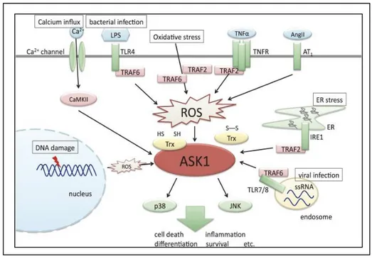
ASK1 is activated in response to various stresses, such as oxidative stress, ER stress, calcium overload and inflammatory signals including those induced by tumor necrosis factor α (TNFα) lipopolysaccharide (LPS), and Ang II (Fig.6) (Hayakawa et al.,2012). Activated ASK1 in turn activates the downstream p38 and JNK pathways and induces various cellular responses, including cell death, inflammation, differentiation and survival ASK1 is involved in the IRE1α pathway, one of the adaptive ER proximal UPR transmembrane proteins. IRE1a collaborates with adaptor-like tumor necrosis factor receptor (TNFR)-associated factor2 (TRAF2) and recruits ASK1 that has been shown to relay various stress signals to the downstream activating among others Jun N-terminal kinase (JNK) and p38 MAP kinase (Derijard et al., 1995; Nishitoh et al., 2002).
Figure 6: Overview of the functions of ASK1.
Unfolded/misfolded/mutated proteins (Hetz et al., 2006), disturbance in cellular redox regulation, endogenous reactive oxygen species (ROS) production (Fedoroff et al., 2006) and hypoxia (Sawada et al., 2008) act as stress signals altering ER homeostasis, if the stress signal is severe and/or prolonged, ER triggers cell death pathways (Szegezdi et al., 2006; Kim et al., 2008; Cheng et al., 2011; Benbrook et al., 2012).
ASK1 is highly conserved among eukaryotes and its activation mechanism appears to be common among the ASK family of proteins, i.e., ASK1, ASK2, NSY-1, and DASK1. ASK1 is a serine/threonine protein kinase activated by phosphorylation of a threonine residue (Thr838 in
24
human ASK1) within the activation loop. In the past decade, various regulatory mechanisms of ASK1 have been elucidated. It has been reported that ASK1 activity is regulated by many ASK1-interacting proteins, among which thioredoxin (Trx) plays an important role. Trx is a redox protein that changes its structure depending on the cellular redox state. Only the reduced form of Trx binds to the N-terminus of ASK1 and inhibits ASK1 activity by inhibiting homophilic interaction through the N-terminal coiled-coil domains in the pre-existent ASK1 oligomer under unstimulated conditions (Hattori et al., 2009).
Upon ROS stimulation, the ASK1 signalosome unbinds from Trx and forms a fully activated higher-molecular-mass complex, in part by recruitment of tumor necrosis factor receptor-associated factor 2 (TRAF2) and TRAF6. However, the precise mechanisms by which Trx inhibits and TRAF2 and TRAF6 activate ASK1 have not been elucidated fully (Fujino et al., 2007; Hattori et al., 2009). ASK1 activation in response to TNF and LPS signaling has been reported to depend on ROS generation, suggesting that ROS play a key role in the regulation of ASK1 activity (Hattori et al., 2009). Investigations will be necessary to determine whether simple steatosis and progression from isolated fatty liver to NASH be preceded by higher ROS generation (Nassir et al., 2014). Kim et al demonstrated that ASK1 is a substrate for phosphorylation by Akt and that this phosphorylation in a consensus sequence at Ser83 level is associated with a decrease in stimulated ASK1 kinase activity. So, the negative regulation of ASK1 activity and consequent activation of downstream signaling molecules can be negatively regulated by Akt stimulation. This regulatory event has measurable consequences for ASK1 downstream signaling, including apoptosis induced by ASK1. ASK1 may be a physiological target of Akt and raise the intriguing possibility that the ability of Akt to inhibit stress-activated kinases in specific cell contexts is a consequence of this interaction (Kim et al., 2001).
Figure 7: The domain structures of ASK1. The binding domains of Trx and TRAF exist in N terminus of
ASK1. Two coiled coil domains (NCC and CCC) are important for the homomeric interaction and activation of ASK1.
25
1.6.4 C-JUN N-TERMINAL KINASES –JNK
C-Jun N-terminal kinases (JNKs), were originally identified as Kinases that bind and phosphorylate c-Jun on Ser 63 and Ser-73 within its transcriptional activation domain. They belong to the mitogen activated protein kinase family, and are responsive to stress stimuli, such as cytokines, ultraviolet irradiation, heat shock, and osmotic shock. They also play a role in T cell differentiation and the cellular apoptosis pathway. Activation occurs through a dual phosphorylation of threonine (Thr) and tyrosine (Tyr) residues within a Thr-Pro-Tyr motif located in kinase subdomain VIII. Activation is carried out by two MAP kinases, MKK4 and MKK4 and JNK can be inactivated by Ser/Thr and Tyr protein phosphatases (Tony et al., 1998).
The c-Jun N-terminal kinases consist of ten isoforms derived from three genes: JNK1 (four isoforms), JNK2 (four isoforms) and JNK3 (two isoforms) (Waetzig et al., 2005). Each gene is expressed as either 46 kDa or 55 kDa protein kinases, depending upon how the 3' coding region of the corresponding mRNA is processed. There have been no functional differences documented between the 46 kDa and the 55 kDa isoform, however, a second form of alternative splicing occurs within transcripts of JNK1 and JNK2, yielding JNK1-α, JNK2-α and JNK1-β and JNK2-β. Differences in interactions with protein substrates arise because of the mutually exclusive utilization of two exons within the kinase domain (Tony et al., 1998). C-Jun N-terminal kinase isoforms have the following tissue distribution:
JNK1 and JNK2 are found in all cells and tissues; JNK3 is found mainly in the brain, but is also found in the heart and the testes (Bode et al., 2007).
Inflammatory signals, changes in levels of reactive oxygen species, ultraviolet radiation, protein synthesis inhibitors, and a variety of stress stimuli can activate JNK. One way this activation may occur is through disruption of the conformation of sensitive protein phosphatase enzymes; specific phosphatases normally inhibit the activity of JNK itself and the activity of proteins linked to JNK activation (Vlahopoulos et al., 2004). JNKs can associate with scaffold proteins JNK interacting proteins as well as their upstream kinases JNKK1 and JNKK2 follows their activation. JNK, by phosphorylation, modifies the activity of numerous proteins that reside at the mitochondria or act in the nucleus. Downstream molecules that are activated by JNK include c-JUN ATF2, ELK1, SMAD4, p53 and HSF1.The downstream molecules that are inhibited by JNK activation include NFAT4, NFATC1 and STAT3. By activating and inhibiting other small molecules in this way,
26
JNK activity regulates several important cellular functions including cell growth, differentiation, survival and apoptosis.
1.7 CELL SURVIVAL SIGNAL MEDIATOR
1.7.1 PHOSPHOINOSITIDE 3-KINASE (PI3K) AND PROTEIN KINASE B (PKB)-AKT AXIS
Phosphoinositide 3-kinases (PI3Ks) are a family of intracellular signal transducers characterized by the capacity of generating phosphatidylinositol (3,4,5)-triphosphate (PIP3) that in turn acts as a second messenger activating several kinases implicated in the regulation of cell proliferation, survival and metabolism (Cantley et al., 2002). The importance of PI3K in preventing hepatic injury has emerged from a number of observations showing that PI3K-mediated signals are important in preventing hepatocytes apoptosis as well as in ameliorating liver reperfusion injury (Webster et al., 2001; Hateno et al, 2002; Muller et al 2003). Consistently, recent studies have shown that ischemic preconditioning activates PI3K signaling in rodent livers, while the block of this kinase abolishes the protective action of preconditioning both in isolated hepatocytes and in the whole organs (Izuishi et al., 2006; Carini et al., 2004).
By using primary rat hepatocytes preconditioned with a brief hypoxia-reoxygenation in the last years we have begun to analyze the signal network that following A2aR stimulation induces hepatocyte resistance to hypoxia. This network involves Gs protein, adenylate cyclase and protein kinase A (PKA) that phosphorylates A2AR and shifts its coupling to Gi protein and Src kinase and activates PI3K and its downstream effector Akt (Alchera et al., 2010)
The lipid product of PI3K, PIP3, facilitates phosphorylation of Akt, also known as Protein kinase B (PKB) by PDK1. This phosphorylation stimulates the catalytic activity of Akt, resulting in the phosphorylation of a host of other proteins that affect cell growth, cell cycle entry, and cell survival (Cantley et al., 2002).
Akt, is a Serine /threonine- specific protein kinase that. It is one of the key molecules downstream of the phosphoinositide 3-kinase (PI3K) signaling pathway. In mammals, Akt comprises of three highly homologous members, including Akt1 (PKBα), Akt2 (PKBβ) and Akt3 (PKBU), which are encoded by three different genes located on different chromosomes (Dillon et al., 2010; Nicholson et al., 2002; Hanada et al., 2004, Schultze et al., 2011). The Akt kinases control an array of diverse functions including cell growth, survival, proliferation and metabolism (Gonzalez et al., 2009).
27
Akt1 and Akt2 are widely expressed, whereas Akt3 expression is restricted to brain, testis, lung, fat, mammary glands and pancreatic islets (Schultze et al., 2011). In the liver, only Akt1 and Akt2 are expressed, with Akt2 as the major isoform (accounting for approximately 70% of total Akt protein) (Schultze et al., 2011). Several growth factors and cytokines are known to confer resistance to Fas-induced liver injury by activation of the Akt pathway (Schulze et al., 2004, Moumen et al., 2007). Akt1 is involved in cellular survival pathways, by inhibiting apoptotic processes. Akt1 is also able to induce protein synthesis pathways and is therefore a key signaling protein in the cellular pathways. Since it can block apoptosis, and thereby promote cell survival, Akt1 has been implicated as a major factor in many types of cancer. Akt (now also called Akt1) was originally identified as the oncogene in the transforming retrovirus AKT8 (Staal et al., 1977). Akt is involved in the PI3K/AKT/mTOR pathway and other signaling pathways. Akt possesses a protein domain known as a PH domain, or pleckstrin homology domain, named after pleckstrin, the protein in which it was first discovered. This domain binds to phosphoinositides with high affinity. In the case of the PH domain of Akt, it binds either PIP3 (phosphotidylinositol (3, 4, 5) -triphosphate, Ptdins (3, 4, 5) P3) or PIP2, phosphotidylinositol (3, 4) bisphosphate, PtdIns (3, 4) P2) (Franke et al., 1997). Once correctly positioned at the membrane via binding of PIP3, Akt can then be phosphorylated by its activating kinases, phosphoinositide dependent kinase 1 (PDPK1 at Thr308) and the mammalian target of rapamycin complex 2 (mTORC2 at Ser473) (Sarbassov et al., 2005).
28
2. AIMS OF THE PROJECT
The increasing prevalence of NAFLD in the general population translates directly into an increasing graft steatosis, affecting both, the quality and the quantity of donor livers available for transplantation. Ischemia-Reperfusion (IR) injury of liver results in hepatocytes irreversible damage occurring during surgical procedures that include hepatic resections and liver transplantation. In particular, the use of steatotic livers for transplantation is associated with an increased risk for primary nonfunction or dysfunction after surgery respect to a normal liver because steatotic livers tolerate poorly I/R injury. Ischemic Preconditioning (IP) has shown to be an effective method that reduces liver injury induced by I/R, but its application to human surgery has given conflicting results. Thus, a better understanding of the mediators and pathways involved in IP might represent a way to optimize strategies against IR damage in normal and fatty livers. On the base of these considerations, the aims of my project were:
To understand the phenotypic changes occurring in the liver during I/R or as a response to preconditioning treatments.
To set up a cellular model able to reproduce “in vitro” the ischemia/reperfusion (I/R) damage using normal and steatotic primary mouse hepatocytes and to characterize the molecular mediators and pathways which could sensitize the steatosis for the exacerbation effect of I/R injury in fatty liver.
To investigate the protective action of different adenosine receptor agonists during hypoxia reoxygenation damage in normal and fatty liver.
29
PAPER-1
Mouse hepatocytes and LSEC proteome reveal novel mechanisms of
ischemia/reperfusion damage and protection by A2aR stimulation
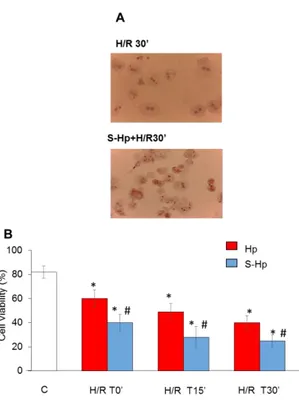
SummaryIschemia-reperfusion (IR) of liver results in irreversible damage of hepatocytes (HP) and sinusoidal endothelial cells (LSEC). Ischemia/reperfusion damage causes up to 10% of early organ graft failure, following liver transplantation, and can lead to a high incidence of both acute and chronic rejections. Minimizing the adverse effects of this injury could significantly increase the number of transplantable livers, improving graft outcome. Previous studies have shown that Ischemic Preconditioning (IP) protects IR damage upon stimulation adenosine A2a receptor (A2aR) enhancing cell tolerance against hepatic IR damage. These effects are also mimicked by the A2aR agonist CGS21680. Understanding the phenotypic changes that underlie hepatocellular damage and protection is critical for optimizing strategies against IR. This work describes, for the first time, the proteome alterations of mouse HP and LSEC isolated from murine livers exposed to IR in the presence or absence of A2aR stimulation, elucidating the liver cell contribution to IR damage and hepatoprotection by pharmacological preconditioning. To this aim the proteome of HP and LSEC isolated from sham or IR exposed mice receiving or not the A2aR agonist CGS21680 (0.5 mg/kg b.w) was analysed by 2-D DIGE/MALDI-TOF. Using this procedure we identified 64 proteins involved in cytoprotection, regeneration, energy metabolism and response to oxidative stress; among them, 34 were associated with IR injury and A2aR protection. The main pathways, down regulated by IR and up regulated by CGS21680 in HP and LSEC, were related to carbohydrate, protein and lipid supply and metabolism. In LSEC, IR reduced stress response enzymes that were instead up regulated by CGS21680 treatment. Functional validation experiments confirmed the metabolic involvement and showed that the inhibition of pyruvate kinase, 3-chetoacylCoA thiolase, and arginase affected the protection exerted by CGS21680 on “in vitro” hypoxia-reoxygenation injury, whereas hepatocyte supplementation with the metabolic products of these pathways reduced IR-induced liver cell damage. Moreover, LSEC, but not HP, were sensitive to H2O2-induced oxidative damage and CGS21680 protected against this effect.
Taken together the results of this study show that IR injury is characterized by specific modifications of HP and LSEC proteomes that are partially reverted by A2aR stimulation providing novel insides in the pathways leading to liver protection by preconditioning treatments.
30
PAPER-1
Mouse hepatocytes and LSEC proteome reveal novel mechanisms of
ischemia/reperfusion damage and protection by A2aR stimulation
Giorgia Mandili1,2, Elisa Alchera3,, Simone Merlin3,, Chiara Imarisio3,, Bangalore R. Chandrashekar3, Chiara Riganti4, Alberto Bianchi3, Francesco Novelli1,2,, Antonia Follenzi3,, Rita Carini3,
Department of Health Science, University of Piedmont Orientale, Novara, Italy Journal of Hepatology 2015 vol. 62: 573–580
ABSTRACT
Background & Aims: Ischemia-reperfusion (IR) of liver results in hepatocytes (HP) and sinusoidal
endothelial cells (LSEC) irreversible damage. Ischemic preconditioning protects IR damage upon adenosine A2a receptor (A2aR) stimulation. Understanding the phenotypic changes that underlie hepatocellular damage and protection is critical to optimize strategies against IR.
Methods: The proteome of HP and LSEC isolated from sham or IR exposed mice receiving or not
the A2aR agonist CGS21680 (0.5 mg/kg b.w.) was analysed by 2-D DIGE/MALDI-TOF
Key results: 64 proteins were identified involved in cytoprotection, regeneration, energy
metabolism and response to oxidative stress; among them, 34 were associated with IR injury and A2aR protection. The main pathways, down regulated by IR and up regulated by CGS21680 in HP and LSEC, were related to carbohydrate, protein and lipid supply and metabolism. In LSEC, IR reduced stress response enzymes that were instead up regulated by CGS21680 treatment. Functional validation experiments confirmed the metabolic involvement and showed that inhibition of pyruvate kinase, 3-chetoacylCoA thiolase, and arginase reduced the protection by CGS21680 of in vitro hypoxia-reoxygenation injury, whereas their metabolic products induced liver cell protection. Moreover, LSEC, but not HP, were sensitive to H2O2-induced oxidative
damage and CGS21680 protected against this effect.
Conclusions: IR and A2aR stimulation produces pathological and protected liver cells phenotypes
respectively characterized by down- and up- regulation of proteins involved in the response to O2
31
synthesis at reperfusion. This provides novel insides in IR hepatocellular damage and protection and suggests additive therapeutic options.
Introduction
Inflow occlusion during liver surgery, with consequent reperfusion, causes liver ischemia-reperfusion (IR) injury. IR causes up to 10% early graft dysfunction or failure during liver transplantation [1]. IR injury is the result of a complex series of alterations that mainly involve hepatocytes (HP) and sinusoidal endothelial cells (LSEC) [2]. Several events contribute to liver damage by IR. The lack of oxygen during the ischemic period is associated with mitochondrial de-energization, ATP depletion that impairs Ca2+, H+, and Na+ homeostasis, with alteration of the volume regulatory mechanisms, and eventually necrosis. Upon oxygen readmission, the uncoupled mitochondria generate reactive oxygen species (ROS) with oxidative stress, mitochondrial permeability transition, and decreased capacity to synthesize ATP. These events, along with caspase activation, lead to cell death by both necrosis and apoptosis. Concomitantly, activation of the inflammatory reactions is also associated with the onset of IR [3,4]. Minimizing the adverse effects of IR could significantly increase the number of transplantable organs and improve the outcome of the grafts [5]. Preconditioning is a powerful protective phenomenon able to activate endogenous systems that make tissues resistant to a subsequent lethal stress [6]. Liver ischemic preconditioning, defined as brief periods of ischemia and reperfusion before sustained hepatic ischemia, can preserve energy loss, reduce transaminases release, inhibit inflammatory reactions, and promote liver regeneration after IR injury [4,7]. The surgical application of ischemic preconditioning represents a promising approach to protect against hepatic IR in humans. However, its use has the main disadvantage of inducing trauma to major vessels and stress to the target organ [8]; clinical studies have given conflicting results preventing the clinical use of ischemic preconditioning [4,8,9]. These observations show the need to explore alternative approaches to activate ischemic preconditioning in patients. To this respect, pharmacological induction of liver preconditioning could represent a more efficient and reliable technique. In vitro and in vivo studies have established a key role of the adenosine A2a receptor (A2aR) stimulation as an approach for pharmacological induction of liver preconditioning [4,10-12]. In fact, even short periods of hypoxia lead to the enhanced breakdown of adenine nucleotides to adenosine, because of the decreased production of ATP. Adenosine accumulation protects tissues from injury upon signalling through the adenosine receptor A2aR [4,12]. Expression of new synthesized