Alma Mater Studiorum – Università di Bologna
Dottorato di Ricerca in
Biologia Cellulare e Molecolare
Ciclo XXXI
Settore Concorsuale di afferenza: 05/A2 – Fisiologia Vegetale Settore Scientifico disciplinare: BIO/04 – Fisiologia Vegetale
Study of carbon metabolism in plants:
from enzymes to the organism, from physiology to stress.
Candidato Supervisore
Dr. Libero Gurrieri Chiar.ma Prof.ssa Francesca Sparla
Coordinatore Dottorato
Abstract
The study of primary carbon metabolism in plants concerns different aspects of plant physiology and plant-environment interactions. Since carbon metabolism starts with CO2 fixation, it involves
one of the biggest process regarding living beings on Earth. Photosynthesis produce carbohydrates that are, directly or indirectly, consumed by the autotrophic organisms and release the oxygen necessary for aerobic production of energy. Thus plants as primary producers are responsible for two fundamental processes for life.
In my 3-year PhD several aspects of plant carbon metabolism have been studied. In vitro characterization of recombinant enzymes highlighted biochemical and regulatory features, while in
vivo analysis on Arabidopsis thaliana pinpointed new functions for enzymes through the
development and in response to stress.
The phosphoribulokinase from the plant Arabidopsis thaliana and the green algae Chlamydomonas
reinhadrtii has been characterized from a structural and biochemical point of view. The
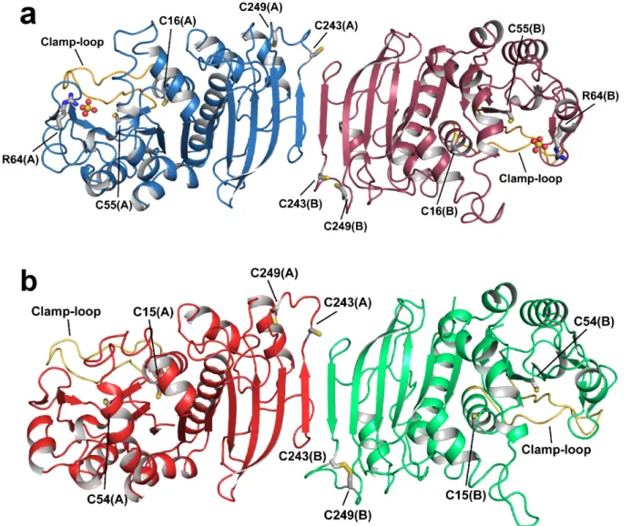
crystallographic structure of both enzymes has been solved, providing the first structural information on redox-sensitive phosphoribulokinase from plants. Both enzymes share a conserved homodimeric structure with 18 strands forming a β-sheet and the two catalytic sites positioned at the edges of the dimer. New hypothesis on the structural base of redox regulation have been inferred. Further characterization has been carried out analyzing enzymes structure in solution by size exclusion chromatography coupled with small angle X-ray scattering and redox properties, like midpoint redox potential and thioredoxin reactivation.
A second in vitro study has been performed on the α-amylase 3 (AtAMY3) from Arabidopsis
thaliana. This enzyme is not necessary for normal starch degradation in mesophyll cells but is
important for starch degradation under osmotic stress (Thalmann et al., 2016) and guard cell functionality (Horrer et al., 2016). AtAMY3 has been found target of glutathionylation in vitro by western blotting and the three glutathionylation sites have been identified by mass spectrometry. Assaying enzymatic activity after incubation with oxidized glutathione showed an inhibitory effect, afterwards the recovery of AtAMY3 functionality upon incubation with reduced glutathione, glutaredoxins and thioredoxin have been investigated. The analysis highlighted different reactivity with these three agents and the possibility of concomitant redox modification, i.e. glutathionylation and disulfide bond, occurring after the exposure to glutathione.
In the last two chapters the focus moved to the in vivo function of carbon metabolism enzymes. In particular in Chapter 4 Arabidopsis thaliana T-DNA lines have been identified and studied to understand which metabolic pathways are involved in carbon mobilization and osmotic stress
A2), SUS1 (Sucrose synthase 1), GWD2 (Glucan, Water Dikinase 2) emerged as interesting. On these lines carbohydrate and amino acids content, especially proline, has been monitored during stress treatment highlighting severe impairments in the accumulation of several sugars and proline. This reduction could affect the recover of osmotic balance and metabolic processes leading to incomplete or weaker stress response. Thus SPSA2, SUS1 and GWD2 resulted important for metabolic adaptation to stress, even if further analysis are needed to understand their specific role. In Chapter 5 the role of the three dikinases from Arabidopsis thaliana have been investigated in plant development, focusing on seed formation. Among these three, GWD1 is the most known and studied, despite its role out of mesophyll cells and tubers is not known. The other dikinases, PWD and GWD2, have been poorly studied and the in vivo function of GWD2 is still unknown. The analysis of T-DNA lines carrying insertions in the corresponding dikinase genes showed same developmental defects and a new role in seed development can be suggested for GWD2 and PWD. On the whole, several sides and features of carbon metabolism in plants still remain to be understood, even for a 70-year topic like the Calvin-Benson cycle, making stimulating the study of a process of primary importance for the life on our planet.
Table of contents
Thesis summary 1
1. Introduction 3
1.1 Carbon and life 3
1.2 Photosynthesis and carbon 3
1.2.1 Photosynthetic electron transport 4
1.2.2 The Calvin-Benson cycle 6
1.2.3 Adaptations to improve CO2 fixation 8
1.2.4 Other pathways for carbon fixation 9
1.3 The metabolism of starch in leaves 11
1.3.1 Diurnal starch biosynthesis 12
1.3.2 Starch degradation at night 14
1.3.3 The cytosolic metabolism of starch-released sugars 17
1.4 Sucrose metabolism 18
1.4.1 Sucrose biosynthesis in photosynthetic tissues 18
1.4.2 Phloem loading and sucrose transport 18
1.4.3 Phloem unloading and sucrose degradation 20
1.5 Carbon fluxes under abiotic stress 21
1.6 Regulation of carbon metabolism 23
1.6.1 The redox regulation 24
1.6.1.1 The thioredoxin system and NADPH-thioredoxin reductase in chloroplast 25
1.6.1.2 Redox modification by glutathione and oxygen and nitrogen reactive species 26
1.6.2 The circadian clock 27
1.6.3 Carbon metabolism controls growth by sugar sensing 29
1.7 Regulation of Calvin-Benson cycle 32
1.8 Regulation of leaf starch metabolism 34
1.8.1 Regulation at protein level 34
1.8.2 Time-dependent regulation of starch metabolism 35
1.9 Regulation of sucrose metabolism and phloem transport 36
1.9.1 Sucrose-phosphate synthases are regulated at different levels 37
1.9.2 Control of sucrose transporters 37
1.9.3 Regulation of sucrose degradation 38
2. Photosynthetic CO2 assimilation:
the last gap in the structural proteome is closed 41
2.1 Abstract 41
2.2 Results and discussion 42
2.3 Materials and methods 51
2.3.1 Protein expression and purification 51
2.3.2 Activity assay and determination of pH- and temperature-dependence 51
2.3.4 Thioredoxin specificity and redox titration 52
2.3.4 Determination of the pKa values of the catalytic cysteines 52
2.3.5 Crystallization and data collection 52
2.3.6 Structure solution and refinement 53
2.3.7 Small angle X-ray scattering data collection 54
2.3.8 Small angle X-ray scattering data analysis 54
2.3.9 Modelling from SAXS data 54
2.3.10 Data availability 55
Addendum 56
3. Glutathionylation of Arabidopsis thaliana AMY3 and regulation by glutaredoxins and thioredoxins in vitro 73
3.1 Brief introduction 73
3.2 Materials and methods 76
3.2.1 Expression and purification of recombinant AMY3 from Arabidopsis thaliana 76
3.2.2 Biotinylated GSSG assay 77
3.2.3 MALDI-ToF Mass Spectrometry 77
3.2.4 AtAMY3 activity assay and oxidative treatments 78
3.2.5 Determination of cysteines pKa 78
3.2.6 Reactivation of glutathionylated AtAMY3 by GSH, GRX and TRX 78
3.3 Results 80
3.3.1 AtAMY3 is target of glutathionylation in vitro 80
3.3.2 Determination of catalytic cysteine pKa 81
3.3.3 Glutathione protects AtAMY3 from irreversible oxidation and inhibition 82
3.3.4 GSH reactivates glutathionylated AtAMY3 and redoxins speed up the process 84
3.4 Discussion and concluding remarks 87
4. Enzymatic components involved in sugar and proline accumulation under osmotic stress in Arabidopsis thaliana 90
4.1 Brief introduction 90
4.2 Materials and methods 93
4.2.1 Plant material and growth conditions 93
4.2.2 Selection of homozygous lines 93
4.2.3 Water content measures 94
4.2.4 Lipid peroxidation measurements 94
4.2.5 Leaf starch quantification 95
4.2.6 Proline and amino acids quantifications 96
4.2.7 Soluble sugar quantification 96
4.2.8 Reducing sugar quantification 97
4.2.9 Glucose and fructose assay 97
4.2.10 Quantification of carbohydrates in the cell wall 98
4.2.11 Quantitative Real Time PCR analysis 99
4.3 Results 101
4.3.1 Water content under osmotic stress 101
4.3.2 Generation of oxidative stress under osmotic stress 101
4.3.3 Proline accumulation under stress and amino acids levels 103
4.3.4 Transitory starch at the end of the day 104
4.3.5 Changing in soluble sugar levels 105
4.3.6 Carbohydrate content in the cell wall 106
4.3.7 Gene expression profile of GWD2, SUS1, SPSA2 and P5CS1 107
4.4 Discussion and concluding remarks 110
4.5 Addendum 113
4.5.2 Selection of homozygous T-DNA lines 113
4.5.3 Oxidative stress levels in T-DNA lines 113
5. The analysis of the different functions of starch-phosphorylating enzymes during the development of Arabidopsis thaliana plants discloses an unexpected role for the cytosolic isoform GWD2 115
5.2 Introduction 116
5.3 Materials and methods 118
5.3.1 Genotype analysis 118
5.3.2 Plant growth conditions 119
5.3.3 Phenotypic characterization 119
5.3.4 Scanning electron microscopy 120
5.3.5 Quantification of starch, protein and lipids in seeds 120
5.4 Results 121
5.4.1 Isolation of homozygous T-DNA lines and leaf starch quantification 121
5.4.2 Seeds shape and composition 121
5.4.3 Seed morphology and permeability 123
5.4.4 Seed viability and primary root growth 125
5.4.5 Transition from vegetative to reproductive growth and plant fitness 125
5.4.6 Rescue by light 126
5.5 Discussion 127
6. Ringraziamenti 131
Thesis summary
During the years of my PhD, plant carbon metabolism has been approached with two different focuses: study the biochemistry of enzymes in vitro and understanding the physiological role of enzymes in vivo. In both cases physiological and stress conditions were considered.
The Chapter 1 is a comprehensive introduction on primary carbon metabolism, passing from the collection of light energy and carbon fixation to starch and sucrose metabolism. In addition, two insights are provided on carbon metabolism under abiotic stress and regulation of carbon metabolism, i.e. Calvin-Benson cycle, starch metabolism, sucrose metabolism.
Starting with the entry of carbon in the living world, an enzyme of Calvin-Benson cycle has been characterized. In Chapter 2, the crystallographic structures and regulatory properties of phosphoribulokinase from the green algae Chlamydomonas reinhadtii and the plant Arabidopsis
thaliana have been studied. The structure have been solved in collaboration with the Dr. Simona
Fermani of Department of Chemistry "G. Ciamician" (University of Bologna) and Dr. Stephane D. Lemaire (CNRS - Paris).
Then in Chapter 3 the regulation of the α-amylase 3 (AtAMY3) of Arabidopsis thaliana by glutathione has been characterized. AtAMY3 is involved in starch mobilization for guard cells movement and in response to osmotic stress. The chapter focuses on glutathionylation, a post-translational redox modifications which can occur when oxidative stress raises inside cellular compartments.
The last two chapters illustrate two studies on Arabidopsis thaliana plants. In Chapter 5 the adaptation of carbon metabolism to osmotic stress has been investigated using T-DNA lines for enzymes involved in carbon metabolism which are not essential for normal plant physiology. In the last chapter, Chapter 6, the focus went back to physiological conditions and three T-DNA lines for the three Arabidopsis dikinases have been studied. One of these enzymes, GWD1, has been examined several times and in different species starting from the '90s. However, the other two, PWD and GWD2, remained poorly studied. In this analysis, the development of plants carrying T-DNA insertions has been monitored and different parameters on seeds and plants have been evaluated to understand the function of GWD1, PWD and GWD2 along the plant life cycle.
1. Introduction
1.1 Carbon and life
Life is based on organic molecules, made of carbon backbones bound to different functional groups. Forming different bonds carbon can produce a wide range of molecules with distinct size and features, like nucleic acids, proteins and carbohydrates. Furthermore, the possibility to react with different elements (hydrogen, oxygen, nitrogen, phosphorus and sulphur) and these reactions can occur in water, making carbon-based molecules suitable for the Earth.
Different strategies evolved among living beings to satisfy the carbon demand. In autotrophy, the organic compounds are produced using inorganic sources from the environment. Reducing power to fix carbon is obtained from the energy of light (photoautotrophs) or oxidation of inorganic compounds (chemoautotrophs). In heterotrophy, carbon is directly obtained from organic sources. For these reasons, the first organisms are called producers and the latter consumers. The photosynthesis fix 1/6 of the atmospheric carbon (Ciais et al., 1997), allowing a massive entry of carbon in the living world. Among the photosynthetic producers land plants, together with algae and cyanobacteria, are the main participants in the entry of carbon into the biosphere.
1.2 Photosynthesis and carbon
The oxygenic photosynthesis evolved on the Earth 2.4 billion years ago (Hohmann-Marriott and Blankenship, 2011), introducing a novel pathway to obtain reducing power and energy and completely modifying the atmosphere of the planet. These great changing allowed the evolution of the life as we know it (Payne et al., 2009). Nowadays, the oxygenic photosynthetic organisms are the most abundant producers and as such extremely important for human beings. Oxygenic photosynthesis takes advantage of the light energy to split water in oxygen, protons and electrons, leading to the production of NADPH and ATP (Figure 1.1). Thus, the chemical energy (ATP) and the reducing power (NADPH) are consumed by the Calvin-Benson cycle in order to produce sugars from CO2 (Figure 1.2).
Depending on the organism, the photosynthetic process has different localization. In cyanobacteria, the carbon fixation is localized in specialized structures called carboxysomes and the photosynthetic
electron transport (PET) occurs in specialized portions of membranes called thylakoids. In algae and land plants, photosythesis is conducted in chloroplasts, specialized organelle originated by cyanobacteria through endosymbiosis. Chloroplasts are characterized by two membranes that delimit an intermembrane space. Inside the inner membrane, the stroma contains the enzymes for the Calvin-Benson cycle and the thylakoids.
1.2.1 Photosynthetic electron transport
The PET makes available the energy of light as chemical energy for the biochemical reactions that are necessary for carbon fixation. The electrons released by the photo-oxidation of water go through a series of protein complexes generating a proton gradient and ending up with the NADP+ reduction
(Figure 1.1). Two different types of protein complexes are responsible for the harvest of light, both need pigments for their functioning. The smaller complexes are the Light-Harvesting Complexes (LHCs) containing chlorophylls and carotenoids. Whereas, the photosystem I and II (PSI and PSII) are the larger complexes. Both LHCs and PSs interact each other and localize in the thylakoid membranes. The pigments in LHCs collect the electromagnetic radiation of light, resulting in excited electrons. The excitation could be transferred between the nearest pigments by dipole-dipole interaction until it is caught by the PSs. If the energy of the excitation move from a pigment to another, it is called Förster resonance energy transfer, otherwise according to the Redfield theory, initially the excitation can oscillate between the donor and the acceptor pigments and then result in the energy transfer (Renger, 2010).
PSs consist of an antenna system and a reaction centre, the antenna system collects the excitation energy and drives it to the reaction centre. In the reaction centre a special pair of chlorophylls is present, which catalyzes the photochemical reaction, consuming the energy of light to release an electron. The special pair are named P700 for PSI and P680 for PSII. The first photochemical reaction occurs inside the PSII, which exists in dimeric and monomeric form in vivo (Takahashi et al., 2009) containing chlorophylls, pheophytins and carotenoids (Guskov et al. 2009). The main structural domains are the transmembrane reaction centre, where is located the special pair P680, the two proteins CP43 and CP47 forming the inner light-harvesting antenna, and a set of proteins associated to the reaction centre in the luminal part of the thylakoids. The Oxygen-Evolving Complex (OEC) is connected to the reaction centre and performs the essential reaction of photo-oxidation of water, releasing O2 and producing 4 electrons and 4 H+ ions.
The excitation energy is directed through the inner antenna to the P680 which release an electron (P680* → P680+ + e-; Bricker and Frankel, 2002). The electron is transferred through cofactors
special pair (P680+) is neutralized thanks to an electron provided by water. An additional energy
transfer to the P680 completes the reduction of PQ to plastoquinol, picking up 2 protons from the stroma (PQH2 ; Satoh, 2008).
PQH2 interacts with another transmembrane protein complex, the Cytochrome b6f (Cyt b6f). The Cyt
b6f has 3 binding sites, one for PQH2, another PQ and the third for plastocyanin (PC). The PQH2 is
oxidized by the Cyt b6f, one electron reduces PC and the other an oxidized PQ, after two oxidation
two PC are completely reduced, one plastoquinone is reduced and 4 H+ are released in the thylakoid
lumen. The release of protons contributes to the formation of a proton gradient across the thylakoid membranes. The reduced PC leaves the Cyt b6f binding site and enter in the binding site of the PSI.
PC is a 10 kDa soluble protein containing a copper ion and belonging to the cupredoxin family (Sigfridsson, 1998), it acts as electron carrier between the Cyt b6f and the PSI in the lumen.
The PSI exists as monomeric complex in plants, diatoms, green and red algae (Gardian et al., 2007; Veith and Buchel, 2007; Germano et al., 2002; Amunts et al., 2007). On one side of the PSI the LHCI interacts with the core, in which more than 100 chlorophylls and 15 carotenoids collect the light energy and transfer it to the reaction centre (Amunts et al., 2007). The PC binds PSI on the luminal side whereas binding site for the terminal acceptor, the Ferredoxin (Fd), is localized on the stromal side of the complex (Busch and Hippler, 2011). The electron emitted by the P700 passes through a series of pigments and iron-sulfur clusters and reduces the ferredoxin (Fd). The positive charge on P700+ is neutralized by reduced PC. In turn, the Fd is oxidized by FNR (Ferredoxin,
NADP+ Reductase), a FAD-enzyme conserved in all the photosynthetic organisms, and NADP+ is
reduced to NADPH.
Figure 1.1 Photosynthetic electron transport chain. From left to right: PSII, photosystem II; PQ, oxidized plastoquinone; PQH2, reduced plastoquinone; Cyt b6f, cytochrome b6f; PC, plastocyanin; PSI, photosystem ; Fd, ferredoxin; FNR, ferredoxin-NADP+ reductase.
The pathway above described is known as Linear Electron Flow (LEF), because the electrons are transferred from the PSII straight to the PSI leading to NADPH production. However, another pathway occurs inside the electron chain. Indeed the PSI electron flow can be directed to the Cyt b6f
thanks to the PGR5-PGRL1 complex resulting in a Cyclic Electron Flow (CEF; Shikanai and Yamamoto, 2017). The CEF specifically contributes to the proton gradient formation and does not lead to NADPH production. Then the gradient is consumed by the F-ATPase on the thylakoid membrane to produce ATP in the stroma. Therefore, the regulation of LEF and CEF contributes to the balance between the production of energy, as ATP, and reducing power, as NADPH, in function of the requirement of the carbon fixation.
1.2.2 The Calvin-Benson cycle
The Calvin-Benson cycle represents the main entrance of inorganic carbon into organic world and is the only pathway occurs in photosynthetic eukaryotes. The reductive pentose phosphate pathway comprises 11 enzymes catalysing 13 reactions that lead to the fixation and reduction of carbon dioxide into triose phosphates (Figure 1.2).
The cycle is classically divided in 3 stages, carboxylation, reduction and regeneration. During these stages the energy and the reducing power provided by PET are consumed to obtain organic carbon. The carboxylation is catalyzed by RuBisCO, acting as carboxylase. RuBisCO is found in eukaryotes and prokaryotes, it is a oligomeric enzymes consisting of 2 types of subunit, the large subunit (50-55 kDa) and small subunit (12-18 kDa), organized in a hexadecameric structure with 8 large and 8 small subunits (Andersson and Backlund, 2008). During the carboxylation, the CO2 is
added to C-2 of a 5-carbon molecule, the ribulose-1,5-bisphosphate (RuBP), which spontaneously splits in two molecules of 3-phosphoglycerate (3-PGA).
Entering in the reduction stage, the two molecules of PGA are phosphorylated by the 3-phosphoglycerate kinase (PGK) consuming ATP producing 1,3-bis3-phosphoglycerate (1,3-BPGA). Then NADPH is oxidized by the photosynthetic isoform of glyceraldehyde-3-phosphate dehydrogenase to reduce 1,3-BPGA yielding glyceraldehyde-3-phosphate (G3P). The carbon fixed by the photosynthesis is mainly provided to the cell metabolism as triose phosphates, G3P or its isomer dihydroxy-acetone-3-phosphate (DHAP). During the day, the triose-phosphate/phosphate translocator (TPT) exports triose phosphates in exchange with inorganic phosphates. TPT is the most abundant integral protein in the chloroplast inner envelope (Flügge et al., 1991). Once in the cytosol, triose phosphates enter in the central metabolism and take part of gluconeogenesis and glycolysis.
Figure 1.2 The Calvin-Benson cycle. Enzymes: RuBisCO, ribulose 1,5-bisphosphate carboxylase/oxygenase; PGK, phosphoglycerate kinase; GAPDH, glyceraldehyde 3-phosphate dehydrogenase; TPI, triose phosphate isomerase; ALD, aldolase; FBPase, fructose bisphosphate phosphatase; TK, transketolase; RPE, ribose phosphate epimerase; SBPase, sedoeptulose bisphosphate phosphatase; PRK, phosphoribulokinase. Intermediates: RuBP, ribulose bisphosphate; 3PGA, 3-phosphoglycerate; 1,3-BPGA, bisphosphoglycerate; G3P, glyceraldehyde 3-phosphate; DHAP, dihydroxyacetone phosphate; FBP, fructose bisphosphate; F6P, fructose 6-phosphate; E4P, erythrose 4-phosphate; SBP, sedoeptulose bisphosphate; S7P, sedoeptulose 7-phosphate; Xu5P, xylulose 5-phosphate; R5P, ribose 5-phosphate; ribulose 5-phosphate. Figure inspired by Michelet et al. (2013).
The regeneration stage is the longest of the cycle and provides the initial substrate, RuBP, allowing the cycle progression. Since the regeneration starts with 3-C molecules and ends with 5-C ones, the aim of the last stage is to rearrange carbon skeletons, thus 5 triose are necessary to yield 3 RuBP. The triose phosphate isomerase interconverts G3P in dihydroxy-acetone-3-phosphate (DHAP), which is condensed with another G3P by the aldolase resulting in a molecule of fructose-1,6-bisphosphate (FBP). The fructose-1,6-bisphostatase hydrolyzes the phosphate group on the C1 of FBP, then fructose-6-phosphate (F6P) is substrate of the transketolase (TK). The TK reaction transfers a 2-C unit from F6P to a third G3P molecule producing xylulose-5-phosphate (X5P) and erythrose-4-phosphate (E4P). Aldolase catalyzes a second reaction in the regeneration stage, E4P is condensed to a fourth G3P and sedoheptulose-1,7-bisphosphate (SBP) is produced. At this point another
phosphatase comes in to play and hydrolizes the phosphate group on the C1 of SBP, the sedoheptulose-7-phosphate produced is the second substrate of the TK. A 2-C unit is moved to a fifth G3P, in this way the TK produces ribose-5-phosphate (R5P) and another X5P. R5P is isomerised by ribose-5-phosphate isomerase (RPI), whereas the two X5P obtained so far are epimerized by phosphate epimerase (RPE), both reactions release ribulose-5-phosphate (Ru5P). The phosphoribulokinase (PRK) catalyzes the last reaction of the regeneration stage consuming ATP to produce RuBP from Ru5P.
1.2.3 Adaptations to improve CO2 fixation
RuBisCO has double activity, as carboxylase participates of the carbon fixation through the Calvin-Benson cycle, but a side-reaction occurs in which the enzyme acts as oxygenase. The oxygenase activity leads to the production of 2-phosphoglycolate (2PG) and (3PG) and is facilitated by a variety of condition, e.g. high temperature (Jordan and Ogren, 1984). 2PG has not metabolic function and the photosynthetic organisms evolved a specific pathway to scavenge this compound and prevent its accumulation, the photorespiration. The photorespiration, so-named because consumes O2 and releases carbon dioxide (and NH3), probably represents the second most important
metabolic flux of the biosphere, after the photosynthesis itself (Bauwe et al., 2010). The photorespiratory reactions involve about 10 enzymes located in chloroplasts, peroxisomes, mithocondria and cytoplasm.
In order to limit the oxygenation of RuBP, some organisms evolved CO2 concentrating mechanisms.
In this way, the carboxylation reaction is promoted due to the accumulation of its substrate. Three different strategies of CO2 concentration were adopted to minimize photorespiration: 1) C4 plants,
adapted to hot environments; 2) Crassulacean Acid Metabolism (CAM), adapted to dry environments, and 3) CO2 pumps, a strategy adopted by aquatic photosynthetic organisms.
In hot environments, the concentration of CO2 lowers more than the O2 concentration (Sage, 2013),
promoting the oxygenation reaction of RuBisCO. To avoid the side-reaction, C4 plants close in RuBisCO from the surrounding environment and concentrate CO2. Indeed the peculiarity of C4
plants is the spatial separation between the site of CO2 intake and the site of carbon fixation. In
mesophyll cells the carbon dioxide (as bicarbonate ion HCO3-) is bound to phosphoenolpyruvate
(PEP) by the PEP-Carboxylase (PEPC) producing oxalacetate (OAA). These cells are more exposed to the atmosphere and so exposed to higher oxygen concentration. OAA is then converted to malate or aspartate, by malate dehydrogenase or aspartate aminotransferase respectively, and transported through the mesophyll to specialized cells in the inner part of the leaf, the bundle sheath cells. These
cells evolved a thicker and suberised cell wall, which becomes almost waterproof and all the exchanges are controlled by diffusion through the plasmodesmata (von Caemmerer and Furbank, 2003). In the bundle sheath aspartate or malate are converted to pyruvate releasing CO2, that is fixed
by the Calvin-Benson cycles while pyruvate returns to the mesophyll cells where is phosphorylated, producing PEP. By means of the C4 strategy plants achieve a 90% reduction of photorespiration (Sage, 2013).
CAM plants adapted to hotter environments than C4 plants. In fact their metabolism takes the name from plants adapted to desert, where the carbon fixation during the day would lead to massive water loss. For these reason CAM plants evolved a mechanism based on PEP caboxylase concentration of CO2 but implemented with a temporal separation between the intake of carbon and its fixation. The
stomata, which allow gas exchange between plant and environment, are open during the night, when the temperatures are lower. Stomata opening allow CO2 diffusion in the photosynthetic tissue,
where is bound to PEP by PEPC and accumulate in the vacuole as malate. During the day, the stomata are closed preventing water loss and malate is decarboxylated releasing CO2 that is fixed
consuming NADPH and ATP produced by the PET. Thanks to this temporal separation CAM plants lose 5-10 times less water than C4 and C3 plants (Taiz and Zeiger, 2010).
1.2.4 Other pathways for carbon fixation
Although all the economically relevant plant species fix CO2 through the Calvin-Benson cycle,
other five carbon fixation pathways are known to occur in different organisms (Ljungdahl, 1986; Buchanan and Arnon, 1990; Berg et al., 2007; Huber et al., 2008; Zarzycki et al., 2009).
Hydrogen (Hedderich and Forzi, 2005; Heim et al., 1998), sulphur, hydrogen sulphide (Friedrich et al., 2005) or sodium gradient are the most common sources of reducing power, while ATP is provided by a variety energetic reactions (Heise et al., 1989; Fuchs, 2011). The five cycles of CO2
fixation take advantage of acetyl-CoA as molecule for carbon uptake, and then utilize it in different ways with different carbon yields and energy costs (Figure 1.3; Fuchs, 2011).
The reductive Coenzyme A pathway (also named Wood-Ljungdahl Pathway, WLP) is present in acetogenic bacteria and produces acetyl-CoA from CO2 (Schuchmann and Müller, 2016). CO2 is
converted into methyl group and carbon monoxide, which are condensed to the acetyl group (Ragsdale and Pierce, 2008). Acetogens do not perform just WLP to sustain their growth because it is uncompetitive if compared to other metabolisms so it is probably integrated with other pathways (Schuchmann and Müller, 2016). Acetogenic bacteria are found in different kind of environments like soils, mammalian and termites gastrointestinal tracts, and high salt and temperature
environments (Simankova et al., 2000; Gössner et al., 1999; Küssel et al., 2001; Kamlage et al., 1997; Graber and Breznak, 2004; Küssel et al., 2000).
Green phototrophic sulfur bacteria are obligate anaerobe and fix CO2 through the reductive citric
acid cycle (RCA) generating acetyl-CoA. Irreversible steps of the Krebs cycle are catalyzed by enzymes with different activity, working in the reductive direction. For example, fumarate reductase replaces succinate dehydrogenase and 2-oxoglutarate dehydrogenase by 2-oxoglutarate synthase (Evans et al, 1966; Aoshima et al., 2007).
Anaerobic and microaerobic Archaea members of Thermoproteales and Desulfurococcales fix carbon via dicarboxylate/4-hydroxybutyrate cycle. The reactions are divided in two parts, the first utilize acetyl-CoA, CO2 and bicarbonate to produce succinyl-CoA, which is converted to
4-hydroxybutyrate in the second part, then yielding two acetyl-CoA molecules (Ramos-Vera et al., 2009; Ramos-Vera et al., 2011).
In the Archaea order Sulfolobales, isolated in aerobic and microaerobic conditions, the 3-hydroxypropionate/4-hydroxybutyrate cycle converts two molecules of bicarbonate to acetyl-CoA. First the bicarbonate ions are fixed to 3-hydroxypropionate, which is processed leading to succinyl-CoA, further processing leads to acetyl-CoA release (Auernik et al., 2008; Ramos-Vera et al., 2011). The last acetyl-CoA utilizing cycle is the 3-hydroxypropionate bi-cycle (Herter et al., 2002; Zarzycki et al., 2009). In the first branch of the cycle glyoxylate is produced from acetyl-CoA and bicarbonate, then the second branch consumes propionyl-CoA produced by the first branch together with glyoxylate yielding pyruvate and again acetyl-CoA.
It is important to highlight how these five pathways are not the only ones possible, and other ways to fix carbon can still be discovered. An interesting example is the reductive hexulose-phosphate pathway (RHP) recently discovered in Methanospirillum hungatei by Kono et al. (2017). The RHP contains both archaeal RuBisCO and PRK allowing fixation of CO2 and regeneration of the initial
substrate. The newly fixed carbon is released as formaldehyde during the reactions in the regeneration phase, which are different from Calvin-Benson cycle. Final steps of the regeneration stage are catalyzed by 6-phospho-3-hexuloisomerase producing hexulose-6-phosphate and D-arabino-3-hexulose-6-phosphate synthase, producing Ru5P then phosphorylated by PRK.
Despite these procaryotic metabolisms do not largely contribute to the global fixation, those reactions are important for new biotechnological applications and in biological niches, in particular anaerobic niches where primary producers like plants and cyanobacteria cannot live (Friedrich et al 2005; Montoya et al., 2012).
Figure 1.3 Schematic representation of carbon fixation pathways in prokaryotes. Combination of paths from 1 to 5 results in all the fixation pathways. Combining path 1 and 5 results in the reductive citric acid cycle. The combination of path 1 and path 3 yields the dicarboxylate/4-hydroxybutyrate cycle. Path 2 and 3 result in the 3-hydroxypropionate/4-dicarboxylate/4-hydroxybutyrate cycle, and combining path 2 and 4 generates the first cycle of the 3-hydroxypropionate bi-cycle, which requires an additional cycle for glyoxylate assimilation. The path 6 represents the reductive acetyl-CoA pathway, which requires the path 1 for the assimilation of acetyl-acetyl-CoA. Oxygen-sensitive enzymes/reactions are represented by red arrows. The figure is adapted from Fuchs (2011).
1.3 The metabolism of starch in leaves
The CO2 fixation in oxygenic photosynthetic organism is a light-driven process, meaning that from
the dark onset until the next day the metabolism need to rest on a different carbon source. For this reason, freshly fixed carbon follows two different directions. Sugars can be immediately consumed to supply biosynthetic and energetic reactions, or they can be transiently immobilized in storage compounds that will be used during the night.
Depending on the plant species, a certain variability in transient carbon storage can be observed. The synthesis of starch in the chloroplast stroma during the day is a conserved feature in land plants (Weise et al., 2011). However, although species like Arabidopsis thaliana, sugar beet and soybean accumulate starch (Zeeman and ap Rees, 1999; Fondy et al., 1989; Upmeyer and Koller, 1973) other species, like Phaseolus vulgaris and spinach, store sucrose (Fondy et al., 1989; Stitt et al., 1983) and cereals and grasses can accumulate oligosaccharides such as fructans and raffinose (Pollock and Cairns, 1991; Bachmann and Keller , 1995; Zeeman et al., 2007).
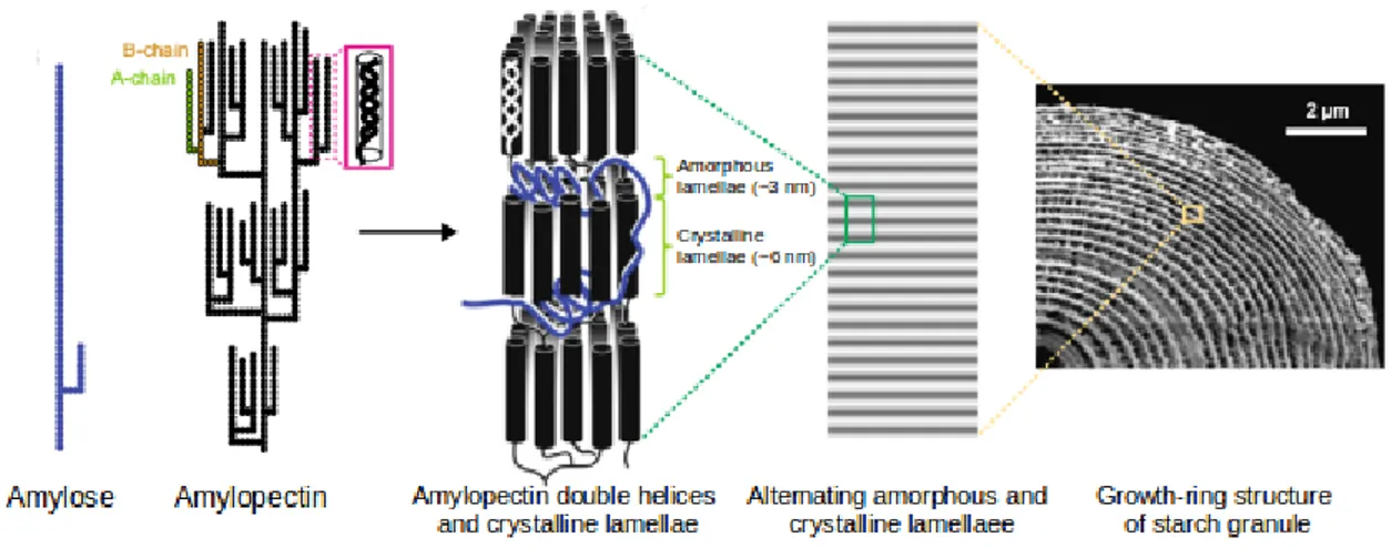
Generally, starch granules are composed by 70-90% of amylopectin and 10-30% of amylose (Tester et al. 2004), which are both polymers of α-D-glucose but with different structural features (Figure 1.4). Amylopectin is made up of α-1,4-linked glucose units forming long chains with regularly arranged α-1,6 branching points. While amylose consists of α-1,4 glucose chains without branching
points. The linear chains are packed in double helices, producing a crystalline insoluble structures defined lamellae. Lamellae alternate with branching points that are grouped in amorphous regions (Santelia and Zeeman, 2011).
Figure 1.4 The structure of starch granule. Figure adapted from Pfister and Zeeman, 2016.
Starch granules are stored both in leaves and storage organs, like tubers and roots, and in seeds, like in grain crops as maize, rice and wheat. However, starch is always stored in plastids: chloroplasts in the leaf, amyloplasts in storage organs. Starch granules have always the same structure but the turnover changes depending on the organ, while leaf starch is degraded at night, starch from storage organs is degraded in specific conditions (e.g. in response to gibberellin during germination).
1.3.1 Diurnal starch biosynthesis
Transitory starch is synthetized in the daylight from photoassimilates. Then during the night, leaf starch content is almost depleted to fuel plant metabolism. Starch biosynthesis starts with F6P released by the FBP during the Calvin-Benson cycle (Figure 1.5). F6P is converted in glucose-6-phosphate (G6P) by the hexose glucose-6-phosphate isomerase (PGI). The glucose-6-phosphate group on C6 is then transferred on C1 by phosphoglucomutase (pPGM).
pPGM is crucial for starch biosynthesis and ppgm knock-out (KO) mutants are described almost starchless (0.3% of wild-type level) and, if starch granules are found, they are tiny compared to wild-type ones (Caspar et al., 1985; Hanson and McHale, 1988; Streb et al., 2009). Many data agree considering pPGM as the main gate for the carbon flux into starch biosynthesis.
Another important enzyme in the biosynthetic pathway is the ADP-glucose pyrophosphorylase (AGPase), an heterotetramer made up of two large subunits, with regulatory function, and two small catalytic subunits, that catalyzes the formation of ADP-glucose from ATP and glucose-1-phosphate, releasing pyrophosphate (PPi) (Streb et al., 2009; Ragel et al., 2013). PPi is then hydrolyzed by
chloroplast pyrophosphatase promoting the synthesis of ADPglucose (ADPglc). Indeed the pyrophosphatase was shown to be essential for chloroplast central metabolism (George et al., 2010). ADPglc is the building block of starch thanks to Starch Synthases (SSs), which add glucose units to the non-reducing end of glucans. In land plants five isoforms of SS have been found (Ball and Morrel, 2003). The Granule-Bound Starch Synthase (GBSS) is the unique starch synthase responsible for amylose synthesis. Indeed gbss knock-out mutants from several species show a full-amylopectin starch (Pfister and Zeeman, 2016). GBSS is embedded in the granule during starch biosynthesis and is not present in soluble form in the stroma (Denyer et al., 2001).
Contrary to GBSS, the other four SS are soluble enzyme. Three of them seem to act mainly on an already existing granule, thus contributing to the growth and the architecture of the starch granule. SS1, SS2 and SS3 elongate amylopectin chains, in contrast SS4 appears to play a crucial role in starch granule initiation, producing glucan primers afterwards elongated by the others SSs. Indeed,
ss4 KO lines have one or no starch granule in contrast to 5-8 granules of the wild-type, and
accumulate almost 200-fold more ADPglc than wild-type plants (Roldán et al., 2007; Crumpton-Taylor et al., 2013; Ragel et al., 2013). Overall, these data reveal how the other SSs have extremely low activity without SS4.
In two recent papers (Seung et al., 2015; Seung et al., 2017) a new protein family named Protein Targeting to Starch (PTST) has been identified by phylogenetic analysis. PTSTs are characterized by coiled-coil domains and Carbohydrate Binding Module 48 (CBM48). First evidences suggest PTSTs support the activity of SSs enzymes, probably providing them substrates. The coilded-coils allow direct interactions with SSs while substrates are bound by PTSTs thanks to a CBM48 domain. In Arabidopsis thaliana 3 PTSTs have been found (Seung et al., 2015; Seung et al., 2017). PTST1 interacts with the unique GBSS, facilitating its amylose-elongating activity (Seung et al., 2015). KO lines for ptst2, and in part for ptst3, showed normal starch content. However, starch granules in
ptst2 are bigger and their number is lower (Seung et al., 2017), moreover these plants accumulate
ADPglc at high levels, even if less than ss4 KO. Considering all the available data, the initial model for PTST2 and 3 function hypothesize that PTST2 interacts with SS4 and provides malto-oligosaccharide subsequently elongated consuming ADPglc, in turn PTST3 binds PTST2 helping its activity. In Seung et al. (2017) was also hypothesised that PTST2 could improve the specificity of SS4 reaction recognising helical glucan chains, since SS4 seems to have no preference for malto-oligosaccharides (Cuesta-Seijo et al., 2016).
For proper amylopectin formation the activity of branching and debranching enzymes (BE and DBE, respectively) are required (Zeeman, Kossmann and Smith, 2010). DBEs hydrolyze
α-1,6-linkages and release linear chains. They can be divided in two classes: isoamylases (ISAs) and limit-dextrinase (LDA).
Figure 1.5 Transitory starch biosynthesis. ADPglc, ADP-glucose; AGPase, ADP-glucose Pyrophosphorylase; BE, Branching Enzyme; F6P, Fructose-6-phosphate; G1P, Glucose-1-phosphate; G3P, Glyceraldehyde-3-phosphate; G6P, Glucose-6-phosphate; GBSS, Granule-Bound Starch Synthase; ISA, Isoamylase (Debranching Enzyme); PGI, Phosphoglucoisomerase; pPGM, plastidial Phosphoglucomutase; PTST, Protein Targeting to Starch; SS, Starch Synthase.
In Arabidopsis ISA1 and ISA2 are involved in amylopectin elongation, while ISA3 and limit-dextrinase (LDA) are primarily involved in starch breakdown (Myers et al., 2000; Pfister and Zeeman, 2016). BEs cleave an α-1,4 glucan chain and transfer the cut to another chain, producing α-1,6 bond. BESs are grouped into two classes according to sequence similarities (Pfister and Zeeman, 2016). Branching activity is required for amylopectin synthesis, albeit BEs are not the sole determinants for the synthesis of crystallization-competent glucans (Pfister and Zeeman, 2016). Simultaneously to BE action, DBEs trim the excess of branching point, allowing SS to elongate just correctly localized branches. In fact, by abolishing the activity of DBEs, the formation of starch granules is impaired due to the impossibility of proper packing of amylopectin (Delatte et al., 2005; Wattebled et al., 2005).
The concurrent action of all these proteins contributes to the production of insoluble and highly ordered starch granules, two important features to improve compactness and allow accumulation of carbohydrates without affecting the osmotic balance.
1.3.2 Starch degradation at night
The breakdown of leaf starch has been mostly characterized in Arabidopsis thaliana, where the key reactions and enzymes have been discovered (Figure 1.6). Transitory starch degradation retains common features with the starch degradation in storage organs and non-photosynthetic tissues
(Zeeman et al., 2007). In fact the first step of the pathway is the phosphorylation of glucan chains on the granule surface. An enzyme called α-glucan, water dikinase (GWD1), initially found in potato and Arabidopsis (Nielsen et al., 1994; Yu et al., 2001), is responsible for this reaction. The phosphorylation occurs at low rate during day and at higher rate during the night (Ritte et al., 2004) and represents a crucial step for the subsequent glucan degradation (Edner et al., 2007). The reaction catalysed by GWD1 starts with the initial phosphorylation of a water molecule, resulting in a release of inorganic phosphate, then the β-phosphate of the ATP is transferred on the C6, preferentially, or on the C3 of the glucose units belonging to a glucan chain (Ritte et al., 2002). Another dikinase is involved downstream GWD1 and phosphorylates amylopectin exclusively in C3 position of C6-phosphorylated glucans, for this reason it has been called phosphoglucan, water dikinase (PWD; Kötting et al., 2005; Baunsgaard et al., 2005). The role of phosphorylation has still to be demonstrated, but a disrupting effect on amylopectin packing has been hypothesized (Yu et al., 2001). The disruption should be due to the hydrophilic properties of phosphates, as consequence glucans in starch become more soluble and accessible to degrading enzymes (Blennow and Engelsen, 2010).
Despite their essential role for glucan accessibility, phosphate groups inhibit the activity of glucan hydrolases involved in starch degradation (Fulton et al., 2008). For this reason, two phosphatases, Starch excess 4 (SEX4) and Like Sex Four 2 (LSF2) evolved to remove phosphate groups from C6 and C3, respectively (Kötting et al., 2009; Meekins et al., 2014; Santelia et al., 2011).
The β-amylases (BAMs) are responsible for most of the transitory starch degradation, they are exo-amylases that start degrading starch from the non-reducing end of linear glucans releasing β-maltose. In Arabidopsis thaliana BAM3 and BAM1 are the two main amylases. Their function is partially redundant, as highlighted by the double KO mutant bam1bam3 which showed a stronger starch excess (sex) phenotype compared to single KO mutants (Fulton et al., 2008). Apparently the roles of the two BAMs do not overlap under stress conditions, BAM1 takes part to osmotic stress response (Zanella et al., 2016) while BAM3 to cold stress response (Kaplan and Guy, 2004).
Since BAMs cannot hydrolize glucan chain at the branching point, debranching enzymes are required. ISA3 and LDA hydrolize the branching points (Wattebled et al., 2005; Delatte et al., 2006) allowing the BAMs to continue. LDA seems to have a minor role in the debranching activity, as highlighted by lda KO lines, which do not display a sex phenotype (Streb et al., 2012).
The pathway for degradation of storage starch, like that in cereal endosperm and tubers, is different and typically performed by α-amylases (Lloyd and Kossmann, 2015). To clarify if α-amylases could have a role also in transitory starch degradation AMY3, the unique plastidial Arabidopsis α-amylases, has been frequently studied. In Yu et al. (2005) emerged as Arabidopsis plants lacking
AMY3 have no alteration of leaf starch content and only minor variations in the sugar amounts. In any case, AMY3 could release phospho-oligosaccharides from leaf starch in vivo, demonstrating that AMY3 attacks starch granules but is not essential for starch degradation (Kotting et al., 2004). Horrer and colleagues (2016) demonstrated that BAM1 and AMY3 have a specialized role in stomata opening, catalyzing the release of carbon in guard cells linked to the opening of stomata pores. On the whole, these data suggest a marginal role of BAM1 and AMY3 in starch degradation at night.
The product of debranching enzymes and α-amylase lead to linear and soluble glucans. Soluble glucans are further degraded by BAMs and by glucan phosphorylase, producing G1P (Smith et al., 2005). Glucan phosphorylase plays a secondary role in the overall degradation, whereas BAMs are the principal enzymes for transitory starch degradation.
The maltose released by BAMs activity is then exported in the cytoplasm by means of MEX1, a maltose transporter localized in the inner membrane of the chloroplast envelope (Niittyla et al., 2004).
Figure 1.6 Transitory starch degradation. From left to right: GWD1, Glucan, Water Dikinase 1; PWD, Phosphoglucan, Water Dikinase; SEX4, Starch Excess 4 (phosphatase); LSF2, Like Sex Four 2 (phosphatase); ISA3, Isoamylase 3 (Debranching enzyme); AMY3, α-amylase 3; DPE1; Disproportionating Enzyme 1; BAM1, β-amylase 1; BAM3, β-amylase 3; MEX1, Maltose Excess 1 (transporter); DPE2, Disproportionating enzyme 2; PHS2, Glucan Phosphorylase 2.
Since BAMs are not active on glucans shorter than 4 glucose units, maltotriose is also produced as byproduct. Maltotriose is hydrolized by the chloroplast disproportionating enzymes (DPE1), an α-1,4-glucanotransferase (Critchley et al., 2001). DPE1 hydrolizes maltotriose into glucose and
maltose. While glucose is exported via glucose exporters, the maltose unit is transferred to another maltotriose molecule (Weber et al., 2000).
Variation on the turnover pathway are possible depending on the species, and homologous enzymes without a clear phenotype in Arabidopsis could have relevant role in other plants (Zeeman et al., 2007).
1.3.3 The cytosolic metabolism of starch-released sugars
Maltose and glucose are the end-products of the starch degradation in leaves, both are exported in the cytoplasm for further processing (respiration, biosynthetic reactions) and transport in other tissues. While the glucose can directly enter in the cell metabolism, maltose needs additional hydrolysis (Figure 1.6). In the cytoplasm two enzymes are responsible for the hydrolysis of maltose to glucose, exploiting cytosolic heteroglycans that act as a sugar buffer, i.e. as donor and acceptor of sugar units. The heteroglycans consist of glucose, galactose, arabinose, mannose, rhamnose and fucose with different abundance and kind of linkages (Lu and Sharkey, 2004; Fettke et al., 2005a). The maltose-processing enzyme is DPE2, a cytosolic isoform of the chloroplast disproportionating enzyme. DPE2 is an α-1,4-glucan transferase which transfers α-1,4-linked glucan from one glucan to another (Chia et al., 2004; Lu and Sharkey, 2004). In the current model maltose is splitted in two glucosyl residues, one is transferred to a heteroglycan while the other contributes to the glucose pool (Fettke et al., 2006). Then, the phosphorylase PHS2 (or PHO2) uses orthophosphate as acceptor molecule of a glucosyl residue from the heteroglycan, releasing glucose-1-phosphate (G1P). The PHS2 reaction is reversible, so it is possible that depending on metabolic conditions of the cell PHS2 catalyses heteroglycan elongation (Fettke et al., 2005b). Glucose molecules are phosphorylated by hexokinase to glucose-6-phosphate (G6P) while G1P can be isomerized to G6P and vice versa by cytosolic phosphoglucomutase (PGM). Additionally, PGI catalyses the interconversion of G6P to F6P. As active sugars, all these hexose phosphates can take part of the cellular metabolism.
G1P could be also converted to UDP-glucose by UDP-glucose pyrophosphorylase (UGPase) (van Rensburg and Van den Ende, 2018), replenishing biosynthetic pathways, like cell wall biosynthesis, although the physiological role of UGPase is still unclear (Meng et al., 2009).
The fate of the hexose pool relies on the metabolic requirements of cell/tissue, typically hexoses are consumed by sucrose synthesis or channelled through the central metabolism via glycolysis, Krebs cycle for biosynthesis or for energetic demands.
1.4 Sucrose metabolism
In photosynthetic tissues, triose phosphates deriving from diurnal carbon fixation and hexoses deriving from starch degradation at night converge in sucrose biosynthesis. Sucrose is transported through the phloem from source tissues (old leaves, storage tissues) to sink tissues (roots, meristems, fruits, flowers and young leaves). In sinks sucrose is degraded fuelling cell metabolism and tissue development providing carbon for the respiratory pathway, biosynthetic reactions and regulating gene expression by itself or through its degradation products by means of sugars sensors and sugar-responsive elements (Li and Sheen, 2016; Koch, 1996; Sheen et al., 1999).
1.4.1 Sucrose biosynthesis in photosynthetic tissues
Leaves constitute the primary carbon source, inside leaves the coupled action of two cytosolic enzymes produce sucrose for sink tissues (Figure 1.7, upper part). Sucrose phosphate synthase (SPS) condenses UDP-glucose and F6P to sucrose-6-phosphate subsequently hydrolyzed to sucrose by the sucrose-phosphate phosphatase (SPP). Another enzyme category can yield sucrose, starting from UDP-glucose and fructose, the sucrose synthase (SUS). Nevertheless it is generally accepted that SPS is responsible for synthesis and SUS preferentially catalyzes the breakdown of sucrose. Indeed several tissues mainly express just one of the two enzymes, but in some cases co-expression has been observed (Huber and Huber, 1996). Co-expression of SPS and sucrose degrading enzymes could lead to sucrose futile cycle which can help in tuning carbon partitioning (Nguyen-Quoc and Foyer, 2000; Ruan, 2014).
Plant SPSs are classified in three evolutionary groups: A, B and C (Langenkämper et al., 2002). From a phylogenetic point of view, specific isoforms for each group are found in dicotyledon and monocotyledon plants (Lutfiyya et al., 2007). The major isoform in tobacco leaves is SPSC (Chen et al., 2005), as well as in Arabidopsis thaliana, in which also SPSA1 acquired a relevant role (Volkert et al., 2014). In Arabidopsis all SPS have overlapping roles and, depending on the isoform, they can completely or partially compensate the absence of the other. In any case, different spatial and temporal expression patterns have been observed (Bahaji et al., 2015).
1.4.2 Phloem loading and sucrose transport
The link between source and sink tissues is the phloem, a vascular tissue constituted by specialized living cells. In flowering plants, the functional unit of phloem are sieve elements and companions cells. The sieve elements are stacked one over the other and connected internally, they contain an
endoplasmic reticulum, rudimentary plastids and mitochondria but no cytoskeleton and nucleus. Being without nucleus they depend on companion cells for maintenance of cellular components. Companion cells are tightly linked to sieve elements through plasmodesmata, providing essential elements like proteins and contributing to the phloem loading. Inside the sieve elements flows the phloem sap, which is loaded by source tissues and delivers nutrients and signals to sink tissues.
Figure 1.7 Sucrose metabolism and transport. Sucrose is synthetized in source tissues (e.g. mesophyll cells) by concerted activity of sucrose phosphate synthase (SPS) and sucrose phosphate phosphatase (SPP). Then sucrose is exported into sieve tube elements (pale yellow) via apoplasmic loading thanks to coupled action of sucrose faciliators (SWEET) and sucrose/H+ symporter (SUT) or via symplasmic loading through plasmodesmata. Phloem sap flows delivering sucrose to sink cells, where sucrose uptake occurs thanks to plasmodesmata (symplasmic unloading) or transporters (SWEETs and SUTs). Sucrose degradation is then performed by Cytosolic Invertases (CINV), Cell Wall Invertases (CWINV) and Sucrose Synthases (SUS).
Sucrose produced in leaves is massively loaded in phloem following two possible mechanism: apoplastic and symplastic loading (Figure 1.7, upper part). In apoplastic loading sucrose is first exported in the apoplast and then is loaded into the phloem against the concentration gradient. It is generally found in herbaceous species and it is the most studied because of agricultural interest (Zhang and Turgeon, 2018). The symplastic loading occurs by diffusion thanks to plasmodesmata
directly linked to the sieve elements of the phloem. Symplastic loading is generally ascribed to trees like poplar (Zhang et al., 2014). The two strategies are not exclusive and the same plant can load phloem in different ways depending on cell and tissue type or developmental stage (Slewinski et al., 2013). In Arabidopsis and crops, like rice, phloem is loaded from the apoplast through SWEET uniporters (Sugar Will Eventually be Exported Transporters) and sucrose/H+ symporters (SUT
-Sucrose Transporter). The first exports sucrose in the apoplast (Chen et al., 2012; Eom et al., 2015), then the latter is localized on sieve elements membranes and loads sucrose together with a proton, consuming the energy of the proton gradient (Julius et al., 2017). SWEET uniporters were recently identified as a family of sugars transporters conserved in plants and animals (Chen et al., 2010), the clade 3 of SWEET family was demonstrated to be the key facilitator of sucrose efflux in the apoplast (Chen et al., 2012). SUTs are divided in 5 evolutionary clades (Kühn and Grof, 2010). SUT1 clade is specific of dicotyledons whereas SUT3 and 5 of monocotyledons, clades 2 and 4 are common of both groups of plants. Different affinities have been measured on these transporters, reflecting different plant- and tissue-related physiological role (Kühn and Grof, 2010). SUT1 has the highest affinity, SUT3 has intermediate values and together with SUT5 they localize in source tissues on plasma membrane of phloem elements. SUT2 showed the lowest affinity as well as SUT4, both localize on plasma membrane in sink tissues, but SUT4 is found in plastid and vacuole membrane inside the cell (Kühn and Grof, 2010). Apparently phloem loading from apoplast is an energy demanding process but transporters different from SUTs have been found, for example in legumes sucrose facilitators localize on sieve element membranes acting in pH- and energy-independent manner and suggesting variations on the classical model (Zhou et al., 2007).
1.4.3 Phloem unloading and sucrose degradation
Due to massive loading in source tissues, the concentration of solutes in sieve elements is higher enough to lower the osmotic potential of phloem in source tissues, in this way water moves into the sieve elements thanks to aquaporins passively driving the sap flow to sink organs, where the osmotic potential is higher as result of phloem unloading (Zhang and Turgeon, 2018). Two models illustrate the unloading process, the apoplasmic and the symplasmic unloading, and a combination of the two was also detected (Figure 1.7, lower part). The two strategies depend more on tissue type than plant type, in fact apoplasmic unloading occurs in stems and roots, and consists of transfer of sucrose from phloem to the apoplast and subsequent transfer into the sink cell. Usually a sucrose facilitator, like AtSWEET11 and 12 (Le Hir et al., 2015), permits sucrose efflux from sieve elements and an active transporter catalyzes the uptake of sucrose into the cell, for example SUT1 in Arabidopsis and SUC1 in Zea mays (Carpaneto et al., 2005; Sivitz et al., 2008). In the
symplasmic unloading the sucrose is directly delivered inside the sink cell through plasmodesmata, this latter case is found in meristems, tubers and root tip (Milne et al., 2018).
Once sucrose is exported, two enzymes are responsible for its degradation, invertases (INV) and sucrose synthases (SUS). INVs catalyze the hydrolysis of sucrose to fructose and glucose. INV are classified on the basis of their subcellular location. Cell wall INVs (CWINV) was demonstrated to be involved in the sucrose breakdown in sinks where the apoplasmic unloading takes place (Ruan, 2014). Apoplast degradation of sucrose leads to lower sucrose concentration, that drives the phloem unloading, and hexoses release. Cytosolic invertases (CINV) could be implicated in sucrose degradation during the symplasmic unloading, although SUSs reaction (see above, Paragraph 1.4.1) are more likely involved in cytosolic sucrose breakdown and are generally considered a marker of sink strength (Koch, 2004).
1.5 Carbon fluxes under abiotic stress
Importance of characterizing plant metabolism under abiotic stress is undoubted. Beyond its relevance for fundamental knowledge, understanding how plants set up their metabolism in adverse conditions is also crucial to select useful traits for crop resistance in challenging environments. Rearrangements in carbon metabolism are not equally studied for all stress conditions and so far most of the available information are from analysis on primary carbon metabolism under drought/osmotic stress. For this reason and for further interest in the thesis work, this paragraph will focus mainly on water deficiency and, when available, further details on other stress will be discussed.
Hummel and colleagues (2010) carried out an extensive survey on metabolites, enzymes and physiological traits in Arabidopsis thaliana under water deficiency. When Arabidopsis plants are exposed to moderate or severe drought (water potential equal to -0.6 MPa and -1.1 MPa respectively) they did not observe significant decrease of net photosynthesis or increase in respiration. Data on CO2 fit well with data on enzyme activities from main carbon pathways like
Calvin-Benson cycle, starch metabolism, sucrose metabolism and tricarboxilic acids cycle. Indeed, monitoring all enzymatic activities in optimal catalytic conditions, enzymes levels were globally stable or just slightly increased (Hummel et al., 2010; Bogeat-Triboulot et al., 2007; Pinhero et al., 2001). Underlining how metabolic response does not need massive changes but more definite and distinctive adaptations (Yu et al., 2003; Bogeat-Triboulot et al., 2007). At the same time enzymes activities can be modulated without affecting enzymes content, for example in Trifolium
subterraneum RuBisCO activity is decreased by drought, indicating some inhibitory effect induced
by the stress (Medrano et al., 1997).
Transitory starch, deriving from recently fixed carbon, is emerging in the last few years as carbon source for abiotic stress response (Thalmann and Santelia, 2017), in addition to its role in carbon metabolism in the night. Indeed, in several challenging conditions transitory starch degradation has been reported in green algae, mosses, cereals and woody plants (Villadsen et al., 2005; Pressel et al., 2006; Goyal, 2007; Damour et al., 2008; Hummel et al., 2010). However, it is should be noted that other studies reported increased starch accumulation under abiotic stress (Kaplan and Guy, 2004; Cuellar-Ortiz et al., 2008; Siaut et al., 2011; Wang et al., 2013). It is still unknown if the balance between starch synthesis and degradation is typical of each stress or it depends more on specific experimental conditions (Thalmann and Santelia, 2017). Despite all, several studies reported a positive correlations between stress tolerance and starch degradation (Nagao et al., 2005; Cuellar-Ortiz et al., 2008; Gonzalez-Cruz and Pastenes; 2012; Zanella et al., 2016; Thalmann et al., 2016). Yano et al. (2005) reported a concerted action of GWD1 and BAM3 in transitory starch mobilization in Arabidopsis plants exposed to cold stress.
Starch degradation pathway under stress does not necessarily follow the canonical pathway (see section 1.3.2). In fact under osmotic stress several experiments showed the importance of BAM1 and AMY3, which gene expression is driven by ABA signalling, typically activated under water stress (Zeller et al., 2009; Valerio et al., 2011; Zanella et al., 2016; Thalmann et al., 2016). Moreover a role for α-glucan phosphorylase PHS1 was suggested under drought and high salinity (Zeeman et al., 2004). These three enzymes are not essential for normal transitory starch degradation at night. It is noteworthy that selection of particular enzyme isoforms is not just a matter of gene expression and distinct biochemical properties should make BAM1 and AMY3 adapted to stress conditions. Indeed both the enzymes are activated by disulphide bond reduction in
vitro, suggesting increased activity in the day, and also pH optima are shifted to more alkaline
values, similar to stromal pH at light (Sparla et al., 2006; Seung et al., 2013).
When water levels are reduced Arabidopsis plants change the growth pattern of the rosette. The rosette expansion is distributed along both day and night, with almost 50% of expansion occurring at night. Under water deficiency the rosette expansion is 40% faster at day and inhibited at night (Hummel et al., 2010). In addition also leaf emergense-expansion rate is lower. In this way plants improve growth during the day when energy and carbon are available and store sources at night for the next day.
Although the rosette growth is decreased, the photosynthesis is not impaired thus a surplus of carbon is generated. Compounds with low molecular mass raise and are probably exported in roots,
since 30% more root mass is observed (Hummel et al., 2010). Raise of sugar content is observed also in maize kernels and seems to be of central importance for drought tolerance (Yang et al., 2018). Accumulation of compounds like sugars can help adjusting the osmotic balance in leaves (Xu et al., 2007; Hagemann and Pade, 2015), but also protect membranes and scavenge reactive species under cold, temperature and salt stress (Garg et al., 2002; Janska et al., 2010; Yuanyuan et al., 2009; Krasensky and Jonak, 2012). In fructan accumulating species, fructants provide freeze resistance thanks to their high solubility (Livingston et al., 2009; Pomerrenig et al., 2018). Sugars are also substrates for sugar alcohols synthesis, typically produced to enhance drought tolerance and scavenge ROS (Smirnoff and Cumber, 1989; Pomerrenig et al., 2018). The accumulation of sugars could be a consequence of several factors like reduced growth of photosynthetic tissues (Hummel et al., 2010), impaired catabolism in sinks and reduced sink strength (Quick et al., 1992) or reduced phloem sap flow due to compensatory water transfer from phloem to xylem (Sevanto et al., 2014). In response to drought Hummel et al. (2010) also describe accumulation of amino acids, fumarate, proline and sucrose, with more fumarate under severe drought. These metabolites are not simply accumulated and stored to recover the osmotic balance, they are also available to plant metabolism. In fact a clear decrease is observed at night for proline, malate and soluble sugars, probably consumed when other carbon sources are not available.
Since drought stress lead to water loss, Hummel et al. (2010) calculated the concentrating effect on osmolytes observed in Arabidopsis and reported that the 66% of the osmotic potential generated by the plant under stress is obtained thanks to the decrease in water content. Anyway, metabolic processes need physiological water levels to work properly and the plant synthesized osmolytes to further counteract the water loss. The osmolytes accumulation is responsible for the 33% of the osmotic potential generated under drought.
Considering the energetic balance of the whole plant, the carbon cost is estimated to be 1-3% of the daily photosynthesic yield (without considering the ATP consumed for biosynthetic reactions), so osmotic adjustment does not need high carbon cost or even lead to carbon depletion (Hummel et al., 2010).
1.6 Regulation of carbon metabolism
In absence of stress, plants manage their carbon to maximize growth (Rasse and Tocquin, 2006). In order to achieve this aim almost all the transitory starch fixed during the day is linearly degraded during the night (Smith, 2012). The nearly depletion of starch ensure the best exploitation of carbon
sources but excessive degradation speed would lead to carbon starvation at the end of the night. Environmental clues and circadian rhythms are complemented to control carbon mobilization and avoid starvation (Lu et al., 2005; Graf et al., 2010; Troein et al., 2009; Farre and Weise, 2012). Management of carbon sources takes place during all the day and is adjusted on the length of the photoperiod. For example, Arabidopsis plants grow preferentially during the light period, but 50% of growth is observed at night under 6 hours photoperiod (Sulpice et al., 2014). Not only the mobilization of carbon is modulated, in the case of starch the biosynthesis is more responsive in short daytime and suddenly starts at the beginning of the day, to ensure maximal exploitation of light energy, whereas when light is not limiting a lag in starch synthesis is observed (Sulpice et al., 2014). Several regulatory strategies are adopted to control carbon metabolism, regulation by redox modifications, circadian control, sugar-sensing and phosphorylation are the most known.
1.6.1 The redox regulation
The sensitivity of enzymes to the redox state of the cell is an important factor, especially in carbon fixation. The redox state is directly linked to the availability of reducing power in the plant and so to the energetic status of cell, tissue or organism. Redox regulation in proteins involves cysteine residues and its reaction with other thiols or reactive species of oxygen and nitrogen (ROS and RNS, respectively), only for RNS regulation other protein residues seem to be involved (Sevilla et al., 2015).
Protein cysteines form disulphide bonds, bind metal ligands, coordinate substrates, have catalytic functions and could be subjected to different post-translational modifications. The sensitivity to redox modifications lies on higher reactivity of specific cysteines, given by lower ionization constant (pKa). Indeed if compared to free cysteines (pKa = 8.3) reactive cysteines have more acidic
constant and propensity to unprotonated form. Several features in the microenvironment determine a lower pKa, like the presence of charged amino acids stabilizing unprotonated thiol, hydrogen
bonds or dipole effect, often generated by N-terminal position of the reactive cysteine residue in α-helix (Couturier et al., 2013).
For what concerns primary carbon metabolism, it is fundamental to couple the photosynthetic energy production to carbon fixation and storage, avoiding energy waste and detrimental reactions. The synchronization is performed by the thioredoxin (TRX) system, which represents the first form of redox regulation discovered (Geigenberger et al., 2017). Further importance is given by the ability redox regulatory systems to react under developmental and stress conditions allowing carbon metabolism to set up in function of internal or external stimuli.