UNIVERSITÀ DEGLI STUDI DI
ROMA
"TOR VERGATA"
FACOLTA' DI SCIENZE MATEMATICHE,
FISICHE E NATURALI
DOTTORATO DI RICERCA IN BIOLOGIA
CELLULARE E MOLECOLARE
XXI CICLO
Inhibition of Telomerase and Hypoxia-Inducible
Factor-1 in Human Glioblastoma Multiforme
Maria Patrizia Mongiardi
A.A. 2008/2009
Docente Guida/Tutor: Dott. Andrea Levi
Coordinatore: Prof. Gianni Cesareni
Table of Contents
ABSTRACT...1 RIASSUNTO ...3 INTRODUCTION ...5 Glioblastoma Multiforme (GBM)...6 Molecular markers ...7 Angiogenesis ...9 Telomerase...13 Telomeres ...13The end replication problem ...16
Telomerase...17
Regulation of telomerase activity ...21
Non canonical functions of telomerase...22
Telomerase and cancer ...23
Telomerase involvement in tumor angiogenesis ...26
Hypoxia-Inducible Factor-1 (HIF-1) ...28
Hypoxia and HIF-1 ...28
Downregulation of HIF-1 protein levels in normoxia ...30
Hypoxic induction of HIF-1 ...33
HIF-1 target genes ...34
Role of HIF in cancer disease ...38
The Warburg effect...39
AIM OF THE PROJECT ...43
Part 1: Telomerase inhibition...43
Part 2: HIF-1 inhibition ...44
MATERIALS and METHODS- Part 1 ...45
Cell cultures ...45
Plasmid ...45
RNA interference...46
Production of viral stocks and cell infections...46
Immunocytochemistry ...47
Semi quantitative RT PCR...48
Real Time RT-PCR ...48
Telomeric Repeat Amplification Protocol (TRAP) Assay. ...49
Growth Curve ...49
FACS analysis ...49
HUVEC and Glioblastoma Cells Xenografting in Immunodeficient Mice...50
Histology, Immunohistochemistry, and Fluorescence Microscopy ...50
Statistical Analyses ... 51
RESULTS- Part 1... 52
Preparation of engineered HUVEC cell populations ... 52
Immunohistochemical characterization of engineered HUVECs ... 56
Could hTERT expression give any proliferative advantage to the cell? ... 57
hTERT protects ECs from oxidative stress induced apoptosis ... 59
hTERT inhibition abolishes the angiogenic behavior of HUVECs in GBM xenografts ... 61
hTERT expression is not sufficient for angiogenic behavior of HUVECs in subcutaneous grafts ... 64
DISCUSSION Part 1 ... 65
MATERIALS and METHODS- Part 2 ... 68
Cell cultures and generation of stably transduced cell lines .... 68
Plasmids ... 68
RNA interference and viral infections ... 69
Real Time RT-PCR ... 70
Western Blotting ... 70
Cell proliferation ... 71
Measurement of cellular ATP ... 71
Determination of Glutathione Content... 72
Glioblastoma Cells Xenografting in Immunodeficient Mice ... 72
Histology, Immunohistochemistry, and Fluorescence Microscopy... 73
Statistical Analyses ... 74
RESULTS Part 2 ... 75
Production of GBM clones with reduced HIF-1a expression .. 75
HIF-1a suppression does not alter ATP concentration, redox status and cell proliferation ... 78
wtTB10 cells out compete siHIF-TB10 cells in mixed culture... 81
Xenografts of mixed population of wtTB10 and siHIF-TB10 cells grow faster than xenografts of either wtTB10 or siHIF-TB10 cells ... 83
DISCUSSION- Part 2... 88
REFERENCES... 92
Abbreviations
ALT: Alternative Lengthening of Telomeres ARNT: Aryl Hydrocarbon Nuclear Translocator bHLH: basic Helix-Loop-Helix
bHLH-PAS: basic-Helix-Loop-Helix-Per-ARNT-Sim CAIX: Carbonic Anidrase 9
DFX: Deferoxamine
DN-TERT: Dominant Negative TERT EPAS: Endothelial Per-ARNT-Sim protein GAPDH: Gliceraldeide 3-Fosfato Deidrogenasi GBM: Glioblastoma Multiforme
GLUT1: Glucose Transporter 1
TERT: Telomerase Reverse Transcriptase HIF-1: Hypoxia Inducible Factor 1 HRE: Hypoxia Response Element HUVEC: Human Vein Endothelial Cells
ODDD: Oxygen-Dependent Degradation Domain
PGK1: Phosphoglycerate Kinase 1 PHD: Prolyl Hydroxylase Domain pVHL: von Hippel-Lindau protein shRNA: short hairpin RNA TBP: TATA Binding Protein
TERT: Telomerase Reverse Trascriptase
TRAP: Telomere Repeat Amplification Protocol VEGF: Vascular Endothelial Growth Factor
ABSTRACT
Glioblastoma Multiforme (GBM) is the most common and the most aggressive glial tumor. It is composed by a heterogeneous tumor cell population, poorly differentiated. GBM (WHO grade IV) develop from low grade astrocytoma (WHO grade I or II) or anaplastic astrocytoma (WHO grade III), but more frequently they arise de novo.
GBM tumors are paradigmatic in their ability to induce neo-angiogenesis, a fundamental process in the growth of solid tumors. Of note, endothelial cells of GBM have the peculiarity to reactivate telomerase, the enzyme necessary to maintain telomere length. Telomerase reactivation is a characteristic feature of tumor endothelial cells, since it was not observed in endothelial cells proliferating in non neoplastic contexts.
One of the most important regulators of angiogenic processes is the transcriptional factor HIF-1, which orchestrates cellular response to hypoxia. Although HIF-1 is universally recognized as a key factor in inducing neo-angiogenesis as a response to reduced oxygen levels, the consequences of HIF-1 inhibition on the growth of solid tumors are not fully elucidated.
The aim of my PhD studies was to explore the consequences of targeting angiogenesis on the development of human GBM. To address this purpose, I selected two parallel strategies: inhibition of telomerase expression in tumor endothelial cells and inhibition of HIF-1 in glial tumor cells.
We developed a controlled in vivo assay of tumor angiogenesis in which primary human umbilical vascular endothelial cells (HUVECs) were subcutaneously grafted with or without human GBM cells in immunocompromised mice as Matrigel implants. We found that primary HUVECs did not survive in Matrigel implants, and that telomerase up regulation had little effect on HUVEC survival. In the presence of GBM cells, however, the grafted HUVECs not only survived in Matrigel implants but developed tubule structures that integrated with murine microvessels. Telomerase up regulation in HUVECs enhanced such effect. More importantly, inhibition of telomerase in
HUVECs completely abolished tubule formation and greatly reduced survival of these cells in the tumor xenografts.
In the second part of this study, we investigated the consequence of downregulating HIF-1 function in a human GBM cell line on cell proliferation in vitro and tumor growth in
vivo. RNA interference targeting the O2-regulated HIF-1α subunit efficiently reduced HIF-1α expression and transcriptional induction of HIF-1α responsive genes. In vitro proliferation rate of HIF-1 α - inhibited cells was not altered. Conversely long term co-cultures of wild type and HIF-1 α - inhibited GBM cells resulted in the overgrowth of the wild type cells. Subcutaneous grafting in nude mice of wild type and HIF-1 α - inhibited GBM cells lead to comparable tumor formation and growth. Surprisingly, co-grafting of HIF-1-positive and HIF1-negative GBM cells resulted in more aggressive tumors, both in terms of tumor appearance and tumor growth. This suggests that heterogeneity of the cellular populations in their ability to mount a response to hypoxia may promote tumor aggressiveness.
RIASSUNTO
Il glioblastoma multiforme (GBM), il più comune e aggressivo dei tumori gliali, è composto da una popolazione eterogenea di cellule tumorali astrocitarie scarsamente differenziate. Questi tumori possono svilupparsi dall’evoluzione maligna di un astrocitoma di più basso grado (grado WHO I o II) o da un astrocitoma anaplastico (grado WHO III), ma più frequentemente si manifestano de novo, senza alcuna evidenza di una neoplasia precedente.
Il GBM è un tumore paradigmatico nella capacità di indurre neo-angiogenesi, processo necessario per la crescita dei tumori solidi. Le cellule endoteliali del GBM hanno la peculiarità di riattivare la telomerasi, enzima deputato al mantenimento dei telomeri. Questa riattivazione della telomerasi da parte delle cellule endoteliali è una caratteristica esclusiva delle cellule endoteliali tumorali: l’attività telomerasica, infatti, non è stata mai osservata in cellule endoteliali che proliferano in contesti di neoangiogenesi non neoplastica.
Uno dei regolatori piu' importanti dei processi angiogenetici è il fattore di trascrizione HIF-1, che coordina la risposta cellulare all’ipossia. Sebbene HIF-1 sia universalmente riconosciuto come un fattore chiave nell’induzione della neo-angiogenesi come risposta all’ipossia, le conseguenze di una sua inibizione nello sviluppo dei tumori solidi non sono ancora del tutto chiare.
Lo scopo dei miei studi di dottorato è stato quello di esplorare le conseguenze dell’inibizione dell’angiogenesi sullo sviluppo del GBM umano. A tale scopo, ho deciso di seguire due strategie parallele: l’inibizione della telomerasi nelle cellule endoteliali del tumore e l’inibizione di HIF-1 nelle cellule tumorali gliali.
Tramite un modello di angiogenesi neoplastica in vivo, messo a punto nel nostro laboratorio, siamo stati in grado di dimostrare che le cellule endoteliali umane primarie HUVEC,
indipendentemente dall’espressione di attività telomerasica, non crescono e non sopravvivono se iniettate nel sottocute murino. Al contrario, se le stesse cellule sono coiniettate insieme al GBM nel sottocute del topo, si evidenzia un significativo aumento della loro sopravvivenza. Inoltre, in questo modello sperimentale, le HUVEC collaborano con le cellule endoteliali dell’ospite per formare i neovasi associati al tumore. Questo effetto è amplificato nel caso in cui le cellule HUVEC esprimono alti livelli di telomerasi. Al contrario, cellule HUVEC in cui, tramite RNAi, è impedita la riattivazione dell’espressione della telomerasi non sopravvivono nemmeno se co-iniettate insieme al GBM.
Tramite l’utilizzo della tecnica dell’RNAi siamo andati poi ad inibire il pathway di HIF nelle cellule di GBM umano. In vitro, mediante curve di crescita e saggi di proliferazione, abbiamo dimostrato che il tasso proliferativo delle cellule in cui HIF-1 è inibito non si presenta alterato. Invece in co-coltura, dopo molte replicazioni cellulari, il numero delle cellule wild type è maggiore rispetto alla controparte con l’espressione di HIF-1 inibita. In modelli di tumorigenesi in vivo in topi nudi, mediante iniezione nel sottocute murino, abbiamo poi dimostrato che la tumorigenicità dei due gruppi è comparabile. Al contrario, inaspettatamente, co-iniettando le due popolazioni cellulari si ottenevano tumori misti con caratteristiche di aggressività accresciute, sia in termini di percentuale di attecchimento dello xenotrapianto, sia in termini di dimensioni raggiunte dal tumore. Questo dato suggerisce che l’eterogeneità della popolazione cellulare nella capacità di rispondere all’ipossia può promuovere l’aggressività tumorale.
INTRODUCTION
The incidence of primary brain tumors worldwide is approximately seven per 100,000 individuals per year, accounting for ~ 2% of primary tumors and 7% of the years of life lost from cancer before the age of 70. The common gliomas affecting the cerebral hemispheres of adults are termed “diffuse” gliomas due to their propensity to infiltrate, early and extensively, throughout the brain parenchyma. These gliomas are classified histologically, immunohistochemically, and/or ultrastructurally as astrocytomas, oligodendrogliomas, or tumors with morphological features of both astrocytes and oligodendrocytes, termed oligoastrocytomas. Tumors are then graded on a WHO consensus-derived scale of I to IV according to their degree of malignancy as judged by various histological features accompanied by genetic alterations (Louis et al., 2007). Grade I tumors are biologically benign and can be cured if they can be surgically resected; grade II tumors are low-grade malignancies that may follow long clinical courses, but early diffuse infiltration of the surrounding brain renders them incurable by surgery; grade III tumors exhibit increased anaplasia and proliferation over grade II tumors and are more rapidly fatal; grade IV tumors exhibit more advanced features of malignancy, including vascular proliferation and necrosis, and as they are recalcitrant to radio/chemotherapy they are generally lethal within 12 months.
My PhD studies are focused on grade IV astrocytomas, i.e. glioblastoma multiforme (GBM).
Glioblastoma Multiforme (GBM)
On the basis of clinical presentation, GBMs have been further subdivided into the primary or secondary GBM subtypes. Primary GBMs account for the great majority of GBM cases in older patients, while secondary GBMs are quite rare and tend to occur in patients below the age of 45 years. Primary GBM presents in an acute de novo manner with no evidence of a prior symptoms or antecedent lower grade pathology. In contrast, secondary GBM derives consistently from the progressive transformation of lower grade astrocytomas, with ~70% of grade II gliomas transforming into grade III/IV disease within 5–10 years of diagnosis. Remarkably, despite their distinct clinical histories, primary and secondary GBMs are morphologically and clinically indistinguishable as reflected by an equally poor prognosis when adjusted for patient age. However, although these GBM subtypes achieve a common phenotypic endpoint, recent genomic profiles have revealed strikingly different transcriptional patterns and recurrent DNA copy number aberrations between primary and secondary GBM as well as new disease subclasses within each category (Maher et al., 2006). These molecular distinctions make obvious the need to change the current standardized clinical management of these diseases toward one of rational application of targeted therapies to appropriate molecular subclasses.
Figure 1. Diagram illustrating the speculated molecular aberrations in the
development of human astrocytomas, involving loss of tumor suppressor genes and gain of tumor promoting genes. At least two molecular pathways have been postulated for GBMs: secondary GBM involving progression from a low- to high-grade astrocytoma and primary GBM involving de novo development of a GBM. WHO = World Health Organization
Molecular markers
Immunohistochemical markers are important and rapidly evolving tools in the classification and neuropathological diagnosis of malignant gliomas. Currently, the most clinically useful and specific of these markers for classification of gliomas are GFAP and OLIG2. GFAP is universally expressed in astrocytic and ependymal tumors and only rarely in oligodendroglial lineage tumors. OLIG2, a more recently discovered stem/progenitor and oligodendroglial marker, is CNS specific and is universally and abundantly expressed in all diffuse gliomas, but is rarely expressed at such high levels in other types of gliomas and CNS malignancies (Ligon et al., 2004). These markers thus serve as effective tools for unequivocal identification of gliomas and their distinction from
non-CNS tumors while aiding the pathologist in distinction of different glioma classes. A recently expanded collection of novel markers has emerged from numerous avenues of research and holds potential to be deployed to improve classification and inform the potential clinical course of glioma patients. Of particular interest are newly discovered stem and progenitor cell markers that, once clinically validated, may aid in the differential diagnosis of these tumors as well as monitoring their responses to therapy. Intensive research efforts are attempting to uncover agents that may target subpopulations of these cells with high tumorigenic potential and increased resistance to current therapies. Along these lines, the cell surface marker, CD133, and other markers of stem cells, such as Nestin and Musashi, have been shown to negatively correlate with outcome parameters. These newly discovered markers suggest that pathologists will soon have at their disposal highly useful tools for improved clinical diagnosis and classification of gliomas. Futhermore, as observed and published by our group, telomerase, a ribonucleoprotein which maintains chromosome length, can be considered another molecular marker for these tumors. Interestingly, expression levels of telomerase correlate with hystological grade of the tumor: in grade I and II gliomas, 30% of cases show telomerase activity, in grade III more than 80% of cases, and 100% in GBM, although the levels of this expression vary among different cells within the same tumor (Falchetti et al., 1999; Falchetti et al., 2000). Indeed, telomerase confers an extended proliferation rate to low-grade tumors by maintainment of telomere length. So the cells that undergo to repeated cell divisions accumulate genetic lesions that allow to anaplastic astrocytomas (AA) or GBM. Moreover, telomerase expression in GBM cells, which show long telomeres (Falchetti et al., 2000), suggests that telomerase may exhibit non-canonical functions.
With the wealth of accumulating profiling and genomic data, an increase in confidence is merited that useful diagnostic, prognostic, and drug response biomarkers will be incorporated into routine clinical management of GBM in the near future.
Angiogenesis
GBMs are among the most highly vascularized of all solid tumors. Microvascular hyperplasia, the defining histopathological phenotype of both primary and secondary GBM, consists of proliferating endothelial cells that emerge from normal parent microvessels as tufted microaggregates (glomeruloid bodies) accompanied by stromal elements, including pericytes and basal lamina (Stiver et al., 2004). Microvascular density, a measure of microvascular proliferation, is an independent prognostic factor for adult gliomas (Leon et al., 1996; Birlik et al., 2006). The idea that angiogenesis is rate limiting for tumor growth, and therefore a rational therapeutic target, is strongly supported by animal studies that have shown that angiogenesis is vital for macroscopic solid tumor growth (Folkman, 2007). One common feature in the transition from low-grade or anaplastic astrocytomas to secondary GBM is a dramatic increase in microvascular proliferation. An equivalently robust microvasculature proliferation phenotype is observed in primary GBM. Since there are marked genomic differences between primary and secondary GBM (Maher et al., 2006), it is likely that different genetic programs converge on a final common angiogenesis pathway involving Hypoxia-Inducible Factor (HIF)-dependent and non-HIF-dependent downstream effectors that include positive (VEGF, PDGF, bFGF,IL-8, SDF-1) and negative (thrombospondin1, thrombospondin2, endostatin, tumstatin, interferons) regulators of this process. A comprehensive understanding of the molecular mechanisms driving angiogenesis in GBM will be necessary for the rational development and deployment of anti-angiogenesis therapies. Interestingly, it is becoming evident that tumor-associated angiogenesis is not simply a physiological adaptation to hypoxia as a result of an increasing tumor cell mass. Rather it appears to be the result of critical genetic mutations that activate a transcriptional program for angiogenesis with local tumor oxygen status further modifying this response. The relative contributions of these two mechanisms are not yet fully
defined, but it is likely that both may operate to different extents in different tumors or even in different regions of the same tumor. Recently, a number of experimental studies have shown that key glioma-relevant mutations—including those in the PTEN, EGFR and CMYC genes—may act as an “angiogenic switch” by stabilizing HIF-1α or one of its downstream targets, VEGF (Watnick et al., 2003; Blum et al., 2005; Phung et al., 2006; Shchors et al., 2006). The distinction between microvascular proliferation being an adaptive response to hypoxia or it being an epiphenomenon of critical genetic mutations that also activate a cascade of proangiogenesis pathways has clinical and therapeutic importance.
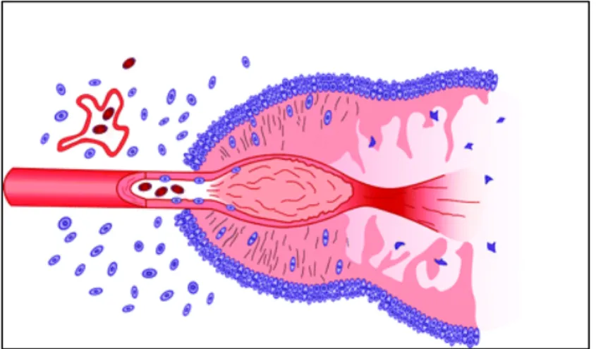
Figure 2. Schematic representation of the formation of a pseudopalisade.
Growth of the glioblastoma stimulates neo-angiogenesis. Prominent secretion of angiogenic factors causes endothelial damage, which, in turn, produces vascular occlusion and hypoxia. Cells unable to survive the hypoxia succumb and form the nidus of coagulation necrosis. Other cells, however, migrate to the periphery of the hypoxic field in waves forming pseudopalisades. The migrating hypoxic cells secrete VEGF, proteases, and other factors that cause further microvascular proliferation and enhanced invasiveness in regions ringing the hypoxic field. These latter effects prompt further aggressive outward expansion of the glioblastoma cells.
Another issue is the functional consequences of tumor angiogenesis, with respect to tissue perfusion (Vogel et al., 2004). Tumor microvessels are highly tortuous with sluggish flow and diminished gradient for oxygen delivery and increasing susceptibility to thrombosis and microhemorrhages (Kaur et al., 2004). Thus, the GBM microvasculature proliferation may provide little support in oxygen/nutrient delivery but rather paradoxically contribute to further exacerbating a metabolic mismatch between the “supply and demand,” leading to progressive hypoxia and eventually necrosis. This scenario is supported by the recent experience with anti-angiogenesis drugs, where their limited clinical benefit seems to be the result of “pruning” immature vessel growth and allowing “normalization” of the pre-existing vasculature (Horsman and Siemann, 2006). In addition to the poor vascular architecture, endothelial cells associated with the tumor vasculature fail to form tight junctions and have few associated pericytes or astrocytic foot processes leaving the integrity of the BBB compromised, resulting in increased interstitial edema. Interstitial edema may further compromise regional blood flow and exacerbate tumor hypoxia leading to areas of necrosis. In addition to these maladapted biophysical properties of GBM microvasculature, specific genetic mutations in GBM likely contribute to compromised tumor bioenergetics, specifically the shift in energy reduction from oxidative phosphorylation to glycolysis (Elstrom et al., 2004; Fantin et al., 2006). These interrelated mechanisms lead to a level of metabolic demand that may exceed the ability of the cerebrovascular system to maintain adequate blood flow to prevent hypoxia and necrosis. The histological evidence of thrombosis and degenerating vessels with microhemorrhages are a common feature of GBM and likely reflect these biological processes.
Our group has a great experience in the study of GBM. In my PhD studies, together with a neurosurgery unit of the Catholic School of Medicine of Rome, we made up in vivo model-based studies that threw light on tumor progression processes and on
the role played by telomerase in these processes. More specifically, I tried to get insight into the complex molecular processes at the basis of GBM progression. Firstly, I focused my attention on the elucidation of telomerase involvement in GBM-induced angiogenesis. Then, in the second part of the work, I studied the consequences of HIF-1-inhibition on the development of GBM tumor. HIF-1 is a transcription factor that orchestrates cellular response to hypoxic conditions. Since histological analysis of GBM sections reveals the presence of extensive hypoxic areas, targeting HIF-1 could be an interesting strategy for GBM therapy.
Telomerase
Telomeres
Telomeres are among the most important structures in eukaryotic cells. Creating the physical ends of linear chromosomes, they play a crucial role in maintaining genome stability, control of cell division, cell growth and senescence. In vertebrates, telomeres consist of repetitive DNA sequences and specific proteins, creating a specialized structure called the telosome that, through mutual interactions with many other factors in the cell, give rise to dynamic regulation of chromosome maintenance. Nearly 60 years ago, the first experiments identified telomeres as specific structures important for genome stability, at the ends of Drosophila and maize chromosomes. Telomeres are dynamic structures and their length varies among organisms or cells of different origin. For example, mice can have telomeres up to 150 kb long, while in humans the length can vary from 3 to 20 kb (Moyzis et al., 1988; Harley et al., 1990). Early analysis of telomeric heterochromatin showed that linear chromosomes end with short tandem repeats consisting of G-rich sequences, such as TTAGGG in vertebrates (de Lange et al., 1990). Electron microscope studies revealed a specific telomere structure in the form of a telomere loop (t-loop) or lariat (Griffith et al., 1999; Rubelj & Vondracek, 1999). The whole t-loop entity is held together by several telomere-specific proteins, as well as a number of other ubiquitous proteins involved in DNA processing and structure organization (de Lange 2005). The subtelomeric region adjacent to the t-loop is heterochromatic and contains repetitive, as well as corrupted telomeric sequences and can stretch up to 10–500 kb towards the centromere (Riethman et al., 2005).
Figure 3. Represents the Telomeric ends forming a structural feature with
their associated proteins that protects their ends.
Special histone and DNA modifications (such as acetylation and methylation) in subtelomeric and telomeric regions can regulate chromatin structure or processes that include replication and homologous recombination (Gonzalo et al., 2006). As part of the chromosome, telomeres contain histones forming nucleosomes, chromosome structural proteins (Nikitina and Woodcock, 2004) and some specific single-stranded DNA binding proteins with a great affinity for single-stranded telomeric sequences. Six proteins are considered to be strictly telomere specific. They meet the following criteria: they are specific for telomere DNA, present throughout the cell cycle and have functions restricted to the telomere. They have important roles in protection, elongation and regulation of the telomere. These proteins form a complex named Shelterin: TRF1, TRF2, hRap, TIN2, POT1, and TPP (Zhong et al., 1992; Bilaud et al., 1997). Each protein has a specific role in the
complex; such as inducing t-loop formation, protecting the telomere from nucleases, checkpoint functions or regulating telomere elongation in telomerase-positive cells.
In normal cells, telomeres shorten with each round of replication, due to the inability of DNA polymerase to synthesize the very end of the DNA lagging strand, as well as due to the presence of 5' exonuclease activity (Makarov et al., 1997). It is estimated that each passage through the cell cycle leads to shortening of telomeres by ~50–150 bp (Olovnikov, 1973; Makarov et al., 1997). Telomere erosion is considered to be a mitotic clock, which regulates the number of divisions before cells enter senescence.
The end replication problem
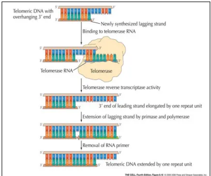
Semi-conservative DNA replication presents a problem when it comes to fully replicating a linear DNA molecule. Infact, DNA polymerase can only synthesize DNA in a 5’-3’ direction and it needs a primer of RNA. This leads to complete replication of leading strand and an incomplete and discontinous synthesis of lagging strand. As originally pointed out by Alexi Olovikov in 1971, in this way, after several rounds of replication, the DNA molecule continues to get smaller and smaller. Telomerase complex solves this question by adding specific nucleotidic repetitions at the end of chromosomes, ensuring the maintenance of telomere length.
Figure 4. DNA polymerase requires an RNA primer to initiate synthesis in
the 5'-3' direction. At the end of a linear chromosome, DNA polymerase can synthesize the leading strand until the end of the chromosome. In the lagging strand, however, DNA polymerase's synthesis is based on a series of fragments, called Okazaki, each requiring an RNA primer. Without DNA to serve as template for a new primer, the replication machinery is
unable to synthesize the sequence complementary to the final primal event. The result is the "end-replication problem" in which sequence is lost at each round of DNA replication
Telomerase
Telomerase was first identified biochemically more than 20 years ago (Greider and Blackburn, 1985) and shown to use an extraordinary mode of synthesis, relying on an intrinsic RNA, TR, that serves as a template for the polymerization of the telomeric DNA sequences, and a catalytic subunit, TERT, that functions as a reverse transcriptase (Greider and Blackburn, 1989; Yu et al., 1990).
TR
Unlike other polymerases responsible for replication of genomic DNA, telomerase activity depends on an essential RNA subunit. Telomerase was first shown to be an RNA-dependent DNA polymerase by characterization of the enzyme isolated from the unicellular ciliate Tetrahymena. The identification of a 159-nucleotide RNA component containing the sequence 5′-CAACCCCAA-3′, complementary to the d(TTGGGG)n telomeric repeat synthesized by Tetrahymena
telomerase, suggested that this region of the RNA provides a template for telomere synthesis (Greider and Blackburn, 1989). The templating region of the telomerase RNA can be dissected into two functionally separable subdomains, employed in primer alignment and primer extension (Gilley and Blackburn, 1996). One end of the RNA template (3′-AAC-5′) serves to align the telomeric DNA primer for the extension step, via basepairing between the 3′ terminus of the primer and a portion of the template. Subsequent elongation occurs by copying the remaining six residues of the template onto this telomeric end. These first round products can be further elongated if the new telomeric terminus is translocated back to the primer alignment site, so that the primer is repositioned for another round of synthesis. Processivity is dictated by more than base pairing interactions at the primer alignment site; telomerase can also interact with telomeric substrates at a second, RNA-independent, primer binding site, called the anchor site, that contributes to processive elongation (Morin, 1989,1991). In addition to alignment and templating functions, several observations indicate that the template region of the RNA directly participates in enzyme action by contributing to both the structure and function of the enzyme active site
Catalytic subunit of telomerase (TERT)
The dependence of telomerase polymerase activity upon RNA formally defined the enzyme as a specialized type of reverse transcriptase (RT). During the last years, the discovery of the
catalytic component of telomerase revealed that it is in fact a reverse transcriptase, related to known RTs by both amino acid sequence and presumably by evolution. This has provided a key step toward a detailed understanding of the mechanism of chromosomal DNA synthesis by telomerase. Identification of this telomerase protein resulted from convergence of complementary biochemical and genetic strategies in the ciliate
Euplotes aediculatus and the yeast S. cerevisiae. In Euplotes,
biochemical fractionation of the enzyme identified two telomerase subunits, p123 and p43, that extensively co-purify with telomerase activity and are in apparent steichiometrically equivalent ratios with the RNA subunit (Lingner and Cech, 1996). An independent genetic approach recovered four yeast
EST (ever shortertelomeres) genes that, when mutated, confer a
telomere replication phenotype in vivo (Lundblad and Szostak, 1989). Comparison of the sequence of the yeast 103-kD Est2 protein with the Euplotes p123 subunit revealed that these two proteins are homologues, sharing ~20% sequence identity and more extensive sequence similarity over the length of both proteins. Most notable was the presence in both of a set of motifs common to RTs, marked by the presence of a number of highly conserved residues. A subset of these amino acid sequence motifs had previously been shown to form a conserved protein fold comprising the active site of reverse transcriptases (Kohlstaedt et al., 1992), with three invariant aspartates that are thought to be critical for catalysis (Larder et al., 1987). Single amino acid changes introduced into the comparable aspartates of the Est2 protein abolished yeast telomerase activity in vitro and conferred an in vivo telomere replication defect, demonstrating that these residues are essential for telomerase catalysis (Counter et al., 1997). Rapid on the heels of the characterization of the telomerase catalytic subunit in Euplotes and yeast has been the identification of the same component in Schizosaccharomyces pombe (Nakamura et al., 1997) and in humans (Harrington et al., 1997). These proteins, like those from S. cerevisiae and Euplotes, also show sequence and mechanistic similarity with known RTs, and hence this protein subgroup has been named TERT (Telomerase Reverse Transcriptases). The TERT protein family is most
similar in sequence to RTs such as non-LTR retrotransposons and group II introns that, like telomerase, extend their RNA-templated polymerization from DNA 3′ hydroxyl primers. However, despite the overall similarities with RNA-dependent polymerases, including the three aspartate residues required for enzyme catalysis, telomerases from disparate organisms are more related to one another than to other polymerases and thus appear to form a distinct subgroup (Nakamura et al., 1997). Several features distinguish telomerase RTs, such as a unique region of sequence conservation termed the “T motif,” as well as a large amino-terminal basic domain (Nakamura et al., 1997). Prior to cloning of any of the TERT proteins, cross-linking studies with Euplotes telomerase indicated that the large subunit of the enzyme (presumably corresponding to the p123 catalytic component) contains a second site for telomeric DNA binding, called the anchor site (Hammond et al., 1997). The anchor site had been functionally defined by studies showing that primer recognition and processivity of the telomerase enzyme are influenced by the presence of G-rich telomeric sequences at the 5′ end of the primer, even when the 3′ terminus is non telomeric (Morin et al., 1989). This site is distinct from the binding that occurs between the 3′ end of the DNA primer and the template region of the RNA. Positioning the 5′ end of the primer in the anchor site is thought to contribute to processivity by preventing dissociation of the primer from the enzyme during translocation on the RNA template of the newly extended 3′ terminus. In studies with the Euplotes enzyme, in which the 3′ end of the primer was bound in the active site, cross-links between DNA and protein were localized 20–22 residues from the 3′ end, consistent with the prediction for an anchor site interaction with its primer (Hammond et al., 1997). Intriguingly, the use of partially duplex substrates with 3′ single-strand overhangs, which should resemble natural telomeres more closely, led to cross-links between protein and the duplex portion of the substrate. As expected, cross-links between the catalytic protein subunit and the telomerase RNA were also observed.
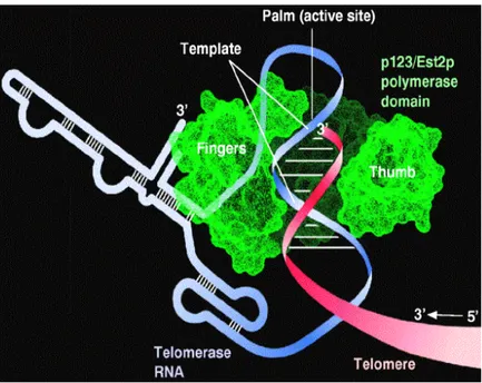
Figure 6. Action of telomerase. (Cooper and Hausman , 2007)
Regulation of telomerase activity
Most normal human cells have undetectable levels of telomerase activity and also fail to express hTERT. Strikingly, enzyme activity in telomerase-negative human cell lines can be restored by the ectopic expression of hTERT (Weinrich et al., 1997). Thus, the catalytic protein is the only limiting factor for telomerase activation in at least a subset of normal human cells, suggesting that regulation of hTERT expression may be a key target during cellular immortalization. By restoring telomerase activity to normal cells, this technique also has laid the groundwork for assessing whether conversion to telomerase
proficiency can reverse the mortal growth characteristics of normal cells. A complement to these in vivo experiments is the demonstration that coexpression of the hTERT protein and the human telomerase RNA in reticulocyte lysates is capable of reconstituting enzyme activity (Weinrich et al., 1997). An additional consequence is that this in vitro system may accelerate the search for inhibitors of telomerase activity; such inhibitors will be critical in testing the hypothesis that telomerase reactivation plays a role in oncogenesis.
Non canonical functions of telomerase
Various studies on telomerase suggest that this complex holds different roles other than canonical telomere maintenance. It was demonstrated that the expression of SV40 early genes, HRAS, and hTERT is sufficient to transform normal human cells (Hahn et al., 2002). Instead, the expression of SV40 and HRAS, together with ALT mechanism, does not induce transformation, suggesting that telomerase could hold other roles different from telomere elongation (Stewart et al., 2003). Moreover, telomerase ectopic expression in epithelial cells HMEC (Human Mammary Epithelial Cell) results in activation of growth promoting genes expression. L’EGFR (Epidermal Growth Factor Receptor) belongs to this group, and it was demonstrated that its inhibition abolished growth increment caused by telomerase (Smith et al., 2003). In previous studies, Li and colleagues analyzed the effects of telomerase inhibition in several human tumor cell lines. They observed that the expression of siRNA directed against TR, results in an inhibition of cell division, but it does not induce DNA damage response genes expression, telomere uncapping or strong shortening of these structures. Furthermore, by microarray, they observed that this inhibition results in an alteration of gene expression profile: as well as down regulation of cell cycle
genes, as Cdc27 and Cyclin G2, and genes involved in angiogenesis and metastatis, as integrin αV (Li et al., 2004; Li et al., 2005). Moreover, new studies about apoptosis revealed involvement of telomerase in these processes. Indeed, human fibroblasts in which telomerase is stably inhibited and treated by irradiation or chemiotherapic reagents, show a DNA damage response more weak than wild type counterpart. In these TERT-inhibited fibroblasts, they observed that chromosome structures are more sensitive to micrococcal DNAse than normal counterpart. These data, and the observations that there is increase of telomerase expression in S phase (Masutomi et al., 2003), demonstrate the involvement of telomerase in chromatidic structures regulation during replication phase (Masutomi et al., 2005).
Telomerase and cancer
Telomerase is expressed in the majority of human cancers, making it an attractive therapeutic target. In cellular in vitro models, for example in the case of CD8positive T cells, hTERT over-expression significantly enhances proliferation and cell survival (Dagarag et al., 2004). Similar observationshave been made with many different cell types. In vivo findingsin animal tumor models showed that mTERC was upregulated early in tumorigenesis and that telomerase became activated in late stages of tumor progression (Blasco et al., 1996). These studies led to the examination of what the effects of constitutive expression or overexpression of TERT would be. mTert overexpression was shown to be associated with spontaneous mammary epithelial neoplasia and invasive carcinomain aged mice (Artandi et al., 2002), while constitutive expression of mTert inthymocytes promotes T-cell lymphoma (Canela et al., 2004). More recently, work on targeted overexpression in specific tissues showed faster wound healing and increased
tumorigenesis in the skin of K5-mTertmice (where mTerc is required for the tumor promoting effect)(Cayuela et al., 2005). In addition, conditional induction (using a tetracycline-induciblesystem) in a mouse model showed that mTert causes the proliferation and mobilization of hair follicle stem cells (Sarin et al., 2005). This was visualized in situ as well as through the observation of exacerbatedhair growth and faster hair regrowth in a manner independentfrom telomere synthesis. How TERT protein can also modulatethe proliferation of stem cells in the skin even in the absence of telomerase RNA is currently not understood.The link between telomere biology and oncogenesis was firstproposed when telomerase expression was found to be a hallmark of human cancer: telomerase expression or reexpression and activitycan be detected in >90% of tumor samples. Telomerase deficiencies and cancer appear to lie at oppositeends of a spectrum similar to p53: loss of p53 is observed in most tumors and is tumor promoting in mouse models, whereas mice with enhanced p53 responses exhibit increased cancer resistance,a shortened life span, and a number of early aging-associated phenotypes (Donehower, 2002). In both models aging appears to be driven in part by a gradual depletion of the functional capacity of stem cells. The link between p53 and telomeres is further illustratedin Li-Fraumeni syndrome (LFS), a cancer predisposition syndromeassociated with germ line TP53 mutations. It was shown that the progressive earlier age of cancer onset in LFS is related to a measurable decrease in telomere length,with each generation providing the first rational biological marker for clinical monitoring of LFS patients (Tabori et al., 2007). Ectopic
hTERT expression can allow post-senescent cells to proliferate
beyond crisis, in a process that could to be independent of catalytic activity (Counter et al., 1998). Tumorigenesis is often associated withthe upregulation of c-Myc that can be induced by retroviral insertion or translocation. c-Myc binding sequences are describedwithin the hTERT promoter, and the MYC protein stimulates hTERTtranscription (Wu et al., 1999), which may in turn contribute to tumorigenesis or tumor progression. The flip side of continued expression or reexpression of hTERT in genetically stable primary cellsand in
animal models is enhanced longevity and a delay of senescence during in vitro culture (Gonzalez-Suarez et al., 2005). However, sustained (over)expression of telomerase in CD4- or CD8-positive T cells over longer periods in culture were shown to promote genomic instability (Roth et al., 2005). This may be directly due to hTERT overexpression or may be aconsequence of extended proliferation and replication errors that may be exacerbated by culture conditions. In addition, gain of expression of hTERC due to the presence of multiple gene copies has also been recently associated with cervicaldysplasia and invasive cancer progression.
Figure 7. In the march to malignancy, there is a progressive shortening of
telomere lengths that is accompanied by an increase in genomic instability. In some cases, this genomic instability can give rise to genetic aberrations that lead to invasive cancer.
Telomerase involvement in tumor angiogenesis
One of the most important demonstrations of telomerase involvement in tumor angiogenesis derives from the observation, made by our group, that hTERT is expressed by the endothelial cells of tumor vessels. This expression is a peculiar feature of tumor vessels, since we didn’t observe any hTERT reactivation in endothelial cells proliferating in contexts of non neoplastic angiogenesis. Moreover, histological analysis of human astrocytomas revealed that hTERT expression by the tumor endothelial cells is related to the histological grade of the tumor itself. Indeed, hTERT is expressed by 100% of endothelial cells in GBM, by 56% in AA, by 29% in low grade astrocytomas. The percentage of low grade astrocytomas that progress to a more aggressive phenotype (29%) is comparable to tumors whose endothelial cells reactivate hTERT expression (Pallini et al., 2001). hTERT reactivation in tumor endothelial cells could be necessary for telomere maintenance of fast growing cells. Indeed, in mTERT knockout mice, the presence of short telomeres inhibits angiogenesis (Franco et al., 2002). But the observation that there is not telomerase reactivation in non neoplastic angiogenesis support the idea that telomerase holds non canonical roles in tumor angiogenesis (Pallini et al., 2001).
B
A
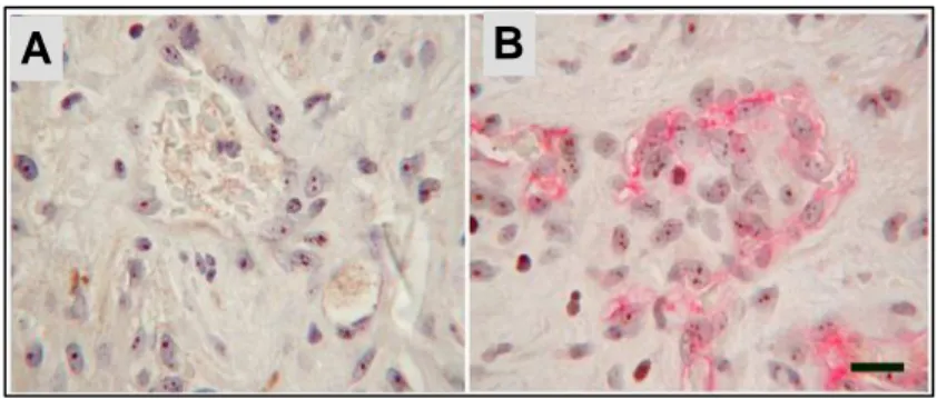
Figure 8: Immunostaining on human glioblastoma section. A: Nuclear staining
with hTERT antibody both on glial neoplastic cells and endothelial cells. B: double immunostaining with hTERT and CD31 antibodies in endothelial cells (Scale bar 40 µm).
Hypoxia-Inducible Factor-1 (HIF-1)
Hypoxia and HIF-1
Oxygen (O2) is essential for the survival of all aerobic organisms. O2 is required for aerobic metabolism that maintains intracellular energy balance. Aerobic energy metabolism is dependent on oxidative phosphorylation, in which the oxido-reduction energy of the mitochondrial electron transport is converted into the high-energy phosphate bond of ATP. In this process, O2 serves as the final electron acceptor. Depending largely on the distance from the nearest functional blood vessel, cells in mammalian tissues typically experience O2 concentrations in the 40–60 mm Hg range. Hypoxia, defined as a state of reduced O2 level below normal values, occurs under various physiological (embryonic development, adaptation to high altitudes, wound healing) as well as pathological (ischemic diseases, cancer) conditions. In order to cope with hypoxia, organisms undergo a variety of systemic and local changes to restore O2 homeostasis and limit the effect of low O2. Systemic adjustments include enhanced O2 delivery by the bloodstream, whereas angiogenesis features prominently among the local adjustments. At the cellular level, the most noticeable response to hypoxia is reduction in oxidative phosphorylation, accompanied by increased glycolysis to compensate for lower ATP production.
Although hypoxia generally inhibits mRNA synthesis, transcription of subsets of genes increases dramatically. At the molecular level, the master switch orchestrating the cellular response to low O2 tension is generally considered to be the transcription factor Hypoxia-Inducible Factor (HIF). HIF is a transcription factor foundin mammalian cells cultured under reduced oxygen tension and plays a key role in the cellular response to hypoxia. HIF is a heterodimer consisting of two subunits, the oxygen-sensitive HIF- and the constitutively
expressed HIF-ß (also known as aryl hydrocarbon receptor nuclear translocator (ARNT),the heterodimeric partner of aryl hydrocarbon receptor (Ahr) (Wang et al., 1995). Both the subunits are members of the basichelix–loop–helix-Drosophila period clock protein (PER)-ARNT-Drosophila single-minded protein (SIM) (HLH-PAS)family of transcription factors (Jiang et al., 1996).
Three HIF- homologues have been identified: HIF-1 , HIF-2 and HIF-3 (Ema et al., 1997; Tian et al., 1997; Gu et al., 1998). HIF-1 and HIF-2 share a high degree of sequence identity, which is highlighted by their common ability to heterodimerize to HIF-ß(Ema et al., 1997; Tian et al., 1997). Heterodimers that contain HIF-1 or HIF-2 appear to have overlapping but distinct tissue-specificexpression patterns and target genes. Less is known about HIF-3 compared with the other HIF- homologues. It has been shown thatthe inhibitory PAS domain protein (IPAS) is an alternativelyspliced variant of HIF-3 and functions as a dominant-negativeregulator of
HIF-, adding to the complexity in the regulation of hypoxia-inducible genes by the HIF family of transcription factors (Makino et al., 2002).
HIF-1 has two transactivation domains located in its COOH-terminal half: the NH2-terminal transactivation domain or (N-TAD) (amino acids 531-575) and the COOH-terminal transactivation domainor C-TAD (amino acids 786-826; Li et al., 1996; Jiang et al., 1997;Pugh et al., 1997).
Figure 9. Hypoxia-inducible factor (HIF)-1a and HIF-1b contain one basic–
helix–loop–helix (bHLH) domain and two PER–ARNT–SIM (PAS1 and PAS2) domains in their N-terminal regions. The positions of post-translational hydroxylation (OH) and acetylation (OAc) of HIF-1a are indicated. Hydroxylation of two proline residues (at P402 and P564) and acetylation of lysine (at K532) within the oxygen-dependent degradation (ODD) domain (residues 401–603) and close to the N-terminal transactivation domain (NTAD) confers recognition by pVHL (the product of the von Hippel–Lindau tumour suppressor gene), leading to degradation of the a-subunit. Hydroxylation at N803 in the C-terminal transactivation domain (CTAD) of HIF-1a inhibits recruitment of coactivators required for HIF-1a transcriptional activity. HIF-1b contains one transactivation domain (TAD) in its C-terminus.
Downregulation of HIF-1 protein levels in normoxia
In normoxia, the von Hippel-Lindau tumour suppressor (pVHL),which is the recognition component of an E3 ubiquitin ligasecomplex, targets HIF-1 (Iwai et al., 1999; Lisztwan et al., 1999), leading to its ubiquitylation and consequent proteasomal degradation(Lisztwan et al., 1999; Cockman et al., 2000; Kamura et al., 2000;Ohh et al., 2000; Tanimoto et al., 2000). The oxygen-dependent degradation domain (ODDD), which overlaps the N-TAD, controlsthe degradation of HIF-1 by the ubiquitin–proteasome pathway. Deletion of this entire domain renders HIF-1 stable even innormoxic cells (Huang et al., 1998). It has been shown that theinteraction of pVHL with HIF-1 is regulated by the hydroxylation of two proline
residues, proline-402 and proline-564, located within the ODDD, which are conserved between HIF-1 and HIF-2 (Ivan et al., 2001; Jaakkola et al., 2001; Masson et al., 2001).Further, detailed analysis has revealed two subdomains within the ODDD: an NH2-terminal subdomain or NODDD (amino acids 380-417;Masson et al., 2001) and a COOH-terminal subdomain or CODDD (amino acids 549–582) that overlaps the N-TAD (Pugh et al., 1997).
In 2001 Epstein and colleagues identified egg-laying defect (EGL)-9 asa prolyl hydroxylase capable of hydroxylating HIF-1 in Caenorhabditiselegans. Three prolyl hydroxylase domain
(PHD) enzymes, known as PHD1, PHD2 and PHD3, were subsequently identified in mammalian cells and shown to hydroxylate HIF-1 , although at varying levels of activity (Bruick & McKnight, 2001; Epstein et al., 2001). This post-translational modification of HIF-1 by the PHDs is oxygen, iron, 2-oxoglutarate and ascorbate dependent, which may be part of the mammalian oxygen-sensing mechanism (Kivirikko & Myllyharju, 1998). Interestingly, each of the PHDs hydroxylates human HIF-1 atproline-564, whereas only PHD1 and PHD2 hydroxylate the secondsite of prolyl hydroxylation, proline-402. Both proline residues subjected to hydroxylation lie within the ODDD, conserved between the worm and the mammalianHIF-1 isoforms (Epstein et al., 2001). The relative role of the three mammalian PHDs in terms of their oxygen-sensing capabilitiesand target proteins for modification remains to be determined.PHD2 has been shown in certain cell lines to be the predominant enzyme that hydroxylates HIF-1 in normoxia, on the basis thatinhibition of PHD2 expression with RNA interference (RNAi) stabilizedHIF-1 in normoxia, where inhibition of PHD1 and PHD3 had noeffect (Berra et al., 2003). PHD genes have been shown to beinduced in hypoxia, and the hypoxic induction is mediatedby HIF-1 (Epstein et al., 2001; Berra et al., 2003;Metzen et al., 2003). The increase in PHD expression under hypoxiamay reflect the function of PHDs in degrading HIF-1 upon reoxygenationor alternatively may be part of the mechanism to balance HIF-1 in hypoxia. Since PHDs are componentsof the HIF pathway, induction of PHDs
by hypoxia suggests a regulatory feedback loop in this signalling pathway.
The interaction of HIF-1 and pVHL was reported to be enhanced by acetylation of lysine-532 through a mouse homologue of an N-acetyltransferase, ADP-ribosylation factor domain protein1 (ARD1) (Jeong et al., 2002). However, further studies havefailed to validate this finding, reporting that human ARD1 (hARD1)does not affect HIF-1 stability (Murray-Rust et al., 2006).
An additional mechanism for negatively regulating HIF-1 proteinlevels involves interaction of the p53 tumor suppressor genewith HIF-1 , either directly or indirectly via Mdm2, itself adownstream target of p53 (Bardos & Ashcroft, 2005).
Figure 10. Under aerobic conditions, HIF-1 is hydroxylated on proline 402
and proline 564. The proline hydroxylations are necessary for binding to von Hippel-Lindau (VHL) and ubiquitin-mediated degradation by the proteasome. The asparagine hydroxylation prevents binding to p300/CBP. A splice derivative of HIF-3 called inhibitory PAS (IPAS), as it only
possesses the PAS domain, competes for HIF-1 binding. Maintenance of cysteine in a reduced state in the transactivation domain (TAD) is essential for p300/CBP binding. Compounds that inhibit thioredoxin inhibit HIF-1 -mediated transactivation. HIF-1 and HIF-1 both translocate to the nucleus to transactivate genes such as vascular endothelial growth factor (VEGF) that possess hypoxia-response elements (HREs). PAS, PER/ARNT/SIM; REF-1, redox factor 1. (Giaccia A et al., 2003).
Hypoxic induction of HIF-1
Inhibition of oxygen dependent hydroxylation
In hypoxia, the proline residues within the ODDD are not hydroxylated and thus HIF-1 is stabilized and the protein levels increase.Stabilized HIF-1 is translocated to the nucleus where it dimerizeswith HIF-1ß (ARNT) and associates with co-activators, such as CREB-binding protein (CBP)/p300.To be transcriptionally active, the HIF complex has to assemble on the HRE in the regulatory regions of target genes. HIF induces the transcription of several hypoxia-response genes, such as the proangiogenic vascular endothelial growth factor (VEGF), by binding to hypoxia-response elements in their promoters. The C-TAD of HIF-1 is involved in modulating the transcriptional activation of HIF-1 under hypoxic conditions, in contrast to the N-TAD, which is involved in the stabilization of HIF-1 . Under hypoxia, the C-TAD is able to interact with transcriptionalco-activators, such as CBP/p300. This interaction is unableto occur under normoxia due to the oxygen-dependent hydroxylation of asparagine-803, located within the C-TAD. Hydroxylation of asparagine-803 is mediated by an asparaginyl hydroxylase, known as factor inhibiting HIF-1 (FIH-1), which prevents HIF-1 from interacting with the transcriptional co-activators CBP/p300. An additional mechanism of controlling HIF-1 transactivation function is through redox (reduction– oxidation)-dependent processes. Transfection of thior-edoxin-1 (Trx-1), belonging to the thioredoxin family of small redox active proteins, increases HIF-1 protein levels under both normoxic and hypoxic conditions. This increase is associated
with increased HIF-1 transactivation and the expression of downstream targets including VEGF andnitric oxide synthase-2 (Welsh et al., 2002). Redox effectorfactor-1 (Ref-1) is a protein which functions not only as aDNA repair endonuclease but also as a redox regulatory factormaintaining transcription factors in an active reduced state. Thiolredox regulation of C-TAD activity by Trx-1 via the Ref-1system has been reported to promote interaction of the C-TADwith CBP/p300 resulting in increased HIF-1 transactivation (Ema et al., 1999).
HIF-1 target genes
Given that cells and organs need to adapt to changes in oxygen supply, it would not be surprising to find that a significant variety of the HIF-1 target genes are regulated in a tissue-specific manner. To date, there are more than 100 HIF-1 downstream genes identified with varying functions. HIF-1 activates the expression of these genes by binding to a 50-base pair cis-acting HRE located in their enhancer and promoter regions (Semenza et al., 1991). Moreover, by using DNA microarrays, it has been reported that more than 2% of all human genes are regulated by HIF-1 in arterial endothelial cells, directly or indirectly (Manalo et al., 2005).
Erythropoiesis/Iron Metabolism
In response to hypoxia, the capacity of red blood cells to transport oxygen is up-regulated by the expression of genes involved in erythropoiesis and iron-metabolism. Hypoxia increases the expression of EPO, which is required for the formation of red blood cells. An increase in the number of erythrocytes enhances the delivery of oxygen to tissues. Products of iron-metabolizing genes control the major erythropoietic rate-limiting step of heme production. Hypoxia up-regulates transferrin (Tf), which transports Fe3+ into cells; the transferrin receptor (Tfr), which binds Tf and enables cellular transferrin uptake; and ceruloplasmin (also known as a
ferroxidase), which is required to oxidize ferrous (Fe2+) to ferric (Fe3+) iron. Increasing of these genes supports iron supply to erythroid tissues (Rolfs et al., 1997).
Angiogenesis
Angiogenesis is a complex process that involves multiple gene products expressed by different cell types. A large number of genes involved in different steps of angiogenesis have been shown to increase by hypoxia challenge. Among them, the vascular endothelial growth factor (VEGF) is the most potent endothelial-specific mitogen, and it directly participates in angiogenesis by recruiting endothelial cells into hypoxic and avascular area and stimulates their proliferation. Therefore, the induction of VEGF and various other proangiogenic factors leads to an increase in the vascular density and hence a decrease in the oxygen diffusion distance. In addition, HIF-1 regulates genes involved in governing the vascular tone such as nitric oxide synthase (NOS2), heme oxygenease 1, endothelin 1 (ET1), adrenomedulin (ADM), and the 1β-adrenergic receptor. Moreover, hypoxia induces genes involved in matrix metabolism and vessel maturation such as matrix metalloproteinases (MMPs), plasminogen activator receptors and inhibitors (PAIs), and collagen prolyl hydroxylase.
Glucose Metabolism
Under low oxygen supply, cells switch their glucose metabolism pathway away from the oxygen-dependent tricarboxylic acid (TCA) cycle to the oxygen-independent glycolysis (Dang and Semenza, 1999). With only 2 ATP molecules from each glucose molecule produced by glycolysis, instead of 38 ATP provided by TCA cycle, hypoxic cells elevate their ability to generate ATP by increasing the glucose uptake. This is achieved by up-regulating the expression of glycolytic enzymes and glucose transporters (Wenger, 2002). Hypoxia and HIF-1 increase virtually all the enzymes in the glycolytic pathway, as well as the glucose transporters 1 and 3
(GLU1, GLU3) (Chen et al., 2001). Furthermore, the glycolysis metabolic products, such as lactate and pyruvate, have been reported to cause HIF-1 accumulation under normoxia and regulate hypoxia-inducible gene expression, hence establishing a potential positive feedback loop (Lu et al., 2002).
Cell Proliferation/Survival
Hypoxia and HIF-1 induce growth factors, such as insulin-like growth factor-2 (IGF2) and transforming growth factor-β (TGF- β) (Krishnamachary et al., 2003). Binding of such growth factors to their cognate receptors activates signal transduction pathways that lead to cell proliferation/survival and stimulates the expression of HIF-1 itself (Semenza, 2003). Cytokines and growth factors, as well as hypoxia in some cell types, can activate signaling pathways MAPK and PI3K, which promote cell proliferation/survival as well as contribute to HIF-1 activity. This leads to increased HIF-HIF-1 transcriptional activity of target genes, including those encoding IGF2 and TGF- , thereby contributing to autocrine-signaling pathways that are crucial for cancer progression (Semenza, 2003).
HIF-1-induced Apoptosis
Paradoxically, cell adaptation to hypoxia leads not only to cell proliferation/survival but also to cell death in some circumstances. Hypoxia has been shown to induce apoptosis, where HIF-1 plays a complex role (Carmeliet et al., 1998). Genetic studies using embryonic stem cells harboring a deletion of HIF-1 showed decreased apoptosis compared with wild type when challenged with low oxygen (Carmeliet et al., 1998). Activation of caspase-3 and Apaf-1-mediated caspase-9, and the release of cytochrome c, have been reported in several cell types under hypoxic conditions. It has also been demonstrated that the expression of HIF-1 significantly correlated with apoptosis and the pro-apoptotic factors, such as caspase-3, Fas, and Fas ligand (Volm and Koomagi, 2000). Moreover, hypoxia depressed the antiapoptotic protein Bcl-2 (Carmeliet et al.,
1998), whereas the proapoptotic protein Bcl-2/adenovirus EIB 19-kDa interacting protein 3 (BNip3) and its homolog Nip3-like protein X (NIX) were up-regulated in a HIF-dependent manner (Bruick, 2000). Some genes involved in cell cycle control, such as p53 and p21, were also found to be HIF-dependent (Carmeliet et al., 1998). In addition, p53 has been implicated in regulating hypoxia-induced apoptosis through induction of apoptosis-related genes such as Bax, NOXA, PUMA, and PERP. In addition to the above classes of genes, HIF-1 also regulated many other target genes implicated in diverse processes such as adipogenesis, carotide body formation, B lymphocyte development, and immune reactions. Although there are some studies showing a role of HIF-2 in the VEGF induction (Akeno et al., 2001; Compernolle et al., 2002), no bona fide target genes have yet been identified for HIF-2 or HIF-3 . However, a recent study using a genetic "knock-in" strategy has shown that targeted replacement of HIF-1 with HIF-2 results in expanded expression of HIF-2 -specific target genes (i.e., Oct-4, a transcription factor essential for maintaining stem cell pluripotency) (Covello et al., 2006).
Figure 11. Under hypoxic conditions the alpha subunit of HIF is stable and
translocates into the cell nucleus where it heterodimerises with the beta subunit inducing binding to the DNA of target genes carrying a hypoxia-response element (HRE). Interaction with the co-activator CBP/p300 initiates the induction or repression of a large number of genes involved in angiogenesis, anaerobic glycolysis, vasodilation and respiration, erythropoiesis, and apoptosis.
Role of HIF in cancer disease
In order for solid tumors to grow, an increase of oxygen delivery to cells, via angiogenesis and activation of glycolysis, have been observed and named, the Warburg effect (Seagroves et al., 2001). Given the importance of HIF-1 in the activation of genes essential to these processes, it is not surprising that both HIF-1 and HIF-2 have been strongly implicated in tumor progression and grade, hence conferring a selective advantage to tumor cells. Overexpression of HIF-1 and HIF-2 was found in various human cancers, probably as a consequence of intratumoral hypoxia or genetic alteration (Talks et al., 2000). The interior of the tumor mass becomes progressively hypoxic as its size increases until adequate blood vessels are obtained by tumors. Hypoxic conditions within tumors can result in increased HIF-1 stability and activity. Immunohistochemical analyses demonstrated that there are detectable levels of HIF-1
protein in benign tumors, elevated levels in primary malignant tumors, and a marked amount in tumor metastases, in contrast to its absence in normal tissues. Expression of HIF target genes is generally consistent with the levels of HIF-1 . There is a remarkable frequency of common genetic alterations in cancer cells associated with increased HIF-1 expression. As mentioned in VHL disease, for example, loss of function of VHL resulted in constitutively expressed HIF-1 (Iliopoulos et al., 1996). In addition, loss of function of wild-type p53, which is inactivated in most of human cancers, increased HIF-1 levels and enhanced HIF-dependent transcription in tumors (Ravi et al., 2000). Loss of function of tumor suppressor gene PTEN in glioblastoma-derived cell line resulted in increased HIF-1 levels and HIF-1-mediated gene expression, probably via activating the PI3K/AKT signaling cascade (Zundel et al., 2000). The transforming potential of the v-Src oncogene is thought to be due in part to its induction of HIF and gain of function of v-SRC increased expression of 1 and HIF-dependent genes (Jiang et al., 1997). Moreover, enhanced HER2 receptor tyrosine kinase signaling has been shown to increase the rate of synthesis of HIF-1 (Laughner et al., 2001). Increased activity of the HER2 receptor tyrosine kinase is a prevalent and important genetic alteration in breast cancer, correlating with tumor aggressiveness and decreased patient survival. Therefore, it seems that HIF-1 overexpression confers selective advantages to tumor cells. A correlation between HIF-1 overexpression and patient mortality, poor prognosis, or treatment resistance has been oserved in many studies (Semenza, 2003).
The Warburg effect
ATP required for normal cell proliferation and survival comes primarily from two sources. The first is glycolysis, which comprises a series of reactions that metabolizes glucose to pyruvate inthe cytoplasm to produce a net of 2 ATP from each
glucose. The other is the tricarboxylic acid (TCA) or Krebs cycle, whichuses pyruvate formed from glycolysis in a series of reactionsthat donate electrons via NADH and FADH2 to the respiratory chain complexes in mitochondria. With oxygen serving as thefinal electron acceptor, electron transfer creates across themitochondrial inner membrane a proton gradient the dissipation of which through ATP synthase forms 36 ATP per glucose molecule.With limited oxygen, such as with muscles that have undergoneprolonged exercise, pyruvate is not used in the TCA cycle and is converted into lactic acid by lactate dehydrogenase (LDH)in a process termed anaerobic glycolysis. Many cancer cells consume glucose avidly and produce lactic acid rather than catabolizing glucose via the TCA cycle, which is the key for generating ATP in nonhypoxic normal cells. The avid uptake of glucose by tumors is the foundation for the detection and monitoring of human cancers by fluorodeoxyglucose positronemission tomography. Many years ago, the renowned biochemist Otto Warburgobservedthat thin slices of human and animal tumors ex vivo displayedhigh levels of glucose uptake and lactate production. The shift toward lactate production in cancers, even in the presence ofadequate oxygen, is termed the Warburg effect or aerobic glycolysis (Warburg, 1956). To account for the conversion of glucose to lactate by cancer cells in the presence of oxygen, Warburg speculated that tumor mitochondria are decreased or functionally impaired. Infact, Warburg suggested that impaired mitochondrial function contributes to tumorigenesis. Recent studies suggest that mutations affecting mitochondrial DNA (mtDNA) or enzymes of the TCA cyclemight contribute to the Warburg effect. Mutations of mtDNA,which are prevalent in human cancers, could render the mtDNAencoded components of the respiratory chain defective complexes.Intriguingly, the subunits of the TCA cycle enzyme SDH (SDHB,SDHC, and SDHD) behave as classic tumor suppressors in pheochromocytoma or paraganglioma. Furthermore, FH is a tumor suppressor in leiomyoma and renal cell carcinoma. Although these observationssuggest that mitochondrial function may be compromised by mutationsin mitochondrial enzymes, there is no direct evidence that thesemutations are sufficient for
tumorigenesis. In this regard, there is no evidence that respiration is, in fact, less activein cancer cells than in normal cells. Hence, further studies are required to address whether impaired mitochondrial function sufficiently contributes to tumorigenesis.Several oncogenes have been implicated in the Warburg effect. The AKT oncogene, which encodes a protein serine-threonine kinase, is associated with enhanced glucose uptake and aerobic glycolysisseemingly independent of HIF-1. The MYC oncogene, which is widely activated in human cancers, has also been implicated in the direct activation of aerobic glycolysis. Elevated and sustained activation of MYC, however, is tightly associated with increased mitochondrial reactive oxygen species, which may cause mtDNA mutations that in turn contribute to dysfunctional mitochondria. In addition to oncogenic activation of aerobic glycolysis, the activation of HIF and its direct transactivation of glycolytic enzyme genes significantly contributes to the conversion of glucose to lactate. Two recent studies provide insight into the Warburg effect via a novel HIF-1-mediated mechanism that actively inhibits mitochondrial function (Papandreou et al., 2006). PDK1, which is one of four family members, was identified as a direct HIF-1 target gene in hypoxic cells.PDK1 phosphorylates and inactivates the mitochondrial pyruvate dehydrogenase (PDH) complex. Suppression of PDH by PDK1 inhibits the conversion of pyruvate to acetyl-CoA, thereby attenuating mitochondrial function and respiration. Because nonhypoxicstabilization of HIF through oncogenic events has been observed in many types of tumors, we hypothesize that PDK1 levels maybe up-regulated in nonhypoxic tumor cells by HIF, which woulddivert pyruvate from PDH and result in the increased lactate production. A recent immunohistochemical study further provided evidence that PDK1 expression is elevated in non–small-cell lung cancers (Koukourakis et al., 2005). Intriguingly, PDH expression seems to be reduced in high PDK1-expressing cancer cells. These studiessuggest that PDK1 activation may be a key regulatory switchcontributing to the Warburg effect. In aggregate, the activation of oncogenes, such as AKT and MYC along with the stabilizationof HIF can enhance aerobic glycolysis or the Warburg effect through
increased glycolytic flux and attenuation of mitochondrial function.
Figure 12. In normal cells the pyruvate generated by glycolysis is
metabolized through the tricarboxylic acid (TCA) cycle and oxidative phosphorylation, which is efficient in energy production. In hypoxic tumour cells pyruvate is converted to lactate because oxidative phosphorylation that requires oxygen is limiting. Since this option is less efficient in producing energy the tumour cells increase their uptake and consumption of glucose through an increase in the production of glucose transporters and enzymes of the glycolytic pathway. The increase in lactate production in turn contributes to the acidosis characteristic of tumours. In addition, one of the metabolites of the TCA cycle the 2-oxoglutarate (2-OG) (α-ketoglutarate) is required for the activity of the PHD and FIH hydroxylases. Catabolism of amino acids is also a source of 2-OG. The production of succinate or fumarate by enzymes of the TCA cycle, respectively succinate dehydrogenase (SDH) and fumarate dehydrogenase (FH) leads to feedback inhibition of these hydroxylases.