© 2008 LANDES BIOSCIENCE. DO NOT
DISTRIBUTE.
While it is known that Retinoic Acid (RA) induces meiosis in mouse female fetal gonads, the mechanisms which regulate this process during spermatogenesis are poorly understood. We show that the All trans RA derivative (ATRA) and Kit Ligand (KL) increase meiotic entry of postnatal mouse spermatogonia in vitro without synergism. Competence to enter meiosis is reached by spermatogonia only at the stage in which they undergo Kit-dependent divisions. Besides increasing Kit expression in spermatogonia, ATRA also upregulates KL expression in Sertoli cells. Both ATRA and KL increase the expression of Stimulated by Retinoic Acid Gene 8 and Dmc1, an early meiotic marker. A specific Kit tyrosine kinase inhibitor prevents the increase in the number of meiotic cells induced by both the two factors, suggesting that they converge on common Kit-dependent signalling pathways. Meiotic entry induced by ATRA and KL is independent from their ability to affect germ cell viability, and is mediated by the activation of PI3K and MAPK pathways through Kit autophosphorylation. ATRA-induced phosphorylation of the two downstream kinases is mediated by a non-genomic mechanism.
These data suggest that RA may control the timing of meiosis by influencing both the somatic and the germ cell compartment of the postnatal testis through the activation of the KL/Kit system.
Introduction
Spermatogenesis is the cyclic differentiative process of male germ cells, in which mitotic, meiotic and spermiogenetic phases orderly occur. During the mitotic phase, spermatogonia proliferate and continuously self-renew to give rise to two sub-populations of germ cells: the differentiated germ cells and the stem cells. One current model proposes that the A single (As) stem cells either renew themselves or divide into paired (Apr) daughter cells that remain connected by an intercellular bridge.1,2 Apr spermatogonia divide
further to form long chains of aligned (Aal) cells, which then generate
the differentiating A1–A4, Intermediate, and Type B spermatogonia. This population of spermatogonia starts expressing the receptor-coupled tyrosine kinase Kit and becomes responsive to Kit Ligand (KL)3 before entering the prophase of meiosis I, in which
sperma-tocytes are formed. During meiosis I homologous chromosomes of spermatocytes align and are kept together tightly by a meiosis-specific nucleoprotein structure known as the synaptonemal complex, which is essential for the homologous recombination process.
Retinoids have been shown to be essential for spermatogenesis progression in human and mice.4 Much of the role of retinoids has
been studied using animal models kept on a diet deficient on vitamin A (VAD) or absent on vitamin A derivatives.5 These animals are
sterile because the seminiferous tubules contain only undifferentiated Kit negative spermatogonia, indicating a role of vitamin A in sper-matogonia differentiation.5 More recently it has been shown that the
vitamin A derivative retinoic acid (RA) (either as all-trans or as 9-cis retinoic acid) promotes the expression of Stimulated by Retinoic Acid Gene 8 (Stra8), a key regulator of mammalian meiosis,6 and Kit
expression in undifferentiated spermatogonia.7,8 RA functions inside
the nucleus recognising two different classes of retinoid receptors. Both classes (RARs and RXRs) consist of three types of receptors, α, β and γ, encoded by distinct genes9 and transduce RA signal by
binding directly to retinoic acid responsive elements (RARE). During post-natal development, each RAR is detected predominantly in a specific cell type of the seminiferous epithelium: RARα in Sertoli cells, RARβ in round spermatids and RARγ in A spermatogonia.10
RARα gene targeting specifically in Sertoli cells (RARα Ser-/-) showed
germ cell apoptosis and seminiferous epithelium dysfunctions related to the disruption of Sertoli cells cyclical gene expression, which preceded testis degeneration.11 Deletion of RAR
β or RARγ, on the
contrary, do not cause primary testis defects.10,12
It has been recently shown that RA produced by mesonephroi causes entry into meiosis of germ cells in the ovary, while its effect is prevented in the fetal testis by the action of the retinoid-degrading enzyme CYP26B1.13,14 It has been proposed that a critical role in
inducing meiosis in both female and male6,15,16 is played by the
RA-stimulated Stra8 gene; however it remains to be elucidated if RA regulates other meiosis promoting genes and which are the precise mechanisms that orchestrate meiotic entry in the postnatal testis.
The Kit (White spotting locus) gene, encoding the transmembrane receptor for Kit Ligand (KL) regulates proliferation, survival and/or
*Correspondence to: Susanna Dolci; Department of Public Health and Cell Biology; University of Rome Tor Vergata; Via Montpellier 1; Bldg. E Nord; Rome 00133 Italy; Tel.: +390672596252; Fax: +390672596268; Email: [email protected] Submitted: 10/01/08; Accepted: 10/26/08
Previously published online as a Cell Cycle E-publication: http://www.landesbioscience.com/journals/cc/article/7262
Report
ATRA and KL promote differentiation toward the meiotic program
of male germ cells
Manuela Pellegrini,1 Doria Filipponi,1 Manuele Gori,1 Florencia Barrios,1 Francesca Lolicato,1 Paola Grimaldi,1 Pellegrino
Rossi,1 Emmanuele A. Jannini,2 Raffaele Geremia1 and Susanna Dolci1,*
1Department of Public Health and Cellular Biology; University of Rome ‘Tor Vergata’; Rome; 2Department of Experimental Medicine; Università dell’Aquila; L’Aquila, Italy Key words: kit, retinoic acid, meiosis, spermatogenesis
© 2008 LANDES BIOSCIENCE. DO NOT
DISTRIBUTE.
migration of stem cells for gametogenesis,17,18 haematopoiesis19 andmelanogenesis.20 In postnatal germ cells, Kit is expressed specifically
in differentiating spermatogonia and is downregulated at the time of meiotic entry.3,21,22 The role of Kit/KL in the maintenance and
proliferation of germ cells has been highlighted by a mouse model with a point mutation of Kit that eliminates the PI3K docking site Y719F.23,24 While PGC proliferation in both sexes is not
compro-mised during embryonic development, Kit(Y719F)/Kit(Y719F) males are sterile due to the lack of spermatogonia proliferation during the prepuberal period and an arrest of spermatogenesis at the premeiotic stages.
In this paper we show that both the retinoic acid derivative All trans Retinoic acid (ATRA) and KL can regulate meiotic entry of isolated mouse spermatogonia cultured in vitro by activating common signal transduction pathways.
Results
ATRA and KL increase meiotic cells in cultures of isolated mouse spermatogonia. It has been recently shown that ATRA
induces meiosis in fetal germ cells in organ-culture systems,13,30
however, it is not clear whether it may also influence meiosis in isolated postnatal germ cells. To define the developmental age at which spermatogonia from the CD1 strain start the expression of Kit, the molecular event that precedes entry into meiosis, we took advantage of the p18 transgenic line25 which has been expanded for
more than eight generations on a CD1 background. This transgenic line express EGFP under the control of c-kit promoter and first intron and show specific expression only in spermatogonia of the prepuberal testis.26 Figure 1 shows that EGFP expression, and thus
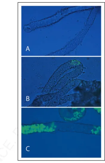
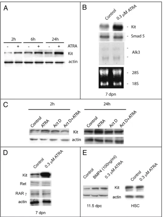
Kit, was absent at 1 dpp (Fig. 1A), appeared at 4 dpp in few small spermatogonial foci within the seminiferous tubules (Fig. 1B), and was spread at 7 dpp in discrete segments of the testicular tubules (Fig. 1C), showing the characteristic “wavy” expression pattern of mouse spermatogenesis. Having established the temporal pattern of Kit expression, we chose 7 dpp as the developmental age in which Kit was uniformly represented within all the tubules.
We isolated spermatogonia from 7 dpn animals and cultured for 24 or 48 h in the presence of increasing concentrations of ATRA. We found that at 0.3 μM concentration, ATRA has a slight negative effect on cell viability compared to the control (75 ± 3% vs 85.5 ± 6% at 24 h, and 68.8 ± 5% vs 78 ± 5% at 48 h) and no significant mitogenic effect on 7 dpn spermatogonia (data not shown). We also stimulated spermatogonia with 100 ng/ml of KL, a growth factor which stimulates proliferation and survival of spermatogonia.21,31,32
Nuclear spreads were prepared from cells at the beginning and at the end of the culture time and probed with antibodies against the synaptonemal complex protein 3 (SCP3) to detect meiotic nuclei and to evaluate the degree of meiotic progression (Figs. 2 and 3B). At the beginning of the culture we found that 4.6% showed prelep-totene morphology (Fig. 2C, in which typical SCP3 spots are found) while 1.8% of the germ cell population showed early leptotene morphology (Fig. 2D). As shown in Figure 3A and B; and Table 1, after 24 h of culture more meiotic nuclei were found in control cells and their number further increased after 48 h of culture (2.5 ± 0.12 and 7 ± 0.11 fold increase vs T0, respectively). Treatment with 0.3 μM ATRA induced a further significant increase of the percentage of meiotic nuclei after 24 and 48 h of culture (4.4 ± 0.12 and 12.8 ± 0.2
folds of increase vs T0, respectively) (Fig. 3A). We determined that this increase was essentially due to cells accumulating at leptotene stage over 48 h of culture (Fig. 3B). Similarly to ATRA, addition of KL also increases the percentage of meiotic nuclei in cultured sper-matogonia as measured by the number of nuclei in leptotene (4.5 ± 0.11 and 12.3 ± 0.6 fold of increase vs T0 after 24 and 48 h, respec-tively) (Fig. 3A and B). Simultaneous addition of both ATRA and KL did not produce any further increase of meiotic nuclei (4 ± 0.1 and 11 ± 0.8 fold of increase vs T0) either after 24 and 48 h of culture (Fig. 3A). The increased percentage of meiotic nuclei induced by ATRA or KL treatments was not associated to the increase of SCP3 protein levels, as evaluated by western blot analysis (data not shown), but rather to the typical pattern of SCP3 organization on the meiotic chromosomes (see Fig. 2). To confirm that ATRA and KL increased the number of meiotic cells in vitro, we also performed a quantitative RT-PCR using primers for Dmc1, which encodes a meiosis specific recombinase expressed during the first meiotic prophase,33 and for
the premeiotic marker Stra8 as positive control (Fig. 3C). Treatment with ATRA or KL for 24 h significantly increased Dmc1 levels (about 2 and 3.5 folds compared to the control, respectively), and as expected, ATRA caused a marked increase of Stra8 mRNA levels
Figure 1. Kit-EGFP expression in neonatal and prepuberal testis tubules. Brightfield and fluorescent merged images of testicular tubules isolated from p18 transgenic mice expressing EGFP under the control of c-kit promoter and 3.5 kb of the first kit intron. The image shows testicular tubules at 1 dpn (A), at 4 dpn (B) and at 7 dpn (C).
© 2008 LANDES BIOSCIENCE. DO NOT
DISTRIBUTE.
increase of Kit expression was exerted at the level of mRNA synthesis rather than of mRNA stabili-zation. Indeed, we found by western blot analysis that pre-incubation with actinomycin D, a strong inhibitor of all RNA polymerases, completely abol-ished the increase of Kit protein levels induced by ATRA (Fig. 5C). ATRA does not affect the levels of GDNF receptor Ret, a marker for undifferentiated spermatogonia,35 neither the levels of RAR
γ, the RA
receptor specifically expressed in spermatogonia10
(Fig. 5D). As in the case of postnatal spermatogonia, we found that ATRA increased Kit expression also in primordial germ cells (PGCs), harvested at 11.5 dpc, during their proliferative period,27 but not in
haematopoietic stem cells, a cell type which also express Kit19 (Fig. 5E).
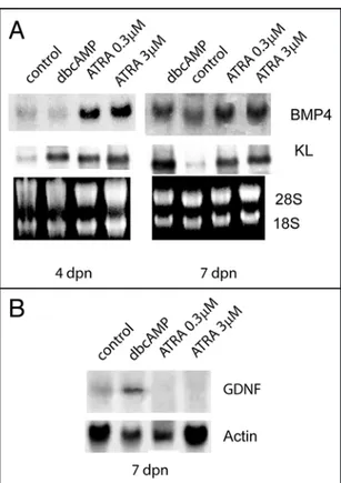
Since Sertoli cells are a known target of retinoic acid,11 we investigated whether ATRA might
regu-late in these cells the production of growth factors essential for germ cells proliferation and/or differen-tiation. Figure 6A shows that, in Sertoli cell cultures obtained from 4 dpn and 7 dpn mice, ATRA strongly upregulated the levels of KL and BPM4 mRNAs after 24 h of culture both at 0.3 and 3 μM concen-tration. As a positive control we included dbcAMP stimulation which we have previously shown is able to upregulate KL levels in Sertoli cells.21 In contrast to dbcAMP stimulation, the levels
of the stem cell growth factor GDNF were downregulated in 7 dpn cultures by ATRA treatment (Fig. 6B).
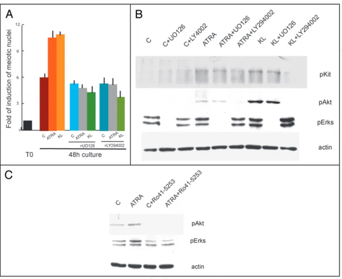
Kit-activated signal transduction pathways are required for both ATRA- and KL-induced of meiotic entry. Since Kit is induced by
ATRA and is the target of KL in 7 dpn spermatogonia, we hypoth-esized that the two factors might converge on Kit signalling pathways to increase the number of meiotic cells in culture. In order to test this hypothesis, we isolated and cultured spermatogonia for 48 h in the presence or absence of 5 μM STI571, a selective inhibitor of Kit tyrosine kinase activity,36 and treated these cells with ATRA or KL.
In cells solely treated with STI571, we found the same percentage of meiotic cells as in the control cultures without the inhibitor. The drug, however, was able to completely revert the increase of meiotic nuclei induced by ATRA and KL (0.9 fold of increase compared to STI571 control sample) (Fig. 7A). Since STI571 inhibited both ATRA- and KL-mediated increase of meiotic figures, we analyzed the levels of phospho-Kit and its downstream targets phospho-Akt and phospho-Erk1/2 in spermatogonia after 15 min stimulation with ATRA and KL pretreated or not for 30 min with STI571. Kit protein levels did not change after this pulse of stimulation while, as previous reported31 we found that KL induced a significant increase of Kit,
Akt and Erk1/2 phosphorylations, completely inhibited by the pres-ence of 5 μM STI571 (Fig. 7B). Interestingly, we observed a positive effect on Kit autophosphorylation, Akt and Erk1/2 phosphorylations after only 15 min stimulation with ATRA, which were all prevented by the presence of the tyrosine kinase inhibitor. To determine which signalling pathway was preferentially activated by ATRA and KL, we cultured spermatogonia in the presence or absence of LY29406 (an inhibitor of PI3K pathways) or of U0126 (an inhibitor of the MAPK pathway). Both these inhibitors completely blocked the increase in the number of meiotic figures induced by ATRA or by KL (3.7 folds vs control). Interestingly, we found that Stra8 mRNA levels
were upregulated also by KL treatment (1.7 fold vs control), and such increase was more evident at the protein level (3.5 fold of increase with respect to control, Fig. 3D).
To verify whether meiotic entry could be stimulated by the two agents also in undifferentiated Kit negative spermatogonia, we treated 4 dpn spermatogonia for 48, 72 and 96 h with ATRA, KL and a combination of both. As previously described,8,30 Kit and Stra8
expression were strongly upregulated in all the culture conditions in which ATRA was present (Fig. 4A and Suppl. Fig. 1C and D), even though cell viability decreased to about 50% after 4 days of culture in all the treatments (data not shown).We did not find meiotic nuclei up to 72 h of culture in all of the conditions tested, however after 96 h of culture a significant number of cells (Fig. 4B) showed a peculiar (early leptotene-like) SCP3 staining (Fig. 4C). Such staining was different from that found in 7 dpn spermatogonia which entered meiosis in vitro (see Figs. 2D and 3B). The SCP3 pattern exhibited nuclear foci as well as short filamentous structures which were less abundant when compared to leptotene nuclei and was reminiscent of that found in ES cells differentiating into germ-cells.34 This pattern
of staining was present in control cultures (15% ± 1), its percentage increased significantly in cultures treated with ATRA (21.7% ± 1.3) but not with KL (14% ± 0.8) and it almost doubled in the presence of both ATRA and KL (30.2 % ± 0.9).
ATRA increases Kit/KL levels in isolated testicular cells. It
has been reported that ATRA stimulates Kit expression in isolated spermatogonia.7,8 We confirmed these results in 4 and 7 dpn
sper-matogonia showing that Kit is induced at doses as low as 0.03 μM and that ATRA effect is not mediated by Sertoli cells (Suppl. Fig. 1). We show that Kit induction is evident as early as after 2 h of ATRA stimulation and increased linearly up to 24 h of culture (Fig. 5A) which corresponded to a significant increase at the RNA level, as shown by Northern blot analysis (Fig. 5B). ATRA-dependent
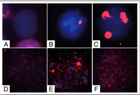
Figure 2. Pattern of SCP3 organization in male germ cells from 7 dpn mice. Representative merged immunofluorescence images showing SCP3 (red) organization on nuclear spreads (blue) in (A) undifferentiated spermatogonia, (B) differentiating spermatogonia, (C) prelep-totene spermatocytes, (D) early-lepprelep-totene spermatocytes (E) lepprelep-totene spermatocytes and (F) zygotene spermatocytes, obtained from freshly isolated or cultured spermatogonia.
© 2008 LANDES BIOSCIENCE. DO NOT
DISTRIBUTE.
(Fig. 8A) and inhibited the increase in phosphorylation levels of Akt and Erk1/2 (Fig. 8B). Since ATRA was activating both PI3K and MAPK pathways, we investigated if the effect was specifically medi-ated by a RAR receptor. We incubmedi-ated spermatogonia in the presence of 20 μM Ro-41-5253, an antagonist of RARα receptor, which at this concentration is also able to block the signalling of RARγ.37 As
shown in Figure 8C, we found that pre-incubation with Ro-41-5253 prevented ATRA-mediated phosphorylation of Akt and Erk1/2.
Figure 3. ATRA and KL promote meiotic in 7 dpn differentiating spermatogonia. (A) Histogram representing the fold of increase of meiotic nuclei in control spermatogonia or in cells stimulated with ATRA, KL or ATRA and KL for 24 h or 48 h. The values were obtained as a ratio of the percentage of nuclei with meiotic SCP3 staining at the different time points with the percentage of nuclei with meiotic SCP3 staining at the beginning of the culture (see Table 1 for absolute percentage values). Bars represent SD. (B) Percentage of leptotene (light green and IF left) and lepto/zygotene (dark green and IF right) nuclei in control cultures or cells treated for 48 h with ATRA, KL or ATRA and KL. Bars represent SD. (C) qRT-PCR for Dmc1 and Stra8 in spermatogonia incubated 24 h with ATRA or KL. (D) Immunoblot analysis of Stra8 and actin expression in spermatogonia cultured for 24 h in the absence or presence of ATRA or KL.
Table 1 Percentage of leptotene and lepto/zygotene
nuclei in culture
Cont ATRA KL ATRA + KL
T0 1.8% ± 0.5 - - -
(leptotene)
24 h 4.5% ± 0.22 7.9% ± 0.25 8.1% ± 0.21 7.2% ± 0.7 48 h 12.6% ± 0.2 23% ± 0.4 22.1% ± 0.9 19.6% ± 1.8
© 2008 LANDES BIOSCIENCE. DO NOT
DISTRIBUTE.
positive Intermediate and/or Type B spermatogonia) and/or that the pathways activated by the two factors might converge at some level of the pro-meiotic signalling cascade. Indeed, several evidences show that Kit positive spermatogonia are sensitive to both KL and ATRA (present results and refs. 8 and 21), which stimulate a significant increase of Stra8 (refs. 7 and 8 and present results), a fundamental regulator of both female and male meiosis.6,15,16 Importantly, ATRA
and KL similarly upregulate Dmc1, an early meiotic marker that is essential for the process of meiotic recombination.33
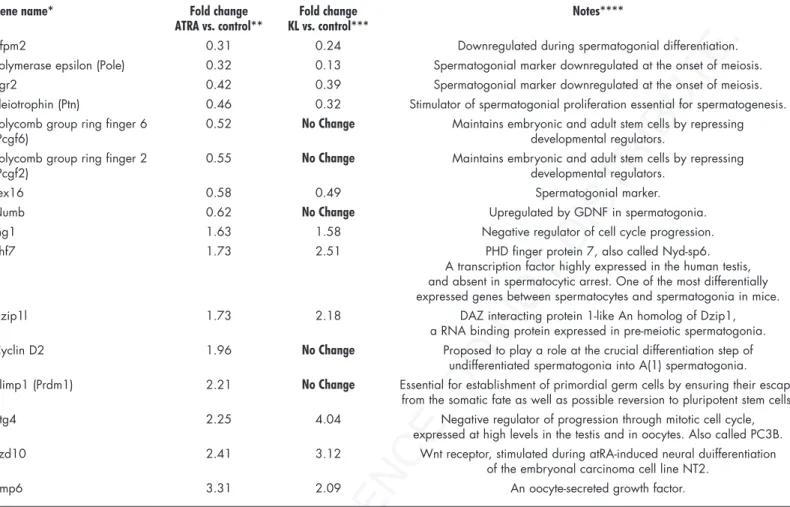
The evidence that ATRA increased the percentage of meiotic nuclei in isolated postnatal male germ cells confirms its role as a meiotic inducing substance. A microarray analysis performed on ATRA-stimulated spermatogonia (the entire set of raw and normal-ized data are available in the Array Express public repository at http:// www.ebi.ac.uk/arrayexpress, with the accession No. E-MEXP-1126) shows changes in the pattern of gene expression compatible with the ongoing spermatogonial differentiation. For instance, we found upregulation of cyclin D2, previously shown to be strongly upregu-lated in spermatogonia of the VAD testis after administration of retinoic acid,38 but also of early meiotic genes, such as Dzip1l, Phf7,
Bmp6 and Btg4 (Table 2). At the same time, ATRA downregulates the expression of stemness genes, such as Pcgf2, Pcgf6 and Numb, and of spermatogonial markers known to be turned off at the onset of meiosis, such as Zfpm2, Pole, Egr2, Ptn, Tex16 (Table 2).
The finding that KL regulates meiotic entry of spermatogonia is a novel finding and is in line with the previously reported in vivo expression pattern of KL (highly expressed in Sertoli cells when leptotene spermatocytes appear), and with the observation that male germ cells co-cultured with a KL expressing cell line can undergo transmeiotic differentiation in vitro.39 These results are also
consis-tent with the data gathered by wide genomic analysis performed with Affymetrix gene chips, which indicate that KL, similarly to ATRA, regulates spermatogonial markers and early meiotic genes.40
By comparing the effect of KL and ATRA on gene expression pattern we found that spermatogonial markers which are normally turned off at the onset of meiosis are downregulated also by KL treatment, whereas genes involved in stemness were not influenced. At the same time inhibitors of the mitotic cell cycle and early meiotic markers were all found to be equally upregulated by ATRA and by KL treat-ment40 (Table 2).
When undifferentiated Kit negative spermatogonia were stimu-lated in vitro with ATRA they underwent a partial differentiation only after 96 h, but they did not enter meiosis correctly. Although ATRA increased Kit and Stra8 levels and the percentage of SCP3-positive nuclei, these cells failed to correctly organize the synaptonemal complex and, as expected, KL did not show any effect. KL synergized with ATRA in the induction of abortive meiosis in 4 dpn sper-matogonia, probably because these cells became Kit positive during the concomitant exposure to ATRA, but they did not undergo a sufficient number of mitotic divisions necessary to become compe-tent to enter meiosis.
We also show that the action of ATRA on spermatogonia is paral-leled by a strong differentiative effect on Sertoli cells. In these cells ATRA induces expression of KL and BMP4 (known to promote proliferation and differentiation of spermatogonia29) and
down-regulation of GDNF, which is essential for stem cell renewal.35
Indeed, it is well established that RA in immature Sertoli cells is
Discussion
It has been recently proposed a model in which RA acts as a meiotic inducing substance (MIS)13 regulating, via its signalling
and metabolism, whether or not female and male germ cells initiate meiosis during embryogenesis.30 In this study we tested in vitro if
its mechanism of action might be conserved in postnatal germ cells. When spermatogonia were stimulated by ATRA, we found that the percentage of meiotic nuclei was significantly increased compared to the control. Interestingly, a similar effect was also exerted by KL, a growth factor essential for spermatogonial survival and proliferation both in vivo and in vitro.21,23,24 Simultaneous addition of ATRA and
KL did not produce any further increase in meiotic nuclei, suggesting that they might be acting on the same cell type (possibly the Kit
Figure 4. ATRA induces an abnormal SCP3 organization in cultured undif-ferentiated spermatogonia. (A) Immunoblot analysis of Kit, Stra8 and actin expression in 4 dpn spermatogonia cultured for 96 h in the absence or pres-ence of ATRA or KL. (B) Histogram representing the percentage of meiotic SCP3 organization in control spermatogonia or in cells stimulated with ATRA, KL or ATRA and KL for 96 h. (C) Representative pictures showing an abortive meiotic organization of SCP3.
© 2008 LANDES BIOSCIENCE. DO NOT
DISTRIBUTE.
synthesis in differentiating spermatogonia (Pellegrini et al., unpub-lished), and because KL has been shown to stimulate DNA synthesis in type A, but not in type B spermatogonia21 which are the
presump-tive target of stimulation of meiotic entry in the present experiments. Thence, we would favour the fourth possibility, i.e., a direct effect of ATRA and KL on the induction of meiotic competence. The fact that, in vivo, Kit positive spermatogonia enter meiosis after a series of controlled Kit-dependent mitotic divisions, and the evidence that Kit requires PI3K and MAPK pathways to regulate both mitotic required to promote spermatogonia
differen-tiation during the prepubertal spermatogenic wave.11
We found that ATRA and KL share a common molecular mechanism of action in inducing meiosis, since a specific inhibitor of the Kit tyrosine kinase (STI571) was able to revert the meiotic increase induced by the two agents. Both agents activate either the PI3K or the MAPK pathways which are essential to mediate meiotic entry of sper-matogonia in vitro. We observed that ATRA not only functions as a genomic inducer of Kit synthesis, but also as a rapid non genomic agent, by inducing Kit phosphorylation and activation. Such non-genomic effect, recently described also in neuronal systems41,42 is
dependent on a RA receptor, since Kit signal-ling pathways activated by ATRA in cultured spermatogonia were prevented by the incuba-tion with a specific RAR inhibitor.
The pro-meiotic effect of ATRA and KL that we observe in vitro might be explained by at least four different possible mechanisms (i) by an increase of survival of already meiosis committed cells (pre-leptotene spermato-cytes), (ii) by an increase of survival of Kit positive spermatogonia, which then might enter meiosis in a cell-autonomous manner; (iii) by an increase of mitotic proliferation of Kit positive spermatogonia, which then might enter meiosis in a cell-autonomous manner; or (iv) by the induction of meiotic competence in Kit positive spermatogonia. We can rule out the first possibility, since we estimated that the germ cell cultures obtained from 7 dpn mice contain only 4.6% of preleptotene spermatocytes, which cannot account for the increase of lepto-tene nuclei observed after 48 h of ATRA or KL stimulation (23%). Moreover, it has been demonstrated that preleptotene sper-matocytes do not express RARs10 and we
found no preleptotene figures in Kit positive immunomagnetic sorted spermatogonia (our unpublished observation). The second possi-bility can also be ruled out by the observation that, while addition of either LY294002 or
U0126 completely abolishes meiotic differentiation, these inhibitors are not able to suppress KL anti-apoptotic effects on cultured sper-matogonia.31 Moreover, we have previously shown that the induction
of an early meiotic gene expression pattern is evident after only 4 hr of KL stimulation, a period of time at which spermatogonial survival is not affected.40 Finally, we observe that ATRA-mediated increase of
meiotic nuclei requires the activation of Kit-dependent pathways, in the absence of any pro-survival effect of ATRA. The third possibility also seems to be unlikely, because ATRA does not stimulate DNA
Figure 5. ATRA increases Kit levels in germ cells during development. (A) Time course curve of ATRA stimulated spermatogonia on Kit protein levels after ATRA treatment. Actin is shown as loading control. (B) Northern blot analysis of Kit mRNA levels in isolated germ cells cultured for 24 h with or without ATRA stimulation. Smad5 and Alk3 were included as specific markers for spermatogonial population.29
28S and 18S are shown as loading and integrity controls. (C) Kit immunoblotting on spermatogonia treated with Actinomycin D and/or ATRA for 2 h or 24 h. Actin is shown as loading control. (D) Kit, Ret and RARγ protein levels after 24 h of ATRA stimulation in 7 dpn spermatogonia. (E) Kit levels in PGCs isolated from 11.5 dpc embryos and in hematopoietic stem cells 24 h after ATRA stimulation.
© 2008 LANDES BIOSCIENCE. DO NOT
DISTRIBUTE.
DNA synthesis31 and meiotic entry of spermatogonia (this paper),
suggest that a link exists between Kit-mediated proliferation and the switch into the meiotic cell cycle. KL induces a gene expression pattern in differentiating spermatogonia40 that is consistent with a
progressive lengthening of the S phase and a progressive shortening of the G2/M transition, both events known to occur in vivo in male germ cells during the switch from mitosis to meiosis.43 Presumably,
during these Kit-mediated mitotic divisions, accumulation of key factors required for meiosis occur. One of these factors is certainly Stra8, which has been proposed to be essential for the last round of pre-meiotic DNA synthesis in female fetal germ cells,6 and to
regu-late the switch from a mitotic pattern of cell division to the meiotic pattern in postnatal male germ cells.15,16
In conclusion, we propose a model (Fig. 9) in which RA acts on the somatic cell compartment of the testis, through the induction of KL and on the germ cell compartment, as a direct pro-meiotic factor sharing with KL the same signalling pathway.
Materials and Methods
Cell isolation and culture. Spermatogonia were obtained as we
previously reported21 by differential enzymatic digestion of testes from
4 to 7 days post natum (dpn) CD1 albino mice. To precisely define the temporal appearance of Kit expressing spermatogonia, a transgenic line (p18) expressing EGFP under the control of the c-Kit promoter and expanded on a CD1 background,25,26 was used. Isolation of Kit
positive spermatogonia was performed by using magnetic-activated cell sorting (MACS) with CD117 conjugated microbeads (Miltenyi Biotec, Germany). Isolated germ cells were then treated with different factors and cultured for 24 h or 48 h prior to immunofluorescence, western blotting analysis or mRNA preparation. Fresh medium and growth factors were replaced after 24 h when culturing for longer times. PGCs were isolated from 11.5 days post coitum (dpc) embryos as we previously reported in ref.27 Spermatogonia were cultured in
modified Earle medium with 100 U/ml penicillin, 100 μg/ml strep-tomycin, 20 mM glutamine without serum supplementation, while
PGCs were cultured in spermatogonia medium supplemented with 10% FCS. KL-dependent SV40T-immortalized hematopoietic cells from bone marrow were cultured in RPMI supplemented with 10% FCS and 10 ng/ml KL. Sertoli cell cultures were prepared according to reference 21.
All-trans-retinoic acid (ATRA, Sigma) was dissolved in ethanol and used in a range of 0.03–3 μM. KL and BMP4 were purchased from Società Italiana Chimici (Rome, Italy) and used at 100 ng/ml. STI571, a generous gift of Dr. L. Gnessi (University of Rome “La Sapienza”), was used at 5 μM concentration and Ro 41-5253 (Biomol, DBA Italy) at 7 and 20 μM. DibutyrilcyclicAMP (Sigma) was used at 1 mM concentration. The MAPK inhibitor U0126 (Promega, Italy) and PI3K inhibitor LY294002 (Alexis, Italy) were used at 10 μM concentration.
Figure 6. ATRA regulates in Sertoli cells the expression of specific growth factors required for germ cells differentiation. Northern blot analysis (A) for BMP4 and KL, and (B) for GDNF expression in Sertoli cells. SC cultures were obtained from 4 dpn and 7 dpn testes and stimulated o/n with dibutyril-cAMP (dbdibutyril-cAMP, 1 mM), with 0.3 μM or 3 μM ATRA. 28S and 18S or actin were shown as loading and integrity controls.
Figure 7. Inhibition of Kit signalling by STI571 completely reverts meiotic progression induced by ATRA and KL. (A) Histogram representing the folds of induction of meiotic nuclei, evaluated as above, in control spermatogonia or in cells stimulated with ATRA, KL or ATRA and KL for 24 h or 48 h in the presence or absence of 5 μM STI571. (B) Immunoblot analysis of Kit activated pathways in control spermatogonia or in cells stimulated for 15 minutes with ATRA or KL, pretreated for 30 minutes with 5 μM STI571.
© 2008 LANDES BIOSCIENCE. DO NOT
DISTRIBUTE.
nitrocellulose membrane (Amersham). The membrane was blocked in PBS-5% skim milk powder for 1 h. Incubation of the membrane with the primary antibody was carried out at 4°C o/n in PBS-5% BSA and then with the appropriate horseradish peroxidase-conju-gated secondary antibody (SantaCruz). Anti-Kit rabbit polyclonal (sc-6283), anti phospho-Kit (Tyr 721) (sc-23766) rabbit polyclonal, phospho Erk1/2 mouse monoclonal antibody (sc-7383), anti-actin rabbit polyclonal (sc-7210), anti-Ret rabbit polyclonal (sc-167), anti-RARγ mouse monoclonal (sc-7387) were from SantaCruz, anti-phospho Akt (Ser-473) was from New England Biolabs, anti Stra-8 antibodies were from Abcam. The horseradish peroxidase conjugate was detected by chemioluminescence with an ECL Kit (Amersham) and autofluorography. For each experiment western blotting analysis were repeated at least four times and densitometry was performed using a Molecular Dynamics Densitometer and ImageQuant soft-ware.
Northern blotting and quantitative RT PCR analyses.
Spermatogonia were collected into Trizol (Invitrogen) and RNA was purified according to manufacturers suggestions. For Northern blot analysis, total RNA was run in a 1% formaldehyde-agarose gel and blotted onto a Nylon membrane (Hybond-N, Amersham, Viability of 4 and 7 dpn spermatogonia was evaluated by Trypan
blue (Sigma) exclusion.
Immunofluorescence and western blotting. For cell spreads,
spermatogonia cultured for 24, 48 or 96 h were prepared and stained essentially as described in ref.28 Slides were washed twice in PBS,
and incubated o/n at 4°C with SCP3 rabbit polyclonal anti-body (Abcam, Cambridge, UK) diluted in blocking solution (10% goat serum, 3% BSA, 0.5% Triton X-100 in PBS). After washing, secondary antibody was added for 1 h at 37°C. The slides were washed and allowed to dry. Vectashield Mounting Medium with DAPI (Vector Laboratories, Burlingame, CA, USA) was added and the slides were viewed using a Leica microscope. Spreads analysis was performed in five independent experiments. A representative morphological classification of SCP3 organization in various types of germ cells obtained in vitro is reported in Figure 2. For immu-nofluorescence, spermatogonia cell suspensions were adhered onto poly-L-lysine coated slides and treated as previously described.29
For western analysis cells were lysed in 1% Triton X-100, 150 mM NaCl, 15 mM MgCl2, 15 mM EGTA, 10% Glycerol, 50 mM Hepes (pH 7.4) with protease inhibitors. Proteins were separated by SDS-10% polyacrylamide gel electrophoresis and transferred to
Figure 8. MAPK and PI3K signalling pathways activated by ATRA and KL are required for meiotic entry of spermatogonia. (A) Histogram representing the folds of induction of meiotic nuclei, evaluated as above, in control spermatogonia or in cells stimulated with ATRA and KL for 48 h in the presence or absence of UO126 or LY29406. (B) Western blot analysis of Kit activated pathways in control spermatogonia or in cells stimulated for 15 minutes with ATRA or KL, pretreated with 10 μM UO126 or 10 μM LY29406. (C) Western blot analysis of Kit activated pathways in control spermatogonia or in cells stimulated for 15 minutes with ATRA, pre-treated for 30 min with 20 μM Ro 41-5253.
© 2008 LANDES BIOSCIENCE. DO NOT
DISTRIBUTE.
GAT GGC CGG GGC CAT ACC TT and rev 5'-TCA GCG GCA TCC ACA CCC CTC TAC CAC CAT; GDNF for 5'GG AGT TAA TGT CCA ACT GGG and rev 5' TAC ATC CAC ACC GTT TAG CG; Alk3 for 5'-ACT TTA GCA CCA GAG GAT ACC and rev 5'-TTT TCA CCA CGC CAT TTA CCC; Smad5 for 5' AAT GAC GTC AAT GGC CAG CTT and rev 5' AGA AGA AAT GGG GTT CAG CG; β-actin for 5'GGT TCC GAT GCC CTG AGG CTC and rev 5'-ACT TGC GGT GCA CGA TGG AGG (also used for normalization in qRT-PCR).
Statistical analysis. The Student t-test and ANOVA have been
used to assess the significance, set at p < 0.05. All experiments were performed from three to five times and at least in triplicate for each sample. Bars in the histograms represent standard deviations obtained from the indicated independent experiments.
UK) using 10x saline sodium citrate (SSC) buffer. Probes were obtained by RT-PCR from spermatogonia cDNAs (Smad5, Alk3 and Kit) or from Sertoli cell cDNAs (KL, BMP4 and GDNF). PCR products were 32P-labelled with a random prime labelling
system (Perkin Elmer). Hybridization was carried out following Quick Hybrid System’s instruction (Stratagene). For Quantitative PCRs, contaminating genomic DNA was removed using DNase I Amplification Grade (Invitrogen) and cDNA synthesis was subse-quently performed with the SuperScript™ III First-Strand Synthesis System (Invitrogen). Quantitative PCR was performed for Stra8 and Dmc1 genes using SYBR green I fluorescent dye (EPPENDORF) and standard deviations were calculated. Primer sequences for qRT-PCR were: Stra8 for 5'-GTT TCC TGC GTG TTC CAC AAG and rev 5'-CAC CCG AGG CTC AAG CTTC; Dmc1: for 5'-CAT ATC ACT ACT GGG AGC and rev 5'-GTA CTG CTT CAT GGT CTAC. Primer sequences for amplification of probes for Northern blot analysis were: Kit for 5'-TAT GGA CAT GAA GCC TGG CGT and rev 5'-CAT TCC TGA TGT CTC TGG CTA GC; KL for 5'-AAC AGC TAA ACG GAG TCG CC and rev 5'-ACA GTG TTG ATA CAA GCC AC; BMP4 for 5'-TTT GGC CAT
Table 2 List of genes regulated by ATRA treatment of mouse spermatogonia from 7 dpn mice cultured for 24 hr and
comparison with the effect of KL treatment on the same cells
Gene name* Fold change Fold change Notes****
ATRA vs. control** KL vs. control***
Zfpm2 0.31 0.24 Downregulated during spermatogonial differentiation. Polymerase epsilon (Pole) 0.32 0.13 Spermatogonial marker downregulated at the onset of meiosis. Egr2 0.42 0.39 Spermatogonial marker downregulated at the onset of meiosis. Pleiotrophin (Ptn) 0.46 0.32 Stimulator of spermatogonial proliferation essential for spermatogenesis. Polycomb group ring finger 6 0.52 No Change Maintains embryonic and adult stem cells by repressing
(Pcgf6) developmental regulators.
Polycomb group ring finger 2 0.55 No Change Maintains embryonic and adult stem cells by repressing
(Pcgf2) developmental regulators.
Tex16 0.58 0.49 Spermatogonial marker.
Numb 0.62 No Change Upregulated by GDNF in spermatogonia. Ing1 1.63 1.58 Negative regulator of cell cycle progression. Phf7 1.73 2.51 PHD finger protein 7, also called Nyd-sp6.
A transcription factor highly expressed in the human testis, and absent in spermatocytic arrest. One of the most differentially expressed genes between spermatocytes and spermatogonia in mice. Dzip1l 1.73 2.18 DAZ interacting protein 1-like An homolog of Dzip1,
a RNA binding protein expressed in pre-meiotic spermatogonia. Cyclin D2 1.96 No Change Proposed to play a role at the crucial differentiation step of
undifferentiated spermatogonia into A(1) spermatogonia.
Blimp1 (Prdm1) 2.21 No Change Essential for establishment of primordial germ cells by ensuring their escape from the somatic fate as well as possible reversion to pluripotent stem cells. Btg4 2.25 4.04 Negative regulator of progression through mitotic cell cycle,
expressed at high levels in the testis and in oocytes. Also called PC3B. Fzd10 2.41 3.12 Wnt receptor, stimulated during atRA-induced neural duifferentiation
of the embryonal carcinoma cell line NT2. Bmp6 3.31 2.09 An oocyte-secreted growth factor.
*Transcripts up or downregulated at least 1.5-fold by ATRA treatment in both of two separate microarray experiments performed with mouse Genome 430 2.0 GeneChip arrays (Affymetrix Inc.,). RNA for microarray analysis was extracted from spermatogonia cultured for 24 hr in the presence or absence of 0.3 μM ATRA. The analysis was performed on duplicate chip arrays, using cRNAs from two different RNA pools, each obtained from two different culture experiments. The entire set of raw and normalized data are available in the Array Express public repository at http://www.ebi.ac.uk/arrayexpress (Accession No.: E-MEXP-1126). **The indicated value of fold change is the average of the two separate microarray experiments. Validity of the data of microarray analysis was confirmed by performing semi-quantitative RT-PCR analysis for randomly selected transcripts on RNA preparations obtained from different cell preparations (not shown). Details about preparation of the samples, bioinformatic and statistical analysis have been described elsewhere.40 ***KL concentration used was
© 2008 LANDES BIOSCIENCE. DO NOT
DISTRIBUTE.
References
1. Huckins C. Cell cycle properties of differentiating spermatogonia in adult Sprague-Dawley rats. Cell Tissue Kinet 1971; 4:139-54.
2. Oakberg EF. Spermatogonial stem-cell renewal in the mouse. Anat Rec 1971; 169:515-31. 3. Schrans-Stassen BH, van de Kant HJ, de Rooij DG and van Pelt AM. Differential
expression of c-kit in mouse undifferentiated and differentiating type A spermatogonia. Endocrinology 1999; 140:5894-900.
4. Chung SS and Wolgemuth DJ. Role of retinoid signaling in the regulation of spermatogen-esis. Cytogenet Genome Res 2004; 105:189-202.
5. van Pelt AM and de Rooij DG. Synchronization of the seminiferous epithelium after vita-min A replacement in vitavita-min A-deficient mice. Biol Reprod 1990; 43:363-7.
6. Baltus AE, Menke DB, Hu YC, Goodheart ML, Carpenter AE, de Rooij DG, et al. In germ cells of mouse embryonic ovaries, the decision to enter meiosis precedes premeiotic DNA replication. Nat Genet 2006; 38:1430-4.
7. Wang Y and Culty M. Identification and distribution of a novel platelet-derived growth factor receptor beta variant: effect of retinoic acid and involvement in cell differentiation. Endocrinology 2007; 148:2233-50.
8. Zhou Q, Li Y, Nie R, Friel P, Mitchell D, Evanoff RM, et al. Expression of stimulated by retinoic acid gene 8 (Stra8) and maturation of murine gonocytes and spermatogonia induced by retinoic acid in vitro. Biol Reprod 2008; 78:537-45.
Acknowledgements
We thank D. Farini for providing the RA inhibitor Ro 41-5352, L. Gnessi for providing the Kit inhibitor STI571, and S. Ottolenghi for providing the p18 transgenic line; M. De Felici, M. Barchi and A. Celeste for helpful suggestions on the manuscript. This work was supported by grants from the Italian Ministry of University (Prin 2005059793_005, 2003052043_003, and 200788TPYE_002).
Note
Supplementary materials can be found at:
www.landesbioscience.com/supplement/PelligriniCC7-24-Sup.pdf
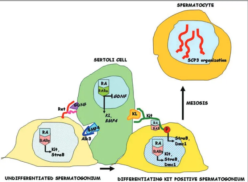
Figure 9. Schematic representation of RA and KL effects on the early steps of spermatogenic differentiation. RA acts in Sertoli cells through the RARα receptor to stimulate the synthesis of growth factors (KL and BMP4), which are essential for germ cell differentiation, and to inhibit the synthesis of the critical growth factor required for spermatogonial stem cell self-renewal (GDNF). RA acts also on undifferentiated and differentiating spermatogonia through the RARγ receptor to stimulate the synthesis of Stra8 and the KL receptor (Kit) but not BMP4 and GDNF receptors (Alk3 and Ret, respectively). Furthermore, similarly to KL, RA directly activates Kit phosphorylation and its signalling cascade in differentiating spermatogonia, through a non genomic mechanism. Activation of Kit dependent signalling is required for differentiation of spermatogonia into spermatocytes , as revealed by the induction of early meiotic markers, such as Dmc1, and by the meiotic organization of nuclear SCP3.
© 2008 LANDES BIOSCIENCE. DO NOT
DISTRIBUTE.
9. Ghyselinck NB, Vernet N, Dennefeld C, Giese N, Nau H, Chambon P, et al. Retinoids and spermatogenesis: lessons from mutant mice lacking the plasma retinol binding protein. Dev Dyn 2006; 235:1608-22.
10. Vernet N, Dennefeld C, Rochette-Egly C, Oulad-Abdelghani M, Chambon P, Ghyselinck NB, et al. Retinoic acid metabolism and signaling pathways in the adult and developing mouse testis. Endocrinology 2006; 147:96-110.
11. Vernet N, Dennefeld C, Guillou F, Chambon P, Ghyselinck NB and Mark M. Prepubertal testis development relies on retinoic acid but not rexinoid receptors in Sertoli cells. Embo J 2006; 25:5816-25.
12. Lufkin T, Lohnes D, Mark M, Dierich A, Gorry P, Gaub MP, et al. High postnatal lethality and testis degeneration in retinoic acid receptor alpha mutant mice. Proc Natl Acad Sci USA 1993; 90:7225-9.
13. Bowles J, Knight D, Smith C, Wilhelm D, Richman J, Mamiya S, et al. Retinoid signaling determines germ cell fate in mice. Science 2006; 312:596-600.
14. MacLean G, Li H, Metzger D, Chambon P and Petkovich M. Apoptotic extinction of germ cells in testes of Cyp26b1 knockout mice. Endocrinology 2007; 148:4560-7.
15. Anderson EL, Baltus AE, Roepers-Gajadien HL, Hassold TJ, de Rooij DG, van Pelt AM et al. Stra8 and its inducer, retinoic acid, regulate meiotic initiation in both spermatogenesis and oogenesis in mice. Proc Natl Acad Sci USA 2008; [Epub ahead of print].
16. Mark M, Jacobs H, Oulad-Abdelghani M, Dennefeld C, Feret B, Vernet N, et al. STRA8-deficient spermatocytes initiate, but fail to complete, meiosis and undergo premature chromosome condensation. J Cell Sci 2008; 121:3233-42.
17. Dolci S, Williams DE, Ernst MK, Resnick JL, Brannan CI, Lock LF, et al. Requirement for mast cell growth factor for primordial germ cell survival in culture. Nature 1991; 352:809-11. 18. Godin I, Deed R, Cooke J, Zsebo K, Dexter M and Wylie CC. Effects of the steel gene
product on mouse primordial germ cells in culture. Nature 1991; 352:807-9. 19. Broudy VC. Stem cell factor and hematopoiesis. Blood 1997; 90:1345-64.
20. Bernex F, De Sepulveda P, Kress C, Elbaz C, Delouis C and Panthier JJ. Spatial and tem-poral patterns of c-kit-expressing cells in WlacZ/+ and WlacZ/WlacZ mouse embryos. Development 1996; 122:3023-33.
21. Rossi P, Dolci S, Albanesi C, Grimaldi P, Ricca R and Geremia R. Follicle-stimulating hormone induction of steel factor (SLF) mRNA in mouse Sertoli cells and stimulation of DNA synthesis in spermatogonia by soluble SLF. Dev Biol 1993; 155:68-74.
22. Sorrentino V, Giorgi M, Geremia R, Besmer P and Rossi P. Expression of the c-kit proto-oncogene in the murine male germ cells. Oncogene 1991; 6:149-51.
23. Blume-Jensen P, Jiang G, Hyman R, Lee KF, O’Gorman S and Hunter T. Kit/stem cell factor receptor-induced activation of phosphatidylinositol 3'-kinase is essential for male fertility. Nat Genet 2000; 24:157-62.
24. Kissel H, Timokhina I, Hardy MP, Rothschild G, Tajima Y, Soares V, et al. Point mutation in kit receptor tyrosine kinase reveals essential roles for kit signaling in spermatogenesis and oogenesis without affecting other kit responses. Embo J 2000; 19:1312-26.
25. Cairns LA, Moroni E, Levantini E, Giorgetti A, Klinger FG, Ronzoni S, et al. Kit regulatory elements required for expression in developing hematopoietic and germ cell lineages. Blood 2003; 102:3954-62.
26. Filipponi D, Hobbs RM, Ottolenghi S, Rossi P, Jannini EA, Pandolfi PP, et al. Repression of kit expression by Plzf in germ cells. Mol Cell Biol 2007; 27:6770-81.
27. Dolci S and De Felici M. A study of meiosis in chimeric mouse fetal gonads. Development 1990; 109:37-40.
28. Romanienko PJ and Camerini-Otero RD. The mouse Spo11 gene is required for meiotic chromosome synapsis. Mol Cell 2000; 6:975-87.
29. Pellegrini M, Grimaldi P, Rossi P, Geremia R and Dolci S. Developmental expression of BMP4/ALK3/SMAD5 signaling pathway in the mouse testis: a potential role of BMP4 in spermatogonia differentiation. J Cell Sci 2003; 116:3363-72.
30. Koubova J, Menke DB, Zhou Q, Capel B, Griswold MD and Page DC. Retinoic acid regulates sex-specific timing of meiotic initiation in mice. Proc Natl Acad Sci USA 2006; 103:2474-9.
31. Dolci S, Pellegrini M, Di Agostino S, Geremia R and Rossi P. Signaling through extracel-lular signal-regulated kinase is required for spermatogonial proliferative response to stem cell factor. J Biol Chem 2001; 276:40225-33.
32. Feng LX, Ravindranath N and Dym M. Stem cell factor/c-kit upregulates cyclin D3 and promotes cell cycle progression via the phosphoinositide 3-kinase/p70 S6 kinase pathway in spermatogonia. J Biol Chem 2000; 275:25572-6.
33. Pittman DL, Cobb J, Schimenti KJ, Wilson LA, Cooper DM, Brignull E, et al. Meiotic pro-phase arrest with failure of chromosome synapsis in mice deficient for Dmc1, a germline-specific RecA homolog. Mol Cell 1998; 1:697-705.
34. Novak I, Lightfoot DA, Wang H, Eriksson A, Mahdy E and Hoog C. Mouse embryonic stem cells form follicle-like ovarian structures but do not progress through meiosis. Stem Cells 2006; 24:1931-6.
35. Meng X, Lindahl M, Hyvonen ME, Parvinen M, de Rooij DG, Hess MW, et al. Regulation of cell fate decision of undifferentiated spermatogonia by GDNF. Science 2000; 287:1489-93. 36. Gambacorti-Passerini C, Tornaghi L, Cavagnini F, Rossi P, Pecori-Giraldi F, Mariani L, et al.
Gynaecomastia in men with chronic myeloid leukaemia after imatinib. Lancet 2003; 361:1954-6.
37. Apfel C, Bauer F, Crettaz M, Forni L, Kamber M, Kaufmann F, et al. A retinoic acid recep-tor alpha antagonist selectively counteracts retinoic acid effects. Proc Natl Acad Sci USA 1992; 89:7129-33.
38. Beumer TL, Roepers-Gajadien HL, Gademan IS, Kal HB and de Rooij DG. Involvement of the D-type cyclins in germ cell proliferation and differentiation in the mouse. Biol Reprod 2000; 63:1893-8.
39. Vincent S, Segretain D, Nishikawa S, Nishikawa SI, Sage J, Cuzin F, et al. Stage-specific expression of the Kit receptor and its ligand (KL) during male gametogenesis in the mouse: a Kit-KL interaction critical for meiosis. Development 1998; 125:4585-93.
40. Rossi P, Lolicato F, Grimaldi P, Dolci S, Di Sauro A, Filipponi D, et al. Transcriptome analysis of differentiating spermatogonia stimulated with kit ligand. Gene Expr Patterns 2008; 8:58-70.
41. Canon E, Cosgaya JM, Scsucova S and Aranda A. Rapid effects of retinoic acid on CREB and ERK phosphorylation in neuronal cells. Mol Biol Cell 2004; 15:5583-92.
42. Masia S, Alvarez S, de Lera AR and Barettino D. Rapid, nongenomic actions of retinoic acid on phosphatidylinositol-3-kinase signaling pathway mediated by the retinoic acid receptor. Mol Endocrinol 2007; 21:2391-402.
43. Monesi V. Autoradiographic study of DNA synthesis and the cell cycle in spermatogonia and spermatocytes of mouse testis using tritiated thymidine. J Cell Biol 1962; 14:1-18.