The Rockefeller University Press $30.00
CD95 predominantly acts as a death receptor
when cross-linked with its CD95 ligand (CD95L)
using a well-characterized pathway. Upon ligand
binding, Fas-associated death domain associates
with CD95, followed by recruitment of the
initi-ator caspase-8 to form the death-inducing
signal-ing complex. Caspase-8 oligomerization initiates
its autocatalytic cleavage, followed by the release
of active caspase fragments into the cytosol,
sub-sequent activation of effector caspases, DNA
frag-mentation, and cleavage of cellular substrates (1).
Together with the Bcl-2 homology 3–only
mol-ecule Bcl-2–interacting mediator of death, the
CD95/CD95L system contributes to the deletion
of activated T cells during the termination phase
of an immune response (2–4). Although the
CD95/CD95L system plays a key role in T cell
apoptosis and immunohomeostasis as indicated
by the phenotype of lymphoproliferation mice
and the induction of autoimmunity and
lympho-proliferation in patients with mutations in either
the receptor or the ligand (5), CD95 mediates
additional functions apart from cell death
induc-tion, including amplification of T cell
prolifer-ation upon co-stimulprolifer-ation with suboptimal doses
of anti-CD3 antibodies (6–8). Nonapoptotic
functions of CD95 have also been identified for
cells of the central nervous system promoting
neuronal development, growth, differentiation,
and regeneration (9, 10). CD95 has also been
re-ported to induce tumor growth in lung, thyroid,
CORRESPONDENCE Gudrun Strauss:
[email protected] Abbreviations used: AAD, amino-actinomycin-D; CD95L, CD95 ligand; DEVD, Asp-Glu-Val-Asp; EMSA, electrophoretic mobility shift assay; IETD, Ile-Glu-Thr-Asp; LAT, linker of activated T cells; m-CD95L, membrane-bound CD95L; MAPK, mitogen-activated protein kinase; PKC, protein kinase C; PLC, phospholipase C; SEE: staphylococcal entero-toxin E; Z-VAD.fmk, Z-Val-Ala-DL-Asp-fluoromethylketone; ZAP70, -chain–associated protein of 70 kD.
J.A. Lindquist and N. Arhel contributed equally to this paper.
CD95 co-stimulation blocks activation
of naive T cells by inhibiting T cell
receptor signaling
Gudrun Strauss,
1Jonathan A. Lindquist,
2Nathalie Arhel,
3Edward Felder,
4Sabine Karl,
1Tobias L. Haas,
5Simone Fulda,
1Henning Walczak,
6Frank Kirchhoff,
3and Klaus-Michael Debatin
11University Children’s Hospital Ulm, 89075 Ulm, Germany
2Institute of Molecular and Clinical Immunology, Otto-von-Guericke University, 39120 Magdeburg, Germany 3Institute of Virology and 4Institute of General Physiology, University of Ulm, 89075 Ulm, Germany 5Division of Apoptosis Regulation, German Cancer Research Center, 69120 Heidelberg, Germany 6Department of Immunology, Division of Medicine, Imperial College London, London W12 ONN, UK
CD95 is a multifunctional receptor that induces cell death or proliferation depending on
the signal, cell type, and cellular context. Here, we describe a thus far unknown function of
CD95 as a silencer of T cell activation. Naive human T cells triggered by antigen-presenting
cells expressing a membrane-bound form of CD95 ligand (CD95L) or stimulated by
anti-CD3 and -CD28 antibodies in the presence of recombinant CD95L had reduced activation
and proliferation, whereas preactivated, CD95-sensitive T cells underwent apoptosis.
Trig-gering of CD95 during T cell priming interfered with proximal T cell receptor signaling by
inhibiting the recruitment of -chain–associated protein of 70 kD, phospholipase-, and
protein kinase C- into lipid rafts, thereby preventing their mutual tyrosine protein
phos-phorylation. Subsequently, Ca
2+mobilization and nuclear translocation of transcription
factors NFAT, AP1, and NF-B were strongly reduced, leading to impaired cytokine
secre-tion. CD95-mediated inhibition of proliferation in naive T cells could not be reverted by the
addition of exogenous interleukin-2 and T cells primed by CD95 co-stimulation remained
partially unresponsive upon secondary T cell stimulation. HIV infection induced CD95L
expression in primary human antigeen-presenting cells, and thereby suppressed T cell
activation, suggesting that CD95/CD95L-mediated silencing of T cell activation represents
a novel mechanism of immune evasion.
© 2009 Strauss et al. This article is distributed under the terms of an Attribu-tion–Noncommercial–Share Alike–No Mirror Sites license for the first six months after the publication date (see http://www.jem.org/misc/terms.shtml). After six months it is available under a Creative Commons License (Attribution–Noncom-mercial–Share Alike 3.0 Unported license, as described at http://creativecommons .org/licenses/by-nc-sa/3.0/).
The Journal of Experimental Medicine
on December 5, 2016
Downloaded from
Supplemental Material can be found at:
on December 5, 2016
Downloaded from
on December 5, 2016
Downloaded from
on December 5, 2016
Downloaded from
on December 5, 2016
Downloaded from
on December 5, 2016
Downloaded from
on December 5, 2016
Downloaded from
signaling events. We also demonstrate that HIV-1 infection
ef-ficiently induced CD95L surface expression in infected
macro-phages and dendritic cells, and that antigen-loaded, CD95L-
expressing macrophages prevent activation of Jurkat cells. Our
data suggest a new function of CD95 as a silencer of T cell
acti-vation, and CD95L expression on APCs might represent a novel
mechanism of viral immune evasion and tolerance induction.
RESULTS
m-CD95L–expressing APCs prevent T cell activation
and proliferation
CD95 sensitivity in human T cells depends on their activation
status. Although naive T cells are CD95 resistant, CD95
sensi-tivity develops 6–7 d after T cell activation (26). We have
pre-viously shown that expression of a noncleavable m-CD95L
and HLA-A1 on APCs efficiently prevents development of
HLA-A1–specific T cells in long-term T cell cultures in vitro
(25). In this study, we analyze whether CD95L-expressing
APCs in fact delete antigen-specific T cells or whether CD95
triggering instead prevents antigen-specific T cell priming in
naive T cells. Therefore, we stimulated CFSE-labeled
HLA-A1
T cells with the lymphoblastoid cell line C1R.A1.CD95L
expressing the alloantigen HLA-A1 and m-CD95L (25). As
controls, we used mock transfectant (C1R.A1.puro;
HLA-A1
+and m-CD95L
) or medium. Activation by HLA-A1 in
the absence of m-CD95L (C1R.A1.puro) induced T cell
pro-liferation in CD4
+and CD8
+T cells starting at day 6.
Prolifer-ation of CD4
+T cells is caused by the expression HLA-Cl II
antigens by the stimulator cells. In contrast, the presence of
m-CD95L (C1R.A1.CD95L) abolished proliferation in both
T cell subsets (Fig. 1 A). Inhibition of proliferation by CD95L
was associated with a significant reduction in the expression of
activation markers on CD4
+and CD8
+T cells at day 6, when
the first T cell divisions were detectable (Fig. 1 B). The
secre-tion of the Th1-specific cytokines IFN- and IL-2 was strongly
reduced in the presence of m-CD95L, and secretion of the
Th2 cytokine IL-10 was totally impaired in T cells activated
with C1R.A1.CD95L after 1, 3, and 5 d compared with
T cells stimulated with the mock transfectant (Fig. 1 C). No
sig-nificant differences were detected for TNF-. These results
demonstrate that the expression of m-CD95L on APCs
im-pairs T cell activation and proliferation.
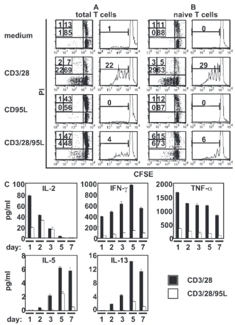
Simultaneous triggering of CD95 and TCR induces
apoptosis in activated human T cells and inhibits
proliferation of naive T cells
HLA-A1–expressing APCs only activate alloantigen-specific
T cells, which constitute 3–10% of total T cells. To stimulate
T cells independently of antigen specificity, in our
experi-ments we substituted APCs with immobilized anti-CD3 and
anti-CD28 antibodies in combination with plate-bound
re-combinant CD95L. Therefore, CFSE-labeled T cells were
stimulated with immobilized CD3 and CD28
anti-bodies in the absence (CD3/28) or presence of CD95L
(CD3/28/95L). After 6 d, proliferation of T cells was
effi-ciently inhibited in the presence of CD95L compared with
and ovarian cancer, and to trigger basal invasion of
glioblas-toma in vivo (11–13). T cells inhibited in caspase activation
(8) or T cells deficient for Fas-associated death domain (14,
15), caspase-8 (16, 17), or flice-like inhibitory protein (18)
exhibited impaired T cell activation and proliferation,
sug-gesting an essential role for molecules downstream of the
CD95 pathway in T cell activation.
T cell activation is initiated by binding of the TCR to the
appropriate antigen presented by HLA molecules, followed by
translocation of the TCR and its associated signaling molecules
into lipid rafts, which are microdomains of the plasma
mem-brane enriched in cholesterol and glycosphingolipids. By
in-ducing close proximity of signaling molecules, rafts serve as
signaling platforms (19, 20). Src family protein tyrosine kinases
(lymphocyte-specific kinase and p59
fyn) subsequently
phos-phorylate the immunoreceptor tyrosine-based activation
mo-tifs of the CD3 chains, followed by recruitment and activation
of -chain-associated protein of 70 kD (ZAP-70). After
phos-phorylation by ZAP-70, the transmembrane adaptor linker of
activated T cells (LAT) and the cytosolic adaptor protein SLP-76
constitute docking proteins (e.g., for PLC-), which then
hydrolyzes phosphatidylinositol 3,4-bisphophate into the
sec-ondary messengers inositol 1,4,5-triphosphate and
diacylglyc-erol to initiate Ca
2+influx and activation of protein kinase C-
(PKC-) and the mitogen activated protein kinase (MAPK)
cascade, finally resulting in the activation of transcription
through NFAT, NF-B, and AP-1 (21). In contrast to TCR
signaling, the requirement for lipid raft formation in CD95
signaling is controversially debated. Although the association
of CD95 with lipid rafts was reported to define the CD95
sen-sitivity of T cells and to render activated T cells sensitive to
apoptosis after TCR stimulation (22), no requirement for raft
formation in CD95-mediated death was reported in a B cell
line (23). Recent studies, however, suggest that molecules of
the CD95 pathway, such as caspase-8 and the c-flice–like
in-hibitory protein
L, are essential components of rafts induced
after TCR ligation and are associated with NF-B adaptors
during T cell activation (24).
T cell activation and T cell death are tightly controlled
pro-cesses to guarantee both efficacy of the immune response and
prevention of autoimmunity. Whether or not cells undergo
apoptosis or start proliferation is defined by cell type, activation
status, and co-stimulatory signals. We have previously shown
that APCs expressing a membrane-bound form of CD95L
(m-CD95L) and the alloantigen HLA-A1 are able to prevent
the outgrowth of HLA-A1–specific T cells in long-term T cell
stimulation cultures and to efficiently induce apoptosis in
pre-activated CD95-expressing T cells (25), suggesting that
HLA-A1–specific T cells were deleted after antigen-specific priming
by the CD95/CD95L system. In this study, we analyzed the
ef-fect of CD95 triggering during the activation of primary human
T cells using CD95L-expressing APCs or CD3 and
anti-CD28 antibodies together with recombinant CD95L. While
activated, CD95-sensitive T cells underwent apoptosis, naive
T cells were inhibited in proliferation, and CD95 triggering
dur-ing T cell primdur-ing directly interfered with early proximal TCR
on December 5, 2016
Because T cells used in this study were purified from human
buffy coats and contained on average 30–50% preactivated,
CD95-sensitive T cells, constitutive CD95 sensitivity may
account for the observed high levels of apoptosis induction.
Therefore, we purified naive T cells from the T cell pool by
CD3/28-stimulated T cells (4 vs. 22%). Triggering of T cells
with CD95L alone did not affect proliferation (0%).
How-ever, the presence of CD95L-induced apoptosis of 43–47%
of nondividing T cells (Fig. 2 A). Thus, the lack of
prolifera-tion might be caused by an increase in apoptosis inducprolifera-tion.
Figure 1. m-CD95L–expressing APCs prevent T cell activation and proliferation. Purified CFSE-labeled HLA-A1 T cells were stimulated with me-dium, C1R.A1.puro (puro), or C1R.A1.CD95L (CD95L) cells. (A) After 6 and 10 d, T cells were counterstained for CD19, CD2, CD4, CD8, and 7-AAD. Stimula-tor cells were excluded by gating on CD19 cells and blots represent proliferation and death induction in the different T cell subsets. (B) After 6 d, the expression of activation markers was determined in CD4+ and CD8+ T cell subsets. (C) After 1, 3, and 5 d, supernatants were analyzed for cytokine content. Cytokine expression in mitomycin c–treated stimulator cells or T cells alone was used as control. All data are representative of one experiment out of three. Values represent the mean of triplicates ± SD.
on December 5, 2016
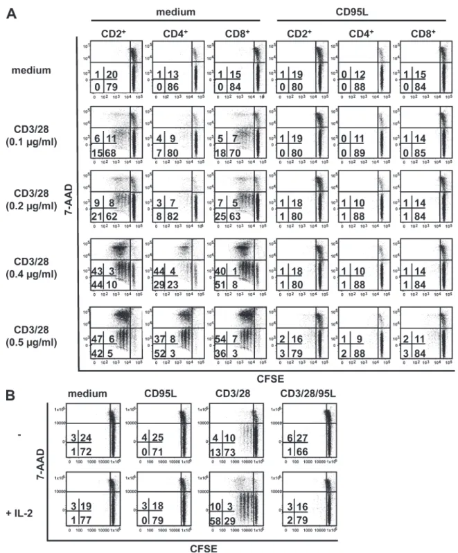
from naive T cells compared with naive T cells stimulated
with CD3/28 alone (Fig. 2 C). To test, whether the
effi-ciency in inhibition of T cell proliferation by CD95L is
de-pendent on the strength of the T cell activating stimulus, we
stimulated naive CFSE-labeled T cells with increasing
con-centrations of immobilized CD3 plus CD28
anti-bodies. CD95L completely prevented T cell proliferation in
CD4
+and CD8
+T cells at all antibody concentrations tested
(Fig. 3 A). At CD3/38 concentrations of 0.2 µg/ml T cells,
proliferation is totally inhibited in the presence of CD95L
compared with T cells stimulated with CD3/28 (CD2
+:
depleting cells expressing the activation markers CD25 and
CD45RO. CFSE-labeled CD45RO
CD25
T cells showed
a significant inhibition of proliferating, live cells in the
pres-ence of CD95L compared with T cells stimulated with CD3/28
(CD3/28 = 29% versus CD3/28/95L = 6%), whereas
apop-tosis induction was only moderately increased (CD3/28 =
8% versus CD3/28/95L = 21%; Fig. 2 B). Although naive
T cells are CD95 resistant, they express CD95 on the cell
sur-face; however, at lower levels than compared with activated
T cells (Fig. S1). The presence of CD95L during T cell
acti-vation prevents secretion of Th1- and Th2-specific cytokines
Figure 2. Simultaneous triggering of CD95 and TCR induces apoptosis in activated T cells and inhibits proliferation and cytokine secretion of naive T cells. CFSE-labeled purified T cells (total T cells; A) or naive T cells (B) were stimulated with immobilized anti-CD3 and anti-CD28 mAb in the absence (CD3/28) or presence of CD95L (CD3/28/95L) or with medium or CD95L alone. After 6 d, T cells counterstained with PI were analyzed by flow cytometry. The experiment is representative of three experiments carried out. (C) Purified naive T cells were incubated with immobilized anti-CD3 and anti-CD28 mAb in the absence or presence of CD95L. Cytokine secretion was determined in the supernatants at different time points. Secretion of cytokines by T cells cultured in medium or on CD95L-coated plates alone was determined, but expression was below the detection level of the assay (0.5 pg/ml), and therefore not included into the graphs. Data are representative of one experiment out of two carried out. Values present the mean of triplicates ± SD.
on December 5, 2016
Figure 3. Independent of the strength of the TCR- mediated signal, proliferation of naive T cells is prevented by CD95 co-stimulation and could not be reverted by IL-2. (A) CFSE-labeled naive T cells were incubated on plates coated with increasing amounts of CD3 and CD28 anti-bodies in the absence (medium) or presence of CD95L. After 6 d, T cells were stained for CD2, CD4, CD8, and 7-AAD, and T cell proliferation and death induction were determined in the different T cell subsets. After 6 d of culture, cell recovery was 84% on average in medium-treated cells, 70% in CD95L, 79% in 0.1 µg/ml CD3/28, 102% in 0.2 µg/ml CD3/28, 139% in 0.4 µg/ml CD3/28, 145% in 0.5 µg/ml CD3/28, 77% in 0.1 µg/ml CD3/28 + CD95L, 81% in 0.2 µg/ml CD3/28 + CD95L, 78% in 0.4 µg/ml CD3/28 + CD95L, and 80% in 0.5 µg/ml CD3/28 + CD95L. (B) CFSE-labeled naive T cells were stimulated with immobilized anti-CD3 and anti-CD28 mAb (0.1 µg/ml) (CD3/28) or CD3/28 plus CD95L (CD3/28/95L), or medium or CD95L alone in the presence or absence of recombinant IL-2 (30 U/ml). After 5 d, cells were stained for CD2, and proliferation and cell death induction was determined for CD2+ T cells. All data are representative of one out of three experiments.
on December 5, 2016
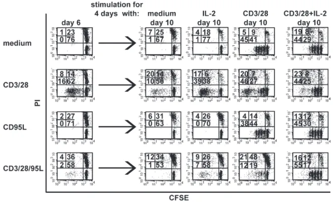
Figure 4. Co-stimulation of CD95 during T cell priming induces T cell unresponsiveness. Naive T cells were CFSE labeled and cultured on plates coated with anti-CD3 and anti-CD28 antibodies in the absence (CD3/28) or presence of CD95L (CD3/28/95L) or CD95L alone or medium. After 6 d, T cells were harvested and 2 × 10⁵ cells of each stimulation group were stained with propidium iodide (PI) and analyzed by flow cytometry (day 6). The remain-ing cells from each stimulation group were restimulated on 0.2 µg/ml CD3/28-coated plates in the presence or absence of 30 U/ml IL-2 or with 30 U/ml IL-2 alone or left untreated (medium). After 4 d, cells were harvested, stained with PI, and analyzed by flow cytometry (day 10). The experiment is repre-sentative for one out of three experiments done.
CD3/28 = 30% versus CD3/28/95L = 2%), whereas
apop-tosis induction was not increased (CD2
+: CD3/28 = 17%
versus CD3/28/95L = 19%). At higher CD3/28
concentra-tions, dividing T cells probably die because of a limited
sup-ply of IL-2, as assays were performed in the absence of
exogenous IL-2. First cell divisions were measurable after day
2 of T cell stimulation (unpublished data). Independent of
whether isolated CD4
+and CD8
+T cells were activated
sep-arately or in co-culture, CD95 co-stimulation totally
inhib-ited their proliferation in both culture systems (Fig. S2).
Because of the usage of T cells from different donors, we
al-ways observed slight differences in proliferation, apoptosis
induction, and efficiency of inhibition. However, in all
ex-periments performed, CD95 co-stimulation significantly
pre-vents T cell proliferation between 80 and 100%. Interestingly,
adding exogenous recombinant IL-2 could not reverse the
inhibitory effect of CD95L on T cell activation. Although
the addition of IL-2 efficiently enhanced proliferation in
CD3/28-stimulated naive T cells, CD3/28/95L-stimulated
T cells did not proliferate irrespective of the presence of IL-2
(Fig. 3 B). No effect of IL-2 was observed in T cells cultured
in medium or in the presence of CD95L alone.
Membrane-bound CD95L is cleaved by proteases from the cell surface
(27), and soluble CD95L might interfere with the
antiprolif-erative effect of immobilized CD95L. However, the addition
of soluble CD95L to our culture system did not prevent the
inhibition of T cell proliferation induced by immobilized
CD95L (Fig. S3). These results clearly show that T cell
prim-ing can be efficiently suppressed by CD95 co-stimulation.
CD95 triggering in combination with CD3/28 activation
induces T cell unresponsiveness
Because CD95L prevents proliferation of CD3/28-activated
naive T cells, we raised the question of whether T cell
silenc-ing requires the constitutive presence of CD95L in the T cell
stimulation culture. We therefore activated naive T cells with
CD3/28, in the absence or presence or CD95L, with CD95L
or medium alone. After 6 d, 2 × 10
5T cells of each stimulation
group were analyzed (day 6). The presence of CD95L
effi-ciently prevented T cell proliferation compared with
CD3/28-stimulated T cells (Fig. 4). The remaining cells from each
stimulation condition were restimulated with immobilized
anti-CD3 and -CD28 antibodies in the presence or absence of
IL-2, and proliferation was measured 4 d later (day 10).
Re-stimulation of previously CD3/28-activated, medium, or
CD95L-stimulated T cells with anti-CD3 and anti-CD28
an-tibodies induced efficient T cell proliferation (day 10). In
con-trast, the proliferation of T cells cultured for 6 d in the presence
of CD3/28/95L and restimulated with CD3/28 was strongly
reduced compared with T cells previously cultured in the
on December 5, 2016
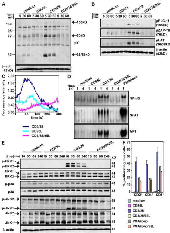
Figure 5. Inhibition of proximal TCR signaling events in T cells activated in the presence of CD95L. (A and B) Purified T cells were activated on plates coated with anti-CD3 and anti-CD28 antibodies in the absence or presence of CD95L. Expression of total protein tyrosine phosphorylation (pY; A) or of specific phosphorylated proteins (B) was determined by Western blot analysis at different time points. Arrows indicate differences in protein tyrosine phos-phorylation. Experiments were performed three times with similar results. (C) Fluo-4 labeled T cells were seeded on poly-l-lysine–coated slides and antibodies were given to cells directly before the measurement. For measuring the intracellular calcium, a time series of one confocal image plane was recorded. Image acquisition was started immediately after applying the antibodies. After subtracting the background fluorescence, the mean of all cells at each time point was calculated. Each curve represents the mean of three independent experiments. (D) Purified naive T cells were activated with immobilized CD3 plus anti-CD28 antibodies in the presence or absence of CD95L. After 1 and 4 h, nuclear extracts were prepared and analyzed by EMSA for DNA-binding activity of NFAT, AP1, and NF-B. PMA/ionomycin stimulation served as a positive control. The experiment is representative of three different experiments performed. (E) Purified T cells stimulated with immobilized anti-CD3 and anti-CD28 mAb in the absence or presence of CD95L were lysed after 10, 30, 40, and 240 min, and lysates were subjected to Western blot analysis to determine the expression of phosphorylated and nonphosphorylated MAPKs (ERK1/2, p38, Jnk1/2). -Actin expression served as a loading control. The experiment shown is representative of two experiments done. (F) CFSE-labeled naive T cells were stimu-lated with immobilized anti-CD3 and anti-CD28 mAb or with PMA and ionomycin in the absence (CD3/28, PMA/iono) or presence of CD95L (CD3/28/95L, PMA/iono/95L) or with medium or CD95L alone. After 5 d, T cells were stained with 7-AAD, CD2, CD4, and CD8 and analyzed by flow cytometry. Data present the percentage of proliferating, live (7-AAD) T cells in the different T cell subsets. Experiments were performed three times with similar results.
on December 5, 2016
presence of CD95L alone (12 vs. 38%). However, the
sus-tained inhibition of proliferation by CD95L was reversed by
the addition of exogenous IL-2. In summary, these data
indi-cate that CD95 co-stimulation of CD3/28-activated naive
T cells induces an unresponsive state, which can be overcome
by IL-2 after release from CD95L-mediated suppression.
T cells activated in the presence of CD95L exhibited
impaired protein tyrosine phosphorylation, Ca
2+mobilization, mitogen-activated protein (MAP) kinase
activation, and nuclear translocation of transcription factor
We next estimated whether CD95 co-stimulation would
in-terfere with early signaling events after TCR engagement.
Af-ter 5 min, CD3/28 stimulation induced a strong tyrosine
phosphorylation of proteins with apparent molecular weights
of 150, 70, and 40 kD, respectively, which was still
detect-able after 60 min and completely blocked in the presence of
CD95L (Fig. 5 A). Western blot analysis using phosphospecific
antibodies showed that the presence of CD95L during TCR
triggering completely abolished the phosphorylation of LAT,
ZAP-70, and phospholipase (PLC)-1 (Fig. 5 B). Triggering
of CD95L alone did not induce protein tyrosine
phosphoryla-tion. Because of the lack of protein tyrosine phosphorylation,
T cells activated with anti-CD3 and anti-CD28 antibodies in
the presence of CD95L showed no Ca
2+influx compared with
T cells activated with CD3/28 (Fig. 5 C). Inhibition of protein
tyrosine phosphorylation and decreased Ca
2+-influx was
re-flected by impaired nuclear translocation of transcription
fac-tors NF-B, AP1, and NFAT in CD3/28/95L-stimulated
T cells compared with CD3/28- or PMA/ionomycin-activated
T cells after 1 and 4 h of activation (Fig. 5 D). T cells
stimu-lated with CD95L alone exhibited no differences to untreated
T cells. Also, mitogen-activated protein kinase (MAPK)
acti-vation known to be induced after TCR stimulation (28) was
totally abolished in T cells activated in the presence of CD95L
after 10, 30, 60, and 240 min (Fig. 5 E). Although
phosphory-lation of ERK1/2 and JNK1 was decreased after 30 min in
CD3/28-activated samples, phosphorylation of p38 remained
constant. Phosphorylation of JNK2 was constitutively
inde-pendent of the stimulus and CD95L treatment alone did not
induce MAPK activation. To further prove that CD95
trigger-ing durtrigger-ing T cell activation interferes with proximal T cell
re-ceptor events, we activated naive T cells with PMA and
ionomycin known to directly activate PKC and Ca
2+mobili-zation independent of TCR stimulation. Although
CD3/28-mediated T cell proliferation was completely blocked in the
presence of CD95L, the proliferation induced by
PMA/iono-mycin treatment could not be inhibited (Fig. 5 F). In
sum-mary, we could show that proximal T cell receptor signaling is
severely disabled by CD95 co-stimulation.
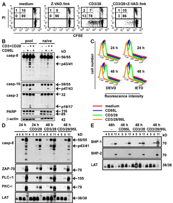
Co-stimulation of CD95 during T cell activation prevents
caspase cleavage and formation of a functional
signaling platform
Caspase activity is a hallmark of apoptosis induction, but
also a prerequisite for T cell proliferation. CD3/28-mediated
T cell proliferation was abrogated by the broad caspase
inhibi-tor Z-Val-Ala-DL-Asp-fluoromethylketone (z-VAD.fmk;
Fig. 6 A). Cleavage of caspase-3, -8, and -10 was detected in
CD3/28-stimulated total T cells (pool) or naive T cells
iso-lated from one buffy coat. Surprisingly, the presence of CD95L
totally inhibited processing of all caspases examined (Fig. 6 B).
No caspase cleavage was detected in T cells stimulated with
CD95L alone or medium. To determine caspase activity,
na-ive T cells were incubated for 24 or 48 h with the cell
per-meable, rhodamine 110-based caspase substrates DEVD
(Asp-Glu-Val-Asp; specific for caspase-3, -6, -7, -8, and -10)
and Ile-Glu-Thr-Asp (IETD; specific for caspase-8 and
gran-zyme B). The presence of CD95L completely blocked
CD3/28-mediated caspase activity at both time points, whereas activation
by CD95L alone or medium had no effect (Fig. 6 C). Despite
caspase activation in CD3/28-treated T cells, no increase in
PARP cleavage compared with untreated cells was observed
(Fig. 6 B), suggesting that processed caspases might be
se-questered to special membrane compartments during T cell
activation to prevent induction of apoptosis. Because
TCR-mediated signaling preferentially occurs within lipid rafts, we
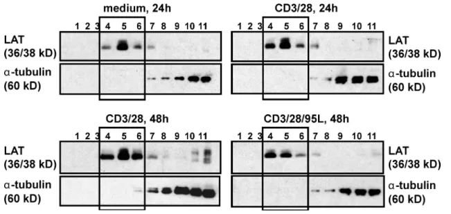
isolated lipid rafts by discontinuous sucrose gradient from
T cells stimulated in the presence or absence of CD95L.
West-ern blot analysis using LAT as a raft marker and the
cytoplas-mic protein -tubulin as a nonraft marker identified fraction
4–6 as raft and fraction 11 as nonraft (Fig. S4).
CD3/28-acti-vated T cells exhibited recruitment of PLC-, PKC-,
and ZAP-70 into lipid rafts, whereas nontreated T cells did
not. Importantly, triggering CD95 during T cell activation
(CD3/28/95L) efficiently prevented recruitment of PLC-,
PKC-, and ZAP-70 into the raft fraction (Fig. 6 D).
Un-treated T cells and CD95L-Un-treated T cells exhibited no
differ-ence in the recruitment of T cell–associated molecules into
lipid rafts (Fig. S5). Recruitment of the protein tyrosine
phos-phatase Scr homology-2 domain-containing phosphos-phatase-1,
which is known to be predominantly expressed in cells of the
hematopoietic system and to function as a negative regulator
of TCR-mediated signaling (29, 30), was not increased in the
raft fraction of CD3/28/95L-activated T cells compared with
CD3/28-treated T cells. Additionally, Scr homology-2
domain-containing phosphatase-2 was not detectable in the raft fractions
independent of the activating stimulus (Fig. 6 E). LAT
constitu-tively expressed in lipid rafts was detected in all raft fractions
in-dependent of the stimulation signal. Because caspase-8 is required
for T cell and NF-B activation (16) and active caspases were
reported to localize in lipid rafts in activated T cells (24, 31), we
determined caspase-8 expression in the lipid raft fractions. The
proform of caspase-8 (56/55) was found in all lipid raft fractions
independent of the stimulus, whereas the intermediate cleavage
product (p43/41) was only found in the raft fraction of
CD3/28-activated T cells (Fig. 6 D). In contrast to the finding that the
presence of CD95L prevents caspase-8 cleavage (Fig. 6 B), we
observed caspase-8 cleavage in the nonraft fraction of CD3/28/
95L-stimulated T cells (Fig. 6 D). However, in Fig. 6 D, T cells
cultured in medium alone already showed a weak caspase-8
ac-tivation, which was not increased in the presence of the CD95L
on December 5, 2016
CD95L up-regulation on APCs may constitute a mechanism
of viral immune evasion
Inhibition of T cell proliferation caused by CD95 triggering
during T cell priming may help invading pathogens to avoid
activation. These differences are caused by varying T cell
prepa-rations derived from different donors. Collectively, these results
clearly indicate that CD95 triggering during T cell activation
prevents the formation of a functional signaling platform.
Figure 6. CD95 triggering during T cell activation prevents caspase activation and translocation of TCR-associated molecules into lipid rafts. (A) CFSE-labeled naive T cells were activated with immobilized anti-CD3 plus anti-CD28 antibodies (CD3/28) in the presence of absence of Z-VAD. fmk (50 µM). After 6 d, cells were stained with propidium iodide (PI) and analyzed by flow cytometry. (B) Purified T cells (pool) and naive T cells (naive) from one buffy coat were activated with immobilized anti-CD3 plus anti-CD28 mAb in the presence or absence of CD95L. After 24 h, cells were harvested and protein lysates were subjected to Western blot analysis. -Actin served as a loading control. (C) Naive T cells were activated with immobilized anti-CD3 plus anti-CD28 antibody in the absence (anti-CD3/28) or presence of CD95L (anti-CD3/28/95L) or with medium or CD95L alone. After 24 and 48 h, cells were incubated with caspase substrates DEVD-D2R and IETD-D2R and caspase activity was determined by flow cytometry. (D and E) T cells were activated on anti-CD3 plus anti-CD28–coated plates in the absence (CD3/38) or presence of CD95L (CD3/28/95L) or with CD95L alone or medium. After 24 or 48 h, cells were harvested and lysed, and lysates were separated on a discontinuous sucrose gradient. Proteins from fraction 4–6 (raft fraction) and fraction 11 (nonraft fraction) were analyzed by Western blot analysis. All data shown are representative of three independent experiments.
on December 5, 2016
Figure 7. HIV-1 infection induces CD95L surface expression in APC and lymphocytes and thereby prevents T cell activation. (A) HIV-1 infec-tion of macrophages, DCs, and activated lymphocytes was determined 4–5 d after infecinfec-tion by GFP expression, and CD95L expression was determined on the GFP+ population for infected cells and on the entire population for noninfected cultures. Results shown are representative of three independent ex-periments. (B) The percentage of CD95L-expressing cells (left) and the total CD95L expression, defined as the product between percentage of CD95L+ cells and mean fluorescence intensity (right), was compared for HIV-1 and HIV-1 Nef*–infected samples, noninfected (n.i.), and isotype control (iso). HIV-1– infected values were set as 100%. Results shown are a mean of three independent experiments ± the SD. (C) Monocyte-derived macrophages (MDM)
on December 5, 2016
were infected with HIV-1 IRES-mCherry or left uninfected and stained for surface CD95L 4 d later. Images show CD95L staining below and a merge of CD95L (green), mCherry (red), and Nomarski phase contrast above. Bars, 20 µm. (D) MDM infected with HIV-1 IRES-mCherry, HIV-1 Nef* IRES-mCherry were separated at day 4 after infection into CD95L+ and CD95L MDM and loaded with SEE. The graphs show the extent of T cell activation after co-culture of infected CD95L+ and CD95L MDM, or uninfected MDM with Jurkat-NFAT-Luc cells (left) or Jurkat-NFAT-YFP (right). 50 ng/ml PMA/1 µM iono and unstimulated Jurkat cells serve as positive and negative control. Each graph represents the mean of three independent experiments performed on different blood donors ± SD.
detection and elimination by the host immune system. It has
been previously shown that the accessory HIV-1 Nef protein,
which is required for effective viral persistence and greatly
ac-celerates progression to AIDS (32), induces CD95L expression
on T cells (33). HIV-1 also infects macrophages and DCs.
Thus, Nef-mediated up-regulation of CD95L on APCs to
suppress T cell responses might represent a novel mechanism
by which HIV-1 ensures immune evasion. To assess this
pos-sibility, we determined the effect of wild-type and Nef-
defective HIV-1 infection on surface CD95L expression in
macrophages and DCs. Monocyte-derived macrophages, DCs,
and lymphocytes were obtained from healthy blood donors
and characterized for phenotypic markers immediately before
infection (Fig. S6). Uninfected cells (mock) or cells infected
with HIV-1 constructs expressing eGFP alone (Nef-deleted
HIV-1 Nef
*) or together with Nef (wild-type HIV-1) were
examined for CD95L surface expression 4–5 d after infection.
Infection rates were typically between 10 and 20% (Fig. 7 A).
CD95L surface expression was determined on GFP
+HIV-1–
infected cells. For all three cell types, HIV-1 infection
consis-tently enhanced both the percentage of CD95L-expressing
cells and the levels of cell surface expression by up to two
or-ders of magnitude (Fig. 7 B). Nef significantly contributed to
induction of CD95L expression in macrophages and
lympho-cytes, but not so in DCs (Fig. 7 B). To directly determine
whether HIV-induced up-regulation of CD95L on the surface
of APCs suppresses their capability to stimulate T cells, we
ex-amined the effect of CD95L
+and CD95L
macrophages on
NFAT activation in two different Jurkat lines containing
ei-ther the YFP NFAT-YFP) or the luciferase
(Jurkat-NFAT-Luc) reporter genes under the control of an NFAT-
dependent promoter. Both T cell lines express a functional
TCR and can be stimulated by staphylococcal enterotoxin
E (SEE) superantigen. This allowed us to measure both the
overall levels of luciferase activity by plate luminometry as well
as the proportion of activated YFP
+T cells by flow cytometry.
To avoid crossover between GFP and YFP fluorescences, we
infected macrophages with HIV-1 IRES-mCherry viruses and
verified HIV infection–induced CD95L expression by
fluo-rescent microscopy (Fig. 7 C). After sorting the infected
mac-rophages into CD95L
+and CD95L
populations and loading
with SEE superantigen, we found that HIV-1–infected CD95L
macrophages caused approximately two- to threefold higher
levels of T cell activation compared with noninfected
macro-phages (Fig. 7 D). In strict contrast, co-culture with CD95L
+-infected macrophages resulted in levels of NFAT activation
similar to those observed with uninfected macrophages in both
readout systems and irrespective of Nef expression. Thus,
HIV-induced CD95L expression on the surface of
macro-phages clearly suppresses their ability to stimulate T cells. These
results suggest that the up-regulation of CD95L on APCs by
HIV-1 to silence T cell activation may suppress immune
re-sponses, facilitate viral immune evasion, and play a role in the
pathogenesis of AIDS.
DISCUSSION
CD95, which was originally characterized as a death receptor
inducing apoptosis after CD95L binding, is now increasingly
recognized as a surface receptor with additional functions apart
from cell death induction depending on cell type, stimulation,
and cellular environment. In this study, we demonstrate for the
first time that co-stimulation of CD95 during T cell activation
induces inhibition of T cell proliferation in naive T cells by
in-terfering with proximal TCR signaling events, and that CD95
can act as a silencer of the immune response. This novel role of
CD95 in down-modulating T cell responses was determined
in two different systems: (a) an antigen-specific system using
APCs expressing A1 and m-CD95L to activate
HLA-A1
T cells and (b) for antigen-independent T cell activation
with immobilized CD3 and CD28 antibodies in the presence
of recombinant CD95L. The presence of CD95L during T cell
activation prevented proliferation of CD4
+and CD8
+T cells
in both systems. Whereas preactivated CD95-sensitive T cells
immediately undergo apoptosis after CD95 stimulation, naive
T cells were inhibited in proliferation. These results appear to
be in contrast to previous data showing that CD95
anti-bodies or cross-linked CD95L can augment proliferation of
CD3-activated primary human T cells (6, 8). However, in
these experiments, T cell activation was suboptimally induced
by low concentrations of anti-CD3 antibodies in the absence
of co-stimulatory signals, and naive and preactivated T cells
were not analyzed separately. Inhibition of T cell activation by
CD95L in our system, however, was effective in naive T cells
activated in the presence of co-stimulation and was
indepen-dent of the strength of the activating signal (Fig. 3 A).
Al-though CD95 triggering induces apoptosis in activated T cells,
it prevents proliferation during T cell priming in naive T cells,
indicating that the time point of CD95 triggering determines
the subsequent fate of the T cell. Dependence on the time
point of receptor triggering was also reported for signals
deliv-ered via CD95L. Although CD95L may promote reverse
sig-naling and T cell co-stimulation directly after antigen-induced
CD95L expression, CD95L also mediates apoptosis in T cells
at the termination phase of the immune response (34).
T cell activation is initiated by the recognition of antigen
through the TCR and subsequent reorganization of signaling
on December 5, 2016
macrophages (49), although the relevance of this effect and the
impact of Nef remained unclear. In this study, we confirmed
that HIV infection leads to effective CD95L up-regulation in
macrophages and T lymphocytes and show for the first time
that this effect is also observed in DCs. Furthermore, we
dem-onstrate that Nef plays an important role in CD95L
enhance-ment in macrophages and T lymphocytes, but not in DCs, and
that HIV-induced CD95L expression on macrophages
signifi-cantly prevents the activation of Jurkat T cells. Thus,
up-regu-lation of CD95L expression on APCs by HIV-1 infection
might contribute to viral immune evasion by inhibiting T cell
activation and promoting unresponsiveness. This mechanism
may also have been adopted by other pathogens. Influenza
in-fection in mice induced the expression of CD95L in lymph
node–resident DCs (50) and up-regulation of CD95L was
ob-served in human DCs after human cytomegalovirus infection
(51). Interestingly, although human cytomegalovirus–infected
CD95L-expressing DCs induced apoptosis in activated T cells,
some T cells survived contact with infected DCs and exhibited
suppressed proliferation after secondary stimulation in the
ab-sence of the virus (51). This is in line with our observation that
T cells that have encountered CD95L during T cell priming
showed decreased proliferation after restimulation. This
un-responsive state was partially reverted by the addition of
ex-ogenous IL-2. Interestingly, lack of anergy induction was also
reported for CD95-deficient lymphoproliferation mice (52).
On the basis of our findings, we propose a novel role for
CD95 in naive T cells as a silencer of T cell activation and
proliferation by down-regulation of TCR signaling upon
co-stimulation. Virus-induced up-regulation of CD95L on the
surface of APCs may suppress immune responses, and thereby
support immune evasion and tolerance induction.
MATERIALS AND METHODS
Cell culture. All cell lines were grown in complete RPMI 1640 medium (Invitrogen) supplemented with 10% FCS (Lonza), 2 mM l-glutamine, and 1 mM sodium pyruvate at 37°C in a humidified atmosphere containing 7.5% CO2. Establishment of C1R.A1.puro and C1R.A1.CD95L cells were de-scribed previously (25). T cells were cultured in complete RPMI 1640 me-dium supplemented with 2% FCS.
T cells were isolated from PBMCs of healthy HLA-A1 donors by Ro-setteSep (StemCell Technologies). Institutional ethic committee approved the usage of buffy coats for T cell isolation. Purity varies between 92–97%. Naive T cells were separated from the T cell pool by using anti-CD45RO (Cl. UCHL1) and anti-CD25 (Cl. M-A251; BD) antibodies, and subsequent incubation with Dynabeads goat anti–mouse IgG (Invitrogen). Purity was determined by flow cytometry using CD45RA-FITC (Cl. ALB11; Immuno-tech) and ranged between 90–97%. For proliferation assays and cytokine assays, plates were coated with 0.1 µg/ml anti-CD3 (cl. OKT3) and 0.1 µg/ml anti-CD28 (cl.15E8). In all other assays, 0.25 µg/ml OKT3 and 0.25 µg/ml 15E8 were used. To trigger CD95, we used 2 µg/ml immobilized recombi-nant CD95L (Alexis Biochemicals) or 2 µg/ml Fc-CD95L supernatant pro-vided by H. Walczak (Imperial College London, London, UK) (53). For TCR-independent T cell activation, we used 0.8 ng/ml PMA (Calbiochem) and 20 nM ionomycin (Calbiochem). CFSE-labeled HLA-A1 T cells were stimulated with medium or mitomycin c–treated C1R.A1.puro or C1R. A1.CD95L cells at a ratio of 1:1.
For HIV infection experiments, monocytes were separated from PBMCs of healthy donors by plastic adherence. PBLs were activated for 3 d with
molecules into lipid rafts. Although productive T cell
activa-tion induced translocaactiva-tion of ZAP-70, PKC-, and PLC-
into lipid rafts, triggering CD95 during T cell activation
pre-vented their recruitment. However, molecules constitutively
associated with rafts, such as LAT and lymphocyte-specific
ki-nase (35), were recruited, indicating that CD95 triggering does
not induce a global change in raft composition. The
mecha-nism by which CD95 signaling prevents translocation of
TCR-associated molecules into the signaling platform remains elusive.
Changes in the composition of raft-associated molecules were
also observed in anergic T cells, where LAT could not be
re-cruited to lipid rafts because of a selective palmitoylation defect
(36), or upon anti-CD4 antibody treatment, which inhibited
ZAP-70 translocation into lipid rafts, thereby preventing NF-B
activation (37). Steric hindrance of TCR signaling by
recruit-ment of CD95 into lipid rafts after CD95 co-stimulation did
not account for the inhibition of T cell proliferation, as no
increase of CD95 in lipid rafts was detected in CD95L-treated
T cells (unpublished data). The inhibitory effect of CD95
trig-gering on T cell activation was also analyzed on downstream
events. Thus, protein tyrosine phosphorylation of ZAP-70,
LAT, and PLC-1 was totally inhibited in the presence of
CD95L. We further observed a significant decrease in Ca
2+-mobilization and inefficient nuclear translocation of
transcrip-tion factors NF-B, AP-1, and NFAT in T cells triggered by
CD95 during T cell activation. Impaired activation of NF-B,
AP-1, and NFAT was confirmed by the reduced expression of
activation markers and a decrease in cytokine secretion. CD95
co-stimulation during T cell priming also prevents MAPK and
caspase activation, both known to be required for efficient
T cell activation (8, 28). Block of calcium channel, Ca
2+mobili-zation, and NFAT activation caused by activation of acidic
sphingomyelinase and ceramide release was also observed in
T cells stimulated by CD95 alone (38).
Our data add a new function to the already known role of
CD95 in apoptosis induction and T cell proliferation (7) by
defining CD95 as a T cell silencer. The newly identified
immunosuppressive function of CD95 co-stimulation might
point to a role of elevated CD95L serum levels in several types
of cancers (39–41) or might explain the requirement for CD95L
expression on veto cells (42). In the murine system, CD95L
expression on veto cells was predominantly correlated with
apoptosis induction of CD95
+T cell precursors (42–44).
How-ever, whereas naive murine T cells are constitutively CD95
sensitive (45), naive human T cells are CD95 resistant (26).
Therefore, human veto cells may not only induce apoptosis,
but may additionally interfere with T cell activation. Although
CD95L is predominantly expressed on T cells and NK cells,
expression of CD95L was reported in human immature blood
DCs, blood monocytes, or activated Langerhans cells (46, 47).
Our findings suggest that CD95L expression on APCs might
represent a novel mechanism to silence T cells and to support
immune evasion. It is known that the HIV-1 Nef up-regulates
CD95L in infected T lymphocytes, possibly to induce
apopto-sis of bystander cytotoxic T cells (33, 48). It has also been
shown that HIV-1 infection leads to CD95L up-regulation in
on December 5, 2016
software were used. For image analysis, the cells were digitally outlined and the mean fluorescence intensity was determined for each cell at each single time point (ImageJ software). After subtracting the background fluorescence, the mean fluorescence intensity of all cells at each time point was determined. Determination of cytokine concentrations in cell culture supernatants. T cells were cultured in medium alone or activated with stimulator cells at a ratio of 1:1 or with antibodies and recombinant CD95L. Supernatants were collected at the indicated time points and immediately stored at 80°C. All supernatants were analyzed simultaneously by Immuned GmbH using Bio-Plex technology. Nuclear protein extraction and electrophoretic mobility shift assay. Purified T cells were activated with immobilized antibodies and recombinant CD95L or with 10 ng/ml PMA and 500 nM ionomycin for 1 h. Nuclear ex-tracts and electrophoretic mobility shift assays (EMSAs) were performed as previously described (54). For EMSA, the following oligonucleotides were used: NF-B, 5-AGTTGAGGGGACTTTCCCAGGC-3 (sense); NF-AT, 5-TCTAAGAGGAAAATTTCATG-3 (sense); AP-1, 5-CGCTTGAT-GAGTCAGCCGGAA-3 (sense).
Analysis of protein tyrosine phosphorylation. Purified T cells were serum starved overnight in complete medium supplemented with 0.5% FCS, and subsequently activated on antibody-coated plates and lysed in 50 mM Hepes, 100 mM NaCl, 1% IGEPAL CA-630, 1% laurylmaltoside, 1 mM PMSF, 5 mM EDTA, 1 mM Na3VO4, 50 mM NaF, and 10 mM Na4O7P2 × 10 H2O for 15 min, and protein lysates were subjected to Western blot analysis.
Isolation of lipid rafts. T cells were lysed in 50 mM Hepes, 100 mM naCl, 3% Brij 58, 1 mM PMSF, 5 mM EDTA, 1 mM Na3VO4, 50 mM NaF, and 10 mM Na4O7P2 × 10 H2O for 10 min on ice. The lysate was mixed 1:1 with ice-cold 80% sucrose diluted in 25 mM MES, 5 mM EDTA, and 150 mM NaCl, transferred to Ultra-Clear centrifuge tubes (Beckman Coulter), and over-laid with ice-cold 30% sucrose, followed by ice-cold 5% sucrose. After centrifu-gation in a Beckman Coulter SW41 rotor, 40,000 rpm, for 20 h at 4°C, 11 1-ml-fractions were collected and subjected to aceton precipitation.
Western blot analysis. Western blot analysis was described previously (55). The following antibodies were used: anti-phosphotyrosine (cl. 4G10), LAT (cl. 2E9) (Millipore), phospho-PLC1 (Tyr783), phospho-LAT (Tyr191), phospho-Zap-70(Tyr319)/Syk(Tyr352), caspase-3, PARP, PLC1 (Cell Signaling Technology), ZAP 70 (cl. 29), PKC- (cl. 27), JNK1/JNK2 (G151-666; BD), -actin (AC-15), ERK-1/ERK2 (Sigma-Aldrich), cas-pase-8 (12F5; Alexis), caspase-10 (cl. 4C1; MBL), ERK1/2(phospho-Thr202/Tyr204), p38 MAPK (phospho-Thr180/Tyr182; Assay Design), anti-active JNKpAb (Promega), p38 (Stressgen) goat anti–mouse IgG-HRP, goat ant–rabbit IgG –HRP, SH-PTP1 (C-19), and SH-PTP2 (B-1; Santa Cruz Biotechnology, Inc.).
Online supplemental material. Fig. S1 compares the expression of CD95L on naive and activated human T cells. Fig. S2 compares inhibition of T cell proliferation by CD95 co-stimulation in CD4+ and CD8+ T cells cultured isolated or together. Fig. S3 shows that soluble CD95L could not reverse the inhibitory effect of immobilized CD95L during T cell activation. Fig. S4 shows the expression of LAT and -tubulin in fractions derived from discontinuous sucrose gradients. Fig. S5 depicts the expression of TCR-associated molecules in raft and nonraft fractions of unstimulated and CD95L-stimulated T cells. Fig. S6 shows the phenotype of primary macrophages, DC, and lymphocytes before HIV infection. The online supplemental material is available online at http://www.jem.org/cgi/content/full/jem.20082363/DC1.
We thank Ingrid Knape for excellent technical assistance. Jurkat-NFAT-YFP cells were a kind gift of Claire Hivroz.
The authors have no conflicting financial interests. J.A.L. is a member of MaCS and SYBILLA [EU 7FP].
Submitted: 20 October 2008 Accepted: 27 April 2009
10 µg/ml PHA before infection. Monocytes were differentiated into imma-ture DCs by culturing for 5 d with 50 ng/ml recombinant human GM-CSF and 20 ng/ml IL4 (Immunotools) or into macrophages by culturing for 7 d with 50 ng/ml human recombinant GM-CSF alone. Phenotypic character-ization before infection was done by flow cytometry using CD3, CD4, HLA-DR, CD14, CD1a, CD80, and CD86 antibodies (BD).
Virus production and infection. Wild-type NL43-Nef-IRES-GFP or Nef-IRES-mCherry (referred to as HIV-1) and Nef-deleted NL43-Nef*-IRES-GFP or NL43-Nef*-IRES-mCherry (referred to as HIV-1 Nef*) viruses were produced by transient calcium phosphate cotransfection of 293T cells with proviral plasmid and an expression plasmid for the vesicular stomati-tis envelope glycoprotein following standard protocols. Cell supernatants con-taining virions were collected at 48 h after transfection and used immediately to infect PBL, immature DCs, or macrophages and replaced after 3 h for monocyte-derived cells (or after 6 h for PBLs) by RPMI medium with the appropriate cytokines.
Microscopy. Macrophages grown on glass coverslips were stained for surface CD95L (NOK-1) followed by goat anti–mouse Alexa Fluor 647 (Invitrogen) at 4 dpi and analyzed by Apotome structured illumination fluorescent microscopy using an Axiovert microscope equipped with a Plan-Apochromat 63×, NA 1.4, oil objective and a MRm charge-coupled device camera piloted by Axiovision software (all from Carl Zeiss, Inc.).
Activation of Jurkat-NFAT by HIV-infected macrophages. Infected macrophages labeled with anti-CD95L mAb (NOK-1) followed by rat anti– mouse microbeads (Miltenyi Biotec) were isolated into CD95L+ and CD95L populations by MACS according to the manufacturer’s instructions. CD95L+ and CD95L macrophages were loaded with 1 µg/ml SEE superantigen (Toxin Technology) and placed in co-culture at a 1:2 ratio with Jurkat-NFAT-Luc cells or with Jurkat-NFAT-YFP cells. After 6 h, Jurkat-NFAT-Luc cells were lysed and tested for luciferase by plate bioluminescence using the Promega Luciferase Assay System according to the manufacturer’s instructions. Jurkat-NFAT-YFP cells were fixed and analyzed by flow cytometry for YFP expression. Incubation was restricted to a 6-h period to avoid interference with CD95-mediated apop-tosis in Jurkat targets.
Flow cytometry. In general, 5 × 10⁵ cells were stained with the following anti-bodies: CD25-PE, CD69-PE, CD71-FITC, CD95-FITC, HLA-DR, DP, DQ-FITC, CD95L (NOK-1), CD2-APC, CD3-PE, CD4-PE, CD4-Pacific blue, CD8-PE, CD8-FITC, CD8-PE-Cy-7 (BD), CD4-PE-Cy5.5 (Caltag), and goat anti–mouse F(ab)2-PE (Dako). Dead cells were visualized using 1 µg/ml propidium iodide (Sigma-Aldrich) or 1 µg/ml 7-amino-actinomycin-D (AAD; Sigma-Aldrich). To measure caspase activity fluorometrically, T cells were incu-bated with a 100-µM solution of the rhodamine 110-based, cell membrane– permeable caspase substrates DEVD or IETD (Invitrogen). Analysis was done on FACScan or LSR II flow cytometer (BD).
CFSE labeling. 2 × 10⁶ T cells/ml were labeled with 100 µl of 50 µM CFSE (Cell Trace CFSE Cell proliferation kit; Invitrogen) for 10 min at 37°C. Cells were immediately washed 4 times with ice-cold PBS-5% FCS and subsequently used for proliferation assays.
Measurement of intracellular calcium. T cells were labeled for 20 min at 37°C with 1 µM Fluo-4 and seeded on poly-l-lysine–coated slides (POLY-PRE slides; Sigma-Aldrich). Nonadherent cells were washed away and anti-bodies were given to cells directly before the measurement. Recombinant CD95L was preincubated for 15 min at 37°C before adding CD3 and CD28 antibodies. To measure the intracellular calcium increase, we recorded a time series (duration, 6 min; interval, 1 s) of one confocal image plane. Images were acquired with an Infinity multibeam confocal device (Visitron) mounted be-tween an Axiovert 200 and a charge-coupled device camera (CascadeII 512). Fluo-4 was excited with a 488-nm Argon/Krypton laser and detected with a 500–550-nm emission filter. A 10× plan Neofluar objective and Metamorph
on December 5, 2016
22. Muppidi, J.R., and R.M. Siegel. 2004. Ligand-independent redistribu-tion of Fas (CD95) into lipid rafts mediates clonotypic T cell death. Nat.
Immunol. 5:182–189.
23. Algeciras-Schimnich, A., L. Shen, B.C. Barnhart, A.E. Murmann, J.K. Burkhardt, and M.E. Peter. 2002. Molecular ordering of the initial signal-ing events of CD95. Mol. Cell. Biol. 22:207–220.
24. Misra, R.S., J.Q. Russell, A. Koenig, J.A. Hinshaw-Makepeace, R. Wen, D. Wang, H. Huo, D.R. Littman, U. Ferch, J. Ruland, et al. 2007. Caspase-8 and c-FLIPL associate in lipid rafts with NF-kappaB adaptors during T cell activation. J. Biol. Chem. 282:19365–19374.
25. Strauss, G., W. Osen, I. Knape, E.M. Jacobsen, S.M. Muller, and K.M. Debatin. 2007. Membrane-bound CD95 ligand expressed on human an-tigen-presenting cells prevents alloantigen-specific T cell response without impairment of viral and third-party T cell immunity. Cell Death Differ. 14:480–488.
26. Klas, C., K.M. Debatin, R.R. Jonker, and P.H. Krammer. 1993. Activation interferes with the APO-1 pathway in mature human T cells. Int. Immunol. 5:625–630.
27. Askenasy, N., E.S. Yolcu, I. Yaniv, and H. Shirwan. 2005. Induction of tolerance using Fas ligand: a double-edged immunomodulator. Blood. 105:1396–1404.
28. Zhang, Y.L., and C. Dong. 2005. MAP kinases in immune responses.
Cell. Mol. Immunol. 2:20–27.
29. Kosugi, A., J. Sakakura, K. Yasuda, M. Ogata, and T. Hamaoka. 2001. Involvement of SHP-1 tyrosine phosphatase in TCR-mediated signaling pathways in lipid rafts. Immunity. 14:669–680.
30. Neel, B.G., H. Gu, and L. Pao. 2003. The ‘Shp’ing news: SH2 domain-containing tyrosine phosphatases in cell signaling. Trends Biochem. Sci. 28:284–293.
31. Koenig, A., J.Q. Russell, W.A. Rodgers, and R.C. Budd. 2008. Spatial differences in active caspase-8 defines its role in T-cell activation versus cell death. Cell Death Differ. 15:1701–1711.
32. Kirchhoff, F., M. Schindler, A. Specht, N. Arhel, and J. Munch. 2008. Role of Nef in primate lentiviral immunopathogenesis. Cell. Mol. Life
Sci. 65:2621–2636.
33. Xu, X.N., B. Laffert, G.R. Screaton, M. Kraft, D. Wolf, W. Kolanus, J. Mongkolsapay, A.J. McMichael, and A.S. Baur. 1999. Induction of Fas ligand expression by HIV involves the interaction of Nef with the T cell receptor zeta chain. J. Exp. Med. 189:1489–1496.
34. Suzuki, I., and P.J. Fink. 2000. The dual functions of fas ligand in the regu-lation of peripheral CD8+ and CD4+ T cells. Proc. Natl. Acad. Sci. USA. 97:1707–1712.
35. Drevot, P., C. Langlet, X.J. Guo, A.M. Bernard, O. Colard, J.P. Chauvin, R. Lasserre, and H.T. He. 2002. TCR signal initiation machinery is pre-assembled and activated in a subset of membrane rafts. EMBO J. 21:1899–1908.
36. Hundt, M., H. Tabata, M.S. Jeon, K. Hayashi, Y. Tanaka, R. Krishna, L. De Giorgio, Y.C. Liu, M. Fukata, and A. Altman. 2006. Impaired activation and localization of LAT in anergic T cells as a consequence of a selective palmitoylation defect. Immunity. 24:513–522.
37. Chentouf, M., S. Ghannam, C. Bes, S. Troadec, M. Cerutti, and T. Chardes. 2007. Recombinant anti-CD4 antibody 13B8.2 blocks mem-brane-proximal events by excluding the Zap70 molecule and downstream targets SLP-76, PLC gamma 1, and Vav-1 from the CD4-segregated Brij 98 detergent-resistant raft domains. J. Immunol. 179:409–420.
38. Lepple-Wienhues, A., C. Belka, T. Laun, A. Jekle, B. Walter, U. Wieland, M. Welz, L. Heil, J. Kun, G. Busch, et al. 1999. Stimulation of CD95 (Fas) blocks T lymphocyte calcium channels through sphingomy-elinase and sphingolipids. Proc. Natl. Acad. Sci. USA. 96:13795–13800. 39. Mizutani, Y., F. Hongo, N. Sato, O. Ogawa, O. Yoshida, and T. Miki.
2001. Significance of serum soluble Fas ligand in patients with bladder car-cinoma. Cancer. 92:287–293.
40. Taylor, D.D., C. Gercel-Taylor, K.S. Lyons, J. Stanson, and T.L. Whiteside. 2003. T-cell apoptosis and suppression of T-cell receptor/CD3-zeta by Fas ligand-containing membrane vesicles shed from ovarian tumors. Clin.
Cancer Res. 9:5113–5119.
41. Tsutsumi, S., H. Kuwano, T. Shimura, N. Morinaga, E. Mochiki, and T. Asao. 2000. Circulating soluble Fas ligand in patients with gastric carcinoma. Cancer. 89:2560–2564.
REFERENCES
1. Jin, Z., and W.S. El-Deiry. 2005. Overview of cell death signaling path-ways. Cancer Biol. Ther. 4:139–163.
2. Hutcheson, J., J.C. Scatizzi, A.M. Siddiqui, G.K. Haines III, T. Wu, Q.Z. Li, L.S. Davis, C. Mohan, and H. Perlman. 2008. Combined deficiency of proapoptotic regulators Bim and Fas results in the early onset of sys-temic autoimmunity. Immunity. 28:206–217.
3. Weant, A.E., R.D. Michalek, I.U. Khan, B.C. Holbrook, M.C. Willingham, and J.M. Grayson. 2008. Apoptosis regulators bim and fas function concur-rently to control autoimmunity and CD8(+) T cell contraction. Immunity. 28:218–230.
4. Hughes, P.D., G.T. Belz, K.A. Fortner, R.C. Budd, A. Strasser, and P. Bouillet. 2008. Apoptosis regulators fas and bim cooperate in shutdown of chronic immune responses and prevention of autoimmunity. Immunity. 28:197–205.
5. Bidere, N., H.C. Su, and M.J. Lenardo. 2006. Genetic disorders of programmed cell death in the immune system. Annu. Rev. Immunol. 24:321–352.
6. Alderson, M.R., R.J. Armitage, E. Maraskovsky, T.W. Tough, E. Roux, K. Schooley, F. Ramsdell, and D.H. Lynch. 1993. Fas transduces activation signals in normal human T lymphocytes. J. Exp. Med. 178:2231–2235. 7. Budd, R.C. 2002. Death receptors couple to both cell proliferation and
apoptosis. J. Clin. Invest. 109:437–441.
8. Kennedy, N.J., T. Kataoka, J. Tschopp, and R.C. Budd. 1999. Caspase ac-tivation is required for T cell proliferation. J. Exp. Med. 190:1891–1896. 9. Tamm, C., J.D. Robertson, E. Sleeper, M. Enoksson, M. Emgard, S. Orrenius, and S. Ceccatelli. 2004. Differential regulation of the mitochon-drial and death receptor pathways in neural stem cells. Eur. J. Neurosci. 19:2613–2621.
10. Desbarats, J., R.B. Birge, M. Mimouni-Rongy, D.E. Weinstein, J.S. Palerme, and M.K. Newell. 2003. Fas engagement induces neurite growth through ERK activation and p35 upregulation. Nat. Cell Biol. 5:118–125. 11. Mitsiades, C.S., V. Poulaki, G. Fanourakis, E. Sozopoulos, D. McMillin,
Z. Wen, G. Voutsinas, S. Tseleni-Balafouta, and N. Mitsiades. 2006. Fas signaling in thyroid carcinomas is diverted from apoptosis to proliferation.
Clin. Cancer Res. 12:3705–3712.
12. Lee, J.K., T.J. Sayers, T.C. Back, J.M. Wigginton, and R.H. Wiltrout. 2003. Lack of FasL-mediated killing leads to in vivo tumor promotion in mouse Lewis lung cancer. Apoptosis. 8:151–160.
13. Kleber, S., I. Sancho-Martinez, B. Wiestler, A. Beisel, C. Gieffers, O. Hill, M. Thiemann, W. Mueller, J. Sykora, A. Kuhn, et al. 2008. Yes and PI3K bind CD95 to signal invasion of glioblastoma. Cancer Cell. 13:235–248. 14. Zhang, J., D. Cado, A. Chen, N.H. Kabra, and A. Winoto. 1998.
Fas-mediated apoptosis and activation-induced T-cell proliferation are defec-tive in mice lacking FADD/Mort1. Nature. 392:296–300.
15. Walsh, C.M., B.G. Wen, A.M. Chinnaiyan, K. O’Rourke, V.M. Dixit, and S.M. Hedrick. 1998. A role for FADD in T cell activation and devel-opment. Immunity. 8:439–449.
16. Su, H., N. Bidere, L. Zheng, A. Cubre, K. Sakai, J. Dale, L. Salmena, R. Hakem, S. Straus, and M. Lenardo. 2005. Requirement for caspase-8 in NF-kappaB activation by antigen receptor. Science. 307:1465–1468. 17. Salmena, L., B. Lemmers, A. Hakem, E. Matysiak-Zablocki, K. Murakami,
P.Y. Au, D.M. Berry, L. Tamblyn, A. Shehabeldin, E. Migon, et al. 2003. Essential role for caspase 8 in T-cell homeostasis and T-cell-mediated im-munity. Genes Dev. 17:883–895.
18. Chau, H., V. Wong, N.J. Chen, H.L. Huang, W.J. Lin, C. Mirtsos, A.R. Elford, M. Bonnard, A. Wakeham, A.I. You-Ten, et al. 2005. Cellular FLICE-inhibitory protein is required for T cell survival and cycling. J. Exp.
Med. 202:405–413.
19. Xavier, R., T. Brennan, Q. Li, C. McCormack, and B. Seed. 1998. Membrane compartmentation is required for efficient T cell activation.
Immunity. 8:723–732.
20. Montixi, C., C. Langlet, A.M. Bernard, J. Thimonier, C. Dubois, M.A. Wurbel, J.P. Chauvin, M. Pierres, and H.T. He. 1998. Engagement of T cell receptor triggers its recruitment to low-density detergent-insoluble membrane domains. EMBO J. 17:5334–5348.
21. Simeoni, L., M. Smida, V. Posevitz, B. Schraven, and J.A. Lindquist. 2005. Right time, right place: the organization of membrane proximal signaling. Semin. Immunol. 17:35–49.
on December 5, 2016
42. Reich-Zeliger, S., Y. Zhao, R. Krauthgamer, E. Bachar-Lustig, and Y. Reisner. 2000. Anti-third party CD8+ CTLs as potent veto cells: coexpression of CD8 and FasL is a prerequisite. Immunity. 13:507–515. 43. Rich, R.F., and W.R. Green. 1999. Antiretroviral cytolytic T-lym-phocyte nonresponsiveness: FasL/Fas-mediated inhibition of CD4(+) and CD8(+) antiviral T cells by viral antigen-positive veto cells. J. Virol. 73:3826–3834.
44. Reich-Zeliger, S., J. Gan, E. Bachar-Lustig, and Y. Reisner. 2004. Tolerance induction by veto CTLs in the TCR transgenic 2C mouse model. II. Deletion of effector cells by Fas-Fas ligand apoptosis.
J. Immunol. 173:6660–6666.
45. Suda, T., M. Tanaka, K. Miwa, and S. Nagata. 1996. Apoptosis of mouse naive T cells induced by recombinant soluble Fas ligand and ac-tivation-induced resistance to Fas ligand. J. Immunol. 157:3918–3924. 46. Lu, G., B.M. Janjic, J. Janjic, T.L. Whiteside, W.J. Storkus, and N.L.
Vujanovic. 2002. Innate direct anticancer effector function of human immature dendritic cells. II. Role of TNF, lymphotoxin-alpha(1)beta(2), Fas ligand, and TNF-related apoptosis-inducing ligand. J. Immunol. 168:1831–1839.
47. De Panfilis, G., M. Venturini, A. Lavazza, M.A. Mommaas, D. Semenza, C. Torresani, and G. Pasolini. 2003. The tolerogenic molecule CD95-L is expressed on the plasma membrane of human activated, but not rest-ing, Langerhans’ cells. Exp. Dermatol. 12:692–699.
48. Baumler, C.B., T. Bohler, I. Herr, A. Benner, P.H. Krammer, and K.M. Debatin. 1996. Activation of the CD95 (APO-1/Fas) system in T cells from human immunodeficiency virus type-1-infected children. Blood. 88:1741–1746.
49. Dockrell, D.H., A.D. Badley, J.S. Villacian, C.J. Heppelmann, A. Algeciras, S. Ziesmer, H. Yagita, D.H. Lynch, P.C. Roche, P.J. Leibson,
and C.V. Paya. 1998. The expression of Fas Ligand by macrophages and its upregulation by human immunodeficiency virus infection. J. Clin.
Invest. 101:2394–2405.
50. Legge, K.L., and T.J. Braciale. 2005. Lymph node dendritic cells control CD8+ T cell responses through regulated FasL expression. Immunity. 23:649–659.
51. Raftery, M.J., M. Schwab, S.M. Eibert, Y. Samstag, H. Walczak, and G. Schonrich. 2001. Targeting the function of mature dendritic cells by human cytomegalovirus: a multilayered viral defense strategy. Immunity. 15:997–1009.
52. Bossu, P., G.G. Singer, P. Andres, R. Ettinger, A. Marshak-Rothstein, and A.K. Abbas. 1993. Mature CD4+ T lymphocytes from MRL/lpr mice are resistant to receptor-mediated tolerance and apoptosis. J. Immunol. 151:7233–7239.
53. Geserick, P., C. Drewniok, M. Hupe, T.L. Haas, P. Diessenbacher, M.R. Sprick, M.P. Schon, F. Henkler, H. Gollnick, H. Walczak, and M. Leverkus. 2008. Suppression of cFLIP is sufficient to sensitize human melanoma cells to TRAIL- and CD95L-mediated apoptosis. Oncogene. 27:3211–3220.
54. Kasperczyk, H., K. La Ferla-Bruhl, M.A. Westhoff, L. Behrend, R.M. Zwacka, K.M. Debatin, and S. Fulda. 2005. Betulinic acid as new acti-vator of NF-kappaB: molecular mechanisms and implications for cancer therapy. Oncogene. 24:6945–6956.
55. Strauss, G., M.A. Westhoff, P. Fischer-Posovszky, S. Fulda, M. Schanbacher, S.M. Eckhoff, K. Stahnke, N. Vahsen, G. Kroemer, and K.M. Debatin. 2008. 4-hydroperoxy-cyclophosphamide mediates caspase-independent T-cell apoptosis involving oxidative stress-induced nuclear relocation of mitochondrial apoptogenic factors AIF and EndoG.
Cell Death Differ. 15:332–343.
on December 5, 2016
The J
our
nal of Exper
imental Medicine
Figure S1. CD95 is up-regulated on preactivated, CD45RO+ T cells. Purified T cells isolated from a buffy coat of a healthy donor were stained with
CD45RA-FITC, CD45RO-PE (A), and APO-1 (anti-CD95) supernatant or IgG3 isotype control, followed by biotinylated polyclonal rabbit anti-mouse F(ab)2 and streptavidin-APC. Cells were counterstained with 7-AAD to gate on live cells. Expression of CD95 and IgG3 is shown on the CD45RA+ and CD45RO+ T cell population (B). Experiment is representative of two experiments carried out with T cells from different donors.
Proliferation and cell death in total naive T cells was determined by gating on CD4+ (A) and CD8+ (C) T cells and compared with isolated CD4+ (B) and isolated CD8+ (D) T cells. This experiment is representative of two experiments.