How to monitor pregnancies complicated by fetal growth
restriction and delivery before 32 weeks: post-hoc analysis of
TRUFFLE study
W. GANZEVOORT
1, N. MENSING VAN CHARANTE
1, B. THILAGANATHAN
2, F. PREFUMO
3,
B. ARABIN
4, C. M. BILARDO
5, C. BREZINKA
6, J. B. DERKS
7, A. DIEMERT
8, J. J. DUVEKOT
9,
E. FERRAZZI
10, T. FRUSCA
11, K. HECHER
8, N. MARLOW
12, P. MARTINELLI
13,
E. OSTERMAYER
14, A. T. PAPAGEORGHIOU
2, D. SCHLEMBACH
15, K. T. M. SCHNEIDER
14,
T. TODROS
16, A. VALCAMONICO
11, G. H. A. VISSER
7, A. VAN WASSENAER-LEEMHUIS
17,
C. C. LEES
18,19and H. WOLF
1, on behalf of the TRUFFLE Group#
1Department of Obstetrics and Gynecology, Academic Medical Centre, Amsterdam, The Netherlands;2Fetal Medicine Unit, St George’s, University of London & St George’s University Hospitals NHS Foundation Trust, Molecular and Clinical Sciences Research Institute, London, UK;3Maternal Fetal Medicine Unit, University of Brescia, Brescia, Italy;4Center for Mother and Child of the Phillips University, Marburg, Germany;5Fetal Medicine Unit, Department of Obstetrics and Gynaecology, University Medical Centre Groningen, Groningen, The Netherlands;6Obstetrics and Gynecology, Medical University of Innsbruck, Innsbruck, Austria;7Perinatal Center, Wilhelmina Children’s Hospital, Utrecht, The Netherlands;8Department of Obstetrics and Fetal Medicine, University Medical Center
Hamburg-Eppendorf, Hamburg, Germany;9Division of Obstetrics and Prenatal Medicine, Department of Obstetrics and Gynaecology, Erasmus MC, Rotterdam, The Netherlands;10Children’s Hospital, Buzzi, University of Milan, Milan, Italy;11Department of Obstetrics and Gynecology, Maggiore Hospital, University of Parma, Parma, Italy;12University College London Institute for Women’s Health Ringgold Standard Institution – Neonatology, London, UK;13Department of Gynecology and Obstetrics, University Federico II of Naples, Naples, Italy;14Section of Perinatal Medicine, Department of Obstetrics and Gynecology, Technical University, Munich, Germany;15Department of Obstetrics, Vivantes Clinic Neuk ¨olln, Berlin, Germany;16Department of Obstetrics and Gynecology, University of Turin, Turin, Italy; 17Department of Neonatology, Emma Children’s Hospital Academic Medical Centre, Amsterdam, The Netherlands;18Centre for Fetal Care, Queen Charlotte’s and Chelsea Hospital, Imperial College London, London, UK;19Department of Development and Regeneration, KU Leuven, Leuven, Belgium
K E Y W O R D S: cardiotocography; ductus venosus; fetal growth restriction; intrauterine growth restriction
ABSTRACT
Objectives In the recent TRUFFLE study, it appeared
that, in pregnancies complicated by fetal growth restriction (FGR) between 26 and 32 weeks’ gestation, monitoring of the fetal ductus venosus (DV) waveform combined with computed cardiotocography (CTG) to determine timing of delivery increased the chance of infant survival without neurological impairment. However, concerns with the interpretation were raised, as DV monitoring appeared to be associated with a non-significant increase in fetal death, and some infants were delivered after 32 weeks, at which time the study protocol no longer applied. This secondary sensitivity analysis of the TRUFFLE study focuses on women who delivered before 32 completed weeks’ gestation and analyzes in detail the cases of fetal death.
Correspondence to: Dr C. C. Lees, Centre for Fetal Care, Queen Charlotte’s and Chelsea Hospital, Du Cane Road, Imperial College Health NHS Trust, London, W12 0HS, UK (e-mail: [email protected]) and Prof. H. Wolf, Department of Obstetrics and Gynecology, Academic Medical Centre, H4-278, Meibergdreef 15, 1007 MB Amsterdam, The Netherlands (e-mail: [email protected])
#TRUFFLE Group collaborating authors are listed at the end of the article. Accepted: 23 January 2017
Methods Monitoring data of 317 pregnancies with FGR
that delivered before 32 weeks were analyzed, excluding those with absent outcome data or inevitable perinatal death. Women were allocated randomly to one of three groups of indication for delivery according to the following monitoring strategies: (1) reduced fetal heart rate short-term variation (STV) on CTG; (2) early changes in fetal DV waveform; and (3) late changes in fetal DV waveform. Primary outcome was 2-year survival without neurological impairment. The association of the last monitoring data before delivery and infant outcome was assessed by multivariable analysis.
Results Two-year survival without neurological
impair-ment occurred more often in the two DV groups (both 83%) than in the CTG-STV group (77%), however, the difference was not statistically significant (P= 0.21).
Among the surviving infants in the DV groups, 93% were free of neurological impairment vs 85% of surviving infants in the CTG-STV group (P= 0.049). All fetal deaths (n= 7) occurred in the groups with DV monitoring. Of the monitoring parameters obtained shortly before fetal death in these seven cases, an abnormal CTG was observed in only one case. Multivariable regression analysis of factors at study entry demonstrated that a later gestational age, higher estimated fetal weight-to-50th percentile ratio and
lower umbilical artery pulsatility index (PI)/fetal middle cerebral artery-PI ratio were significantly associated with normal outcome. Allocation to DV monitoring had a smaller effect on outcome, but remained in the model (P < 0.1). Abnormal fetal arterial Doppler before delivery was significantly associated with adverse outcome in the CTG-STV group. In contrast, abnormal DV flow was the only monitoring parameter associated with adverse outcome in the DV groups, while fetal arterial Doppler, STV below the cut-off used in the CTG-STV group and recurrent decelerations in fetal heart rate were not.
Conclusions In accordance with the findings of the
TRUFFLE study on monitoring and intervention manage-ment of very preterm FGR, we found that the proportion of infants surviving without neuroimpairment was not significantly different when the decision for delivery was based on changes in DV waveform vs reduced STV on CTG. The uneven distribution of fetal deaths towards the DV groups was probably a chance effect, and neurologi-cal outcome was better among surviving children in these groups. Before 32 weeks, delaying delivery until abnor-malities in DV-PI or STV and/or recurrent decelerations in fetal heat rate occur, as defined by the study protocol, is likely to be safe and possibly benefits long-term outcome. Copyright© 2017 ISUOG. Published by John Wiley & Sons Ltd.
INTRODUCTION
No cure exists for fetal growth restriction (FGR). Only timely diagnosis, fetal surveillance and the decision to deliver the baby when the fetal condition deteriorates can reduce the risk of mortality and neurological impairment. No consensus exists for the best way to monitor and when to trigger delivery in early preterm FGR, although optimal timing of delivery could be crucial for the chance of healthy survival.
The Trial of Umbilical and Fetal Flow in Europe (TRUFFLE) study, carried out in 20 European perinatal centers, explored whether a monitoring method using abnormal ductus venosus (DV) Doppler measurements (defined as ‘early’ when pulsatility index (PI) was > 95th percentile and ‘late’ when the A-wave was absent) as an indication for delivery could increase the chance of healthy infant survival in pregnancies complicated by FGR between 26 and 32 weeks’ gestation compared with the standard monitoring method of abnormal findings on computed cardiotocography (CTG)1,2. Survival without neurological impairment occurred more often in the group
delivered for late DV changes than in the CTG group, and differences between the early and late DV groups were minimal2. However, reservations in the interpretation of the data were raised by the fact that only a proportion of fetuses allocated to delivery for DV changes actually delivered according to this criterion, the majority having been delivered according to safety-net criteria that were applied to all patients irrespective of their allocated group. In addition, all fetal deaths occurred in the DV groups. Differences in outcome between the DV groups were minimal and a proportion of the infants were delivered after 32 weeks, at which time the study protocol was no longer followed.
The primary aim of this post-hoc analysis was to assess the association between the most recent monitoring data before delivery and long-term infant outcome to elucidate how allocation to combined DV and CTG monitoring could have improved this in comparison to CTG monitoring alone. The secondary aim of the study was to analyze monitoring data in cases of fetal death. METHODS
The study design has been described previously1,2. Briefly, pregnant women with a singleton fetus at 26–32 weeks’ gestation with very preterm FGR (fetal abdominal circumference < 10th percentile and umbilical artery (UA) PI > 95th percentile) were included in a 20-center European study (ISRCTN 56204499). Baseline maternal and fetal data were obtained from secure internet datasets. Eligible women were allocated at an even ratio from randomly-sized blocks, stratified for gestational age (< 29 or ≥ 29 weeks’ gestation) and for participating center, to one of three monitoring strategies for delivery: (1) reduced fetal heart rate short-term variation (STV) (< 3.5 ms before 29 weeks and < 4.0 ms thereafter) on CTG; (2) early DV Doppler changes (PI > 95th percentile – ‘DV-p95’ group); and (3) late DV Doppler changes (A-wave at or below baseline – ‘DV-no-A’ group). Abnormal DV measurements were confirmed by a repeat measurement within 24 h, if CTG results allowed this. In all groups, the timing of delivery could also be decided by safety-net criteria if the CTG showed recurrent decelerations in fetal heart rate or when STV in the DV groups was very low (STV < 2.6 ms before 29 weeks and < 3.0 ms thereafter).
The primary outcome was survival at 2 years of age without cerebral palsy, severe neurosensory impairment or low score (< 85) on the Bayley Scales of Infant Development.
This post-hoc analysis focused on determining an association between fetal monitoring data (CTG-STV, DV-PI, DV A-wave, UA-PI, middle cerebral artery (MCA)-PI and the UA-PI/MCA-PI ratio (U/C ratio)) that were available shortly before delivery and outcome (2-year neurodevelopmental outcome and fetal, neonatal and infant death).
Because the study protocol was restricted to manage-ment before 32 weeks and monitoring data thereafter
were not stored (and DV waveform was not measured), we analyzed only the data of women who delivered before 32 weeks. Five women with inevitable fetal death and one with absent neonatal data who had remained in the pri-mary published intention-to-treat analysis were excluded, as these circumstances precluded any exploration of an association between monitoring data and outcome. In five of these women, fetal death occurred because they declined intervention. In one case, no neonatal data could be provided after transfer to a neonatal intensive care unit in another hospital immediately after delivery. Fur-thermore, short-term data on 33 (9%) surviving infants who did not participate in follow-up after 2 years were excluded from endpoint data analysis.
Statistical analysis
Cut-off values for fetal monitoring data were those defined by the study protocol. For estimated fetal weight and birth weight, the ratio of weight to the 50th-percentile weight adjusted for gestational age, maternal ethnicity, weight and height and infant gender (EFW-p50 and BW-p50, respectively) was calculated3.
The effect of the most recent monitoring data before birth on long-term outcome was evaluated by univariable and multivariable analysis. Univariable analysis was performed using ANOVA, the chi-square test or the Kruskal–Wallis test, as appropriate. Multivariable analy-sis allowed for adjustment for relevant clinical details that were found to be significantly different between outcome categories in the univariable analysis. Significance levels for inclusion in, and exclusion of potential variables from, the model were set at P= 0.05 and P = 0.10, respectively. IBM SPSS version 22 (IBM Corp., Armonk, NY, USA) was used for statistical analysis.
RESULTS
Three hundred and seventeen of the original 503 FGR infants with known outcome (either perinatal death or follow-up examination at 2 years) included in the TRUFFLE study were delivered before 32 weeks and included in this post-hoc analysis (Table 1). For the purposes of further analysis, the two DV monitoring groups were combined to assess more precisely the association of abnormal DV Doppler with infant outcome. The primary outcome (2-year survival without neurological impairment) occurred more often in the DV groups (both 83%) than in the CTG-STV group (77%), although this difference was not statistically significant (P= 0.21). Nevertheless, when analyzing the group of surviving infants, the prevalence of neurological impairment in those with DV monitoring was half that in the CTG-STV group (14/190 (7%) vs 14/95 (15%); relative risk, 0.50 (95% CI, 0.25–1.00); P= 0.049; number-needed-to-treat, 13).
Table 2 shows demographic, obstetric and neonatal data for infants with normal and impaired neurological development at 2 years of corrected age and those
with perinatal mortality. At 2-year follow-up, 32 (10%) infants had died (seven fetal and 25 neonatal deaths). Causes of neonatal death that were not included in the study definition of severe neonatal morbidity were acute respiratory distress, multiorgan failure and clinical sepsis. Twenty-eight (9%) infants had impaired neurological development.
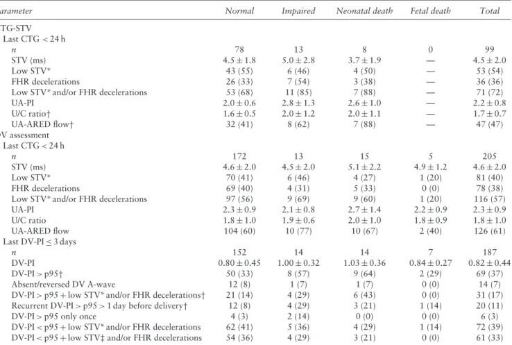
Pregnancies with normal infant outcome had been randomized at a later gestational age, and the EFW-p50 and BW-p50 ratios were larger than for those in which the infant died or had impaired development. All cases of fetal death (n= 7) occurred in pregnancies allocated to DV monitoring. Assessment of the monitoring parameters obtained shortly before fetal death showed that in only one case STV was below the cut-off used in the CTG-STV group, and the DV waveform was normal (Table 3). All other cases of fetal death had either no STV assessment within 24 h before death or normal CTG according to the CTG-STV group protocol. In two cases, the last DV-PI measurement before death had been > 95thpercentile, but these cases had been allocated to the DV-no-A group.
Infants with a normal outcome were born at a later gestational age, with a higher birth weight and BW-p50 ratio, and had a low Apgar score less frequently than those with impaired outcome or death (Table 2). Severe composite morbidity at discharge was less likely in infants with normal outcome. Specifically, cerebral hemorrhage and periventricular leukomalacia were more frequent in infants with impaired outcome (21%) than in the normal outcome group (2%). Eighty-three percent of the liveborn infants survived without neurological impairment, although 28% of these had severe morbidity in the neonatal period. In contrast, 46% of the surviving infants with neurological impairment did not have severe morbidity during the neonatal period.
There were no differences in demographic, obstetric or neonatal characteristics between the monitoring groups and between infants who did and those who did not undergo follow-up at 2 years of age, corrected for prematurity (data published previously2). Infants delivered after 32 weeks were included in the study at a later gestational age, with a larger estimated fetal weight and better outcomes than those included earlier, as would be expected.
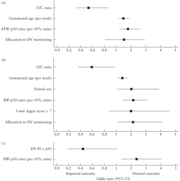
Multivariable regression analysis of parameters at study inclusion demonstrated that gestational age, larger EFW-p50 ratio and lower U/C ratio were significantly associated with 2-year survival and normal outcome (Figure 1a). Allocation to DV monitoring had a smaller effect (P < 0.1), but remained in the model. Multivariable analysis of parameters at delivery demonstrated that pregnancies with normal outcome were more likely to have been allocated to the DV groups, have a lower U/C ratio and higher birth weight and Apgar score, and more often delivered a female neonate (Figure 1b). When this analysis was repeated including only the DV-PI of women with a last DV Doppler measurement within 3 days before delivery or fetal death (n= 180), a last DV-PI measurement > 95th percentile was associated
Table 1 Summary of inclusion of cases in post-hoc analysis of TRUFFLE study on preterm pregnancies with severe fetal growth restriction according to monitoring strategy for delivery: reduced fetal heart rate short-term variation on cardiotocography (CTG-STV), ductus venosus (DV) pulsatility index (PI) > 95thpercentile (p95) or absent DV A-wave
Parameter CTG-STV (n= 166) DV-PI > p95 (n= 167) No DV A-wave (n= 170) Total (n= 503) Excluded from post-hoc analysis
Inevitable perinatal death 2 (1) 1 (1) 2 (1) 5 (1)
Neonatal data missing 1 (0.6) 0 (0) 0 (0) 1 (0.2)
Alive but no 2-year follow-up 12 (7) 13 (8) 8 (5) 33 (7)
Delivered≥ 32 weeks 46 (28) 51 (31) 50 (30) 147 (29)
Included in post-hoc analysis 105 (63) 102 (61) 110 (65) 317 (63)
GA at delivery (weeks) 29.7 (28.5–30.9) 29.9 (28.7–30.9) 29.9 (28.7–30.7) 29.9 (28.6–30.9)
Birth weight (g) 888± 202 887± 220 876± 208 884± 209
Fetal death 0 (0) 3 (3) 4 (4) 7 (2)
Neonatal death 10 (10) 6 (6) 9 (8) 25 (8)
Alive at 2-year follow-up 95 (90) 93 (91) 97 (88) 285 (90)
Alive without neurological impairment
% of infants evaluated 81 (85)* 85 (91) 91 (94) 257 (90)
% of all included infants 81 (77)† 85 (83) 91 (83) 257 (81)
Data are given as n (%), median (interquartile range) or mean± SD. Percentages for fetal death and neonatal data were calculated from total number of included cases. Pearson’s chi-square test used for comparison of CTG-STV group with both DV groups combined. *P= 0.049; †P= 0.21. GA, gestational age.
with impaired outcome, and a larger BW-p50 ratio was associated with normal outcome, while the other parameters were rejected from the model (Figure 1c).
The association between the last monitoring data before delivery or fetal death and the primary outcome is detailed separately for the CTG-STV and DV groups in Table 4. The association of abnormal monitoring results with the primary outcome differed between these groups. In the CTG-STV group, absent or reversed end-diastolic (ARED) flow in the UA and a high U/C ratio were negatively associated with the primary outcome (Figure 2a). In the DV groups, DV-PI > 95th percentile was negatively associated with the primary outcome, and this effect was more pronounced for recurrent elevated DV-PI > 95th percentile for more than 1 day (which was allowed by the study protocol) (Figure 2b). The negative effect of DV-PI > 95th percentile was not further enhanced when it occurred in combination with a STV below the cut-off used for the CTG-STV group and below the safety-net cut-off used in the DV groups and/or fetal heart-rate decelerations. Although the U/C ratio in the DV groups was the same as in the CTG-STV group and the incidence of UA-ARED flow was similar, the negative association of these parameters with the primary outcome that was observed in the CTG-STV group was absent in the DV groups.
DISCUSSION
This secondary sensitivity analysis of the data of the TRUFFLE study strengthens the conclusion of their primary intention-to-treat analysis, that perinatal outcomes are improved if DV Doppler measurements are combined with CTG-STV in the monitoring of fetuses with severe preterm FGR. Our analysis targeted infants who were delivered before 32 weeks in order to focus on
the effect of the different monitoring techniques on infant outcome. We carried out an in-depth study of perinatal deaths and the association between the last measurements of fetal monitoring parameters with the primary outcome. In this post-hoc analysis, both DV groups were com-bined to explore the association of 2-year neurodevelop-mental outcome with DV Doppler measurements. This was justified because survival with normal neurodevelop-ment at 2 years of age, corrected for prematurity, was equal in both DV groups (83% in infants with known outcome). Normal outcome at 2 years of age was less fre-quent in the CTG-STV group (77%), but this difference was not statistically significant.
Perinatal mortality was similar between the CTG-STV and DV groups (10%); however, all fetal deaths occurred in the DV groups. Analysis of this antenatal mortality suggested a spurious result: 6/7 cases of fetal death would probably not have been delivered in a timely manner if they had been allocated to the CTG-STV group, as the last STV measurement was above the cut-off limits used for this group. Two cases of fetal death might have benefited from a DV-PI cut-off at the 95th percentile instead of an absent A-wave as indication for delivery.
Multivariable analysis did not demonstrate a significant benefit for normal outcome in those randomized to the DV groups after adjustment for gestational age and EFW-p50 ratio. If analysis was restricted to those who were liveborn, assuming that the uneven distribution of fetal death between groups was by chance, there would be a statistically significant benefit of DV monitoring. This finding is in line with aggregated cohort evidence in a systematic review by Morris et al.4, which showed moderate predictive accuracy of longitudinal DV Doppler measurements for fetal/neonatal wellbeing in high-risk pregnancies (likelihood ratio, 3.15 (95% CI, 2.19–4.54)). Analysis of the results of the different monitoring techniques shows that, with CTG monitoring, heart
Table 2 Demographic, obstetric and neonatal characteristics of 317 infants with severe preterm fetal growth restriction delivered < 32 weeks, according to neurodevelopmental outcome at 2 years of age
Normal Impaired Dead Total
Characteristic (n= 257) (n= 28) (n= 32) (n= 317)
Demographic and clinical characteristics
Maternal age (years) 31± 5 31± 5 30± 5 31± 5
Caucasian ethnicity 220 (86) 25 (89) 29 (91) 274 (86) Nulliparous* 159 (62) 14 (50) 27 (84) 200 (63) BMI (kg/m2) 25± 6 25± 6 25± 5 25± 6 Smoker 30 (12) 6 (21) 4 (13) 40 (13) GA at inclusion (weeks)* 28+ 6 (26+ 0 to 31 + 5) 28+ 1 (26+ 0 to 31 + 0) 27+ 6 (26+ 0 to 31 + 4) 28+ 4 (26+ 0 to 31 + 5) EFW (g)* 852± 193 778± 180 703± 178 833± 202 EFW-p50 ratio* 0.65± 0.09 0.62± 0.08 0.60± 0.08 0.64± 0.09
Uterine artery notching 131 (51) 19 (68) 20 (63) 170 (54)
UA-PI 2.0± 0.5 2.2± 0.7 2.2± 0.7 2.1± 0.6
UA-ARED flow 111 (43) 15 (54) 18 (56) 144 (45)
UA-RED flow 15 (6) 1 (3.6) 3 (9) 19 (6)
U/C ratio* 1.5± 0.5 1.8± 0.6 1.7± 0.8 1.5± 0.6
Any hypertensive morbidity 203 (79) 22 (79) 23 (72) 248 (78)
Pre-eclampsia/HELLP 155 (60) 15 (54) 16 (50) 186 (59)
Antihypertensive medication 162 (63) 16 (57) 16 (50) 194 (61)
Magnesium treatment* 63 (25) 3 (11) 3 (9) 69 (22)
Interval to delivery (days) 7± 6 8± 7 6± 6 7± 7
GA at delivery (weeks)* 30+ 0 (26+ 2 to 31 + 6) 29+ 5 (26+ 1 to 31 + 3) 28+ 5 (26+ 1 to 31 + 6) 29+ 6 (26+ 1 to 31 + 6) Fetal death 0 (0) 0 (0) 7 (22) 7 (2) Neonatal characteristics Live birth 257 (100) 28 (100) 25 (78) 310 (98) Birth weight (g)* 910± 203 804± 170 736± 231 887± 209
Birth weight-p50 ratio* 0.59± 0.09 0.54± 0.07 0.55± 0.10 0.59± 0.09
Male gender* 118 (46) 20 (71) 12 (48) 150 (48)
Apgar score < 7* 29 (11) 9 (32) 6 (24) 44 (14)
UA pH (n= 280) 7.3 (6.8 to 7.4) 7.3 (7.0 to 7.4) 7.3 (6.9 to 7.3) 7.3 (6.8 to 7.4)
UA pH < 7.0 2 (1) 1 (4) 1 (4) 4 (1)
Composite severe morbidity*† 73 (28) 15 (54) 16 (64) 104 (34)
Bronchopulmonary dysplasia* 32 (12) 8 (29) 2 (8) 42 (14)
Proven sepsis* 43 (17) 7 (25) 12 (48) 62 (20)
NEC* 6 (2.3) 0 (0) 5 (20) 11 (4)
GMH Grade III or IV* 4 (1.6) 4 (14) 3 (12) 11 (4)
PVL Grade II or III 2 (0.8) 2 (7) 0 (0) 4 (1)
Data are given as mean± SD, n (%) or median (interquartile range). Percentages for neonatal data were calculated from total number of liveborn infants. *Significant difference between groups, P < 0.05. †Components of severe morbidity: bronchopulmonary dysplasia (supplemental oxygen at 36 weeks), germinal matrix hemorrhage (GMH) Grade III or IV, periventricular leukomalacia (PVL) Grade II or III, necrotizing enterocolitis (NEC) (diagnosed by X-ray or laparotomy) or proven sepsis. ARED flow, absent/reversed end-diastolic flow; BMI, body mass index; EFW, estimated fetal weight; EFW-p50 ratio, ratio of EFW to EFW 50thpercentile; GA, gestational age; PI, pulsatility
index; RED flow, reversed end-diastolic flow; UA, umbilical artery; U/C ratio, umbilical artery-PI/fetal middle cerebral artery-PI ratio.
Table 3 Last measurements of fetal monitoring parameters in seven cases of fetal death in preterm pregnancies with severe fetal growth restriction allocated to Doppler assessment of ductus venosus (DV) as monitoring strategy for delivery
Case Monitoring group GA (weeks) CTG < 24 h before death STV (ms) STV low CTG decelerated DV-PI DV-PI high DV A-wave UA-PI U/C
ratio EDF Comment
1 DV-no-A 29 Yes 5.1 No No 0.30 No Present 1.6 1.2 Present
2 DV-p95 29 Yes 2.7 Yes No 0.57 No Present 3.8 3.5 Absent
3 DV-no-A 28 Yes 5.2 No No 1.01 >p95 Present 2.2 1.5 Absent
4 DV-p95 27 No 6.9 No No 0.77 No Present 1.9 1.3 Absent Abruption
5 DV-p95 29 No 7.5 No No 0.66 No Present 4.8 5.4 Reversed
6 DV-no-A 27 Yes 5.6 No No 1.10 >p95 Present 1.6 1.3 Present
7 DV-no-A 28 Yes 5.8 No No 0.74 No Present 2.0 1.7 Present
Last DV Doppler assessment recorded < 3 days before death and last umbilical artery (UA) Doppler assessment recorded < 1 week before death. CTG, cardiotocography; DV-no-A, absent A-wave in DV; DV-p95, DV pulsatility index (PI) > 95thpercentile; EDF, end-diastolic
0.0 Allocation to DV monitoring EFW-p50 ratio (per 10% ratio) Gestational age (per week) U/C ratio (a) 0.2 0.4 0.6 0.8 1 2 3 4 5 0.0 (c) 0.2 0.4 Impaired outcome
Odds ratio (95% CI)
Normal outcome
0.6 0.8 1 2 3 4 5
0.0 Allocation to DV monitoring
5-min Apgar score > 7 BW-p50 ratio (per 10% ratio)
BW-p50 ratio (per 10% ratio) Female sex Gestational age (per week) U/C ratio
DV-PI > p95 (b)
0.2 0.4 0.6 0.8 1 2 3 4 5
Figure 1 Odds ratios for normal outcome at corrected age of 2 years in infants with fetal growth restriction delivered before 32 weeks’ gestation, calculated by multivariable analysis of: (a) parameters at study inclusion (area under receiver–operating characteristics curve (AUC), 0.69); (b) parameters at delivery (AUC, 0.75); and (c) parameters in those with ductus venosus (DV) monitoring and a DV measurement < 3 days before delivery (n= 180; AUC, 0.75). Inclusion in model, P = 0.05; removal from model, P = 0.10. Allocation to DV monitoring forced to stay in model. BW-p50 ratio, ratio of birth weight (BW) to 50th-percentile BW adjusted for gestational age, maternal
ethnicity, weight and height and infant gender; EFW-p50 ratio, ratio of estimated fetal weight (EFW) to 50th-percentile EFW adjusted for
gestational age, maternal ethnicity, weight and height and infant gender; PI, pulsatility index; U/C ratio, umbilical artery-PI/fetal middle cerebral artery-PI ratio.
rate decelerations and UA-ARED flow are negatively associated with normal outcome, while this is not found for combined monitoring of DV waveform and CTG-STV. It might be that those at risk for neurological impairment with UA-ARED flow are delivered in a more timely fashion in the DV groups because of an abnormal DV measurement, although we cannot prove this because DV was not measured in the CTG-STV group after inclusion in the study.
Typically, abnormalities in UA/MCA flow precede abnormalities in DV flow pattern5,6. Elevated U/C ratio and UA-ARED flow are known to be associated with adverse outcome in pregnancies with FGR7. Our findings confirmed this, but only in the CTG-STV group.
It is possible that Doppler assessment of the DV allowed ‘fine tuning’ of the timing of delivery and selection of a subgroup of fetuses with severe redistribution (U/C ratio)
and placental impairment (UA-ARED flow) that were most at risk for cerebral damage.
The observations that elevated DV-PI or
absent/reversed A-wave is associated with increased neonatal morbidity and adverse long-term infant out-come, and that abnormalities in DV flow are a stronger predictor of these outcomes than are abnormalities in UA flow have been noted previously8,9. The current analysis, in which an abnormal DV flow pattern in the DV groups was associated with impaired neurological outcome, is consistent with these observations.
The difference in the associations of monitoring data and outcome between the CTG-STV group and DV groups, and the lower prevalence of neurological impairment among survivors in the DV groups, may support the hypothesis that, in some early preterm growth-restricted infants, cardiac dysfunction (abnormal
Table 4 Last measurements of fetal monitoring parameters before delivery or fetal death according to neurodevelopmental outcome in preterm pregnancies with severe fetal growth restriction allocated to monitoring strategy for delivery of reduced fetal heart rate (FHR) short-term variation on cardiotocography (CTG-STV) or Doppler assessment of ductus venosus (DV)
Parameter Normal Impaired Neonatal death Fetal death Total
CTG-STV Last CTG < 24 h n 78 13 8 0 99 STV (ms) 4.5± 1.8 5.0± 2.8 3.7± 1.9 — 4.5± 2.0 Low STV* 43 (55) 6 (46) 4 (50) — 53 (54) FHR decelerations 26 (33) 7 (54) 3 (38) — 36 (36)
Low STV* and/or FHR decelerations 53 (68) 11 (85) 7 (88) — 71 (72)
UA-PI 2.0± 0.6 2.8± 1.3 2.6± 1.0 — 2.2± 0.8 U/C ratio† 1.6± 0.5 2.0± 1.2 2.0± 1.1 — 1.7± 0.7 UA-ARED flow† 32 (41) 8 (62) 7 (88) — 47 (47) DV assessment Last CTG < 24 h n 172 13 15 5 205 STV (ms) 4.6± 2.0 4.5± 2.0 5.1± 2.2 4.9± 1.2 4.6± 2.0 Low STV* 70 (41) 6 (46) 4 (27) 1 (20) 81 (40) FHR decelerations 69 (40) 4 (31) 5 (33) 0 (0) 78 (38)
Low STV* and/or FHR decelerations 97 (56) 9 (69) 9 (60) 1 (20) 116 (57)
UA-PI 2.3± 0.9 2.1± 0.8 2.7± 1.4 2.2± 0.9 2.3± 0.9
U/C ratio 1.8± 1.0 1.9± 0.6 2.0± 1.0 1.8± 0.9 1.8± 1.0
UA-ARED flow 104 (60) 10 (77) 10 (67) 2 (40) 126 (61)
Last DV-PI≤ 3 days
n 152 14 14 7 187
DV-PI 0.80± 0.45 1.00± 0.32 1.03± 0.36 0.84± 0.27 0.82± 0.44
DV-PI > p95† 50 (33) 8 (57) 9 (64) 2 (29) 69 (37)
Absent/reversed DV A-wave 12 (8) 1 (7) 1 (7) 0 (0) 14 (7)
DV-PI > p95+ low STV*and/or FHR decelerations† 21 (14) 4 (29) 6 (43) 0 (0) 31 (17) Recurrent DV-PI > p95 > 1 day before delivery† 12 (8) 4 (29) 3 (21) 1 (14) 20 (11)
DV-PI > p95 only once 4 (3) 2 (14) 0 (0) 0 (0) 6 (3)
DV-PI < p95+ low STV*and/or FHR decelerations 62 (41) 5 (36) 4 (29) 1 (14) 72 (39) DV-PI < p95+ low STV‡ and/or FHR decelerations 54 (36) 4 (29) 3 (21) 0 (0) 61 (33) Data are given as n (%) or mean± SD. Participants included if last CTG was < 24 h, last DV assessment was ≤ 3 days or last fetal arterial Doppler assessment was within 1 week of delivery or fetal death. Percentages total > 100 because some fetuses had multiple test results recorded within the relevant time period. Eight cases from CTG-STV group and eight cases from DV groups excluded because last CTG was≥ 24 h before delivery. Twenty cases from DV groups excluded because last DV pulsatility index (PI) measurement was > 3 days before delivery. *STV cut-off for CTG-STV group. †Comparison of all outcomes: P < 0.05. ‡STV cut-off of safety-net criteria for DV groups. ARED, absent/reversed end-diastolic; UA, umbilical artery; U/C ratio, umbilical artery-PI/fetal middle cerebral artery-PI ratio.
DV assessment) can precede cerebral dysfunction (low CTG-STV). Thus, timely detection of these changes by DV monitoring (and subsequent action) can prevent neurological impairment in some fetuses. In others, this sequence can occur the other way round, with earlier STV abnormality or recurrent heart rate decelerations being the indication for delivery. In five cases of fetal death, the last DV measurement was within the normal range, but in two cases it was higher than the 95th percentile. Frauenschuh et al.10 found that, in four cases of severe placental insufficiency, DV flow prior to intrauterine fetal death was unaffected. Thus, it is possible that there is some variation in the effects of malnutrition and hypoxia on FGR fetuses, and the onset of organ damage may not follow the same pattern in all fetuses.
We included in our analysis only infants delivered before 32 weeks because, according to the study protocol and in actual practice, DV assessment contributed only to the decision to deliver before 32 weeks’ gestation. The potential bias introduced by excluding differential delivery after 32 weeks by trial allocation group is likely
to be small as inclusion and outcome parameters were equally distributed between the groups. Results from this analysis can therefore be applied only to women with FGR before 32 weeks.
This post-hoc analysis highlights some of the effects of DV monitoring that were obscured by the original intention-to-treat analysis in the TRUFFLE study. However, as with all post-hoc analyses, we advocate caution regarding the possibility of bias. Nonetheless, the current findings are consistent with the original data.
In conclusion, in accordance with the results of the overall TRUFFLE study on the monitoring and intervention management of very preterm severe FGR, we found that the difference in the proportion of infants that survived without neuroimpairment was non-significant when comparing timing of delivery with or without changes in the DV waveform. We speculate that the uneven distribution of fetal deaths towards the DV groups was a chance effect, and found that, among surviving children in these groups, neurological outcomes at 2 years of age were better. Adverse neurodevelopmental outcome
0.0 Low STV and/or FHR decelerations
Low STV FHR decelerations UA-ARED flow U/C ratio > 2.0 (a) 0.2 0.4 0.6 0.8 1 2 3 4 5 6 0.0 DV A-wave present Recurrent DV-PI > p95 (> 1 day interval) DV-PI > p95 + (low STV and/or FHR decelerations) DV-PI > p95 Low STV and/or FHR decelerations FHR decelerations Low STV UA-ARED flow U/C ratio > 2.0 (b) 0.2 0.4 0.6 0.8 1 2 3 4 5 6 Impaired outcome
Odds ratio (95% CI)
Normal outcome
Figure 2 Odds ratios from univariate analysis for normal outcome at corrected age of 2 years specified for parameters at last cardiotoco-graphy (CTG) within 24 h, last fetal arterial pulsatility index (PI) assessment within 7 days or last ductus venosus (DV)-PI within 3 days before delivery in infants with fetal growth restriction randomized to CTG monitoring (a) or DV Doppler assessment (b) for delivery indication. ARED, absent/reversed end-diastolic; FHR, fetal heart rate; p95, 95thpercentile; STV, short-term variation; UA, umbilical artery;
U/C ratio, UA-PI/fetal middle cerebral artery-PI ratio.
was significantly associated with abnormal DV-PI before delivery and a lower birth weight in surviving babies. Before 32 weeks, delaying delivery until abnormalities in fetal DV-PI or STV and/or recurrent heart rate decelera-tions occur, as defined by the study protocol, is therefore likely to be safe and possibly benefits long-term outcome. ACKNOWLEDGMENTS
C.C.L. is supported by the National Institute for Health Research Biomedical Research Centre based at Imperial College Healthcare National Health Service Trust and Imperial College London, UK. The Trial of Randomized Umbilical and Fetal Flow in Europe study was supported by ZonMw, 2509 AE Den Haag, The Netherlands (grant 94506556), in The Netherlands. In other countries, the study was not funded. A contribution was made to study funding from the Dr Hans Ludwig Geisenhofer Foundation, Munich, Germany.
TRUFFLE GROUP COLLABORATING AUTHORS
A. Aktas (Marburg, Germany), S. Borgione (Turin, Italy), R. Chaoui (Berlin, Germany), J.M.J. Cornette (Rotter-dam, The Netherlands), T. Diehl (Hamburg, Germany),
J. van Eyck (Zwolle, The Netherlands), N. Fratelli (Bres-cia, Italy), I.C. van Haastert (Utrecht, The Netherlands), S. Lobmaier (Munich, Germany), E. Lopriore (Leiden, The Netherlands), H. Missfelder-Lobos (Cambridge, UK), G. Mansi (Naples, Italy), P. Martelli (Brescia, Italy), G. Maso (Trieste, Italy), U. Maurer-Fellbaum (Graz, Austria), S. Mulder-de Tollenaer (Zwolle, The Netherlands), R. Napolitano (Naples, Italy), M. Oberto (Turin, Italy), D. Oepkes (Leiden, The Netherlands), G. Ogge (Turin, Italy), J.A.M. van der Post (Ams-terdam, The Netherlands), L. Preston (Cambridge, UK), F. Raimondi (Naples, Italy), H. Rattue (London, UK), I.K.M. Reiss (Rotterdam, The Netherlands), L.S. Scheepers (Nijmegen/Maastricht, The Netherlands), A. Skabar (Trieste, Italy), M. Spaanderman (Nijmegen, The Netherlands), N. Weisglas-Kuperus (Rotterdam, The Netherlands), A. Zimmermann (Munich, Germany).
REFERENCES
1. Lees C, Marlow N, Arabin B, Bilardo CM, Brezinka C, Derks JB, Duvekot J, Frusca T, Diemert A, Ferrazzi E, Ganzevoort W, Hecher K, Martinelli P, Ostermayer E, Papageorghiou AT, Schlembach D, Schneider KT, Thilaganathan B, Todros T, van Wassenaer-Leemhuis A, Valcamonico A, Visser GH, Wolf H; TRUFFLE Group. Perinatal morbidity and mortality in early-onset fetal growth restriction: cohort outcomes of the trial of randomized umbilical and fetal flow in Europe (TRUFFLE).
Ultrasound Obstet Gynecol 2013; 42: 400–408.
2. Lees CC, Marlow N, van Wassenaer-Leemhuis A, Arabin B, Bilardo CM, Brezinka C, Calvert S, Derks JB, Diemert A, Duvekot JJ, Ferrazzi E, Frusca T, Ganzevoort W,
Hecher K, Martinelli P, Ostermayer E, Papageorghiou AT, Schlembach D, Schneider KT, Thilaganathan B, Todros T, Valcamonico A, Visser GH, Wolf H; TRUFFLE study group. 2 year neurodevelopmental and intermediate perinatal outcomes in infants with very preterm fetal growth restriction (TRUFFLE): a randomised trial.
Lancet 2015; 385: 2162–2172.
3. Gardosi J, Chang A, Kalyan B, Sahota D, Symonds EM. Customised antenatal growth charts. Lancet 1992; 339: 283–287.
4. Morris RK, Selman TJ, Verma M, Robson SC, Kleijnen J, Khan KS. Systematic review and meta-analysis of the test accuracy of ductus venosus Doppler to predict compromise of fetal/neonatal wellbeing in high risk pregnancies with placental insufficiency. Eur J Obstet Gynecol Reprod Biol 2010; 152: 3–12.
5. Hecher K, Bilardo CM, Stigter RH, Ville Y, Hackeloer BJ, Kok HJ, Senat MV, Visser GH. Monitoring of fetuses with intrauterine growth restriction: a longitudinal study.
Ultrasound Obstet Gynecol 2001; 18: 564–570.
6. Baschat AA, Gembruch U, Harman CR. The sequence of changes in Doppler and biophysical parameters as severe fetal growth restriction worsens. Ultrasound Obstet
Gynecol 2001; 18: 571–577.
7. Flood K, Unterscheider J, Daly S, Geary MP, Kennelly MM, McAuliffe FM, O’Donoghue K, Hunter A, Morrison JJ, Burke G, Dicker P, Tully EC, Malone FD. The role of brain sparing in the prediction of adverse outcomes in intrauterine growth restriction: results of the multicenter PORTO Study. Am J Obstet Gynecol 2014; 211: 288.e1–5.
8. Baschat AA, Viscardi RM, Hussey-Gardner B, Hashmi N, Harman C. Infant neurodevelopment following fetal growth restriction: relationship with antepartum surveillance parameters. Ultrasound Obstet Gynecol 2009; 33: 44–50.
9. Bilardo CM, Wolf H, Stigter RH, Ville Y, Baez E, Visser GH, Hecher K. Relationship between monitoring parameters and perinatal outcome in severe, early intrauterine growth restriction. Ultrasound Obstet Gynecol 2004; 23: 119–125.
10. Frauenschuh I, Frambach T, Karl S, Dietl J, M ¨uller T. [Ductus venosus blood flow prior to intrauterine foetal death in severe placental insufficiency can be unaffected as shown by doppler sonography]. Z Geburtshilfe Neonatol 2014; 218: 218–222.