Mitola, Efrem Foglia, Pieter B. van Loenen, Astrid E. Alewijnse and Marco Presta
Chiara Tobia, Paola Chiodelli, Stefania Nicoli, Patrizia Dell'Era, Simone Buraschi, Stefania
Print ISSN: 1079-5642. Online ISSN: 1524-4636
Copyright © 2012 American Heart Association, Inc. All rights reserved. Greenville Avenue, Dallas, TX 75231
is published by the American Heart Association, 7272 Arteriosclerosis, Thrombosis, and Vascular Biology
doi: 10.1161/ATVBAHA.112.250035
2012;32:e104-e116; originally published online July 26, 2012;
Arterioscler Thromb Vasc Biol.
http://atvb.ahajournals.org/content/32/9/e104
World Wide Web at:
The online version of this article, along with updated information and services, is located on the
http://atvb.ahajournals.org//subscriptions/
at:
is online Arteriosclerosis, Thrombosis, and Vascular Biology
Information about subscribing to
Subscriptions:
http://www.lww.com/reprints
Information about reprints can be found online at:
Reprints:
document.
Question and Answer
Permissions and Rights
page under Services. Further information about this process is available in the
which permission is being requested is located, click Request Permissions in the middle column of the Web Copyright Clearance Center, not the Editorial Office. Once the online version of the published article for
can be obtained via RightsLink, a service of the Arteriosclerosis, Thrombosis, and Vascular Biology
in
Requests for permissions to reproduce figures, tables, or portions of articles originally published
Permissions:
by guest on October 6, 2013 http://atvb.ahajournals.org/
Downloaded from http://atvb.ahajournals.org/ by guest on October 6, 2013 Downloaded from http://atvb.ahajournals.org/ by guest on October 6, 2013 Downloaded from http://atvb.ahajournals.org/ by guest on October 6, 2013 Downloaded from http://atvb.ahajournals.org/ by guest on October 6, 2013 Downloaded from http://atvb.ahajournals.org/ by guest on October 6, 2013 Downloaded from http://atvb.ahajournals.org/ by guest on October 6, 2013 Downloaded from http://atvb.ahajournals.org/ by guest on October 6, 2013 Downloaded from http://atvb.ahajournals.org/ by guest on October 6, 2013 Downloaded from http://atvb.ahajournals.org/ by guest on October 6, 2013 Downloaded from http://atvb.ahajournals.org/ by guest on October 6, 2013 Downloaded from http://atvb.ahajournals.org/ by guest on October 6, 2013 Downloaded from http://atvb.ahajournals.org/ by guest on October 6, 2013 Downloaded from http://atvb.ahajournals.org/ by guest on October 6, 2013 Downloaded from
e104
T
he lysophospholipid sphingosine-1-phosphate (S1P) is a signaling molecule that acts through the binding to a family of 7-transmembrane G-protein–coupled receptors that include 5 high-affinity members (S1P1–5).1 S1P receptorsmod-ulate a variety of biological processes, including cell adhe-sion, migration, and endocytosis with different and sometimes opposing effects on the regulation of cellular functions.2 In
particular, endothelial S1P1 is involved in cytoskeletal rear-rangement and formation of intercellular adherens junctions, its exposure to blood-borne S1P being required for the main-tenance of blood–barrier integrity in defined vascular beds.3,4
Also, genetic deletion demonstrates the nonredundant role of S1P1 in vascular maturation during embryogenesis, leading to embryonic lethality in knockout mice.5 Thus, chemical
modu-lators of S1P1 activity have been developed as putative thera-peutic molecules acting to restore endothelial barrier integrity in various pathological conditions characterized by the dis-ruption of endothelial cell–cell interaction in different organs, including lung, heart, kidney, and brain.4
Endothelial barrier integrity is dependent upon a complex network of molecular interactions involving adherens and tight junctions.6 S1P may affect endothelial cell–cell junctions
by regulating assembly and expression of the major adherens junction component, vascular endothelial adhesion molecule VE-cadherin, and formation of zonula occludens 1 (ZO1)+
tight junctions.7–10 However, the molecular and functional
bases of the impact of the S1P/S1P1 receptor system on endo-thelial barrier integrity remain to be fully elucidated.
The teleost zebrafish (Danio rerio) represents a promising experimental model for in vivo analysis of the molecular and cellular mechanisms underlying blood vessel development and vascular dysfunctions.6 When compared with other vertebrate
model systems, zebrafish offers many advantages, including ease of experimentation, drug administration, amenability to in vivo manipulation, and feasibility of reverse and forward genetic approaches.11 Also, zebrafish possesses a complex
circulatory system similar to that of mammals and the optical transparency and ability to survive for several days without a
Received on: October 24, 2011; final version accepted on: July 13, 2012.
From the Department of Biomedical Sciences and Biotechnology, Unit of General Pathology and Immunology, School of Medicine, University of Brescia, Italy (C.T., P.C., P.D., S.N., S.B., S.M., M.P.); Department of Biology, University of Milan, Italy (E.F.); Department of Pharmacology and Pharmacotherapy, Academic Medical Center, Amsterdam, The Netherlands (P.B.v.L., A.E.A.); Department of Internal Medicine, Yale University School of Medicine, New Haven, CT (S.N.); and Department of Pathology, Anatomy, and Cell Biology, Thomas Jefferson University, Philadelphia, PA (S.B.).
Correspondence to Marco Presta, General Pathology, Department of Biomedical Sciences and Biotechnology, Viale Europa 11, 25123 Brescia, Italy. E-mail [email protected]
© 2012 American Heart Association, Inc.
Arterioscler Thromb Vasc Biol is available at http://atvb.ahajournals.org DOI: 10.1161/ATVBAHA.112.250035
Objective—Endothelial sphingosine-1-phosphate (S1P) receptor-1 (S1P1) affects different vascular functions, including
blood vessel maturation and permeability. Here, we characterized the role of the zS1P1 ortholog in vascular development in zebrafish.
Methods and Results—zS1P1 is expressed in dorsal aorta and posterior cardinal vein of zebrafish embryos at 24 to
30 hours postfertilization. zS1P1 downregulation by antisense morpholino oligonucleotide injection causes early pericardial edema, lack of blood circulation, alterations of posterior cardinal vein structure, and late generalized edema. Also,
zS1P1 morphants are characterized by downregulation of vascular endothelial cadherin (VE-cadherin) and Eph receptor
EphB4a expression and by disorganization of zonula occludens 1 junctions in posterior cardinal vein endothelium, with no alterations of dorsal aorta endothelium. cadherin knockdown results in similar vascular alterations, whereas
VE-cadherin overexpression is sufficient to rescue venous vascular integrity defects and EphB4a downregulation in zS1P1 morphants. Finally, S1P1 small interfering RNA transfection and the S1P1 antagonist (R)-3-amino-(3-hexylphenylamino)-4-oxobutylphosphonic acid (W146) cause EPHB4 receptor down-modulation in human umbilical vein endothelial cells and the assembly of zonula occludens 1 intercellular contacts is prevented by the EPHB4 antagonist TNYL-RAW peptide in these cells.
Conclusion—The data demonstrate a nonredundant role of zS1P1 in the regulation of venous endothelial barrier in zebrafish and identify a S1P1/VE-cadherin/EphB4a genetic pathway that controls venous vascular integrity. (Arterioscler Thromb
Vasc Biol. 2012;32:e104-e116.)
Key Words: sphingosine-1-phosphate ◼ endothelial barrier ◼ zebrafish ◼ VE-cadherin ◼ Eph receptor
Sphingosine-1-Phosphate Receptor-1 Controls Venous
Endothelial Barrier Integrity in Zebrafish
Chiara Tobia, Paola Chiodelli, Stefania Nicoli, Patrizia Dell’Era, Simone Buraschi, Stefania Mitola,
Efrem Foglia, Pieter B. van Loenen, Astrid E. Alewijnse, Marco Presta
functioning circulation make the zebrafish embryo amenable for vascular biology studies.12
Expression of the zebrafish ortholog of the S1P1 receptor (zS1P1) has been observed in the zebrafish embryonic brain,13
whereas zebrafish mutants of the S1P2 receptor ortholog miles
apart (mil) show organogenesis defects of the heart,14
dem-onstrating that bioactive lipid receptors play different roles in zebrafish development. In the present work, we performed the characterization and functional analysis of zS1P1 receptor during vascular development in zebrafish. zS1P1 is transiently expressed in the axial vasculature of the trunk of zebrafish embryos. zS1P1 knockdown caused defects of venous vas-cular integrity, including VE-cadherin downregulation and disorganization of endothelial junctions, lack of blood circu-lation, and late generalized edema. In association with these defects, zS1P1 morphants showed the downregulation of the Eph receptor EphB4a in venous axial endothelium. Similar defects were observed in VE-cadherin morphants and genetic evidence indicate that VE-cadherin is a zS1P1-regulated ele-ment that controls venous endothelial barrier integrity by acting upstream of EphB4a. Finally, S1P1 small interfering RNA (siRNA) transfection and the specific S1P1 antagonist W14615 cause EPHB4 receptor downmodulation in human
umbilical vein endothelial cells (HUVECs). Furthermore, a selective EPHB4 antagonist disrupts ZO1+ intercellular
con-tacts in these cells. Together, the results provide evidence for a nonredundant role for S1P1 in the regulation of blood–barrier integrity via a novel S1P1/VE-cadherin/EphB4a cascade that controls venous endothelial cell–cell junction assembly.
Materials and Methods
Reagents
S1P, dihydro-S1P (dhS1P), and W146 were from Avanti Polar Lipids Inc (Alabaster, AL). 5-[4-phenyl-5-(trifluoromethyl)-2-thienyl]-3-[3-(trifluoromethyl)phenyl]-1,2,4-oxadiazole (SEW2871) was from Calbiochem (Nottingham, UK). Racemic 2-amino-2-[2-(4- octylphenyl)ethyl]-1,3-propanediol mono (dihydrogen phosphate) ester (FTY720-P) and [2-(4-(5-(3,4-diethoxyphenyl)-1,2,4-oxa-diazol-3-yl)-2,3-dihydro-1H-inden-1-yl amino) ethanol (CYM-5442) were synthesized as described.16,17 TNYL-RAW peptide
(YNYLFSPNGPIARAW) and scrambled peptide SCR-WTL (WTLAIFARNYNGPSP)18,19 were kindly provided by Dr O. Salvucci
(Bethesda, MD).
Zebrafish Stocks
Wild-type AB and transgenic tg(fli1:enhanced green fluores-cent protein [EGFP])y120 zebrafish lines were maintained under
standard conditions,21 and embryos were staged by hours
post-fertilization (hpf), as described.22 When specified, fertilized
eggs were maintained in fish water containing 250 mmol/L D-mannitol (Sigma, St. Louis, MO) or treated with 0.66 mg/mL tricaine at different developmental stages and allowed to develop at 28.5°C.
Whole-Mount In Situ Hybridization
Digoxigenin-labeled RNA probes were transcribed from linear cDNA constructs (Roche Applied Science, Indianapolis, IN). Whole-mount in situ hybridization (ISH) was performed as described.23 For
section-ing, fixed embryos were dehydrated in ethanol series, cleared in xilol, and paraffin embedded overnight.
Morpholino-Mediated Knockdown of zS1P1 and VE-cadherin
Antisense morpholino oligonucleotides (MO) (Gene Tools, Corvallis, OR) were directed against the 5ʹ untranslated region spanning the
zS1P1 ATG start codon (zS1P1-MO: 5ʹ-AGTGTCTGGCGATTAGGT CATCCAT-3ʹ) or were designed to complement exon–intron bound-aries of the zebrafish VE-cadherin gene (VE-cadherin-MO: 5ʹ-TTT ACAAGACCGTCTACCTTTCCAA-3ʹ). Standard MO (Std-MO: 5ʹ-CCTCTTACCTCAGTTACAATTTATA-3ʹ) was used as con-trol. Routinely, MOs were microinjected in 4.0 nL volume into 1- to 4-cell stage embryos at the concentration of 0.4 pmoles/embryo for zS1P1-MO or 1.0 pmoles/embryo for VE-cadherin-MO. A sub-set of embryos was coinjected with 0.25 to 0.4 pmoles/embryo of
zS1P1-MO and human S1P1 mRNA (70–100 pg/embryo), murine
VE-cadherin mRNA (40–60 pg/embryo), or human VEGF-A mRNA
(100 pg/embryo). zS1P1-MO shows no significant homology with human S1P1 mRNA and murine VE-cadherin mRNA sequences.
Microangiography
Tetramethylrhodamine isothiocyanate–dextran (molecular weight 2.0×10⁶; Invitrogen, Carlsbad, CA) was dissolved in double
dis-tilled water at 20 mg/mL and microinjected into the sinus venosus of zebrafish embryos at 50 hpf as described.24
Microscopy
Embryos were mounted in agarose-coated dishes and photographed under an epifluorescence Leica MZ16 F stereomicroscope (1× Plan Apo objective; NA, 0.141) equipped with a DFC480 digital cam-era and ICM50 software version 2.8.1 (all from Leica, Wetzlar, Germany). Confocal images were acquired with an LSM 50 META confocal laser microscope (Carl Zeiss, Germany). When specified, dechorionated 28 hpf zebrafish embryos were fixed overnight at 4°C with 1.5% glutaraldehyde plus 4% paraformaldehyde in 0.1 mol/L sodium cacodylate buffer, pH 7.3. Then, embryos were postfixed for 1 hour in sodium-cacodylate–buffered 1% osmium tetroxide, dehy-drated in a graded ethanol series, transitioned to propylene oxide, and embedded in Epon 812-Araldite. Semithin sections (0.8 μm) were obtained using a Reichert UltracutE ultramicrotome, stained with gentian violet and basic fuchsin, observed under a Leica DM 6000B microscope and photographed with a digital camera.
ZO1 Immunofluorescence Analysis of Zebrafish Embryos
Control and MO-treated tg(fli1:EGFP)y1 embryos were fixed at 30 hpf
in 4% paraformaldehyde for 2 hours at room temperature. Whole-mount immunofluorescence analysis was performed as described25
using a mouse anti-ZO1 antibody (Invitrogen) followed by Alexa Fluor 594 anti-mouse IgG (Invitrogen) and embryos were analysed by confocal laser microscopy.
zS1P1 Transfection in Mammalian Cells and cAMP Assay
The polymerase chain reaction (PCR) fragment with the full-length coding sequence of zS1P1 was cloned from the total RNA isolated from zebrafish embryos at 24 hpf (primer set: forward, 5ʹ-TCCATGATGCAGTTTTTGGA-3ʹ; reverse, 5ʹ-AGTTCCATCCCTCCAGTTT-3ʹ) into the pCRII-TOPO vector (Invitrogen). Next, the zS1P1 coding region was subcloned from pCRII-TOPO into the pcDNA5/FRT/TO vector (Invitrogen) using BamHI and XbaI (Fermentas Life Sciences, St. Leon-Rot, Germany). Chinese hamster ovary–FlpIn cells (Invitrogen) were maintained in Ham-F12 with L-glutamine, supplemented with 100 U/mL penicillin, 100 μg/mL streptomycin, and 10% charcoal-stripped fetal calf serum (Invitrogen). Ten μg of plasmid DNA were used for transfection into Chinese hamster ovary–FlpIn cells using Lipofectamine 2000 (Invitrogen). After 24 hours, transfected cells were washed with serum-free medium and serum starved for
16 hours. Next, transiently transfected cells were detached from the surface using cell dissociation buffer (Invitrogen), washed once with Hanks balanced salt solution, and resuspended in 5.0 mmol/L Hepes buffer, containing Hanks balanced salt solution with 0.05% fatty acid–free BSA (Sigma). Stimulation mixtures consisted of stimulation buffer with 3 μmol/L forskolin, 0.5 mmol/L 3-isobutyl-1-methylxanthine (IBMX) (both from Sigma) and the concentration response curve of the indicated compounds. Cells (2500 per well) were added to the stimulation mixtures in a 1:1 ratio in a 384-well Optiplate (Perkin-Elmer, Zaventem, Belgium) and stimulated for 30 minutes. The LANCE cAMP 384 kit (Perkin-Elmer) was used to determine the concentration of cAMP accumulated during stimulation according to the manufacturer’s protocol in a total volume of 20 μL. Measurements were performed 3 hours after adding detection buffer and antibody mixture using a Wallac 1420 Victor2 (PerkinElmer).
Data are expressed as the potency of the indicated compound to inhibit forskolin–induced cAMP accumulation (calculated as pEC50 value, here defined as the negative logarithm of the EC50 value).
Quantitative RT-PCR Analysis
Total RNA was isolated from zebrafish embryos using Trizol reagent (Invitrogen). Approximately 2 μg of total RNA were retrotranscribed using M-MLV Reverse Transcriptase (Invitrogen). Quantitative real-time PCR (qRT-PCR) was performed with a Biorad iCycler iQ Real-Time PCR Detection System using iQ SYBR Green Supermix (Biorad, Hercules, CA) according to manufacturer’s instructions. Primers sequence were as follows:
EphB4 (forward: 5ʹ-GGGGCCTGCAGGAGAGACCA-3ʹ, reverse:
5ʹ-GATCCGCGCTCCTTCTCGCA-3ʹ); VE-cadherin (forward: 5ʹ-TGACGAGGAGGGCGGAGGAG-3ʹ, reverse: 5ʹ-AGGTCCCACA CTGGGCCGAA-3ʹ); β-actin (forward: 5ʹ-CGAGCAGGAGATG GGAACC-3ʹ, reverse: 5ʹ-CAACGGAAACGCTCATTGC-3ʹ).
Samples were analyzed in triplicate, normalized against the thresh-old cycle (Ct) of β-actin, and expressed as changes with respect to the
levels of the gene under investigation in control samples.
HUVEC Cultures
HUVECs were grown in M199 medium (Invitrogen) supplemented with 20% fetal calf serum (Invitrogen), endothelial cell growth fac-tor (100 μg/mL, Sigma), and porcine heparin (100 μg/mL, Sigma). HUVECs were used at early passages (I–V) and grown on plastic sur-face coated with porcine gelatin (Sigma). When indicated, HUVECs were transfected with control-scrambled siRNA or with specific
S1P1 siRNAs (TAGCATTGTCAAGCTCCTAAA, S1P1 siRNAa; and CTCGGTCTCTGACTACGTCAA, S1P1 siRNAb; both from Qiagen, Chatsworth, CA) using the HiPerFect Transfection Reagent (Qiagen) according to the manufacturer’s instructions. Transfection efficiency was equal to 70% to 80% as shown by transfection with a fluorescein-conjugated control siRNA.
Western Blot Analysis of HUVECs
HUVECs were incubated with 30-μmol/L W146 for 48 hours, and cells lysates were probed with anti-EPHB4 antibody in a Western blot (Santa Cruz Biotechnology). Uniform loading of the gels was assessed using anti-focal adhesion kinase antibodies. In the second set of experiments, S1P1 siRNA-transfected HUVECs were analyzed 96 hours after transfection by Western blotting with anti-EPHB4 and anti-S1P1 antibodies (Santa Cruz Biotechnology). The corresponding immunoreactive bands were quantified by computerized image analy-sis and data were normalized for the intensity of the control tubulin band in the same sample.
Immunofluorescence Analysis of HUVECs
Cells were seeded on gelatine-coated coverslips in complete cell culture medium and allowed to reach confluence. Then, confluent cells were incubated for 24 hours in the absence or in the presence of TNYL-RAW or SCR-WTL (both at 100 μmol/L), washed, fixed in 4% paraformaldehyde in PBS, permeabilized with 0.2% Triton-X100,
and blocked with 10% BSA/0.1% Triton-X100 in PBS. Then, cells were incubated with the mouse anti-ZO1 antibody followed by incu-bation with Alexa Fluor 488 anti-mouse IgG (Invitrogen) and nuclear counterstained with 4ʹ,6-diamidino-2-phenylindole (DAPI). Then, cells were photographed using a LSM510 Meta confocal microscope equipped with a Plan-Apochromat ×63/1.4 NA oil objective.
Results
Vascular Expression of zS1P1
The complete coding sequence of zS1P1 (GenBank accession no. NM_131691) was cloned from the total RNA isolated from zebrafish embryos at 24 hpf. In keeping with previous obser-vations,13 zS1P
1 encodes for a putative 362 amino acid protein
highly homologous to human and murine S1P1 receptors (70% and 71% similarity, respectively). Accordingly, both the selec-tive S1P1 receptor agonists SEW287126 and CYM544217 and
the nonselective S1P receptor agonists S1P, dhS1P, and syn-thetic FTY720-P27 potently inhibit forskolin-induced cAMP
accumulation in Chinese hamster ovary–FlpIn cells transiently transfected with the zS1P1 cDNA with the following rank-ing order: FTY720-P>dhS1P≈S1P>CYM5442≈SEW2871 (Table). Similar results were obtained for Chinese hamster ovary–FlpIn cells transiently transfected with the human S1P1 cDNA (Table), thus, confirming that zS1P1 encodes for the bona fide zebrafish ortholog of the human S1P1 gene.
On this basis, we analyzed the expression pattern of the
zS1P1 gene in zebrafish embryos by whole-mount ISH. In agreement with previous observations,13 zS1P
1 expression is
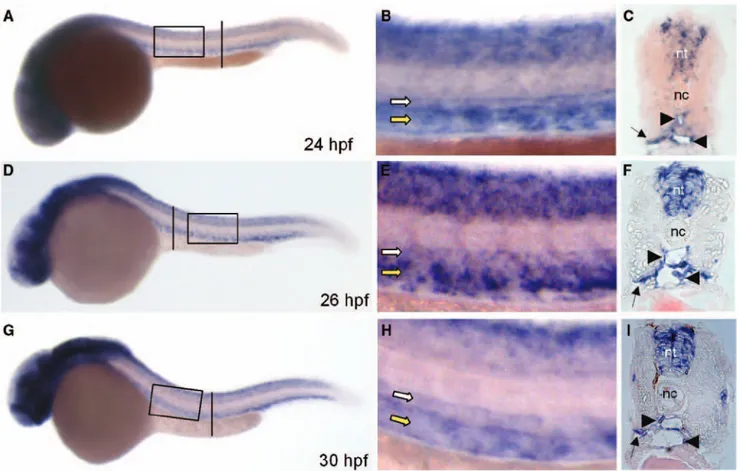
restricted to the diencephalon at early somitogenesis (data not shown) and is widespread in the brain and neural tube at subsequent stages of development (Figure 1). Interestingly, whole-mount ISH showed also a transient but significant expression of zS1P1 transcript in the axial vasculature of the trunk of zebrafish embryos. This vascular pattern expression occurs at 24 hpf (Figure 1A and 1B), is maintained at 26 hpf (Figure 1D and 1E), and decreases at 30 hpf (Figure 1G and 1H), being lost at 48 hpf (data not shown). Analysis of the cross sections of the trunk of 24, 26, and 30 hpf embryos con-firmed that, in addition to the neural tube, zS1P1 marks the
Table. Pharmacological Characterization of zS1P1 Receptor
Receptor Agonist* zS1P1(pEC50)† hS1P1(pEC50)†
S1P 9.0±0.3 9.1±0.2
dhS1P 9.2±0.1 9.7±0.1
FTY720-P 10.5±0.1 10.8±0.4
SEW2871 7.5±0.2 7.9±0.1
CYM-5442 7.7±0.1 8.3±0.1
zS1P1 indicates zebrafish ortholog of the endothelial
sphingosine-1-phosphate receptor-1; dhS1P, dihydro-S1P.
*Chinese hamster ovary–FlpIn cells were transiently transfected with zebrafish zS1P1 or human hS1P1 receptor cDNAs and treated with the indicated
receptor agonists.
†Data are expressed as the potency of the indicated compound to inhibit
forskolin-induced cAMP accumulation (calculated as pEC50 value, here defined
as the negative logarithm of the EC50 value) and represent the mean±SEM of
2 to 3 independent experiments. No effect was observed in mock-transfected cells (data not shown).
endothelium of the main medial dorsal aorta (DA) and pos-terior cardinal vein (PCV), as well as the basal aspect of the somites (Figure 1C, 1F, and 1I).
Endothelial differentiation in zebrafish is under the control of the sonic hedgehog/vascular endothelial growth factor (shh/vegf) genetic pathway.28 Endothelial expression of zS1P
1
in the axial vasculature of the trunk of 26 hpf embryos was abrogated by treatment with cyclopamine, a potent inhibitor of shh signaling29 (data not shown) or after downregulation
of vegf expression by antisense MO injection (Figure 2). Specificity of the effect was demonstrated by the lack of effect of both treatments on somitic and central nervous system expression of zS1P1, thus, indicating that endothelial zS1P1 expression is under the control of the shh/vegf cascade.30
MO Knockdown of zS1P1 Function Results in Venous Vascular Barrier Defects
To assess the functional role of zS1P1 in zebrafish vascu-lar development, we used an antisense MO knockdown approach.31 To this purpose, a MO was designed directed
against the 5ʹ untranslated region spanning the zS1P1 ATG
start codon to inhibit protein translation (zS1P1-MO). Next, tg(fli1:EGFP)y1 transgenic zebrafish embryos in which EGFP
expression is driven by the promoter of the pan-endothelial marker fli-1 were injected at the 1- to 4-cell stage with dif-ferent doses of zS1P1-MO (ranging from 0.1–0.4 pmoles/ embryo) or of control std-MO and the development of EGFP– labeled blood vessels was directly observed in live embryos. Figure 1. Vascular expression of the zebrafish ortholog of the endothelial sphingosine-1-phosphate receptor-1 (zS1P1). A, D, and G,
zS1P1 expression was analyzed in zebrafish embryos by in situ hybridization at the indicated developmental stages. Embryos are anterior to the left and lateral to the top. B, E, and H, High magnification of the area in black boxes in A, D and G. C, F, and I, Transverse sec-tions through the trunk highlighted as vertical black bars in A, D, and G. zS1P1 is expressed by the axial vasculature of the trunk (white arrow indicates dorsal aorta [DA]; yellow arrow, posterior cardinal vein [PCV)). Transverse sections confirm zS1P1 expression in DA and PCV (black arrowheads), in the basal aspect of the somites (black arrows), and in neural tube (nt). nc indicates notocord; hpf, hours postfertilization.
Figure 2. Vascular zebrafish ortholog of the endothelial sphingosine-1-phosphate receptor-1 (zS1P1) expression is mediated by vascular endothelial growth factor (vegf). zS1P1 expression was assessed by in situ hybridization in standard-MO (std-MO) (A) and vegf-MO–injected embryos (B) at 28 hpf. Note the absence of zS1P1 transcript in dorsal aorta (DA) and posterior cardinal vein (PCV) of vegf morphants. White arrow indicates dorsal aorta; yellow arrow, PCV.
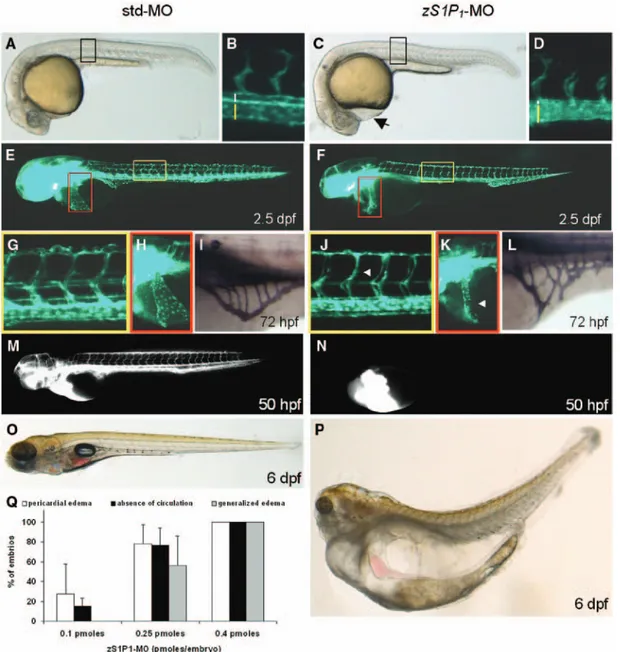
Whole–mount microscopic analysis of 24-hpf embryos injected with 0.4 pmoles/embryo of zS1P1-MO revealed the presence of pericardial edema and enlargement of the PCV (Figure 3C and 3D) whose altered structure was confirmed by the analysis of histological semithin cross sections of the trunk of zS1P1 morphants (Figure 4). At 2.5 days postfertilization (dpf), intersomitic vessels of all zS1P1-MO–injected embryos (n=181) appear slightly thinner than in control Std-MO– injected embryos (Figure 3J). Also, zS1P1 downregulation was
associated with minor defects in the development of the duct of Cuvier and of the subintestinal vein basket in all mophants (Figure 3K and 3L). In keeping with the modest impact of
zS1P1 downregulation on blood vessel development, no differences in the expression of shh, vegf, kdr, and fli-1 genes were observed in zS1P1-MO–injected versus Std-MO–injected embryos (data not shown). These data support previous observations in S1P1-null mice indicating that this S1P receptor is not essential for vasculogenic and angiogenic events
Figure 3. Vascular defects in the zebrafish ortholog of the endothelial sphingosine-1-phosphate receptor-1 (zS1P1) morphants. Vascu-lar morphology in tg(fli1:EGFP)y1 embryos injected with standard-MO (std-MO) or zS1P
1-MO. Morphological appearance of control (A) and zS1P1 morphant (C) embryos at 24 hours postfertilization (hpf). The pericardial edema in zS1P1 morphants is indicated by a black arrow in C. Trunk regions highlighted by black boxes are shown at higher magnification in B and D where dorsal aorta (DA) and posterior cardinal vein (PCV) are indicated by white and yellow bars, respectively; note the abnormal size of PCV in zS1P1 morphants (D). E and F, Epifluorescence microscopy of std-MO– and zS1P1-MO–injected tg(fli1:EGFP)y1 embryos at 2.5 days postfertilization (dpf). Trunk and
Duct of Cuvier regions highlighted by yellow and red boxes, respectively, are shown at higher magnification in (G), (H), (J), and (K); white arrowheads point to thinner intersomitic vessels (J) and to poorly developed Duct of Cuvier (K) in zS1P1 morphants. I and L, Alkaline phosphatase staining of 3-dpf embryos revealed alterations of subintestinal vessel organization in zS1P1 morphants (L). M and N, zS1P1 knockdown causes absence of blood circulation as shown by microangiography performed at 50 hpf. O and P, Morphological appear-ance of control and zS1P1 morphant larvae at 6 dpf; note the generalized edema in zS1P1 morphants (P). Q, Dose-dependent effects of
zS1P1 knockdown: white bars, pericardial edema; black bars, absence of blood circulation; gray bars, late generalized edema (each point is the mean±SD of 97–181 embryos per group).
during embryogenesis.5 However, despite minor alterations in
vascular development, the presence of a beating heart with no major developmental defects (Figure 5) and of a patent lumen in both DA and PCV cross sections (Figure 4), zS1P1-MO– injected embryos showed no apparent blood circulation, as confirmed by the absence of tetramethylrhodamine isothiocyanate–dextran perfusible vessels at 50 hpf (Figure 3N). Of note, 6 dpf zS1P1-MO–injected embryos showed a remarkable generalized edema never observed in control embryos (Figure 3P). The incidence of phenotypic alterations (early pericardial edema, lack of blood circulation, and late generalized edema) was directly related to the dose of injected
zS1P1-MO, 100% of the embryos showing all these defects when injected with 0.4 pmoles/embryo (Figure 3Q). The appearance of the generalized edema, already evident at 4 dpf, was fully prevented when zS1P1 morphants were grown in fish water containing 250 mmol/L D-mannitol. Nevertheless, blood circulation was not restored (data not shown).
To confirm the specificity of the vascular and circulatory defects in zS1P1 morphants, zS1P1-MO was coinjected with a MO-resistant form of the human S1P1 mRNA. In 2 indepen-dent experiments, analysis of the embryos at 30 hpf showed the ability of the human transcript to rescue the zS1P1 -knock-down phenotype, as assessed by the presence of normal blood flow in 76% of rescued embryos when compared with 36% of zS1P1 morphants (P<0.001; Fisher exact test; Figure 6A). Also, analysis of the embryos at 6 dpf showed the ability of the human transcript to rescue the generalized edema observed in zS1P1 morphants. In fact, only 34% of zS1P1 morphants coinjected with the human S1P1 mRNA developed general-ized edema when compared with 57% of zS1P1 morphants (P<0.001; Fisher exact test; Figure 6B).
S1P1 receptor and its ligand S1P play an important role in the control of vascular barrier by affecting endothelial cell–cell junctions,15 including the expression of the
adhe-rens junction component VE-cadherin.10 The phenotype
observed in zS1P1 morphants prompted us to assess the pos-sibility that zS1P1 may control endothelial barrier integrity in zebrafish. On this basis, the effect of zS1P1 downregula-tion on the organizadownregula-tion of endothelial cell–cell juncdownregula-tions was investigated by whole-mount ISH of VE-cadherin32
expression in the axial vasculature of the trunk and by immunofluorescence analysis of ZO1, a tight junction– associated protein functionally critical in regulating S1P-mediated endothelial barrier integrity.33 As shown in Figure
7B, zS1P1 knockdown caused the selective downregulation of VE-cadherin in the PCV but not in the DA of zS1P1 mor-phants. In keeping with the ISH data, the RT-PCR analysis showed a decrease of steady state VE-cadherin mRNA levels in zS1P1 morphants when compared with control embryos (31%±12%, average of the 2 independent experiments). Figure 4. Alterations of posterior cardinal vein (PCV)
morphol-ogy in the zebrafish ortholog of the endothelial sphingosine-1-phosphate receptor-1 (zS1P1) morphants. Histological semithin cross sections of the trunk of 28 hours postfertilization embryos demonstrate the altered structure of the PCV in S1P1 morphants when compared with standard-MO (std-MO)–injected embryos. White arrows indicate dorsal aorta; yellow arrows, PCV.
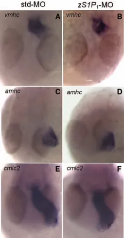
Figure 5. Effect of the zebrafish ortholog of the endothelial sphingosine-1-phosphate receptor-1 (zS1P1) knockdown on car-diac myogenesis gene expression. Standard-MO (std-MO)– and
zS1P1-MO–injected embryos (n=14) were grown in fish water containing 250 mmol/L D-mannitol and analyzed at 28 hours post fertilization (hpf) for the expression of the cardiac myogenesis gene markers ventricular myosin heavy chain (vmhc; A and B),
atrial myosin heavy chain (amhc; C and D), and cardiac myosin
light chain 2 (cmlc2; E and F). No alterations were observed in zS1P1 morphants when compared with controls. At later stages of development (48 hpf), minor defects in cmlc2 expression were observed only in a limited number of zS1P1 morphants (11/33) (data not shown).
Also, confocal microscopy analysis of the vasculature of the trunk of control and zS1P1 morphants at 30 hpf revealed a complete disorganization of the ZO1+ endothelial
junc-tions in the PCV of all the zS1P1 morphants examined (n=5)
(Figure 7D). Instead, no effect on ZO1 immunolocalization was observed in the DA of zS1P1 morphants (Figure 7D) and in both axial vessels of std-MO–injected embryos (Figure 7C). Together, the data indicate a nonredundant
Figure 6. Human hS1P1 overexpression rescues vascular defects in the zebrafish ortholog of the endothelial sphingosine-1-phosphate receptor-1 (zS1P1) morphants.
zS1P1 morphants were injected with hS1P1 mRNA and lack of blood flow (A) and generalized edema (B) were analyzed at 30 hours postfertilization and 6 days post-fertilization (dpf), respectively. hS1P1 over-expression caused a significant increase in the percentage of embryos with normal blood flow (122/161 [76%) vs 42/117 [36%] zS1P1 morphants injected or not with hS1P1 mRNA, respectively; P<0.001, Fisher exact test) and in the percentage of embryos devoid of a significant general-ized edema (100/152 [66%] vs 48/112 [43%] zS1P1 morphants injected or not with hS1P1 mRNA, respectively; P < 0.001, Fisher exact test). Western blot analysis confirmed the increased expression of S1P1 protein in 24 hpf zebrafish embryos
injected with hS1P1 mRNA when com-pared with noninjected animals (inset in A; human umbilical vascular endothelial cells [HUVEC] extract was used as a control and uniform loading of the gel was assessed with anti–α-tubulin [α-tub] antibodies). C, The loss of EphB4a and vascular endo-thelial cadherin (VE-cadherin) expression in posterior cardinal vein (PCV) of zS1P1 morphants (43/60 [72%] and 33/44 [75%) embryos in two different experiments) was reduced to 4/17 (24%) and 9/36 (25%) embryos coinjected with the human S1P1 mRNA (P<0.001, Fisher exact test). White arrow indicates DA; yellow arrow, PCV.
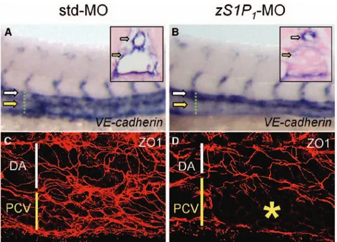
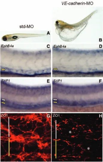
Figure 7. The zebrafish ortholog of the endothelial sphingosine-1-phosphate receptor-1 (zS1P1) knockdown affects Vas-cular endothelial cadherin (VE-cadherin) expression and zonula occludens 1 (ZO1) immunolocalization in posterior cardinal vein (PCV). Standard-MO (std-MO)– and
zS1P1-MO–injected embryos were ana-lyzed at 28 hpf for the expression of
VE-cadherin by in situ hybridization (A and B) and immunostained for ZO1 at 30 hours postfertilization (C and D). Dorsal aorta (DA) and PCV are indicated by white and yellow symbols, respectively. zS1P1 morphants
were characterized by the downregulation of VE-cadherin expression (33/44 embryos examined) and altered ZO1 organization in PCV (asterisk in D) but not in DA (D) (5/5 embryos examined). In contrast, normally organized ZO1+ junctions were observed in
both axial vessels of control embryos (C). Transverse sections highlighted as verti-cal green dotted bars confirm VE-cadherin downregulation in PCV of zS1P1 morphants when compared with controls (insets in A and B).
vascular function for zS1P1 that affects venous endothelial cell–cell barrier in zebrafish.
Downregulation of Venous Endothelial
EphB4a Expression in zS1P1 Morphants
The selective effect of zS1P1 knockdown on PCV structure,
VE-cadherin expression, and ZO1 organization prompted us to investigate the expression of arterial and venous markers in the axial vessels of the trunk in zS1P1-MO–injected embryos. As shown in Figure 8, zS1P1 downregulation does not affect the expression of the specific arterial markers ephrinB230
and crlr34 in the DA of zS1P
1 morphants. Also, zS1P1-MO
injection does not alter the expression of the venous markers
flt435 and dab236 in the PCV. Thus, in keeping with previous
observations in S1P1 null mice,5 the data indicate that zS1P
1
knockdown does not affect arterial/venous differentiation of endothelial cells in zebrafish. However, zS1P1 downregulation caused the loss of the expression of the Eph receptor EphB4a in the PCV of zS1P1-MO–injected embryos (Figure 8J), as confirmed by the decrease of steady state EphB4a mRNA levels in zS1P1-null embryos when compared with controls (38%±9%, average of the 2 independent quantitative RT-PCR experiments). Of note, zS1P1 downregulation did not affect the expression of EphB4a in the gut of zebrafish embryos, thus demonstrating the specificity of the effect of zS1P1-MO on EphB4a expression in PCV (asterisk in Figure 8I and 8J).
The loss of EphB4a and VE-cadherin expression in venous endothelium of zS1P1 morphants (72% and 75% of zS1P1 mor-phants, respectively) was significantly reduced to 24% and 25% for the 2 genes in embryos coinjected with the human
S1P1 mRNA (Figure 6C), thus confirming the specificity of the effect (P<0.001, Fisher exact test). Also, heart beating arrest after treatment with a high dose of ethyl-m- aminobenzoate (tricaine) did not affect EphB4a and VE-cadherin expres-sion in zebrafish embryos (n=22), thus, indicating that the observed downregulation of these genes in zS1P1 morphants is not the direct consequence of the lack of blood flow (Figure 9). Finally, coinjection of the human S1P1 mRNA fully res-cued ZO1 organization in the PCV of zS1P1 morphants (data not shown). These observations point to a role for S1P1 recep-tor in the control of venous endothelial cell functions and gene expression in zebrafish.
VE-cadherin Acts Upstream of EphB4a in
Modulating Venous Vascular Barrier Organization Previous observations had demonstrated a tight relationship between the expression of the members of the cadherin family of adhesion molecules and Eph receptors in epithelial cells.37
On this basis, we addressed the possibility that the venous endothelial barrier defects in zebrafish embryos lacking zS1P1 activity might be the consequence of defects in a putative
zS1P1/VE-cadherin/EphB4a pathway.
To assess this hypothesis, we investigated the effect of
VE-cadherin downregulation on EphB4a expression and vascular integrity in zebrafish morphants.32 As shown in
Figure 10, embryos injected with VE-cadherin–MO (1.0 pmoles/embryo) showed downregulation of EphB4a expression (24/24 embryos), pericardial edema and lack of blood circulation (28/30 embryos, data not shown), and
late generalized edema (19/22 embryos) with no changes in vascular zS1P1 expression (13/13 embryos). Also,
VE-cadherin morphants revealed a complete disorganization of the ZO1+ endothelial junctions in PCV and DA of all the
embryos examined (Figure 10H). These data extend previous observations about the role of VE-cadherin on vascular stability in zebrafish.32,38 On this basis, to assess whether exogenous
VE-cadherin is sufficient to rescue EphB4a expression and vascular integrity in the absence of zS1P1 activity, zS1P1 Figure 8. The zebrafish ortholog of the endothelial sphingosine-1-phosphate receptor-1 (zS1P1) knockdown causes EphB4a downregulation in posterior cardinal vein (PCV). Standard-MO (std-MO)– and zS1P1-MO–injected embryos were analyzed at 28 hours postfertilization for the expression of the indicated mark-ers by in situ hybridization (lateral view of the trunk region; dorsal aorta (DA), white arrow; PCV, yellow arrow). zS1P1 morphants were characterized by the lack of expression of EphB4a in the PCV (J; 43/60 embryos examined). In contrast, arterial markers
ephrinB2 (B) and crlr (D) and venous markers flt4 (F) and dab2 (H) were expressed in 9/10, 9/12, 11/12, and 14/18 zS1P1 mor-phants, respectively. All genes were instead normally expressed in all control embryos examined (A, C, E, G, and I; n=15–30). Note that EphB4a expression, downregulated in the PCV, is maintained in the gut of all the zS1P1 morphants (asterisk in I and J). Transverse sections highlighted as vertical black bars confirm
EphB4a downregulation in the PCV of zS1P1 morphants when
morphants were injected with the mRNA encoding for murine
VE-cadherin.32 As shown in Figure 11B and 11C, the loss of
EphB4a expression in venous endothelium of zS1P1 morphants was significantly reduced in embryos coinjected with the murine VE-cadherin mRNA (78% versus 41% of morphants, respectively; P<0.001, Fisher exact test). Also, injection of VE-cadherin mRNA caused a significant decrease in the number of zS1P1 morphants showing early pericardial edema, lack of blood circulation, and late generalized edema (Figure 11A). Accordingly, VE-cadherin mRNA overexpression rescued the organization of ZO1+ endothelial junctions in
the PCV of zS1P1 morphants (Figure 11E). At variance, no rescue was observed after injection of zS1P1 morphants with
VEGF-A mRNA, thus confirming the specificity of the effect (data not shown). These results suggest that the loss of EphB4a expression in zS1P1 morphants is a result of the VE-cadherin downregulation in PCV endothelium downstream of zS1P1, pointing to the existence of a zS1P1/VE-cadherin/EphB4a pathway modulating venous vascular barrier organization in zebrafish embryo.
EPHB4 Controls Barrier Integrity in HUVECs The segregation of distinct arterial and venous vessels is dis-rupted by EphB4a knockdown in zebrafish,39 hampering the
possibility to use EphB4a morphants to directly assess the role of this receptor in PCV integrity. Also, EphB4a over-expression severely affects the general development of S1P1 morphants (data not shown), forbidding phenotypic rescue experiments in these embryos.
To overcome these limitations and to confirm that a S1P/ Eph receptor cross talk may modulate intercellular contacts also in mammalian venous endothelium, we took advantage of HUVECs that express both S1P140 and the Eph receptor
EPHB4.19 As anticipated, a 1-hour pretreatment of confluent
HUVECs with 30 μmol/L of the selective S1P1 antagonist
W14615 prevents ZO1+ intercellular junction assembly
trig-gered by a 2-hour treatment with 62.5 nmol/L S1P (data not shown) and causes a potent inhibition of EPHB4 expres-sion in both control and S1P-treated HUVECs as assessed by quantitative RT-PCR (87%±2% and 95%±1% inhibition, respectively; n=2). Accordingly, W146 treatment induces a significant inhibition of the levels of EPHB4 receptor protein in these cells (Figure 12A).
Only a limited inhibition of EPHB4 protein levels was observed after transfection with either 1 of the 2 different
S1P1 siRNAs, each causing a partial downregulation of
S1P1 expression (Figure 12B). However, the simultaneous Figure 9. EphB4a and vascular endothelial cadherin
(VE-cadherin) downregulation in zS1P1 morphants is not a result of the absence of blood circulation. In situ hybridization at 28 hour postfertilization revealing EphB4a and VE-cadherin expression in the posterior cardinal vein (PCV) of tricaine-treated embryos (C and D) and absent in the zebrafish ortholog of the endothe-lial sphingosine-1-phosphate receptor-1 (zS1P1)-MO–injected embryos (A and B). White arrow indicates DA; yellow arrow, PCV.
Figure 10. Vascular endothelial cadherin (VE-cadherin)–MO injection phenocopies the zebrafish ortholog of the endothe-lial sphingosine-1-phosphate receptor-1 (zS1P1) knockdown. Standard-MO (std-MO)– and VE-cadherin-MO–injected embryos were analyzed for the appearance of generalized edema at 6 dpf (A and B), EphB4a and zS1P1 expression by in situ hybridiza-tion at 28 hours postfertilizahybridiza-tion (hpf) (C–F) and immunostained with anti-ZO1 antibody at 30 hpf (G and H). Dorsal aorta (DA) and posterior cardial vein (PCV) are indicated by white and yellow symbols, respectively. VE-cadherin morphants were characterized by presence of generalized edema (B, 19/22 embryos), downregulation of EphB4a (D, 24/24 embryos), and altered zonula occludens 1 (ZO1) organization in PCV and DA (asterisks in H). In contrast, normally organized ZO1+ junctions
were observed in both axial vessels of control embryos (G).
VE-cadherin downregulation does not affect zS1P1 expression in DA
transfection with both S1P1 siRNAs allowed a more significant downregulation of S1P1 receptor and resulted in a remarkable decrease of EPHB4 protein levels (Figure 12B), with ≈25% to 30% of the receptor levels detected in cells transfected with the scrambled siRNA as assessed by computerized image analysis of the immunoreactive bands (Figure 12C). Thus, in agreement with the data obtained with zS1P1 morphants, these results indicate that the suppression of S1P1 receptor expression (by siRNA transfection) or activity (by W146 treatment) induces a significant downregulation of EPHB4 in HUVEC.
On this basis, we investigated the effect of the selective EPHB4 receptor antagonist TNYL-RAW peptide and of the control-scrambled peptide SCR-WTL18,19 on ZO1+
intercellu-lar contact organization in HUVECs. Simiintercellu-lar to W146 (data not shown), TNYL-RAW inhibited ZO1 organization in cell– cell junctions of confluent HUVEC monolayers maintained in serum-containing cell culture medium, whereas no effect was exerted by the control-scrambled peptide SCR-WTL (Figure 12D). In keeping with our observations in zebrafish embryos, these data support a direct functional role for the Eph receptor EPHB4 in mediating S1P1-dependent organiza-tion of ZO1+ intercellular junctions in venous endothelium.
Discussion
In this study, we report the characterization and functional analysis of S1P1, a major endothelial S1P receptor, in the development of vascular system in zebrafish embryo. Here we show that S1P1 plays a nonredundant role in the regula-tion of endothelial barrier in zebrafish via a novel S1P1
/VE-cadherin/EphB4a genetic pathway that controls venous vascular integrity.
In agreement with previous observations,13
pharmacologi-cal characterization based on the effect of nonselective and selective S1P1 receptor agonists on forskolin-induced cAMP accumulation demonstrates that the binding properties of the cloned zS1P1 receptor expressed in mammalian cells are simi-lar to those shown by its human counterpart, thus confirm-ing that zS1P1 encodes for the bona fide zebrafish ortholog of the human S1P1 gene. Consistent with a vascular pattern of expression of S1P1 in mouse embryo,5 zS1P
1 is expressed
in the axial vasculature of the trunk of zebrafish embryos, whole-mount ISH showing that zS1P1 transiently marks the endothelium of DA and PCV at 24 to 30 hpf, being lost at 48 hpf. The shh signaling inhibitor cyclopamine or vegf anti-sense MO both abrogate expression of zS1P1 in the axial vas-culature of the trunk, thus indicating that endothelial zS1P1 expression is under the control of the endothelial differen-tiation shh/vegf genetic pathway.30 These data extend
previ-ous observations about the ability of vascular endothelial growth factor to upregulate S1P1 expression in mammalian endothelium.41
Here, we show that zS1P1 morphants are characterized by minor defects in the vascular development of intersomitic vessels, duct of Cuvier, and subintestinal vein basket. This was paralleled by early pericardial edema, lack of blood circulation, alterations of PCV structure, and a dramatic late generalized edema. Interestingly, several of these phenotypic features were observed also in S1P1 null mice,5 as well as in
VE-cadherin zebrafish morphants (C. Tobia, our unpublished observations, 2010).32 However, S1P
1-null mice exhibit
massive embryonic hemorrhage and deficient smooth muscle cell/pericyte recruitment in embryonic vasculature,5 defects
that were not observed in zS1P1 morphants. These species differences may be, at least in part, a result of the fact that zebrafish is characterized by a delay in smooth muscle cell/ pericyte maturation with a very limited PCV coverage with respect to mammals.42 Indeed, transgelin-positive mural cells
are not yet detectable in PCV of zebrafish embryos at 80 hpf Figure 11. Murine vascular endothelial cadherin (VE-cadherin)
overexpression rescues vascular defects in the zebrafish ortholog of the endothelial sphingosine-1-phosphate receptor-1 (zS1P1) morphants. A, zS1P1 morphants were left untreated (n=72) or were injected with murine VE-cadherin mRNA (n=123), and their phenotype was analyzed for the presence of pericardial edema and lack of blood flow at 48 hours post fertilization (hpf) and late generalized edema at 5 dpf. mVE-cadherin overexpression rescued all these vascular alterations in zS1P1 morphants. B–E, Control and mVE-cadherin mRNA-–injected zS1P1 morphants were analyzed at 28 hpf for the expression of EphB4a (B and C) and immunostained for zonula occludens 1 (ZO1) at 30 hpf (D and E). Dorsal aorta (DA) and posterior cardinal vein (PCV) are indicated by white and yellow symbols, respectively. The loss of EphB4a in PCV of zS1P1 morphants (60/77 [78%] embryos in 3 different experiments) was reduced to 31/76 (41%) embryos coinjected with mVE-cadherin mRNA (C; P<0.001, Fisher exact test). Also, normal ZO1 immunolocalization was observed in the PCV of all the observed mVE-cadherin mRNA–injected zS1P1 morphants (E).
or 120 hpf,42 when the vascular defects in zS1P
1 morphants are
already established.
Lack of blood circulation and pericardial edema have been observed also in S1P2 receptor mil zebrafish mutants as a consequence of profound defects in heart organogenesis.14 At
variance with mil mutants, we did not observe major defects in the developing heart of the zS1P1 morphants, as indicated by the normal pattern of expression of the cardiac myogenesis genes ventricular myosin heavy chain, atrial myosin heavy
chain, and cardiac myosin light chain 2 (see Figure 5), demonstrating that the 2 S1P receptors play different roles in zebrafish development.
Edema formation may result from cardiovascular, lym-phatic vascular or excretory system defects. To this respect,
zS1P1 morphants showed an apparently normal lymphatic thoracic duct at 4 dpf (C. Tobia, unpublished observations, 2010). Also, high molecular weight proteins (≥80 000) were present in the extravascular transudate we collected from edematous zS1P1 morphants at 6 dpf but not in the transudate collected from prox-1 morphants showing lack of lymphatic vessel development43 as observed by SDS-PAGE (C. Tobia,
unpublished observations, 2010). Thus, even though the effect of zS1P1 knowdown on cardiac, lymphatic and excre-tory system development and function will deserve further investigation, our observations suggest that alterations of endothelial barrier integrity may contribute, at least in part, to the edematous phenotype observed in zS1P1 morphants.
Actually, in keeping with a role for S1P1 in vascular bar-rier integrity, zS1P1 morphants showed significant altera-tions of vessel morphology, downregulation of VE-cadherin expression, and ZO1 disorganization in PCV but not in DA. It must be pointed out that zS1P1 knockdown did not affect arterial/venous differentiation of endothelial cells in zebraf-ish. Indeed, zS1P1 morphants did not show any change in the levels of expression of shh, vegf, kdr, and fli-1, as well as of the arterial markers ephrinB230 and crlr34 and of the venous
markers flt435 and dab2,36 thus confirming previous
observa-tions in S1P1−/− mice.5 Nevertheless, the expression of the Eph
receptor EphB4a was significantly downregulated in zS1P1 -MO–injected embryos. To this respect, it is interesting to note that EphB4a knockdown after MO injection at 1- to 4-cell stage has a dramatic impact on early vascular development and arterial/venous segregation in zebrafish.39 In contrast, as
stated above, EphB4a downregulation in zS1P1 morphants, which occurs at 28 hpf in the already differentiated PCV, is paralleled only by minor defects in late vascular develop-ment, mostly affecting venous microcirculation (ie, the duct of Cuvier and the subintestinal vein basket) with no apparent effect on arterial/venous differentiation. Taken together, these data suggest that EphB4a may play a dual role in the circula-tory system of zebrafish: an earlier effect on arterial/venous differentiation and a later effect on venous vascular integrity. In keeping with this hypothesis, EphB4a has been shown to regulate venous remodelling in the adult murine vasculature.44
Figure 12. EPHB4 controls barrier integrity in human umbilical vein endothelial cells (HUVECs). A, HUVECs were incubated with vehicle or with 30-µmol/L W146 for 48 hours and cells lysates were probed with anti-EPHB4 antibody in a Western blot. B, HUVECs were left untreated (n.t.), mock transfected, or transfected with control-scrambled short interfering RNA (siRNA), 2 different S1P1 siRNAs (a or b) or the pool of the 2 S1P1 siRNAs (a+b). After 96 hours, cell lysates were probed by Western blotting with anti-S1P1 and anti-EPHB4
antibod-ies. Uniform loading of the gels was assessed with anti-focal adhesion kinase (FAK) (A) or anti-tubulin (B) antibodies. C, The cell lysates of untreated, mock transfected, scrambled siRNA–transfected, and endothelial sphingosine-1-phosphate receptor-1 (S1P1) siRNAs (a+b)–transfected HUVECs were probed by Western blotting with anti-S1P1 and anti-EPHB4 antibodies, and the corresponding immu-noreactive bands were quantified by computerized image analysis and normalized for tubulin levels. The data are the mean±SD of the 3 independent transfection experiments and are expressed as percentage of immunoreactive protein levels in untreated cells. D, Confluent HUVECs were treated with vehicle or with 100 µmol/L control SCR-WTL peptide or TNYL-RAW peptide for 24 hours in complete cell cul-ture medium. Then, cells were immunostained with anti–zonula occludens 1 (ZO1) antibody. Note that ZO1 localization in cell–cell contact regions is lost in TNYL-RAW–treated cells.
Eph receptors have been implicated in the permeability of epithelial and endothelial barriers45 and EphA receptors have
been suggested to act downstream of E-cadherin to mediate epithelial cell-to-cell contacts.37 Our data strongly suggest
that a cadherin/Eph receptor cross talk may exist also in the endothelial cells, genetic VE-cadherin/EphB4a interactions mediating venous vascular integrity downstream of S1P1. Several experimental evidences support this hypothesis. (1) The S1P/S1P1 receptor system modulates VE-cadherin expression and the localization of this adherens junction component at intercellular junctions in mammalian endo-thelium.10 (2) zS1P
1 knockdown causes VE-cadherin and EphB4a downregulation in PCV of zebrafish morphants. (3) Both zS1P1 morphants and VE-cadherin morphants show early pericardial edema, lack of blood circulation, late generalized edema, disorganization of endothelial ZO1+
junctions, and EphB4a downregulation. (4) Exogenous
VE-cadherin is sufficient to rescue EphB4a expression and vascular integrity in the absence of zS1P1 activity. (5) Stable
S1P1 knockdown downregulates VE-cadherin expression in different human endothelial cells lines.46 (6) In keeping
with the data obtained in zS1P1 zebrafish morphants, S1P1 siRNA transfection and the selective S1P1 antagonist W14615
downregulate EPHB4 receptor expression in mammalian HUVECs. (7) Similar to W146, the selective EPHB4 recep-tor antagonist TNYL-RAW peptide18,19 prevents ZO1+
inter-cellular contact organization in the same cells. Together, the data point to the existence of a S1P1/VE-cadherin/EphB4a genetic pathway modulating vascular barrier organization in venous endothelium.
At present, the molecular mechanism(s) orchestrating this genetic pathway remain unravelled. To this respect, the capac-ity of zS1P1 in modulating intercellular barrier appears to be restricted to venous endothelium. Indeed, even though zS1P1 receptor is expressed in both DA and PCV endothelial cells, its knockdown results in VE-cadherin downregulation with consequent alterations of vessel morphology and disorgani-zation of ZO1+ endothelial junctions only in PCV, with no
apparent alterations of arterial endothelium. This indicates that VE-cadherin expression and endothelial barrier integrity are controlled by different mechanisms in venous and arterial endothelium of zebrafish embryo.
Endothelium demonstrates remarkable heterogeneity in structure and function.47 Within the microvasculature,
junc-tions are tighter in arterioles compared with capillaries and are quite loose in venules, likely reflecting the role of postcapil-lary venules in mediating inflammation-induced extravasation of leukocytes and plasma constituents.48 Accordingly,
differ-ences in the expression of adherens junctional proteins have been reported in arterial versus venous endothelial cells.49,50
Several transcription factors, including members of the E-26 and Twist/Slug/Snail families, and Wnt/β-catenin signaling regulate VE-cadherin expression.51 Further experiments are
required to elucidate the transcriptional and signaling mecha-nisms responsible for the observed modulation of VE-cadherin expression in human46 and zebrafish (present work)
endothe-lium by S1P1 knockdown and the molecular bases responsible for the different responses of venous and arterial vascular beds to zS1P1 downregulation in zebrafish embryo.
Alterations of the endothelial barrier integrity may occur in different pathological conditions, including acute respiratory distress syndrome in the lung, ischemia-reperfusion stresses in the kidney and myocardium, and experimental autoimmune encephalomyelitis in the brain. In all these conditions, S1P receptor activation may favour vascular integrity, synthetic chemical agonists/antagonists of S1P receptors representing therapeutic modulators of the endothelial barrier.4 Zebrafish
is suitable for high-throughput screening of chemical com-pounds using robotic platforms.52,53 Also, zebrafish has been
proposed as a suitable animal model of human vascular mal-formation disorders.6 Our findings indicate that this animal
model may provide useful information about the molecular mechanisms regulating vascular barrier integrity and may be used for the screening of novel endothelial barrier-targeting therapeutics.
Acknowledgments
We thank Dr Franco Cotelli (University of Milan, Italy) for the help-ful discussions and criticisms.
Sources of Funding
This work was supported, in part, by grants from Ministero dell’Istruzione, Università e Ricerca (Centro IDET, FIRB project RBAP11H2R9 2011), Lombardy Innate Immunity Network (LIIN), and Associazione Italiana per la Ricerca sul Cancro (AIRC grant no. 10396) to M. Presta and AIRC MFAG grant no. 9161 to S. Mitola.
Disclosures
None.References
1. Rosen H, Goetzl EJ. Sphingosine 1-phosphate and its receptors: an auto-crine and paraauto-crine network. Nat Rev Immunol. 2005;5:560–570. 2. Spiegel S, Milstien S. Sphingosine-1-phosphate: an enigmatic signalling
lipid. Nat Rev Mol Cell Biol. 2003;4:397–407.
3. Rosen H, Sanna MG, Cahalan SM, Gonzalez-Cabrera PJ. Tipping the gatekeeper: S1P regulation of endothelial barrier function. Trends
Immunol. 2007;28:102–107.
4. Marsolais D, Rosen H. Chemical modulators of sphingosine-1-phos-phate receptors as barrier-oriented therapeutic molecules. Nat Rev Drug
Discov. 2009;8:297–307.
5. Liu Y, Wada R, Yamashita T, Mi Y, Deng CX, Hobson JP, Rosenfeldt HM, Nava VE, Chae SS, Lee MJ, Liu CH, Hla T, Spiegel S, Proia RL. Edg-1, the G protein-coupled receptor for sphingosine-1-phosphate, is essential for vascular maturation. J Clin Invest. 2000;106:951–961. 6. Dejana E, Tournier-Lasserve E, Weinstein BM. The control of vascular
integrity by endothelial cell junctions: molecular basis and pathological implications. Dev Cell. 2009;16:209–221.
7. Komarova YA, Mehta D, Malik AB. Dual regulation of endothelial junc-tional permeability. Sci STKE. 2007;2007:re8.
8. Hla T. Signaling and biological actions of sphingosine 1-phosphate.
Pharmacol Res. 2003;47:401–407.
9. Lee MJ, Thangada S, Claffey KP, Ancellin N, Liu CH, Kluk M, Volpi M, Sha’afi RI, Hla T. Vascular endothelial cell adherens junction assem-bly and morphogenesis induced by sphingosine-1-phosphate. Cell. 1999;99:301–312.
10. Wang L, Dudek SM. Regulation of vascular permeability by sphingosine 1-phosphate. Microvasc Res. 2009;77:39–45.
11. Thisse C, Zon LI. Organogenesis–heart and blood formation from the zebrafish point of view. Science. 2002;295:457–462.
12. Weinstein B. Vascular cell biology in vivo: a new piscine paradigm?
Trends Cell Biol. 2002;12:439–445.
13. Im DS, Ungar AR, Lynch KR. Characterization of a zebrafish (Danio rerio) sphingosine 1-phosphate receptor expressed in the embryonic brain. Biochem Biophys Res Commun. 2000;279:139–143.
14. Kupperman E, An S, Osborne N, Waldron S, Stainier DY. A sphingosine-1-phosphate receptor regulates cell migration during vertebrate heart development. Nature. 2000;406:192–195.
15. Sanna MG, Wang SK, Gonzalez-Cabrera PJ, Don A, Marsolais D, Matheu MP, Wei SH, Parker I, Jo E, Cheng WC, Cahalan MD, Wong CH, Rosen H. Enhancement of capillary leakage and restoration of lymphocyte egress by a chiral S1P1 antagonist in vivo. Nat Chem Biol. 2006;2:434–441.
16. Albert R, Hinterding K, Brinkmann V, Guerini D, Müller-Hartwieg C, Knecht H, Simeon C, Streiff M, Wagner T, Welzenbach K, Zécri F, Zollinger M, Cooke N, Francotte E. Novel immunomodulator FTY720 is phosphorylated in rats and humans to form a single ste-reoisomer. Identification, chemical proof, and biological characteriza-tion of the biologically active species and its enantiomer. J Med Chem. 2005;48:5373–5377.
17. Gonzalez-Cabrera PJ, Jo E, Sanna MG, Brown S, Leaf N, Marsolais D, Schaeffer MT, Chapman J, Cameron M, Guerrero M, Roberts E, Rosen H. Full pharmacological efficacy of a novel S1P1 agonist that does not require S1P-like headgroup interactions. Mol Pharmacol. 2008;74:1308–1318.
18. Koolpe M, Burgess R, Dail M, Pasquale EB. EphB receptor-binding peptides identified by phage display enable design of an antagonist with ephrin-like affinity. J Biol Chem. 2005;280:17301–17311.
19. Salvucci O, de la Luz Sierra M, Martina JA, McCormick PJ, Tosato G. EphB2 and EphB4 receptors forward signaling promotes SDF-1-induced endothelial cell chemotaxis and branching remodeling. Blood. 2006;108:2914–2922.
20. Lawson ND, Weinstein BM. In vivo imaging of embryonic vascular development using transgenic zebrafish. Dev Biol. 2002;248:307–318. 21. Westerfield M. The Zebrafish Book. Eugene, OR: University of Pregon
Press; 1995.
22. Kimmel CB, Ballard WW, Kimmel SR, Ullmann B, Schilling TF. Stages of embryonic development of the zebrafish. Dev Dyn. 1995;203:253–310. 23. Paffett-Lugassy NN, Zon LI. Analysis of hematopoietic development in
the zebrafish. Methods Mol Med. 2005;105:171–198.
24. Isogai S, Horiguchi M, Weinstein BM. The vascular anatomy of the developing zebrafish: an atlas of embryonic and early larval develop-ment. Dev Biol. 2001;230:278–301.
25. Blum Y, Belting HG, Ellertsdottir E, Herwig L, Lüders F, Affolter M. Complex cell rearrangements during intersegmental vessel sprouting and vessel fusion in the zebrafish embryo. Dev Biol. 2008;316:312–322. 26. Hale JJ, Lynch CL, Neway W, et al. A rational utilization of
high-throughput screening affords selective, orally bioavailable 1-benzyl-3-carboxyazetidine sphingosine-1-phosphate-1 receptor agonists. J Med
Chem. 2004;47:6662–6665.
27. Brinkmann V, Davis MD, Heise CE, Albert R, Cottens S, Hof R, Bruns C, Prieschl E, Baumruker T, Hiestand P, Foster CA, Zollinger M, Lynch KR. The immune modulator FTY720 targets sphingosine 1-phosphate receptors. J Biol Chem. 2002;277:21453–21457.
28. Lawson ND, Vogel AM, Weinstein BM. sonic hedgehog and vascular endothelial growth factor act upstream of the Notch pathway during arte-rial endothelial differentiation. Dev Cell. 2002;3:127–136.
29. Cooper MK, Porter JA, Young KE, Beachy PA. Teratogen-mediated inhibition of target tissue response to Shh signaling. Science. 1998;280:1603–1607.
30. Torres-Vázquez J, Kamei M, Weinstein BM. Molecular distinction between arteries and veins. Cell Tissue Res. 2003;314:43–59.
31. Nasevicius A, Ekker SC. Effective targeted gene ‘knockdown’ in zebraf-ish. Nat Genet. 2000;26:216–220.
32. Montero-Balaguer M, Swirsding K, Orsenigo F, Cotelli F, Mione M, Dejana E. Stable vascular connections and remodeling require full expres-sion of VE-cadherin in zebrafish embryos. PLoS ONE. 2009;4:e5772. 33. Lee JF, Zeng Q, Ozaki H, Wang L, Hand AR, Hla T, Wang E, Lee MJ.
Dual roles of tight junction-associated protein, zonula occludens-1, in
sphingosine 1-phosphate-mediated endothelial chemotaxis and barrier integrity. J Biol Chem. 2006;281:29190–29200.
34. Nicoli S, Tobia C, Gualandi L, De Sena G, Presta M. Calcitonin recep-tor-like receptor guides arterial differentiation in zebrafish. Blood. 2008;111:4965–4972.
35. Thompson MA, Ransom DG, Pratt SJ, et al. The cloche and spadetail genes differentially affect hematopoiesis and vasculogenesis. Dev Biol. 1998;197:248–269.
36. Song HD, Sun XJ, Deng M, et al. Hematopoietic gene expression pro-file in zebrafish kidney marrow. Proc Natl Acad Sci USA. 2004;101: 16240–16245.
37. Cheng N, Brantley DM, Chen J. The ephrins and Eph receptors in angio-genesis. Cytokine Growth Factor Rev. 2002;13:75–85.
38. Mitchell IC, Brown TS, Terada LS, Amatruda JF, Nwariaku FE. Effect of vascular cadherin knockdown on zebrafish vasculature during develop-ment. PLoS ONE. 2010;5:e8807.
39. Herbert SP, Huisken J, Kim TN, Feldman ME, Houseman BT, Wang RA, Shokat KM, Stainier DY. Arterial-venous segregation by selective cell sprouting: an alternative mode of blood vessel formation. Science. 2009;326:294–298.
40. Hla T, Lee MJ, Ancellin N, Liu CH, Thangada S, Thompson BD, Kluk M. Sphingosine-1-phosphate: extracellular mediator or intracellular sec-ond messenger? Biochem Pharmacol. 1999;58:201–207.
41. Igarashi J, Erwin PA, Dantas AP, Chen H, Michel T. VEGF induces S1P1 receptors in endothelial cells: Implications for cross-talk between sphingolipid and growth factor receptors. Proc Natl Acad Sci USA. 2003;100:10664–10669.
42. Santoro MM, Pesce G, Stainier DY. Characterization of vascular mural cells during zebrafish development. Mech Dev. 2009;126:638–649. 43. Yaniv K, Isogai S, Castranova D, Dye L, Hitomi J, Weinstein BM.
Live imaging of lymphatic development in the zebrafish. Nat Med. 2006;12:711–716.
44. Muto A, Yi T, Harrison KD, Dávalos A, Fancher TT, Ziegler KR, Feigel A, Kondo Y, Nishibe T, Sessa WC, Dardik A. Eph-B4 prevents venous adaptive remodeling in the adult arterial environment. J Exp Med. 2011;208:561–575.
45. Ivanov AI, Romanovsky AA. Putative dual role of ephrin-Eph receptor interactions in inflammation. IUBMB Life. 2006;58:389–394.
46. Krump-Konvalinkova V, Yasuda S, Rubic T, Makarova N, Mages J, Erl W, Vosseler C, Kirkpatrick CJ, Tigyi G, Siess W. Stable knock-down of the sphingosine 1-phosphate receptor S1P1 influences multiple functions of human endothelial cells. Arterioscler Thromb Vasc Biol. 2005;25:546–552.
47. Aird WC. Phenotypic heterogeneity of the endothelium: I. Structure, function, and mechanisms. Circ Res. 2007;100:158–173.
48. Curry FR, Adamson RH. Vascular permeability modulation at the cell, microvessel, or whole organ level: towards closing gaps in our knowl-edge. Cardiovasc Res. 2010;87:218–229.
49. Herwig MC, Müller KM, Müller AM. Endothelial VE-cadherin expres-sion in human lungs. Pathol Res Pract. 2008;204:725–730.
50. Kevil CG, Okayama N, Trocha SD, Kalogeris TJ, Coe LL, Specian RD, Davis CP, Alexander JS. Expression of zonula occludens and adherens junctional proteins in human venous and arterial endothelial cells: role of occludin in endothelial solute barriers. Microcirculation. 1998;5:197–210.
51. Harris ES, Nelson WJ. VE-cadherin: at the front, center, and sides of endothelial cell organization and function. Curr Opin Cell Biol. 2010;22:651–658.
52. Pichler FB, Laurenson S, Williams LC, Dodd A, Copp BR, Love DR. Chemical discovery and global gene expression analysis in zebrafish.
Nat Biotechnol. 2003;21:879–883.
53. Ny A, Autiero M, Carmeliet P. Zebrafish and Xenopus tadpoles: small animal models to study angiogenesis and lymphangiogenesis. Exp Cell