• Non ci sono risultati.

Expression of the K303R estrogen receptor alfa mulation induces hormonal resistance in breast cancer

N/A
N/A
Protected

Academic year: 2021

Condividi "Expression of the K303R estrogen receptor alfa mulation induces hormonal resistance in breast cancer"

Copied!
142
0
0

Testo completo

(1)
(2)

Introduction …...……….1

Materials and Methods …………...……….10

¾ Reagents, hormones and antibodies ………...10

¾ Plasmids ……...………...10

¾ Cell culture …...………..11

¾ Quantitative image analyses by high throughput microscopy (HTM) …...……12

¾ Immunoprecipitation and immunoblot analysis ……...………..13

¾ Cell proliferation assays ……...………..14

¾ Blocking peptide delivery ……...………15

¾ Aromatase activity assay ………...……….15

¾ Tumor xenografts …...……….16

¾ Apoptosis assays …...………..16

¾ Statistical analysis .………....16

Results

¾ Ligand-independent signaling to the K303R ER

α mutant reduces Tam

Sensitivity ………..……….17

¾ MCF-7 K303R-ER

α mutant expressing cells exhibit altered growth factor

signalling ………..……….……….20

¾ Tamoxifen fails to inhibit anchorage-independent growth induced by heregulin

in MCF-7 K303R ERα-overexpressing cells ……...………...27

¾ Phosphorylation at serine residue 305 (S305) of ER

α K303R mutant is involved

in growth factor signaling up-regulation …...………30

(3)

Discussion ………..39

References ……….49

Scientific Publication

1. Catalano S., Malivindi R., Giordano C., Gu G., Panza S., Bonofiglio D.,

Lanzino M., Sisci D., Panno M.L. and Andò S. Farnesoid X Receptor, through

the binding with steroidogenetic factor 1 responsive element, inhibits aromatase

expression in tumor Leydig cells. The Journal Of Biological Chemistry 2009.

In revision.

2. Catalano S., Barone I., Giordano C., Rizza P., Qi H., Gu G., Malivindi R.,

Bonofiglio D. and Andò S. Rapid estradiol/ERα signaling enhances aromatase

enzymatic activity in breast cancer cells. Mol Endocrinol, 23(10):1634–1645,

Oct 2009.

3. Barone I., Cui Y., Herkyn MH., Corona-Rodriguez A., Giordano C., Selever J.,

Beyer A., Andò S., Fuqua S.A. Expression of K303R Estrogen Receptor α

Breast Cancer Mutation Induces Resistance to an Aromatase Inhibitor via

Addiction to the PI-3K/Akt Kinase Pathway. Cancer Research, 69(11):4724-32,

Jun 2009.

4. Giordano C., Cui Y., Barone I., Andò S., Mancini M.A., Berno V., Fuqua S.A.

Growth Factor-Induced Resistance to Tamoxifen is Associated with Mutation of

Estrogen Receptor α and its Phosphorylation at Serine 305. Breast Cancer Res

Treat, (Epub ahaed of print), Feb 2009.

5. Catalano S., Giordano C., Rizza P., Gu G., Barone I., Bonofiglio D., Giordano

F., Malivindi R., Gaccione D., Lanzino M., De Amicis F., Andò S. Evidence

that leptin through STAT and CREB signaling enhances Cyclin D1 expression

and promotes human endometrial cancer proliferation. J Cell Physiol. John

Wiley & Sons, Inc (USA), 218(3):490-500, Mar 2009.

6. Mauro L., Catalano S., Bossi G., Pellegrino M., Barone I., Morales S.,

Giordano C., Bartella V., Casaburi I. and Andò S. Evidences that leptin

upregulates E-cadherin expression in breast cancer: effects on tumor growth and

progression. Cancer Research, American Association for Cancer Research,

Philadelphia (USA), 67: 3412-3421, 2007.

7. Catalano S., Rizza P., Gu G., Barone I., Giordano C., Marsico S., Casaburi I.,

Middea E., Lanzino M., Pellegrino M., Andò S. Fas Ligand expression in TM4

Sertoli cells is enhanced by estradiol “in situ” production. Journal of Cellular

Physiology, John Wiley & Sons, Inc (USA), 211: 448-456, 2007.

(4)

INTRODUCTION

Estrogens play a crucial role in regulating the growth and differentiation of

normal breast epithelium and also of breast cancers, with approximately two-thirds of

all breast cancers dependent for their growth on a functional estrogen receptor α (ERα).

ERα is a member of the nuclear hormone receptor superfamily that regulates

transcription of ER target genes by binding with specific estrogen response elements

(ERE) (classical action), or without directly interacting with DNA, via tethering to other

transcription factors such as activator protein-1 (1), SP-1 (2) and others (nonclassical

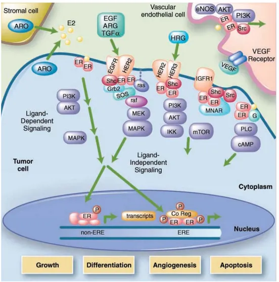
action) (3, 4). Furthermore estrogens also clearly have some nongenomic actions (fig.1),

which are manifested in the rapid induction of kinase signalling cascade and are

probably mediated by the receptors’ activity in the vicinity of the cytoplasic membrane

(5, 6).

Fig.1 The multifaceted mechanisms of estrogen receptor signalling.

Current endocrine therapies offered to women with ERα-positive breast cancer

are based mainly on targeting the ER signalling pathway by either: antagonizing ER

function with antiestrogens such as tamoxifen (Tam), or down-regulating ER levels

with pure antiestrogens such as fulvestrant (Flasodex); or reducing levels of estrogens

(5)

using aromatase inhibitors. At present, a nonsteroidal SERM (Selective Estrogen

Receptor Modulator) Tamoxifen, remains the most frequently prescribed drug for the

treatment of all stages of breast cancer. ERα posses tow major trancriptional activation

domains residing in their NH2 and the COOH termini which harbour, respectively, the

constitutively active, ligand-independent AF-1 and the ligand-dependent AF-2 functions

(Fig.2).

ER

α

Fig. 2 Structure and functional domains of ER

α. Domain structures of steroid receptors. The

numbers of the amino acids found at the boundaries in the individual receptors between the

NTD (AF-1, amino-terminal domain), DBD (DNA binding domain), hinge region (H), and

ligand binding domain (LBD, AF-2) are indicated in the figure. Also shown are the best

characterized phosphorylation sites in the human steroid receptors.

A key feature of the estrogen agonist-receptor structure is the ability of the ligand to be

enveloped in a hydrophobic pocket that is closed by helix 12 in the ligand-binding site

of ERα. Helix 12 positioning over the hydrophobic pocket is critical for the recruitment

of coactivators to the AF-2 site and subsequent initiation of RNA polymerase activity.

The repositioning of helix 12 after binding has been proposed as an important

mechanisms for full estrogen action at ERα (7). SERMs like as 4-hydroxitamoxifen

(4-OHT) fit into the hydrophobic pocket of the ligand binding domain, but the antiestrogen

side chain prevents the reorientation of helix 12 that must seal the ligand into the

receptor before coactivators can bind and produce a transcriptional complex. Tamoxifen

treatment, then, inhibits breast cancer growth mainly through competitive blocking of

(6)

the ER activity, thereby inhibiting estrogen-induced growth. However, although

tamoxifen is initially effective in many patients, and in general is very tolerated, a major

obstacle to its use is tumor resistance. Almost 50% of breast cancers, despite the

presence of ERα, fail to respond to tamoxifen (termed de novo resistance); furthermore,

even patients who initially respond eventually acquire tamoxifen resistance, leading to

tumor progression and death. In general, acquired resistance to tamoxifen is not

attributable to loss of or alteration in the ERα, and resistant tumors often respond to

second-line endocrine therapy (8).

In previous work Fuqua et al. identified an A to G somatic mutation at ERα

nucleotide 908 (A908G) from several usual ductal hyperplasias, which are early

premalignant breast lesions (9). This transition introduces a lysine to arginine

substitution at residue 303 (K303R ERα) within exon 4, at the border between hinge

domain D and the beginning of hormone-binding domain E of the ERα. Growth studies

in breast cancer cells stably transfected with the mutated ERα, demonstrated that the

K303R ERα mutation confers increased sensitivity to subphysiological levels of

estrogen (9). To date, no other ERα mutation has been identified in more that a few

invasive breast cancers (10). Others have reported that the K303R ERα mutation was

not present in invasive tumors (11, 12), but Fuqua et al. have demonstrated that the

detection method used by these other investigators, standard dye-terminator sequencing,

was not sensitive for detection of this specific mutation (13). In addition, Conway et al

have identified this mutation in 6% of breast cancers using a different detection method

(14). The mutation resides at major post-translational modifications sites (acetylation,

ubiquitination, methylation) adjacent to a protein kinase A (PKA) phosphorylation site

at serine residue 305 (S305). There are four potential sites for phosphorylation by PKA

in ERα (serine S236, S305, S338, and S518), with S236 being the best characterized

(7)

site regulating receptor dimerization and DNA binding (15). These serines are all within

the cAMP- dependent protein kinase phosphorylation sequences XRRXSX or

SKKXSX. Interestingly, it has been shown that the mutation of the SKKXSX

recognition sequence to SKRXSX increases the kinetics of PKA phoaphorylation (16)

and predicts that the K303R mutation in ERα may generate a more efficient substrate

for PKA phosphorylation at S305. Recently, Cui et al. have demonstrated that this

naturally-occurring mutation is a more efficient substrate for phosphorylation by PKA,

and is hypoacetylated which subsequently alters estrogen sensitivity (17). Michalides et

al. have suggested that phosphorylation of ER

α S305 by PKA induces a switch from

antagonistic to agonistic effects of Tam, which induces resistance to this antiestrogen

(18). It has also been shown that the ERα S305 site can be an in vivo substrate for

p21-activated kinase 1 (PAK 1)-mediated phosphorylation (19), and its overexpression

correlates with resistance to tamoxifen in breast cancer patients (20). These findings

identify S305 as a crucial site in ERα that upon phosphorylation by either PKA or

PAK-1 is responsible for resistance to tamoxifen. Therefore, this phosphorylation

affects conformational changes in ERα following binding to tamoxifen, which alters its

orientation toward the co-activator SRC-1 resulting in the recruitment of RNA

polymerase II, thereby stimulating estrogen receptor-driven transcription by the

anti-estrogen tamoxifen (19, 21).

We know that estrogens regulation of breast cancer cell growth can also be

modulated by complex interactions with a variety of peptide growth factors. A large

body of evidence supports the idea that rapid membrane effects of ERα (also called

non-genomic effects of ER) may activate various components of growth factor tyrosine

kinase signaling, such as that from insulin-like growth factor-IR (IGF-IR), epidermal

growth factor receptor (EGFR), and c-erbB2/HER2 (22-24). Furthermore, the kinase

(8)

cascade signaling initiated by growth factor receptors can activate ERα (termed

ligand-independent effects) and its coregulatory proteins, causing an interdependent loop of

cross-talk that leads to enhanced tumor cell survival and proliferation (8, 25-27). The

genomic and non-genomic mechanisms of action of ERα are not mutually exclusive,

and many interactions between these two pathways exist. For example, ER induces the

expression of transcripts for both transforming growth factor α (TGFα) and

amphiregulin (AR) (28, 29). TGFα and AR are both able to bind and activate EGFR

thus laeding to activation of MAPK and AKT signalling (30). In addition, ERα binds to

caveolin-1 at the cell membrane and activates specific G protein (23), this phenomenon

leads to activation of src, which in turn activates matrix metalloproteinases that cleave

transmembrane precursors of heparin binding-EGF (HB-EGF), an EGFR ligand (31).

Therefore, both genomic and non-genomic activities of ERα can lead to increased

activation of EGFR and its downstream effectors. Several preclinical and clinical

studies suggest that overexpression of EGFR or HER2, and/or high levels of

phosphorylated Akt and extracellular signal-regulated kinases (ERKs) in breast cancers

contribute to Tam resistance (27, 32-36). Particularly, the membrane tyrosine kinase

HER2 (c-ErbB2, HER2/neu) is gene-amplified in 20-25% of ER-positive breast cancer,

and there is clinical evidence that tamoxifen is less effective in HER2-positive tumors

(37). Furthermore, preclinical models show that HER2 overexpression can cause

tamoxifen-stimulated growth as a mechanism of de novo resistance and that the HER

family receptors are also implicated in acquired resistance to this drug (27, 38). The

ErbB/HER family is composed of four growth factor receptors that share a high degree

of sequence homology: EGFR (or erbB1 or HER1), erbB2 (or HER2; or neu in rodents),

erbB3 (or HER3), and erbB4 (or HER4). Signaling through EGFR, HER2, and HER3

has been shown to promote tumor cell proliferation and survival in a variety of

(9)

epithelial malignancies (39). These receptors are transmembrane proteins with

intracellular tyrosine kinase activity (except for HER3), and their activation results from

the formation of homodimers and heterodimers, which is dependent on their ligands

(except for HER2). Four subdomains have been identified in the extracellular domain of

these proteins: subdomain I and III for ligand binding and the cystein-rich subdomains

II and IV for receptor dimerization (40) (Fig. 3). The EGF family of ligand can be

divided into three groups: EGF, transforming growth factor (TGF)-α, and amphiregulin,

which bind exclusively to EGFR; betacellulin, heparin-binding EGF-like growth factor,

and epiregulin, which bind to both EGFR and HER4; and the neregulin 1 and 2, which

bind to both HER3 and HER4, and neregulin 3 and 4, which bind to HER4 (40). The

extracellular domain of HER2 is unable to bind extracellular ligand and depends on

heterodimerizing with other ligand-activated receptors.

Fig. 3 Epidermal growth factor family of ligands and the ErbB gene family. The inactive

ligand-binding domains of ErbB2 and the inactive kinase domain of ErbB3 are denoted with an

X.

(10)

Overexpression of HER2 leads to activation of the HER2-mediated signalling pathway,

even in the absence of ligand. Changes in HER2 auto-activated conformation might

facilitate dimerization that includes the transmembrane and kinase domains but not

extracellular domain homodimers. HER2 cooperating with other receptors mediates

multiple downstream signalling pathways, including the mitogen-activated protein

kinase (MAPK) and phosphatidylinositol-3-kinase (PI3K) pathways. These signaling

pathways play a key role in tumorogenesis by promoting cell proliferation,

differentiation, angiogenesis, invasion, and survaival (fig. 4). Advanced studies of ER

biology have related new insights into ER action in breast cancer and have highlighted

the role of an intimate crosstalk between the ER and the epidermal growth factor

receptor (EGFR/HER1)/HER2 signaling pathways as a fundamental contributor to the

development of resistance to endocrine therapies (37) (fig. 4).

We therefore hypothesized that the K303R mutation adapts ERα for enhanced

reception of intracellular signal transduction, which leads to antiestrogen resistance. To

test this hypothesis we used as an experimental model MCF-7 breast cancer cells stably

transfected with either wild-type (WT) or the K303R mutant ERα. We found that cells

expressing the estrogen hypersensitive K303R ERα mutant showed elevated levels of

growth factor signaling, and enhanced crosstalk between the mutant and the HER2

growth factor receptor. Furthermore, in the second part of this study, we focused our

attention on the possible role of K303R mutation in the resistance on aromatase

inhibitors. It is well know that aromatase inhibitors (AI) are rapidly becoming the first

choice for hormonal treatment of ERα-positive breast cancer in postmenopausal

women. However, de novo and acquired resistance frequently occurs. To understand

resistance mechanisms, several laboratories have developed in vitro cell line models to

study the molecular changes associated with long-term estrogen deprivation (41-44).

(11)

Fig. 4 Interactions of estrogen and growth factor receptor signaling in human tumors.

AI resistance has also been esamine using aromatase-overexpressing MCF-7 breast

cancer cell line models grown as xenografts in athymic nude mice (45), or breast cancer

cells made resistant to AIs via long-term treatment with these drugs (46, 47). These

models suggest the hypothesis that resistance to endocrine therapy may be through the

acquisition of estrogen hypersensitivity, whereby low subphysiologic levels of

estrogens remaining after estrogen deprivation are sufficient for maintenance of tumor

growth. Thus, we questioned whether resistance to AIs could arise in breast cancer cells

expressing the ERα mutation. Particularly, we hypothesized that such a mutant could

provide a continuous mitogenic stimulus to the breast even during phases of low

circulating hormone, such as menopause, thus affording a proliferative advantage

(12)

possibility, we generated K303R-overexpressing MCF-7 cells stably transfected with an

aromatase expression vector. Cells were stimulated with the aromatase substrate,

androstenedione, with or without the AI anastrozole (Ana). We found that Ana

decreased androstenedione-stimulated growth of wild-type cells, whereas

K303R-expressing cells were resistant to the inhibitory effect of Ana on growth.

Taken together, the results obtained in this study contribute to amplify the

knowledge of the mechanisms by which breast cancer cells become resistant to

endocrine therapy, and suggest that the presence of the A908G ERα somatic mutation

may be useful as a predictive marker of hormonal response in breast cancer patients

whose tumors exploit ERα and/or growth factor cross-talk to evade treatment.

(13)

MATERIALS AND METHODS

Reagents, hormones and antibodies

17β-Estradiol (E

2

), 4-Hydroxytamoxifen (4-OH), Epidermal growth factor

(EGF), Insulin like growth factor -1 (IGF-1), and Heregulin (H),

4-androstene-3,17-dione (Ana) were from Sigma (St. Louis, MO). Anastrozole was provided by

AstraZeneca. Herceptin was form Genentech (San Francisco, CA). Antibodies used for

immunoblotting were: ERα (6F11) from (Novocastra, Newcastle, United Kingdom);

progesterone receptor (PR) from DAKO (Carpinteria, CA); Rho GDIα from Santa Cruz

Biotechnology (Santa Cruz, CA); total MAPK, total Akt, total c-Src, phosphorylated

p42/44 MAPK (Thr

202

/Tyr

204

), Akt (Ser

437

), c-Src (Tyr

416

)and poly(ADP

ribose)polymerase (PARP) from Cell Signaling Technology (Beverly, MA); total HER2

from NeoMarker (Fremont, CA); phosphorylated HER2 (Tyr

1248

), phosphor-ER-S305

from UPSTATE (Temecula, CA); Living colors

TM

Full Length polyclonal antibody

(Clontech, Mountain View, CA); cytochrome P450 aromatase was from Serotec; Bax

and Bcl-2 were from Calbiochem.

Plasmids

Generation of yellow fluorescent protein (YFP)-tagged expression construct, YFP-WT

and K303R ERα, has been previously described (17).

Full-length human aromatase cDNA was amplified from the pCMV6-Arom plasmid

(OriGene Technologies) by PCR using the following primers: forward

ACACTAGTATGGTTTTGGAAATGCTGAACCC-3’ and reverse

5’-ACGCGGCCGCCTAGTGTTCCAGACACCTGTCT-3’. This PCR product was

subcloned into the Spe I/Not I sites of the pZeoSV2-vector (Invitrogen). The resulting

(14)

pZeoSV2-aromatase expression vector (pZeo- Arom) sequence was confirmed by DNA

sequencing.

Cell culture

MCF-7 breast cancer cells, originally obtained from Dr. Benita

Katzenellenbogen (University of Illinois, Urbana, IL), were maintained on plastic in

minimal essential medium (MEM, Invitrogen) supplemented with 10% fetal bovine

serum (FBS; Summit Biotechnology, Fort Collins, CO), 0.1 nmol/L nonessential amino

acid, 2 mmol/L L-glutamine, 50 units/ml penicillin/streptomycin, at 37°C with 5% CO

2

/

95% air. HeLa cells were obtained form American Type Culture Collection (Manassas,

VA), and were maintained in the same media. Chinese Hamster Ovary (CHO) cells

were cultured in Kaighn’s Modification F-12 media (F-12K, Invitrogen) supplemented

with 10% fetal bovine serum (FBS), 0.1 nmol/L nonessential amino-acid, 2 mmol/L

L-glutamine, and 50 units/ml penicillin/streptomycin.

MCF-7 wild-type (WT) and K303R ERα-expressing cells were generated as following

described. MCF-7 cells were stably transfected yellow fluorescent protein (YFP) ERα-

and K303R ERα-tagged vectors, using Fugene 6 according to the manufacturer’s

instructions (Roche, Indianapolis, IN), and individual clones were isolated and

expanded with G418 selection. MCF-7 parental and YFP-K303R ERα clones were

stably transfected with the pZeo-Arom expression vector using Fugene 6 reagent

according to the manufacturer, and individual clones were isolated and expanded with

Zeocin selection. In some experiments we used a pools of stably transfected cells

selected for one week with G418 or Zeocin antibiotics. Chinese hamster ovary (CHO)

stably transfected with YFP-WT ERα and YFP-K303R ERα expression vectors, were

also used. Stably transfected clones and pools of stably transfected cells were screened

(15)

for expression of exogenous and endogenous ERα using immunoblot analysis before

each experiment.

Quantitative image analyses by high throughput microscopy (HTM)

High-throughput microscopy for biological purposes may be broadly defined as

an automated image acquisition system coupled to cell identification and cell

morphological detection/measurement algorithms. Particularly,

HTM is an analytical

tool to study steroid nuclear receptor and nuclear receptor coregulator biology at the

single cell level.

The molecular mechanisms of action for estrogens and antiestrogens at

the single cell level are still being determined. Single cell studies of ERα show that, in

the absence of ligand, its subcellular distribution is predominantly a diffuse nuclear

pattern nuclear. The addition of ligand causes a redistribution of ERα into discrete foci.

However, the nuclear redistribution of ERα does not always correlate well with

transcription, for example, E

2

and tamoxifen both induce subnuclear redistribution,

however, in HeLa, the latter does not induce transcription. Therefore, Berno et al.

have

developed a model system (PRL-HeLa cell line) that is conducive to HTM analysis in

order to study ERα transcriptional properties at the single cell level.

PRL-Hela is a cell line specifically engineered for the single cell study of ER function

(48). PRL-Hela cells contain multiple genomic integrations of a replicated prolactin

(PRL) promoter/enhancer. The multiple integrations (PRL array) are spatially confined

and are visualized by the accumulation of fluorescently tagged ERα. PRL-Hela cells

transiently expressing YFP-WT ERα or YFP K303R ERα, pretreated with forskolin (10

µM) for 15 minutes, and then treated with increasing concentration of E

2

or Tam for 30

minutes, were fixed and DAPI-stained as previously reported (48, 49). The cells were

imaged using the Cell Lab IC 100 Image Cytometer (Beckman Coulter, Fullerton, CA)

(16)

with a Nikon 40X Plan S flour 0.90 NA objective. Two channels were imaged: channel

0 (DAPI stain) was used to find the focus and nuclei, when channel 1 was used to image

YFP ERα. A proprietary algorithm (GPRC) developed at Beckman Coulter was used to

identify and quantify the YFP-ERα-targeted PRL array. The parameters for the GPRC

algorithm were: object scale=30 and minimum peak height=10. Foci identified by the

GPRC algorithm were masked. The area of the mask in pixels was the measure of PRL

array size. Channel 1 was offset 2µm from DAPI focused for cells in all treatment

conditions. After image acquisition and application of the GPRC algorithm, the total

cell populations for each treatment were progressively filtered (gated) using the same

criteria. Nuclei clusters, and mitotic cells were filtered from the total cell population

using an intersection of DNA content and DNA clusters gates. In addition, low YFP

ERα expression and low aggregate number gates were generated and applied to produce

the final cell population to be analyzed. From the final population of cells, the array size

was determined using the GPRC mask (50). The images and masks were visually

inspected for accuracy. Unpaired student’s t-test assuming equal variance was

performed to determine statistical significance (two-tailed, p<0.05). Standard deviations

are shown.

Immunoprecipitation and immunoblot analysis

Cells were starved in phenol red free MEM with 5% charcoal-stripped FBS for

48 h and treated as indicated before lysis [50 mmol/L Tris-HCl (pH 7.4), 150 mmol/L

NaCl, 2% NP40, 0,25% deoxycholic acid, 1 mmol/L EDTA, 1 mmol/L Na

3

VO

4,

and

1:100 protease inhibitors cocktail; Calbiochem]. For coimmunoprecipitation

experiments, we used 1 mg of total cellular protein and a 1:200 dilution of anti-Living

colors Full Length polyclonal antisera (Clontech) that recognizes native and denatured

forms of recombinant YFP fusion proteins expressed in mammalian cells, and 2 µg of

(17)

HER2 polyclonal antisera overnight, followed by protein A/G precipitation with

rotation at 4°C for 2 h. Immunoprecipitated proteins were washed thrice with lysis

buffer. Equal amounts of cell extract and immunoprecipitated proteins were resolved

under denaturing conditions by electrophoresis in 8% to 10% polyacrylamide gels

containing SDS (SDS-PAGE), and transferred to nitrocellulose membranes (Schleicher

& Schuell, Keen, NH) by electroblotting. After blocking the transferred nitrocellulose

membranes were incubated with primary antibodies overnight at 4°C, with secondary

antibodies goat anti-mouse or goat anti-rabbit antisera (1:3000; Amersham Bioscences;

Piscataway, NJ) for 1 h at room temperature and developed with enhanced chemi

luminescence reagents (Alpha Innotech, San Leandro, CA).

Cell proliferation assays

Anchorage-independent soft agar growth assays: cells (5000 per well) were

plated in 4 ml of 0.35% agarose with 5% charcoal-stripped FBS in phenol red-free

MEM, in a 0.7% agarose base in six-well plates. Two days after plating, media

containing control vehicle or hormonal treatments was added to the top layer, and the

appropriate media was replaced every two days. In some experiments a pools of stably

transfected cells were used. After 14 days, 150 µl of MTT was added to each well and

allowed to incubate at 37°C for 4 h. Plates were then placed in 4°C overnight and

colonies > 50 µm diameter from triplicate assays were counted. Data are the mean

colony number of three plates and representative of two independent experiments

analyzed for statistical significance (p<0.05) using a two-tailed student’s Test,

performed by Graph Pad Prism 5 (GraphPad Software, Inc., San Diego, CA). Standard

deviations are shown.

MTT assay: Seven hundred cells were plated into 96-well plates in phenol red–

free MEM containing 5% charcoal-stripped fetal bovine serum (FBS). After 24 h, cells

(18)

were treated as indicated. Nine days later, proliferation was assessed using

3-(4,5-dimethylthiazol-2-yl)-2,5- diphenyltetrazolium bromide (MTT) reagent. Briefly,

MTT

was added to each well and incubated at 37°C and 5% CO

2

for 2 h followed by medium

removal and solubilization in 100 µl DMSO. The resulting color change was read at 570

nm and calculated as absorbance above background.

Blocking peptide delivery

A blocking peptide of 13 residues (IKRSKKNSLALSC) from the sequence

(residues 298-310) surrounding the S305 residue (in bold) of the human ERα was

transferred into cells using a cationic amphiphile molecule, PULSin

TM

delivery reagent

(Polyplus transfection, Illkirch, France), as suggested by manufacturer. Briefly, cells

were plated in a 6-well plate with regular growth media, and then starved for 48 h in a

phenol red free MEM with 5% charcoal stripped FBS. After starvation cells were

washed with PBS to remove all traces of serum, and fresh phenol-red free media

without serum was added. The mixture containing the S305 blocking peptide (4

µg/well) diluted in 200 µl of 20 nM Hepes, and 16 µl of PULSin

TM

was incubated for

15 min at room temperature, and then added to the cells. The media was changed after 4

hours of incubation, cells were treated as indicated, and cellular extracts were prepared.

Delivery of R-phycoerythrin was used as a positive control and live cells were observed

by fluorescence microscopy after 4 h.

Aromatase activity assay

Aromatase activity was evaluated using a

3

H water release assay using 0.5

µmol/L of [1β -

3

H]-androst-4-ene-3,17-dione as substrate (51). The incubations were

performed at 37°C for 1 h. The results were expressed as fmol-pmol/h/mg protein.

(19)

Tumor xenografts

All animal studies were carried out according to the guidelines and with the

approval of the Baylor College of Medicine Animal Care and Use Committee. Female

nude ovariectomized athymic mice were injected with MCF-7 WT and

YFP-K303R ERα–expressing cells or MCF-7 Arom 1 and YFP-K303R Arom 1–expressing cells

as described (52). Animals were supplemented with estrogen tubing releasing ~80

pg/mL, or supplemented daily with injections of the aromatase substrate

androstenedione (AD; 100 µg).

Apoptosis assays

Cells were seeded into six-well plates (10

5

/well) in phenol red–free MEM with

5% charcoal-stripped FBS for 48 h and then incubated with different treatments for 72

h. Apoptosis was measured using the Cell Death Detection ELISA Kit (Roche). Data

are representative of two independent experiments performed in triplicate.

Statistical analysis

Data were analyzed by Student’s t test using the GraphPad Prism5 software

program.

(20)

RESULTS

Ligand-independent signaling to the K303R ER

α mutant reduces Tam sensitivity

We have previously shown that K303R ERα mutant-overexpressing cells

display enhanced ligand-independent activity when stimulated with cyclic AMP, in part

because the mutation generates a more efficient substrate for PKA-mediated signaling

(17). It is well known that ligand-independent signaling can influence cellular

responsiveness to Tam (53). For instance, PKA-mediated phosphorylation of ERα at

S305 allows the antagonist Tam to behave as an agonist, which then results in

ERα-dependent transactivation (18). Here we have utilized the stable cell line PRL-Hela

containing a multi-copy integrated prolactin enhancer/promoter DNA array that is

responsive to ER such that when the receptor is expressed as a YFP-fusion protein, the

integration site can be easily visualized (PRL-array) (48). High-throughput microscopy

has been used to identify ligand-independent changes in the size of the PRL-array which

is an indicator of the chromatin condensation status at the promoter (50). Arrays rapidly

decondense after E

2

treatment or condense after anti-estrogen treatment (both within

minutes). Thus, in PRL-Hela cells, the array size is an indicator of receptor-mediated

transcription function in response to different treatments and allows direct and live

observation of ER-dependent chromatin remodelling. Therefore using a

mammalian-based, stably transfected prolactin promoter array system, we analyzed live cell

dynamics on an estrogen-responsive promoter, to visualize chromatin remodelling

induced by E

2

and Tam. We transiently transfected PRL-Hela cells with YFP

tagged-ERα WT or the YFP-tagged-ERα K303R mutant receptor expression vector, and examined E

2

(Fig 1a) and Tam’s (Fig 1b) effect on chromatin remodelling in the presence of

forskolin, an activator of PKA signaling. Using high through-put microscopy, the

amount of chromatin remodelling was quantified (50). 24h after transfection cells were

(21)

pretreated with forskolin (FSK), and then treated with E

2

or Tam at different doses, as

indicated, for 30 minutes. Over 60 nuclei in two separate experiments were analyzed to

determine the size of the PRL-reporter array. Vehicle control showed similar values for

the array size in the presence of the WT receptor and the mutant (Fig. 1). In PRL-Hela

cells expressing the WT receptor, estrogen treatment increased array size in a

dose-dependent manner in estrogen concentrations between 10

-12

M and 10

-9

M.

Mutant-expressing cells showed a linear dose-response in the range of 10

-17

M through 10

-11

M

of estrogen. Indeed, mutant ERα expressing cells exhibited a much lower EC50 (3 X

10

-13

M) compared with WT ERα-expressing cells (5.1 X 10

-11

M). These data

demonstrate that K303R mutant-expressing cells are hypersensitive to estrogen and

form larger arrays in the presence of low estrogen concentrations. With forskolin

treatment, WT and mutant ERα cells stimulated with estrogen both displayed

significantly increased array sizes. WT ERα-expressing cells demonstrated a one-log

reduction in the EC50 (4.2 X 10

-12

M ± 0.0038 compared to 5.1 X 10

-11

M ± 0.0132 in

the absence of forskolin) and a significantly increased average array size (Fig. 1a).

Mutant-expressing cells also displayed a significantly increased average size, but

without a significant change in the EC50 (8 X 10

-13

M ± 612.5 compared to 3 X 10

-13

M

± 0.0004 in the absence of forskolin). Thus, forskolin increased average array size, and

reduced the EC50 of WT ERα cells thus enhancing estrogen sensitivity, whereas

forskolin treatment of mutant-expressing cells only increased the array size, suggesting

that these cells displayed an inherent hypersensitivity to estrogen. As shown in Fig. 1b,

Tam treatment decreased the array size in WT ERα-expressing cells, however the

mutant receptor was significantly less responsive to Tam (p<0.0001). Forskolin

treatment blocked Tam-induced promoter condensation (reduced size of the array), and

the mutant receptor array size was not affected by physiological levels of Tam (100 nM)

(22)

in the presence of forskolin. All together these results suggest that

hormone-independent kinase signaling to the mutant receptor K303R ERα may confer resistance

to antiestrogen treatment in breast cancer cells.

Fig. 1 E

2

and Tam effects on large-scale chromatin structure in PRL-Hela cells. Cells

transiently expressing YFP-ERα WT (WT) or the YFP-K303R ERα (K303R) mutation were

pretreated with forskolin (FSK) for 15’ minutes and then were treated with E

2

(a) or Tam (b) at

different doses for 30 minutes. After fixing and counter-staining with DAPI, cells were imaged

and array size was quantified using HTM as described in Materials and Methods. Results are

expressed as array size normalized to control cells obtained from three independent

experiments.

(23)

MCF-7 K303R-ER

α mutant expressing cells exhibit altered growth factor

signaling

To further explore ligand-independent activation of signaling and estrogen

hypersensitivity in mutant-expressing breast cancer cells, we developed MCF-7

ERα-positive human breast cancer cell lines overexpressing either WT or the K303R ERα

mutant. We chose to introduce the mutant receptor into parental WT ERα-expressing

cells to simulate the situation in invasive human breast tumors, where WT receptor is

most frequently co-expressed along with the mutant (13). To differentiate the

exogenously expressed receptor, we tagged the vector with YFP. Stably transfected

clones were screened for expression of ERα using immunoblot analysis (Fig. 2a).

Parental MCF-7 cells are shown along with one clone stably expressing YFP-WT, and

three clones expressing YFP-K303R ERα (MCF-7 K303R-1-3). First, to directly

demonstrate “in vivo” the biological consequences of the K303R ERα mutation, we

examined growth of stably transfected cells as tumor xenografts in ovariectomized

athymic nude mice. MCF-7 cells stably transfected WT and K303R ERα were injected

into the number 4 glands of ovariectomized athymic nude mice supplemented with 80

pg/ml of estradiol, representing low premenopausal levels (Fig. 2b). K303R ERα

tumors grew faster than the WT ERα tumors (P=0.0466). Thus, expression of the

mutant generated an estrogen hypersensitive phenotype.

It is well accepted that the response to endocrine therapies in human breast cancer

patients correlates with ERα and progesterone receptor (PR) levels. Several studies

have shown that patients with ERα/PR-positive breast cancers derive greater benefit

from adjuvant hormonal therapy than those patients whose tumors lack PR (54-56)

however it must be noted that other studies have not always found this result (57). It has

been hypothesized that high growth factor signaling activity in breast cancers may be

(24)

associated with decreased PR levels (for a review see (58)) and elegant studies have

shown that up-regulation of pMAPK Erk1/2 leads to a loss of PR via degradation by the

26S proteasome (59). In clinical samples we have previously reported a borderline

significant inverse correlation between the presence of the K303R ERα mutation and

the PR-B isoform (13), and we have shown that a reduction in PR-B levels was

associated with a poorer response to endocrine therapy (60). Therefore, we first

evaluated the levels of PR-A and B in our parental MCF-7 cells, and two stably

transfected clones (Fig. 2c). Parental MCF-7 cells expressed both PR isoforms, with

PR-A as the predominant form in these cells. PR is a known estrogen-induced gene, and

as expected we saw higher levels of both PR-A and B in MCF-7 cells after estrogen

treatment. In comparison, MCF-7 WT-overexpressing cells exhibited slightly less PR

induction with E

2

. In contrast, in basal conditions, K303R ERα mutant-overexpressing

cells demonstrated much lower levels of both PR isoforms, and less were induced with

E

2

treatment. Lower levels of PR mRNA levels were also detected in

estrogen-stimulated mutant cells using Real-Time polymerase chain reaction (qPCR), (p=0.0008,

data not shown). These results suggest that growth factor signals might be altered in the

mutant-overexpressing cells.

To test for altered intracellular signaling, we next examined the effects of short-term

treatments with E

2

, heregulin (H), EGF (E), and IGF-1 (I) on phosphorylation levels of

downstream growth factors signaling components, such as HER2, Akt, and MAPK

using immunoblot analysis (Fig. 3a) which is quantitatively represented in Fig. 3b.

Cells were maintained under estrogen-depleted conditions for 2 days, and then treated

for 10 minutes with E

2

or different growth factors as indicated, and cellular extracts

were prepared. MCF-7 K303R mutant cells showed constitutively higher levels of

phosphorylated HER2 as well as total HER2, compared to MCF-7 WT

ERα-overexpressing cells (Fig. 3a). To begin to understand the mechanism associated with

(25)

higher levels of total HER2 in the mutant cells we analyzed the expression level of

HER2 mRNA in different clones of mutant ERα expressing cells, by qPCR; but we did

not found differences in HER2 mRNA levels between WT and K303R ERα expressing

cells (data not shown).

A

B

C

Fig. 2 Characterization of MCF-7 K303R ER

α-expressing cells. (a) MCF-7 cells stably

transfected with a yellow-fluorescent protein (YFP)-tagged expression construct YFP-WT ERα

(MCF-7 WT) and different clones transfected with the mutant YFP-K303R ERα construct

(MCF-7 K303R-1, -2, -3) were screened for expression of exogenous (96 kDa) and endogenous

ERα (66 kDa) by immunoblot analysis using an antibody against human ERα. Parental cells

(MCF-7) were used as a control for endogenous ERα expression. Rho GDIα was used as a

control for equal loading and transfer. Numbers below the blot represent the ratio between

YFP-ERα and endogenous YFP-ERα protein expression. (b) WT YFP-ERα and K303R YFP-ERα cells were

injected into mice (n = 5/group) supplemented with E

2

. Tumor growth was plotted as the time

for tumor tripling [months (mos)]. (c) MCF-7 parental, MCF-7 WT, and MCF-7 K303R-1 cells

were serum-starved for 48h, and then treated with or without 1 nM E

2

for 24 hours before lysis.

Equal amounts of total cellular extract were analyzed for progesterone receptor (A and

PR-B) levels by Western blotting.

This result suggests that post-translational modification and increase in protein

stability could be involved in the HER2 up-regulation that we found in mutant

ERα-expressing cells, and we are currently exploring this possibility. Treatment with E

2

,

(26)

heregulin, EGF, and IGF-1 induced higher levels of pHER2 in mutant-overexpressing

cells, but only a small induction was seen in MCF-7 WT cells which contain low

endogenous levels of HER2. Treatment with heregulin or EGF led to increased

phosphorylation of the downstream signaling molecules Akt and MAPK in

mutant-overexpressing cells compared with MCF-7 WT cells. IGF-1 treatment also induced

enhanced phosphorylation of Akt, but increased levels of phosphorylated MAPK were

only detected in K303R ERα mutant-overexpressing cells. In both cell lines, no

enhancement of Akt and MAPK phosphorylation was seen with estrogen treatment.

A

B

Fig. 3 Growth factor signaling in MCF-7 WT-ER

α and MCF-7 K303R ERα-expressing

cells. (a) MCF-7 WT and MCF-7 K303R-2 cells were treated with vehicle ethanol (C), 1 nM

E

2

, 2 ng/ml heregulin (H), 10 ng/ml EGF (E) and 10 ng/ml IGF-1 (I) for 10 min before lysis.

Levels of phosphorylated (p) HER2 (Tyr

1248

), Akt (Ser

473

), and MAPK (Thr

202

/Tyr

204

), at the

indicated residues, and total non-phosphorylated protein were measured in cellular extracts by

immunoblot analysis. (b) Quantitative analysis of the blots shown above.

(27)

To investigate if the kinetics of growth factor signaling might be altered in

mutant-expressing cells, we performed a time-course study with heregulin treatment in WT

ERα and K303R ERα mutant cells (Fig. 4). After two days of starvation cells were

treated for 1, 3, 5, 10, or 20 minutes with heregulin, and then lysed as described in

Materials and Methods. As shown in Fig. 4 a rapid response to heregulin was seen

within 1’ in MCF-7 WT cells with activation of HER2, and

phospho-AKT/phosphoMAPK within 10-20’ (numerical quantitation relative to control WT is

shown beneath each immunoblot). In contrast, mutant-expressing cells exhibited

enhanced activation of pHER2, pAKT, and pMAPK. Compared with WT-expressing

cells, these molecules were all stimulated at earlier time points in mutant cells;

heregulin induced phosphorylation of MAPK and Akt by 5 minutes, but levels of these

phosphorylated kinases were not detectable at this time-point in WT ERα cells. We saw

that the non receptor tyrosine kinase c-Src also exhibited an earlier time-point of

activation (by 3 min), and higher phosphorylation levels at the activating c-Src tyrosine

residue 416 (Tyr

416

) was seen in K303R ERα cells.

These collective data demonstrate that the mutant-overexpressing cells are

hypersensitive to growth factor signal transduction, and suggest that the mutant receptor

could impact on ligand-independent signaling pathways commonly known to be

employed to evade anti-hormonal therapeutic strategies in breast cancer.

(28)

Fig. 4 Time-course study on growth factor signaling in MCF-7 WT-ER

α and MCF-7

K303R ER

α-expressing cells. MCF-7 WT and MCF-7 K303R-2 .cells were treated with

heregulin (H) at 2 ng/ml for different times before lysis. Cellular extracts were analyzed as in

panel c, and for p-Src (Tyr

416

) at the indicated residues. Numbers below the blot represent fold

change in protein expression of MCF-7 K303R-2 cells compared to MCF-7 WT cells.

Previous reports have shown that ERα and growth factor pathways can interact at

different levels, through a direct association or complex formation of ERα with key

signaling molecules such as c-Src, Shc, and the p85α regulatory subunit of PI3K

(61-64). C-Src family tyrosine kinases are involved in signaling of a number of different

growth factor receptors, including EGFR/HER2, in breast cancer cells. In previous work

we have reported that MCF-7 cells stably expressing the K303R ERα mutant receptor

exhibited increased c-Src kinase activity and c-Src tyrosine phosphorylation, when

compared with WT ERα-expressing cells (65). Therefore we next addressed whether

the mutation might alter the ability of the receptor to bind with the HER2 tyrosine

kinase receptor, which is expressed at higher levels in mutant cells (Fig. 3a). To

evaluate potential protein-protein interactions between WT and mutant receptors with

(29)

HER2 in our model system, we treated MCF-7 cells overexpressing WT or the mutant

for 5’ with EGF 100 ng/ml and then lysates were prepared (Fig. 5). Equal amounts of

protein were immunoprecipitated with either anti-YFP antisera or anti-HER2 antisera

followed by immunoblot for HER2 and YFP-ERα. As shown in Fig. 5, similar amounts

of input from the whole-cell lysates were used. In the absence of treatment, both WT

ERα and HER2 resided in a protein complex; and EGF treatment slightly increased the

amount of the protein in the complex. The levels of mutant receptor bound to HER2

under basal conditions were higher, and EGF treatment did not further increase the

amount of receptor in the complex. The bottom panel shows that equal amounts of

YFP-ERα and HER2 were immunoprecipitated under all conditions tested. These results

suggest that the K303R ERα mutant may be constitutively associated with HER2,

which could be involved in the enhanced bidirectional crosstalk that we report between

ERα and growth factor receptor signaling pathways.

Fig. 5 Protein-protein study interactions between WT and mutant receptors with HER2.

MCF-7 WT (WT) and MCF-7 K303R-3 (K303R) cells after 48h of starvation were treated with

or without EGF 100 ng/ml for 5 min before lysis. YFP-WT ERα and YFP-K303R ERα

proteins were immunoprecipitated using an YFP polyclonal antibody (IP:YFP), or an

anti-HER2 polyclonal antibody (IP:anti-HER2), and immunoblotted (IB) with anti-HER2 and anti-ERα

antibodies, respectively. Whole-cell lysates (Input) were used as input controls. Rho GDIα was

used as a control for equal loading and transfer. Immunoblots are representative of three

separate experiments.

(30)

Tamoxifen fails to inhibit anchorage-independent growth induced by heregulin in

MCF-7 K303R ER

α-overexpressing cells

It is already appreciated that altered crosstalk between several receptor tyrosine

kinases and ERα may contribute to endocrine resistance (8), and it has been shown that

elevated levels of HER2 contribute to resistance in a tamoxifen-resistant subline of

MCF-7 cells compared to unselected parental cells (66). Since the K303R ERα

mutation conferred enhanced heregulin-mediated signaling, we next addressed whether

this alteration might confer a decrease in response to antiestrogen therapy. To test this

question we next performed anchorage-independent growth assays using our two

ERα-overexpressing models. Cells were plated in soft agar and then treated with estrogen

(E

2

, 1 nM) or heregulin (H, 2 ng/ml), EGF (10ng/ml, E), IGF-1 (10ng/ml, I) in the

presence or absence of tamoxifen (Tam, 100 nM). After 14 days of growth colonies

>50 µM in diameter were counted (Fig. 6).

Fig. 6 Heregulin treatment reduced the ability of tamoxifen to inhibit

anchorage-independent growth of MCF-7 K303R cells. MCF-7 WT and MCF-7 K303R-2 stably

transfected cells were seeded (5000/well) in 0.35% agarose and then treated with vehicle (C), E

2

(1 nM), heregulin (2 ng/ml, H), EGF (10ng/ml, E), IGF-1 (10ng/ml, I) with or without Tam

(100 nM). Cells were allowed to grow for 14 days and the number of colonies >50 µm were

quantified and the results were graphed. *p=0.0001 vs control (C) of MCF-7 WT cells.

(31)

Control (C) basal growth of mutant-expressing cells was higher compared to

WT-expressing cells (p=0.001); as expected treatment with estrogen as well as heregulin

increased the number of colonies in both cell lines, while EGF and IGF-1 slightly

increased proliferation but only in MCF-7 K303R ERα-overexpressing cells. Tam

treatment induced a significant reduction in the number of colonies in MCF-7 WT cells

under control conditions, and was able to inhibit growth of cells stimulated with

estrogen or heregulin (reduction in colonies 78% and 96%, respectively). In contrast

Tam treatment was less efficient at inhibiting estrogen or heregulin-stimulated

proliferation in mutant-overexpressing cells (reductions were only 54% and 21%,

respectively), but effectively reduced EGF and IGF-1 stimulated growth in these cells

Thus, response to Tam was greatly affected by the enhanced growth factor signaling by

heregulin in mutant cells; this enhanced sensitivity prevented the major antagonistic

activity of tamoxifen on the proliferation of mutant-expressing cells.

Next, we examined whether the HER2 pathway found to be up-regulated in the

mutant-expressing cells, was responsible for the higher growth of mutant cells

compared to WT ERα cells. We therefore evaluated anchorage-independent growth of

either mutant or WT ERα cells after herceptin treatment. Herceptin is a humanized

monoclonal antibody directed against the extracellular domain of HER2, and was

developed as an agent to inhibit the growth of HER2-overexpressing tumor cells (67,

68). Cells were plated in soft agar and then treated with heregulin in the presence or

absence of herceptin (10 µg/ml). After 14 days of growth, colonies were counted (Fig

7). As expected since HER2 levels are low in WT cells, herceptin had no effect on their

growth either under basal conditions or with heregulin treatment. In contrast, herceptin

significantly inhibited anchorage-independent growth of K303R mutant cells in control

untreated conditions, and with heregulin treatment (*p=0.0002 vs control; **p=0.03 vs

(32)

for the higher constitutive growth of mutant-overexpressing cells rendering these cells

more sensitive to the inhibitory effect of this selective HER2-targeted agent.

aaaaaaaaaaaaa

Fig. 7 Anchorage-independent growth of either mutant or WT ER

α cells after herceptin

treatment. Immunoblot analysis showing YFP-ER

α and endogenous ERα protein expression

(upper panel) in the pool of stable transfectants. Numbers below the blot represent fold change

in protein expression of MCF-7 K303R-2 cells compared to MCF-7 WT cells. MCF-7 WT-P

and MCF-7 K303R-P pool of transfected and overexpressing cells were plated in soft agar and

then untreated or treated with heregulin 2 ng/ml (H) in the presence or absence of Herceptin (10

µg/ml). *p=0.0002 vs control (C); **p=0.03 vs heregulin (H) treated cells (bottom panel);

standard deviations are shown.

(33)

Phosphorylation at serine residue 305 (S305) of ER

α K303R mutant is involved in

growth factor signaling up-regulation

ERα is a known target of several post-translational modifications, such as

phosphorylation, sumoylation, and acetylation (17, 69-71). For instance, receptor

phosphorylation, which regulates receptor affinity, coregulator protein binding, and

transcriptional activity, can be induced in the absence of ligand via cross-talk with

various signal transduction pathways (72). We previously have reported that the K303R

ERα mutant is a more efficient substrate for phosphorylation by PKA at S305 which

enhanced hormone sensitivity and stimulated cellular growth (17). We hypothesize that

the phosphorylation status of S305 in the mutant receptor may control receptor activity,

and be a conduit for enhanced downstream cross-talk with growth factor signaling

networks. To explore this possibility, we first evaluated the phosphorylation status of

the S305 residue in either WT ERα or K303R mutant cells after estrogen treatments

between 5 minutes to 2 hours. Cellular extracts were subjected to immunoblot assay

using a specific anti-phospho-S305 ERα antibody (Fig. 8a). Estrogen treatment

enhanced the phosphorylated levels of S305 within 15’ in YFP-WT-expressing cells,

and these levels remained elevated at 2 hours. In contrast, K303R ERα

mutant-expressing cells exhibited elevated levels of pS305 under basal control conditions, and

this elevated phosphorylation remained constant with longer estrogen treatments.

Since, it is well established that ERα can be activated in a ligand-independent manner

by MAPK (73) at serine 118 (S118), we also evaluated the levels of phospho-S118 in

WT ERα or K303R ERα-overexpressing cells under the same conditions described

above. We did not detect significant changes in S118 phosphorylation patterns between

the two cell lines (data not show). These results suggest that constitutively higher

phosphorylation of S305 in the mutant receptor might play a role in the

(34)

ligand-independent activation of the receptor itself. It is possible that this enhanced S305

phosphorylation within the mutant might play a key role in the observed up-regulation

of growth factor signaling cascades seen in these cells as well. To test this hypothesis

we performed immunoblot analysis to evaluate the phosphorylation levels of a number

of growth factor signaling components after incubation with an S305 blocking peptide.

To block phosphorylation at S305 we delivered a peptide (S305 peptide, residues 298 to

310) to the cells. After peptide delivery, the cells were subjected to short term

treatments (10 min) with heregulin and then growth factor signaling molecules were

analyzed by immunoblot analysis (Fig. 8b). Heregulin enhanced S305 phosphorylation

in K303R mutant-overexpressing cells, but had no effect WT receptor. Phosphorylation

of the mutant receptor was abrogated by the S305 peptide. Addition of the S305

blocking peptide also inhibited heregulin-induced phosphorylation of HER2, Akt, and

MAPK in both cell lines. Interestingly, reduction in phospho-HER2 and MAPK levels

were more pronounced in mutant-expressing cells compared with WT, suggesting that

the mutant cells were more sensitive to the inhibitory effect of S305 blocking peptide.

These data indicate that phosphorylation of the S305 residue may be crucial in

mediating enhanced cross-talk between HER2 and mutant ERα, and suggest that

phosphorylation blockade might be a potential therapeutic strategy to block mutant

function.

(35)

Fig. 8 Serine residue 305 (S305) in the K303R ER

α mutant is involved in growth factor

signal up-regulation. (a) MCF-7 WT and MCF-7 K303R-1 cells were treated for different

times with E

2

(1nM) before lysis. Cellular extracts were analyzed for phosphorylation levels of

S305 YFP ERα (pS305) and total non-phosphorylated YFP-ERα. (b) Cells were incubated

with the S305 peptide (4 µg/well) for 4 hours in serum-free media and then treated with or

without heregulin (2 ng/ml) for 10 min before lysis. Levels of phosphorylated (p) S305

YFP-ERα, HER2 (Tyr

1248

), Akt (Ser

473

), and MAPK (Thr

202

/Tyr

204

), and total non-phosphorylated

proteins were measured in cellular extracts by immunoblot analysis. Blots are representative of

three separate experiments. Rho GDIα was used as a control for equal loading and transfer

(36)

Expression of the mutation and AI resistance

Estrogen-deprived MCF-7 cell lines (41-43, 74), and breast cancer cells resistant to

aromatase inhibitors (AI

R

) have been generated (45, 46); one hypothesis emerging is

that resistance may result from adaptive estrogen hypersensitivity or

estrogen-independent ERα activation. This hypothesis led us to question if the K303R ERα

mutant might play a role in AI

R

due to its estrogen-hypersensitive phenotype. To test for

AI

R

, cells were stably transfected with an aromatase cDNA expression vector. Figure 9a

shows aromatase protein levels in a vector control clone (MCF-7 V), one clone stably

expressing aromatase (MCF-7 Arom 1), and two clones coexpressing the YFPK303R

mutant ERα along with aromatase (K303R Arom 1 and Arom 2). These cells

overexpressed aromatase protein and activity at ~1,000 times more than vector control

cells (0.032 versus 29–65 pmol/h/mg protein) (data not shown).

We next evaluated proliferative responses in anchorage-independent assays (Fig.

9b). K303R Arom 1–expressing cells exhibited significantly enhanced control growth

compared with WT ERα MCF-7 Arom1–expressing cells, and growth was further

increased by AD treatment.

Fig. 9 Characterization of K303R Aromatase-expressing cells. (a) immunoblotting

showing aromatase (Arom), ER

α

and Rho GDI

α

. Numbers below blots represent aromatase

activity. (b) anchorage-independent growth assay in cells treated with vehicle or

4-androstene-3,17-dione (AD, 1, 10, and 25 nmol/L). Bars, SD (*, P < 0.05; **, P < 0.01; ***, P < 0.0005).

K303R Arom 1 group versus the respective treatment in MCF-7 Arom 1 group.

(37)

MCF-7 Arom 1 and K303R Arom 1 cells were also injected into mice to monitor

xenograft growth; a significant increase in the growth of AD-treated mutant tumors was

seen (Fig. 10).

Fig. 10 Tumor xenograft studies. MCF-7 Arom 1 and K303R Arom 1 cells were injected into

mice, and supplemented with AD. Tumor growth was plotted as the percentage of change in

mean tumor volume/animal (n = 5) compared with day 0 of treatment ± SD (*, P < 0.05; **, P

<0.01 K303R Arom 1 group versus MCF-7 Arom 1 group).

To investigate whether the mutant hypersensitive phenotype could cause AI

R

, we

examined the effects of the nonsteroidal aromatase inhibitor Ana on cell growth using

MTT assay (Fig. 11a). Growth of MCF-7 vector (V) cells was significantly enhanced by

E

2

, but as expected, they did not respond to AD treatment. AD treatment enhanced

growth to the same extent as E

2

in MCF-7 Arom 1 cells, as well as K303R Arom 1–

expressing cells. Ana treatment decreased AD-stimulated growth at ~40% in MCF-7

Arom 1–expressing cells, but had no effect on the growth of mutant cells. To extend the

MTT data, we performed anchorage-independent soft agar growth assays (Fig. 11b). E

2

and AD treatments enhanced colony numbers, and treatment with Ana completely

abrogated AD-stimulated growth of MCF-7 Arom 1 cells. In contrast, basal colony

number was increased 10-fold in K303R Arom 1 and Arom 2 clones, and Ana was

unable to inhibit AD-induced colony growth.

(38)

Fig. 11. K303R ER

α

mutation confers resistance to anastrozole. (a) MTT growth assays in

cells treated with vehicle, E

2

(1 nmol/L), AD (10 nmol/L), and/or anastrozole (Ana 100 nmol/L,

1 µmol/L, 10 µmol/L). Cell proliferation is expressed as fold change relative to vehicle treated

cells. Data are representative of three independent experiments, performed in quadruplicate.

Columns, mean; bars, SD (**, P < 0.005; n.s., nonsignificant AD versus Ana + AD). (b)

anchorage-independent growth assay in cells treated with vehicle, E

2

(1 nmol/L), AD (10

nmol/L) ± Ana (1 µmol/L). Bars, SD (*, P = 0.01; n.s., nonsignificant AD versus Ana + AD).

Because it was possible that overexpression of exogenous ERα alone might

stimulate cell growth and contribute to the AI

R

phenotype, we also transfected MCF-7

Arom 1–expressing cells with an expression vector for YFP-WT ERα. Aromatase

(39)

activity levels and exogenous YFP-ERα expression levels are shown in Fig. 12a, top

panel. Pools expressing exogenous WT or mutant receptor were then evaluated in soft

agar assays (Fig. 13a, bottom panel). The basal growth of K303R ERα-Arom pools (P)

was significantly higher than WT pools. Inhibition of aromatase activity by Ana caused

a significant reduction in AD-stimulated growth in WT ERα Arom cells, but Ana was

unable to reduce AD-stimulated mutant cell growth, confirming our stably transfected

clones results. In the mutant pool AD + Ana stimulated growth compared with AD

treatment alone, which may be a variable clonal effect. We also generated stable pools

of YFP-tagged ERα in ER-negative, aromatase-positive CHO cells (Fig. 12b). Ana did

not reverse AD-stimulated growth in CHO mutant cells, confirming that the AI

R

phenotype was associated with expression of the mutant in a number of different

cellular backgrounds.

Fig. 12 AI

R

phenotype in stably transfected pools. Anchorage-independent growth assay in

MCF-7 Arom P (a, bottom) and CHO P (b, bottom) pools treated with vehicle, AD (10 nmol/L)

± Ana (1 µmol/L). Bars, SD (**, P < 0.01; n.s., nonsignificant AD versus Ana + AD). a and b,

ERα and Rho GDIα (top). Numbers below blots represent aromatase activity

(40)

K303R ER

α-aromatase cells exhibited altered apoptotic responses

It is well known that AIs induce cell death by apoptosis (75). This suggestion led us

to question if the K303R ERα mutation could protect cells from apoptosis induced by

Ana, providing these cells with a potential survival advantage. We first evaluated PARP

proteolysis, and found a reduction in the basal levels of the proteolytic form of PARP

(85 kDa) in mutant cells under control conditions (Fig. 13a). AD treatment reduced its

levels in a time-dependent fashion in WT and mutant cells, but this reduction was less

pronounced in WT cells. E

2

was able to reduce PARP-cleavage levels only in mutant

cells, suggesting that reduced apoptosis may underlie their hypersensitivity. Ana

treatment of MCF-7 Arom 1–expressing cells increased proteolysis compared with AD

treatment; cleavage was unchanged in mutant cells (Fig. 13b).

Fig. 13 PARP-cleavage in mutant cells. a and b, immunoblotting showing PARP and Rho

GDIα in cells treated with vehicle, E

2

1 nmol/L (24 h), AD 10 nmol/L (12, 24, and 48 h, a), or

AD 10 nmol/L and AD + Ana 1 Amol/L (24 h, b).

In addition, we found an increase in the Bcl-2/Bax ratio in K303R Arom 1–

expressing cells, which was further increased with AD and Ana treatments (Fig. 14a and

b). To determine the levels of cellular apoptosis, we also used ELISA cell death

detection assays (Fig. 14c), and found that AD-stimulated mutant cells exhibited a

lower apoptosis compared with MCF-7 Arom 1–expressing cells, suggesting that the

proliferative advantage provided by the mutation may be achieved by a decreased

(41)

apoptotic response of these cells. In addition, Ana treatment induced an increase in cell

apoptosis only in MCF-7 Arom 1 cells. We hypothesize that mutant-expressing cells are

resistant to AI-induced cellular apoptosis.

A B

C

Fig. 14 Altered apoptotic response in mutant cells. (a) Bcl-2, Bax, and Rho GDI

α in cells

treated with vehicle, AD 10 nmol/L ± Ana 1 µmol/L (24 h). (b) quantitative analysis is the fold

difference in Bcl-2/Bax ratio relative to vehicle-treated MCF-7 Arom 1 cells. (c) ELISA cell

detection assay in cells treated with AD 10 nmol/L ± Ana 1 µmol/L. Columns, mean; bars, SD

(*, P < 0.05, **, P < 0.005, n.s., nonsignificant AD versus Ana + AD).

Figura

Fig. 3 Epidermal growth factor family of ligands and the ErbB gene family. The inactive
Fig. 4 Interactions of estrogen and growth factor receptor signaling in human tumors.
Fig. 1   E 2  and Tam effects on large-scale chromatin structure in PRL-Hela cells. Cells  transiently expressing YFP-ERα WT (WT) or the YFP-K303R ERα (K303R) mutation were  pretreated with forskolin (FSK) for 15’ minutes and then were treated with E 2  (a
Fig. 2  Characterization of MCF-7 K303R ER α-expressing cells.    (a) MCF-7 cells stably
+7

Riferimenti

Documenti correlati

Mario Molteni presenta l’articolo “Organizzazioni non profit e sviluppo: il caso del Cono Norte di Lima”, pubblicato da Clara Caselli nel 2005 nella rivista Non profit, edita da

È giusto altresì tenere a mente che nei libri sul folklore di Cocchiara si valuta Corso come studioso delle tradizioni popolari e non come folklorista di guerra, d’altronde avremo

• For hemispherical domes, the ratio between the buckling loads P cr and F cr (respectively generated by a uniform external pressure and a uniform load over a

Senza un chiaro quadro delle caratteristiche dell’antecedente latino, infatti, risulta diffi- cile stabilire il confine tra l’effettiva attività di compendio del volgarizzatore,

Così come per i musei e tutti gli altri luoghi della cultura, anche le sale da concerto, i teatri o le piazze che portano in scena l’opera lirica o la musica classica

[r]

Lo scopo di questo sistema è quello di individuare ed identificare strutture ed oggetti sotterranei utilizzando la propagazione di onde elettromagnetiche ad alta

Despite these limitations, we have observed that among apparently normal sized fetuses a depressive effect of antenatal steroids is more likely if the weight percentile is in the