Università della Calabria
Facoltà di Farmacia e Scienze della Nutrizione e della Salute
Dipartimento Farmaco-Biologico
Dottorato di Ricerca in “Biochimica Cellulare ed
Attività dei Farmaci in Oncologia” (XX ciclo)
Leptin activates cyclin D1 promoter gene in Ishikawa
endometrial cancer cells: role of STAT and cAMP
response element.
Coordinatore
Ch.mo Prof.Sebastiano Andò
Docente Tutor Dottorando
Ch.mo Prof.ssa Stefania Catalano Dott. Guowei Gu
INDEX
Summary ………. Pag. 4
Introduction ……….. 6
Materials and Method ………... 14
¾ Materials 14
¾ Plasmids 15
¾ Site-directed mutagenesis 15
¾ Cell Culture 16
¾ DNA Flow Cytometry 16
¾ Total RNA extraction and reverse transcription-PCR assay 17
¾ Immunoblotting 18
¾ Transient transfection assay 19
¾ Electrophoretic mobility shift assay 19
¾ ChIP assay 21
Results ……… 23
¾ Leptin modulates cell cycle progression in endometrial cancer cells 23
¾ Leptin enhances cyclin D1 and down-regulates p21WAF1/Cip1 expression in Ishikawa cells 23 ¾ Leptin-induced cyclin D1 expression is STAT, MAPK and
¾ Effects of leptin on activity of human cyclin D1
promoter/luciferase reporter gene constructs in Ishikawa cells 28
¾ Leptin increases STAT3-DNA and CREB-DNA binding activity to cyclin D1 promoter 32
¾ Leptin enhances recruitment of STAT3 and CREB to the promoter region of cyclin D1 32
Discussion ……….…... 37
References ………..………. 42
Scientific Publication .……….. 57
¾ Fas Ligand expression in TM4 Sertoli cells is enhanced by estradiol “in
situ” production. Journal of Cell Physiology. 211:448-456, 2007
¾ Fas Ligand in TM4 cells is up-regulated by estradiol through Estrogen Receptor α interaction via sp-1. The Endocrine Society’s 87th Annual Meeting, San Diego, June 4-7, 2005
¾ Evidence that PI3K/Akt pathway is involved in the short non genomic autocrine loop between 17 β-estradiol and aromatase activity in breast cancer. San Antonio Breast Cancer Simposium, San Antonio, December 8-11, 2005
¾ Evidence that estradiolo, through a short non genomic loop,
downregulates PTP1B and enhances aromatase activity in MCF-7 Cells. The Endocrine Society’s 89th Annual Meeting, Toronto, Canada, June 2-5, 2007
Summary
Leptin, a cytokine mainly produced by adipocytes, in addition to the control weight homeostasis by regulatingfood intake and energy expenditure, isimplicated in multiple biological actions. Epidemiological studies demonstrate a positive association between obesity and an increased risk of developing different cancers include breast, prostate, colon and endometrial cancer in both pre- and postmenopausal women. It has been shown that leptin receptors ObR (short and long isoforms) are expressed in both cancer and non-cancer endometrium and a recent study demonstrated that leptin promotes endometrial cancer growth and invasiveness through STAT/MAPK and Akt pathways. However, the involvement of leptin in endometrial carcinogenesis still needs to be elucidated.
In this study we evaluated the molecular mechanism underlying the proliferative role of leptin in Ishikawa human endometrial cancer cells analyzing cell-cycle profile with flow cytometric analysis and the expression of cell-cycle regulators with RT-PCR and Western blotting analysis. Leptin treatment significantly reduced the numbers of G0/G1-phase cells associated with the increase of cell population in S and G2/M phases.
Upon leptin exposure we evidenced an up-regulation of cyclin D1 expression together with a down-regulation of cyclin-dependent kinase inhibitor p21 WAF1/CIP1.
Mutagenesis studies, eletrophoretic mobility and chromatin immunoprecipitation assay revealed that cyclic AMP-responsive element binding
(CREB) protein and activation of signal transducers and activators of transcription 3 (STAT3) binding protein motifs, present on cyclin D1 promoter, were important for the up-regulatory effects induced by leptin on cyclin D1 expression in endometrial cancer Ishikawa cells.
In conclusion, our findings for the first time demonstrated that the increased proliferation by leptin in human endometrial cancer cells is due, at last in part, to the up-regulation of the cell-cycle positive regulator cyclin D1. This gives a great emphasis to the role of leptin in promoting endometrial cancer establishing a direct association between obesity and endometrial carcinogenesis.
Introduction
Leptin, a product of the obese (ob) gene, mainly secreted by adipocytes, is involved in the control of body weight and results strongly correlated to the body fat mass (Zhang et al, 1994; Bray 2002). In addition to its regulatory role in energy metabolism, it is implicated in the modulation of many other processes such as reproduction (Brann et al, 2002), lactation (Neville et al, 2002), hematopoiesis
(Bennett et al, 1996),immune responses (Lord et al, 1998), cell differentiation (Motta
et al, 2007) and proliferation (Chen et al, 2007).
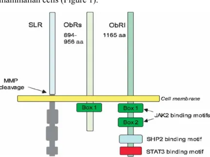
Leptin circulates as a 16 kDa protein particularly bound to plasma protein and exerts its action through the trans-membrane leptin receptor (ObR) which belongs to a family of class I cytokine receptors (Tartaglia 1997). Several isoforms of the leptin receptor, generated by mRNA alternative splicing have been discovered but ObRs, the short form, and ObRl, the long form, are the two major isoforms prevalently expressed in mammalian cells (Figure 1).
Figure 1. Schematic representation of leptin receptors, ObRs (short form) ObRl (long form). ObRl and ObRs share a common extracellular leptin-binding domain, but
ObRl is highly expressed in the hypothalamus (Bjorbaek et al, 1997), but lower levels of this receptor have been identified in many peripheral organs, such as pancreas (Morton et al, 1999), prostate (Stattin et al, 2001), keratinocytes (Frank et al,
2000), vagal afferent neurons (Buyse et al, 2001), stomach mucosa cells (Goiot et al, 2001), placenta (Ebenbichler et al, 2002), bone (Lee et al, 2002) and endometrial cells
(Yuan et al, 2004).
The short forms of ObRs are ubiquitously expressed (Fei et al, 1997). Their functions are not clear, but there is evidence that ObRs can be involved in intra- and trans-cellular leptin transport (Hileman et al, 2000).
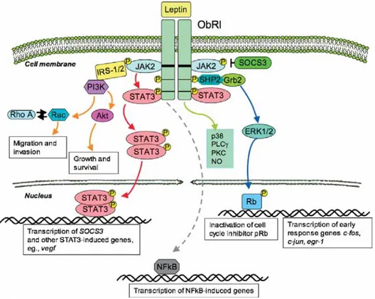
Only the long form, has the intracellular motifs necessary for the activation of signaling pathways (Tartaglia et al, 1997). As with other class I cytokine receptors, the leptin signaling is thought to be transmitted mainly by the JAK/STAT (Janus Kinase /Signal Transducers and Activators of Transcription) pathway (Bahrenberg et
al, 2002; Ahima & Osci et al, 2004). JAKs associate constitutively with conserved
Box 1 and 2 motifs in the intracellular domain of ObRl. Binding of leptin to ObRl results in autophosphorilation of JAK 1 and JAK 2 as well as phosphorilation of the cytoplasmatic domain of ObRl and the downstream transcription factors STATs (Ahima & Osci et al, 2004). The leptin signal is terminated by induction of SOCS3 (suppressor of cytokine signaling), a member of a family of proteins which inhibits the JAK/STAT signaling cascade (Bjorback et al, 1999, Emilsson et al, 1999). SOCS proteins have a variable amino-terminal domain, a central SH2 domain and a carboxy-terminal domain termed the SOCS-box motif. They are induced by cytokines
and act in a negative feedback loop to inhibit the receptor. It has been shown that overexpression of SOCS-3 inhibits leptin mediated tyrosine phosphorilation of JAK 2 (Bjorback et al, 1999, Emilsson et al, 1999).
In addition to STAT3 activation, leptin regulates other key signaling pathways: Ras/ERK1/2 (Ras/Extracellular signal-regulated kinase 1/2) cascade and PI-3K/Akt/GSK3 (phosphoinositide 3 kinase/ protein kinase B/glycogen synthase kinase 3) growth/antiapoptotic pathway. Besides, leptin has been found to induce phospholipase C (PLC)-gamma, protein kinase C (PKC), nitric oxide (NO) and p38 kinase (Bjorbaek et al, 1997; Sweeney et al, 2002; Zabeau et al, 2003). Ultimately, induction of ObRl can activate several genes involved in cell proliferation, including c-fos, c-jun, junB, and egr-1 (Sweeney et al; 2002; Zabeau et al, 2003; Frankenberry
et al, 2004). (Figure 2)
Recently, leptin is considered as a new growth factor. In fact, many studies have demonstrated that this cytokine is able to stimulate the proliferation of various cell types and it plays an important role in the development and progression of several cancer cells such as breast (Okumura et al, 2002), prostate (Somasundar et al, 2004), ovarian (Choi et al, 2004), colorectal (Hardwick et al, 2001), pancreatic (Morioka et
al, 2007), and lung cancers (Tsuchiya et al, 1999).
Previous studies suggested that leptin signaling can crosstalk with both polypeptide growth factor signaling and with steroid receptor function. For instance, insulin is known to increase leptin expression (Cusin et al, 1995; Saladin et al, 1995;
Hardie et al, 1996; Leroy et al, 1996), but it can also induce leptin resistance by the
inhibition of leptin signaling through JAK2 (Kellerer et al, 2001).
Of particular interest is the link between leptin activity and ERα. Recent reports demonstrated that ERα and ObR are coexpressed in malignant mammary tissue and breast cancer cell line (Dieudonne et al, 2002; Hu et al, 2002; Laud et al, 2002). Notably, mitogenic effects of leptin and leptin-dipendent activation of STAT3 require SRC-1, a member of the p160 family of steroid receptor modulators (Yin et al, 2004), which might represent crosstalk between steroid receptor and leptin-induced transcriptional mechanisms. Furthermore, leptin has been found to modulate both estrogen syntesis and ERα activity. Our previous works demonstrated in breast cancer epithelial cells that leptin is an amplifier of E2 signaling through a double mechanism:
an enhanced aromatase gene expression (Catalano et al, 2003) and a direct trans-activation of ERα (Catalano et al, 2004). In addition, leptin and E2 enhance
primary tumor mass either in vivo in MCF-7 cell tumor xenograft and in vitro in MCF-7 three dimensional cultures through an up-regulation of E-cadherin expression (Mauro et al, 2007).
The level of serum leptin is strongly correlated to body fat content, thus hyperleptinemia is a common feature of obese patients. Epidemiological studies have suggested a positive correlation between obesity and an increased risk of developing different cancers, include breast, prostate, colon and endometrial. There is convincing and consistent evidence from both case-control and cohort studies that obesity is tightly related to endometrial cancer in both pre- and postmenopausal women (Calle
and Thun et al, 2004).
Endometrial cancer is the most common gynaecological malignancy and the fourth most common malignancy in women in the developed world after breast, colorectal and lung cancer. The incidence is estimated at 15-20 per 100,000 women per year (Ryan et al, 2005). The majority of cases can be divided into two broad categories based on clinic-pathological and molecular characteristics: type I oestrogen-dependent with endometriod morphology and Type II non-oestrogen-dependent with serous papillary or clear cell morphology.
Increased endometrial cancer risk has been associated with early menarche and late menopause, suggesting a relationship of risk with greater lifetime exposure to estrogens at pre-menopausal levels. Other hormone-related factors associated with risk are parity and use of exogenous estrogens for oral contraception or postmenopausal replacement therapy (Emons et al, 2000). Furthermore, risk has been
related to plasma concentrations of estrogens, progesterone, androgens, SHBG, and insulin (Potischman et al, 1996; Troisi et al, 1997). Although, it is generally thought that excess weight influences endometrial cancer risk through changes in endogenous hormone metabolism, an additional candidate that may play a crucial role in the same scenario could be leptin (Kaaks et al, 2001; Key et al, 2001).
Indeed, several studies demonstrated that serum leptin levels among cases with endometrial cancer were significantly higher compared to controls (Petridou et al,
2002; Yuan et al, 2004).
Expression of leptin and its functional receptors include short and long isoforms (ObRl and ObRs) has been shown in both cancer and non-cancer endometrium (Gonzales et al, 2000; Yuan et al, 2004; Sharma et al, 2006; Koda et al, 2007). The levers of ObRl was similar in cancer and normal tissue, but the short isoforms were significantly decreased in malignant cells. Moreover, induction of the expression of this receptor resulted in inhibited proliferation of cancer cells due to delayed start of the mitotic S phase suggesting that loss of ObRs in endometrial cancer might contribute to malignant progression (Yuan et al, 2004).
A recent report demonstrated that leptin promotes endometrial cancer growth and invasiveness through STAT/MAPK and Akt pathways. Particularly, treatment with leptin resulted in increased proliferation and induces invasion of ECC1 and Ishikawa cells (Sharma et al, 2006).
However, the molecular mechanism involved in leptin induced endometrial cancer cell proliferation still needs to be elucidated.
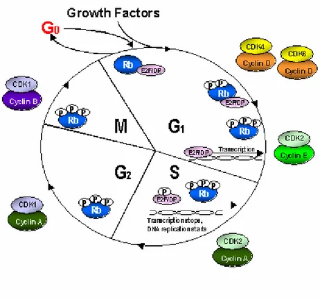
In recent years, a large body of evidence has shown that disruption of cell cycle control mechanism is a common pathway in human cancer and over-expression of cyclin D1 is one of the most commonly observed alterations. Cyclin D1, an important cell cycle regulator is required for completion of the G1/S transition in normal mammalian cells (Fu et al, 2004). (Figure 3)
Figure 3. The mammalian cell cycle.
This cycle modulator increases from normal endometrium to hyperplasia and carcinoma, suggesting that it may play a role in endometrial carcinogenesis.
In this study, we explored the molecular mechanism eliciting the biological effect of leptin in endometrial carcinoma cells’ growth. By performing a panel of different assays, we have demonstrated that leptin enhances cyclin D1 expression
within the cyclin D1 promoter. Our findings have provided evidence for better understanding of the association between obesity and endometrial cancer progression.
Materials and Methods
Materials
Dulbecco's modified Eagle's medium (DMEM), L-glutamine, penicillin, streptomycin, fetal bovine serum (FBS), bovineserum albumin (BSA), phosphate-buffered saline were purchasedfrom Eurobio (Les Ullis Cedex, France).
Triazol reagent by Invitrogen(Carlsbad, CA).
FuGENE 6 by Roche Applied Science (Indianapolis,IN).
TaqDNA polymerase, RETROscript kit, TnT® T7/T3 coupled rabbit reticulocyte lysate system, 100-bp DNA ladder, Dual Luciferase kit,and TK Renilla luciferase plasmid were provided by Promega (Madison,WI).
Aprotinin, leupeptin, phenylmethylsulfonyl fluoride (PMSF),sodium orthovanadate, H89 and recombinant human leptin were purchasedby Sigma (Milan, Italy).
Antibodies against phospho-CREB (ser133) (1B6), CREB (48H2), phospho p44/42 MAPK (Thr 202/Tyr 204) (#9101S), p44/42 MAPKinase (#9102) and U0126 (inhibitor of MAPK) were provided by Cell Signaling.
AG 490 were provided by Biomol (Milan, Italy).
Antibodies against Cyclin D1 (M-20), GAPDH (FL-335), P21 (H-164), pSTAT3 (B-7), and STAT3 (F-2) were provided by Santa Cruz Biotechnology.
An ECL system, [ 32P]ATP, and SephadexG-50 spin columns were purchased from Amersham Biosciences (Buckinghamshire,UK).
QuickChange kit (Stratagene, La Jolla, CA)
RNase A (Calbiochem, La Jolla, CA).
Plasmids
The plasmids containing the human cyclin D1 promoter or its deletions (p-2966/+142, p-944/+142, p-848/+142, p-136/+142) were kindly provided by Prof A. Weisz (University of Naples, Italy). These fragments were inserted into the luciferase vector pXP2.
Site-directed mutagenesis
The cyclin D1 promoter plasmids bearing STAT3 binding recognition site (GAS) mutated site (pGAS mut) and cyclic AMP-responsive element (CRE) mutated site (pCRE mut) were created by site-directed mutagenesis using QuickChange kit.
Briefly, this was based on a PCR reaction with two complementary oligonucleotide primers containing the mutation. The PCR was performed with the Pfu DNA polymerase during 16 cycles (30sec at 95°C, 30sec at 55°C and 8min at 68°C), using as template the human cyclin D1 promoter P-136/+142 and the following mutagenic primers:5'-CGGACTACAGGGGAGTAGCGTTGAAGTTGCAAAGTCCTGGAG-3' and 5'-CTCCAGGACTTTGCAACTTCAACGCTACTCCCCTGTAGTCCG-3' (GAS MUT); 5'-GATCTTTGCTTAACAACAGTAACTCTACACGGACTACAGGGGAG -3'and 5'-CTCCCCTGTAGTCCGTGTAGAGTTACTGTTGTTAAGCAAAGATC-3' (CRE MUT). To create the plasmid mutated in both responsive elements (pGAS/CRE mut), we used as template pCRE mut and the primers for GAS site above mentioned. The PCR products were then incubated with DpnI which only digests the parental methylated cDNA and the constructed mutated expression vectors were confirmed by DNA sequencing.
Cell Culture
Ishikawa human endometrial cancer cells were obtained from D. Picard (University of Geneva, Geneva, Switzerland). Ishikawa cells were maintained in
DMEM without phenol red supplemented with 10% fetal bovine serum, 1%
L-glutamine and 1% penicillin/streptomycin. Cells were switched to medium without serum 48h before each experiment.
Ishikawa cells at 50-60% confluence were shifted to serum-free medium (SFM) for 48 h and then untreated or treated with 1000 ng/mL leptin in SFM for 24h. Thereafter, cells were trypsinized, centrifuged at 1500 rpm for 3 min, washed with PBS, and then treated with 20 μg/ml RNase A. DNA was stained with 100µg/ml propidium iodide for 30 min at 4 °C protected from light, and cells were analyzed with the FAC-Scan (Becton Dickingson and Co., Franklin Lakes, NJ).
Total RNA extraction and reverse transcription-PCR assay
Total cellular RNA was extracted from Ishikawa cells using Triazol reagent as suggested by the manufacturer.The purity and integrity of the RNA were checked spectroscopically and by gel electrophoresis before carrying out the analytical procedures. The evaluation of genes expression was performed by the reverse
transcription-PCR method. cDNA was synthesized by oligo (dT) using a
RETROscript kit as suggested by the manufacturer.The cDNAs obtained were further
amplified by a PCR using thefollowing primers: 5'-TCTAAGATGAAGGAGACC
ATC-3' and 5'-GCGGTAGT AGGACAGGAAGTTGTT-3' (cyclin D1); 5'-GCTTC ATGCCAGCTACTTCC-3' and 5'-CTGTGCTCACTTCAGGGTCA-3' (p21); 5'-CTC
AACATCTCCCCCTTCTC-3' and 5'-CAAATCCCATATCCTCGTCC-3'(36B4).
The PCR was performed for 30 cycles for cyclin D1 (94°Cfor 1 min, 60°C for 1 min, and 72°C for 2 min), 30 cycles for p21 (94°Cfor 1 min, 58°C for 1 min, and 72°C for 2 min) and 15 cycles (94°C for 1 min, 58°C for 1 min,and 72°C for 2 min) to amplify 36B4 in the presence of 1μl of first strand cDNA, 1 µM each of the primers
mentionedabove, 0.5 mM dNTP, Taq DNA polymerase (2 units/tube), and2.2 mM magnesium chloride in a final volume of 25 µl.To check for the presence of DNA contamination, a reverse transcription-PCRwas performed on 1 µg of total RNA without Moloney murineleukemia virus reverse transcriptase (the negative control).
The PCR products were analyzed on 2% agarose gel and stained with ethidium
bromide. DNA quantity in each lane was analyzed by scanning densitometry.
Standard DNA (100-bp DNA ladder) wasrun to provide the appropriate size marker.
Immunoblotting
Ishikawa cells were grown in 10 cmdishes to 50–60% confluence and lysed in 500 µlof 50 mM HEPES, pH 7.5, 150 mM NaCl, 1.5 mM MgCl2, 1 mM EGTA,10%
glycerol, 1% Triton X-100, and a mixture of protease inhibitors(aprotinin, PMSF, and sodium ortho-vanadate). Equal amounts oftotal protein were resolved on an 11% SDS-polyacrylamide gel.The proteins were transferred to a nitrocellulose membrane, probed with rabbit polyclonal antiserum directed against thehuman cyclin D1 (1:1000) and p21(1:1000). The antigen-antibodycomplex was detected by incubation of the membranes for 1 h at room temperature with peroxidase-coupled goat anti-rabbitIgG and revealed using the ECL System. The blots were thenexposed to film, and the bands of interest were quantifiedby densitometer (model 620; Bio-Rad). The results obtainedas optical density arbitrary values were transformed to percentages of the control (percent control) taking the samples from cellsnot treated as 100%.
Transient transfection assay
Ishikawa cells were starved with serum free medium for 24 h and then transfected using the FuGENE 6 reagent with the mixture containing 0.25 µg of human cyclin D1 promoter constructs. Twenty-four hours after transfection, the cells were untreated or treated with 1000 ng/mL leptin for 6, 12 and 24h. TK Renilla luciferase plasmid (10 ng per each well) was used. Firefly and Renilla luciferase activities weremeasured by Dual Luciferase kit. The firefly luciferase datafor each sample were normalized based on the transfection efficiencymeasured by Renilla luciferase activity.
Electrophoretic mobility shift assay
Nuclear extracts from Ishikawa cells wereprepared as previously described (Anderws et al, 1991). Briefly, Ishikawa cells plated into 60 mm dishes were scraped into 1.5 ml of cold phosphate-buffered saline (PBS). Cells were pelleted for 10 sec and resuspended in 400 μl cold buffer A (10 mM HEPES-KOH pH 7.9 at 4°C, 1.5 mM MgCl2, 10 mM KCl, 0.5 mM dithiothreitol, 0.2 mM PMSF, 1 mM leupeptin) by
flicking the tube. The cells were allowed to swell on ice for 10 min and then vortexed for 10 sec. Samples were then centrifuged for 2 min and the supernatant fraction was discarded. The pellet was resuspended in 50 μl of cold Buffer B (20 mM
HEPES-KOH pH 7.9, 25% glycerol, 1.5 mM MgCl2, 420 mM NaCl, 0.2 mM EDTA,
0.5 mM dithiothreitol, 0.2 mM PMSF, 1 mM leupeptin) and incubated on ice for 20 min for high-salt extraction. Cellular debris was removed by centrifugation for 2 min
at 4°C and the supernatant fraction (containing DNA binding proteins) was stored at –70°C. The probe was generated by annealing single-stranded oligonucleotides, labeled with [ 32P] ATP and T4 polynucleotide kinase, and then purified using
Sephadex G50 spin columns. The DNA sequences used as probe or as cold
competitors are as follows: 5'-AGGGGAGTTTTGTTGAAGTTGCAAA-3' and 5'-TTTGCAACTTCAACAAAACTCCCCT-3' (GAS); 5'-TTAACAACAGTAACGT CACACGGACTA-3' and 5'-TAGTCCGTGTGACGTTACTGTTGTTAA-3' (CRE);
5'-AGGGGAGTAGCGTTGAAGTTGCAAA-3' and 5'-TTTGCAACTTCAACGCT
ACTCCCCT-3' (GAS MUT); 5'-CTTAACAACAGTAATTGCACACGGACTA-3'
and 5'-TAGTCCGTGTGCAATTACTGTTGTTAAG-3' (CRE MUT).
In vitro transcribed and translated CREB protein was synthesized using the T7
polymerase in the rabbit reticulocyte lysate system. The protein-binding reactions were carried out in 20 mL of buffer [20 mmol/L HEPES(pH 8), 1 mmol/L EDTA, 50 mmol/L KCl, 10 mmol/L DTT, 10% glycerol,1 mg/mL BSA, 50 µg/mL poly(dI/dC)] with 50,000 cpm oflabeled probe, 20 µg of Ishikawa nuclear protein or an appropriate amount of CREB protein and 5 µg of poly (dI-dC). The mixtures were incubated at room temperature for 20 min in the presence or absence of unlabeledcompetitor oligonucleotides. For experiments involving STAT3 and CREB antibodies, the reaction mixture was incubated with these antibodies at 4°C for 12h. The entire reaction mixture was electrophoresed througha 6% polyacrylamide gel in 0.25x Tris borate-EDTA for 3 h at150 V.Gel was dried and subjected to autoradiography at -70°C.
Chromatin immunoprecipitation assay
According to the ChIP assay procedure previously described (Shang et al,
2000), Ishikawa cellswere grown in 100 mm dishes to 50-60% confluence, shifted to serum free medium (SFM) for 24 hours and then untreated or treated with 1000 ng/mL leptin for 10 min, 30 min and 1 h. Thereafter, cells were washed twice with PBS and crosslinked with 1% formaldehyde at 37°C for 10 min. Next, cells were washed twice with PBS at 4°C, collected and resuspended in 200 μl of lysis buffer (1% SDS, 10 mM EDTA, 50 mM Tris-HCl pH 8.1) and left on ice for 10 min. Then, cells were sonicated four times for 10 sec at 30% of maximal power (Sonics, Vibra Cell 500W) and collected by centrifugation at 4°C for 10 min at 14 000 rpm. The supernatants were diluted in 1.3 ml of IP buffer (0.01% SDS, 1.1% Triton X-100, 1.2 mM EDTA, 16.7 mM Tris-HCl pH 8.1, 16.7 mM NaCl) and immunocleared with 80 μl of sonicated salmon sperm DNA/protein A agarose for 1 hour at 4°C. The precleared chromatin was immunoprecipitated with a specific anti-STAT3, anti-CREB antibodies and with a normal mouse serum IgG (Nms) as negative control. At this point, 60 µl salmon sperm DNA/protein A agarose were added and precipitation was further continued for 2 hours at 4°C. After pelleting, precipitates were washed sequentially for 5 min with the following buffers: Wash A (0.1% SDS, 1% Triton X-100, 2 mM EDTA, 20 mM Tris-HCl pH 8.1, 150 mM NaCl), Wash B (0.1% SDS, 1% Triton X-100, 2 mM EDTA, 20 mM Tris-HCl pH 8.1, 500 mM NaCl), and Wash C (0.25 M LiCl, 1% NP-40, 1% sodium deoxycholate, 1 mM EDTA, 10 mM Tris-HCl pH 8.1), and then twice with TE buffer (10 mM Tris, 1 mM EDTA). The
immunocomplexes were eluated with elution buffer (1% SDS, 0.1 M NaHCO3),
reverse crosslinked by heating at 65°C and digested with proteinase K (0.5 mg/ml) at 45°C for 1 hour. DNA was obtained by phenol/chloroform/isoamyl alcohol extraction. 2 µl of 10 mg/ml yeast tRNA were added to each sample and DNA was precipitated with 95% EtOH for 24 hours at –20°C, and then washed with 70% EtOH and resuspended in 20 µl of TE buffer. 3 µl of each sample were used for PCR amplification with the following primers flanking GAS/CRE sequence present in the cyclin D1 promoter region: 5’-TGCGCCCGCCCCCGCCCCCCTC-3’ and 5’-TGTTCCATGGCTGGGGCTCTT-3’. The PCR conditions were 1 min at 94°C, 1 min at 65°C, and 2 min at 72°C. The amplification products obtained in 35 cycles were analyzed in a 2% agarose gel and visualized by ethidium bromide staining.
Statistical Analysis
Each datum point represents the mean ± S.E. of three different experiments. Data were analyzed by ANOVA test using the STATPAC computer program.
Results
Leptin modulates cell cycle progression in endometrial cancer cells
On the basis of previous studies demonstrating the expression of the Ob and ObR in both cancer and non-cancer endometrium (Gonzalez et al, 2000; Kitawaki et
al, 2000; Koshiba et al, 2001), and the stimulatory effects of leptin on proliferation of
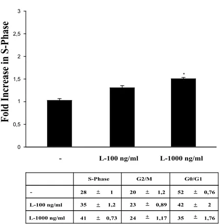
Ishikawa and ECC1 cells in a time- and dose-dependent manner (Sharma et al, 2006), we first investigated the role of leptin on Ishikawa cell cycle progression. Cells were synchronized with serum starvation for 48h and then induced to re-enter the cell cycle by treatment with hormone. Flow cytometric analysis revealed that 24h leptin 1000 ng/ml treatment significantly reduced the numbers of G0/G1-phase cells accompanied with the increase of cell population in S phase, compared with control group (Fig. 4).
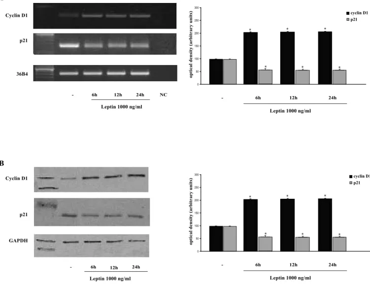
Leptin enhances Cyclin D1 and down-regulates p21WAF1/Cip1 expression in Ishikawa cells
Considering that the cyclin D1 is a critical modulator in the cell cycle G1/S transition and its over-expression is one of the most commonly observed alterations in human endometrial cancers (Horn et al, 2006), we aimed to examine the potential ability of leptin to modulate cyclin D1 mRNA and protein content in Ishikawa human endometrial cancer cells. Results of RT-PCR showed an increased cyclin D1 mRNA after the treatment with leptin 1000 ng/ml for 6, 12 and 24 h. mRNA expression of the cyclin D1 gene was normalized using the human housekeeping gene 36B4 (Fig. 5A). The leptin-induced expression of cyclin D1 was confirmed at protein level, at all
F
ol
d
In
cr
ea
se
in
S
-P
ha
se
0 0,5 1,5 2 2,5 3 L-100 ng/ml -1 L-1000 ng/ml * L-100 ng/ml L-1000 ng/ml -S-Phase G2/M G0/G1 28 35 41 20 23 24 52 35 42 -+ -+ -+ -+ -+ -+ -+ -+ -+ 1 1,2 0,73 1,2 0,89 1,17 0,76 2 1,76Figure 4 Leptin increases the fraction of Ishikawa cells in the S phase of the cell cycle.
Ishikawa cells were synchronized in serum-free medium for 48 h and were exposed to 100 ng/ml and 1000 ng/ml leptin for 24 h or left untreated (-). The distribution of Ishikawa cells in the cycle was determined by flow cytometry using propidium-iodide stained nuclei. The results indicate the fold increase of Ishikawa cells in S phase after serum starvation or leptin treatment. The histograms represent the means S.E. of three separate experiments done in triplicate. * P <0,01, compared with serum-starved condition. The table shows the distribution of Ishikawa cells in the various phases of cell cycle.
times investigated, by Western blotting analysis (Fig. 5B).
To further study the involvement of leptin in cell cycle progression, the expression of p21WAF1/Cip1, which plays as a major negative regulator in the G1 checkpoint, was analyzed. p21WAF1/Cip1 mRNA and protein levels were decreased in leptin treated samples than untreated cells (Figs. 5A and 5B).
These observations indicate that leptin by stimulating expression of the positive regulator, cyclin D1, and inhibiting the expression of the negative regulator p21WAF1/Cip1, promotes the entry of G1 into S phase in cell cycle progression.
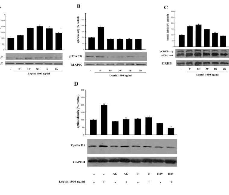
Leptin-induced cyclin D1 expression is STAT, MAPK and PKA dependent in proliferating Ishikawa cells.
Leptin exerts its biological functions through binding to its receptors that mediate a downstream signal by activating multiple signaling pathways (Garofalo et
al, 2006). To gain insight into the mechanism underlying the modulatory role of leptin
on cyclin D1 expression in endometrial cancer cells, we examined the changes in signal transductional pathways involved in mediating leptin action. Cellular proteins were extracted from Ishikawa cells treated with 1000 ng/ml leptin for various time periods, and by western blotting analysis we determined the status of STAT3 and ERK1/2 phosphorylation. As shown in Figure 6A, leptin significantly induced phosphorylation of STAT3 within 15 minutes of treatment while an increased phosphorylation of ERK was observed after 5 minutes of leptin stimulation followed by a decline (Fig. 6B). Besides, we also examined effect of leptin on phosphorylated
A B Leptin 1000 ng/ml -Cyclin D1 GAPDH 6h 12h 24h Leptin 1000 ng/ml -Cyclin D1 36B4 6h 12h 24h NC p21 o pt ic al d en si ty (a rb itr ar y un its ) 0 50 100 150 200 250 300 * * * Leptin 1000 ng/ml - 6h 12h 24h * * * o pt ic al d en si ty (a rb itr ar y un its ) 0 50 100 150 200 250 300 * * * Leptin 1000 ng/ml - 6h 12h 24h * * * p21 cyclin D1 p21 cyclin D1 p21
Figure 5 Effects of Leptin on cyclin D1 and p21WAF1/Cip1expression in Ishikawa cells.
Ishikawa cells were serum-starved for 48 h followed by treatment with 1000 ng/ml leptin for 6, 12 and 24 h or left untreated (-). A, total RNA was isolated from Ishikawa cells and reverse transcribed. cDNA was subjected to PCR using specific primers for cyclin D1 (30 cycles), p21 (30 cycles) or 36B4 (15 cycles). NC: negative con-trol, RNA sample without the addition of reverse transcriptase. B, Protein extracts obtained from Ishikawa cells were immunoblotted with rabbit polyclonal antiserum against human cyclin D1 and p21. GAPDH served as loading control. The histograms represent the means S.E. of three separate experiments in which band intensities were evaluated in terms of optical density arbitrary units and expressed as percentages of the control, which was assumed to be 100%. * p<0.01 compared to vehicle.
B op tic al de ns ity (% co nt ro l) 0 50 100 150 200 250 * Leptin 1000 ng/ml 5' 1h 2h - 15' 30' pMAPK MAPK A 0 50 100 150 200 250 * * * * Leptin 1000 ng/ml 5' 1h 2h - 15' 30' STAT pSTAT op tic al de ns ity (% co nt ro l) op tic al de ns ity (% co nt ro l) 0 50 100 150 200 250 300 * * CREB pCREB ATF-1 Leptin 1000 ng/ml 5' 1h 2h - 15' 30' C D 0 50 100 150 200 250 300 * - - U U H89 H89 + - + - - + AG AG - + Cyclin D1 GAPDH Leptin 1000 ng/ml op tic al de ns ity (% co nt ro l)
Figure 6 Activation of leptin signaling in up-regulation of cyclin D1 expression.
Ishikawa cells were serum-starved for 48 h and treated with 1000 ng/ml leptin for various time intervals or left un-treated (-). Protein extracts obtained from Ishikawa cells were immunoblotted with a specific antibodies against total or phosphorylated (p) forms of STAT3 (A), MAPK (B) and CREB/ATF-1 (C). The histograms represent the means S.E. of three separate experiments in which band intensities were evaluated in terms of optical density arbi-trary units and expressed as percentages of the control, which was assumed to be 100%. * p<0.05 compared to vehicle. (D) Ishikawa cells were serum-starved for 48 h and treated with 1000 ng/ml leptin for 24 h or left un-treated (-). For combined treatment, cells were preun-treated with AG490 (20 uM), U0126 (10 uM) and H89 (10 uM) for 30 min followed leptin treatment. The histograms represent the means S.E. of three separate experiments in which band intensities were evaluated in terms of optical density arbitrary units and expressed as percentages of the control, which was assumed to be 100%. * p<0.01 compared to vehicle; ** p<0.01 compared to leptin treatment.
**
** **
CREB/ATF-1, a downstream substrate of MAPK, but also an effector of PKA (Delghandi et al, 2005). The CREB Ab produces two bands and recognizes both CREB (upper band) and ATF-1 (lower band). Notably, leptin exposure for 15 minutes significantly induced phosphorylation of CREB/ATF1 (Fig. 6C).
Leptin had no effect on total STAT3, ERK and CREB protein expression levels. Next, to investigate the signal transduction pathways involved in leptin-induced cyclin D1 expression, chemical inhibitor of JAK/STAT (AG490), ERK1/2 (U0126) and PKA (H89) were added to serum starved Ishikawa cells before the treatment with 1000 ng/ml of leptin. Our results revealed that, AG490, U0126 and H89 effectively prevent leptin induction of cyclin D1 expression level (Fig. 6D).
Effects of leptin on activity of human cyclin D1 promoter/luciferase reporter gene constructs in Ishikawa cells.
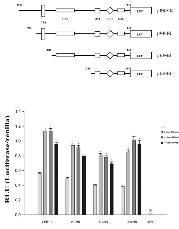
To evaluate whether leptin is able to activate cyclin D1 promoter, we transiently transfected Ishikawa cells with a luciferase reporter construct containing the upstream region of the cyclin D1 gene spanning from -2966 to + 142. As shown in Figure 7, a significant increase in promoter activity was observed in the transfected cells exposed to leptin 1000 ng/ml for 6, 12 and 24 h.
Cyclin D1 promoter contains multiple regulatory elements, including binding sites for AP-1, STATs, NF-kB, Oct-1, Sp1, CRE and TCF/LEF (Saxena et al, 2007;
Bartusel et al, 2005; Brockman et al, 2005; Natsume et al, 2003; Allan AL et al, 2001).
p-2966/+142 GAS CRE TRE SP-1 LUC +142 -2966 GAS p-944/+142 LUC +142 -944 p-848/+142 LUC +142 -848 p-136/+142 LUC +142 -136 A B C 24h Leptn 1000 ng/ml 12h Leptn 1000 ng/ml 6h Leptn 1000 ng/ml p-2966/+142 p-944/+142 p-848/+142 p-136/+142 0,0 0,2 0,4 0,6 0,8 1,0 1,2 1,4 pXP2 * * * * * * * * * * * ** R L U ( L u ci fe ra se /r en il la )
Figure 7 Leptin enhances human cyclin D1 promoter in Ishikawa cells.
A. Schematic representation of human cyclin D1 promoter fragments used in this study. All of the promoter constructs contain the same 3' boundary (+142). The 5' boundaries of the promoter fragments varied from -2966 to -136. Each fragment was subcloned into the pXP2 vector. B. Transcriptional activity of Ishikawa cells with promoter constructs is shown. Ishikawa cells were serum-starved for 24 h, transfected for 24 h and left untreated (-) or treated with 1000 ng/ml leptin for 6, 12 and 24 h. The values represent the means S.E. of three separate experiments. In each experiment, the activities of the transfected plasmid was assayed in triplicate transfections. pXP2: basal activity measured in cells transfected with pXP2 basal vector. * p<0.01 compared to vehicle.
To delimit the cis-element involved in cyclin D1 transcriptional activation by leptin, we transiently transfected Ishikawa cells with plasmids containing a series of 5’ deleted segments of human cyclin D1 promoter. Schematic representation of constructs is shown in Figure 7A.
In transfection experiments performed using p-944/+142, p-848/+142 and p-136/+142 the responsiveness to leptin was still observed at all time investigated (Fig. 7B), suggesting that the region from -136 to +142 was required for the trans-activation of cyclin D1 by leptin.
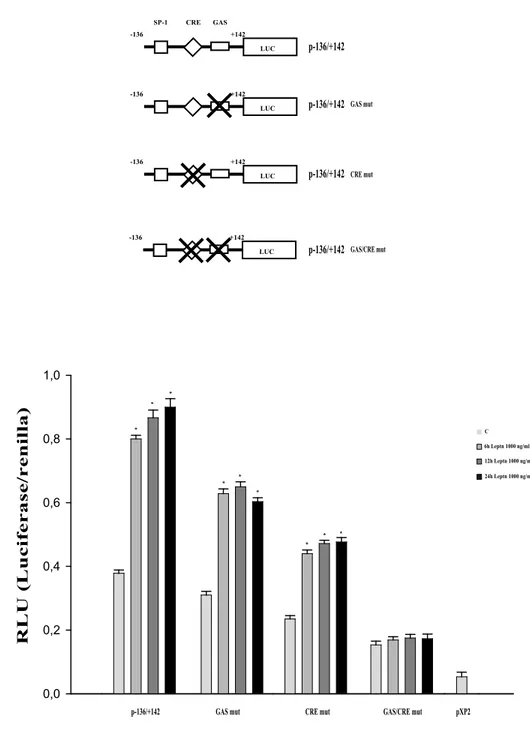
The nucleotide sequence analysis of this region evidenced STAT3 binding motif (GAS) and a cyclic AMP-responsive element (CRE) located at position -52 and -27 respectively, putative effectors of leptin signaling as previously demonstrated in other systems (Mauro et al, 2007, Saxena et al, 2007). Thus, mutation analysis of the GAS and CRE sites on cyclin D1 promoter was carried out to define further their role for functional interaction with leptin.
The effects of leptin on different GAS and CRE mutant constructs showed that mutation of the GAS site and CRE alone moderately affected the regulation of cyclin D1 promoter activity by leptin (Fig. 8B). In contrast, mutation of both GAS and CRE completely abolished leptin responsiveness of cyclin D1 promoter in Ishikawa cells (Fig. 8B). These results suggest that leptin signaling pathways stimulate cyclin D1 transcription through both GAS and CRE motifs. Whereas either of these two sites may be sufficient alone to induce cyclin D1 promoter activity, both of them are necessary for optimal leptin action.
LUC +142 -136 CRE SP-1 GAS LUC +142 -136 LUC +142 -136 p-136/+142 GAS mut CRE mut GAS/CRE mut p-136/+142 p-136/+142 p-136/+142 LUC +142 -136 A B C 24h Leptn 1000 ng/ml 12h Leptn 1000 ng/ml 6h Leptn 1000 ng/ml
p-136/+142 GAS mut CRE mut GAS/CRE mut
0,0 0,2 0,4 0,6 0,8 1,0 pXP2 * * * * * * * * * R L U ( L u ci fe ra se /r en il la )
Figure 8 Leptin transactivates cyclin D1 gene promoter.
A. Schematic representation of the mutated plasmids used in this study. B.Transcriptional activity of Ishikawa cells with promoter constructs is shown. Ishikawa cells were serum-starved for 24 h, transfected for 24 h and left untreated (-) or treated with 1000 ng/ml leptin for 6, 12 and 24 h. The values represent the means S.E. of three separate experiments. In each experiment, the activities of the transfected plasmid was assayed in triplicate transfections. pXP2: basal activity measured in cells transfected with pXP2 basal vector. * p<0.01 compared to vehicle.
Leptin increases STAT3-DNA and CREB-DNA binding activity to cyclin D1 promoter.
To further define whether GAS and CRE are responsible for the transcriptional activation of cyclin D1 by leptin, we performed EMSA experiments.
Using synthetic oligodeoxyribonucleotides corresponding to the GAS and CRE motifs, we observed the formation of a complex in nuclear extract Ishikawa cells (Figs. 9 and 10, lane 1), which was abrogated by 100 fold molar excess of unlabeled probe (Figs. 9 and 10, lane 2) demonstrating the specificity of the DNA binding complex. This inhibition was not observed when a mutated oligodeoxyribonucleotides were used as competitor (Figs. 9 and 10, lane 3). Leptin induced both GAS and CRE activation compared with untreated at the same time-point (Figs. 9 and 10, lane 4, 5,
6). Incubation of anti-STAT3 with the nuclear extracts resulted in a greatly reduced
band, indicating the presence of STAT3 protein in the complex (Fig. 9, lane 7). Similarly, incubation of anti-CREB with the nuclear extracts resulted in reduced and supershifted bands (Fig. 10, lane 7). IgG did not affect either GAS or CRE complex formation (Figs. 9 and 10, lane 8). Using transcribed and translated in vitro CREB protein, we obtained a complex migrating at the same level as that of Ishikawa nuclear extracts (Fig. 10, lane 9).
Leptin enhances recruitment of STAT3 and CREB to the promoter region of cyclin D1.
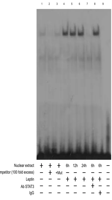
Nuclear extract
Ab STAT3
+Mut Leptin
Competitor (100 fold excess)
6h
IgG
12h 24h 6h 6h
1 2 3 4 5 6 7 8 9
Figure 9 Effects of in vitro leptin treatment on STAT3-DNA binding activity in Ishikawa cells.
Nuclear extracts from Ishikawa cells were incubated with a double-stranded STAT3-specific consensus sequence probe labeled with [32P] ATP and subjected to electrophoresis in a 6% polyacrylamide gel (lane 1). Competition experiments were done by adding as competitor a 100-fold molar excess of unlabeled probe (lane 2) or a 100-fold molar excess of unlabeled oligonucleotide containing a mutated GAS (lane 3). Ishikawa nuclear extracts treated with 1000 ng/ml leptin for 6, 12 and 24 h incubated with probe (lane 4, 5 and 6). The specificity of the binding was tested by adding to the reaction mixture a STAT3 antibody (lane 7). IgG did not affect either GAS complex formation (lane 8). Lane 9 contains probe alone.
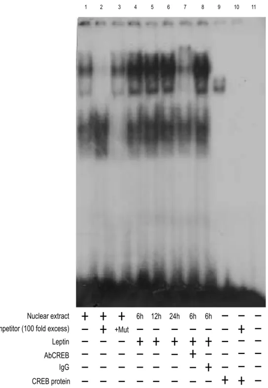
Nuclear extract
AbCREB
+Mut Leptin
Competitor (100 fold excess)
6h
IgG
12h 24h 6h 6h
CREB protein
1 2 3 4 5 6 7 8 9 10 11
Figure 10 Effects of in vitro leptin treatment on CREB-DNA binding activity in Ishikawa cells.
Nuclear extracts from Ishikawa cells were incubated with a double-stranded CREB-specific consensus sequence probe labeled with [32P] ATP and subjected to electrophoresis in a 6% polyacrylamide gel (lane 1). Competition experiments were done by adding as competitor a 100-fold molar excess of unlabeled probe (lane 2) or a 100-fold molar excess of unlabeled oligonucleotide containing a mutated CRE (lane 3). Ishikawa nuclear extracts treated with 1000 ng/ml leptinfor 6, 12 and 24 h incubated with probe (lane 4, 5 and 6). The specificity of the binding was tested by adding to the reaction mixture a CREB antibody (lane 7). IgG did not affect either CRE complex formation (lane 8). We used as positive control a transcribed and translated in vitro CREB protein (lane 9) or in combination with 100-fold molar exess of unlabeled probe (lane 10). Lane 11 contains probe alone.
Although our findings clearly demonstrated the role of STAT3 and CREB in leptin mediated regulation of cyclin D1 promoter, we further sought to determine that STAT3 and CREB directly participate in leptin mediated cyclin D1 gene regulation using chromatin immunoprecipitation assay. Using specific antibodies against STAT3, formaldehyde cross-linked protein-chromatin complexes were immunoprecipitated from Ishikawa cells cultured with or without leptin 1000 ng/ml for various periods. The resulting precipitated genomic DNA was then analyzed by PCR using primers spanning the STAT3 binding elements in the promoter region of the cyclin D1. As shown in Figure 11A, ChIP analysis with anti-STAT3 antibodies revealed that treatment with leptin for 1h increased STAT3 recruitment to cyclin D1 promoter. Interestingly, we also observed upon leptin stimulation a significant increase in CREB recruitment to the cyclin D1 promoter as evidenced by ChIP analysis using anti-CREB antibody (Fig. 11B). Our data suggest that cyclin D1 may be a target for leptin mediated growth stimulation of Ishikawa cells and molecular mechanisms involve recruitment of STAT3 and CREB transcription factors.
A
B
INPUT ChIP: STAT3
Leptin
MIN 10' 30' 60' 10' 30' 60' C
Leptin
MIN 10' 30' 60' 10' 30' 60' C
INPUT ChIP: CREB
* 0 50 100 150 200 250 300 o pt ic al d en si ty ( ar bi tr ar y un it s) Leptin 1000 ng/ml - 10' 30' 60' * 0 50 100 150 200 250 300 o pt ic al d en si ty ( ar bi tr ar y un it s) Leptin 1000 ng/ml - 10' 30' 60'
Figure 11 Recruitment of STAT3 and CREB to the cyclin D1 promoter in Ishikawa cells.
The cells were serum-starved for 48 h and left untreated (-) or treated with 1000 ng/ml leptin for various time intervals. The preacleared chromatin was immunoprecipitated with specific antibody anti-STAT3 and anti-CREB. Cyclin D1 promoter sequences contain GAS and CRE sites was detected by PCR with specific primers, as detailed in Materials and Methods. To determine input DNA, the cyclin D1 promoter fragment was amplified from 3 l, purified soluble chromatin before immunoprecipitation. PCR products obtained at 35 cycles. ChIP with non-immune IgG was used as negative control (C-). The histograms represent the means S.E. of three separate experiments in which band intensities were evaluated in terms of optical density arbitrary units and expressed as percentages of the control, which was assumed to be 100%. * p<0.01 compared to vehicle.
Discussion
Increasing epidemiologic data in humans and many in vitro investigative reports have linked obesity with various disease states and suggested a strong link between leptin and tumor progression (Garofalo et al, 2006; Somasundar et al, 2003). Indeed, several reports have described a mitogenic effect of leptin on gastric (Goiot et
al, 2001), breast (Okumura et al, 2002), ovarian (Choi et al, 2004) and prostate cancer
cells (Somasundar et al, 2004).
Leptin levels have been shown to have a positive correlation with endometrial cancer (Petridou et al, 2002; Yuan et al, 2004). For instance, both short and long isoforms of leptin receptor mRNA and proteins, were expressed in endometrial cancer (Gonzales et al, 2000; Yuan et al, 2004; Sharma et al, 2006; Koda et al, 2007). However, even thought, a growth stimulatory effect of leptin in human endometrial cancer cells was recently proposed (Sharma et al, 2006), the molecular mechanisms remains to be fully elucidated.
Thus, in the present study, we focused, in Ishikawa endometrial cancer cells, leptin signaling on cell cycle progression. Our experimental results showed that leptin treatment is able to speed up the progression reducing G0/G1 arrest with the increase of cell population in S phase, in a dose-dependent manner.
The cell cycle is regulated by the coordinate action of cyclin-dependent kinases (cdk), specific cyclin proteins and cdk inhibitors (Hilakivi-Clarke et al, 2004). Cyclin D1 and cyclin-dependent kinases are required for completion of the G1/S transition in normal mammalian cells (Fu et al, 2004).
In recent years, a large body of evidences have shown that overexpression of cyclin D1 is one of the most commonly observed alterations in human cancer, bringing cell cycle as a critical interface between hormonal signaling and tumorgenesis (Chen et al, 2007). Particularly, cyclin D1 overexpression in endometrial glands increases progressively in intensity and extent from normal endometrium to complex hyperplasia and carcinoma (Ruhul et al, 2001).
Of interest, we found that leptin exposure up-regulates both cyclin D1 mRNA and protein levels at all time investigated with a concomitant decrease of p21WAF1/Cip1 expression.
In addition, in our study, we demonstrated that leptin stimulated cyclin D1 expression requires JAK/ STAT, MAPK and PKA activation, as it emerges by the observation that the chemical inhibitors of the above mentioned pathways completely reversed the increase of cyclin D1 protein levels.
It is worth noting that our findings recall previous reports indicating the involvement of JAK/STAT and MAPK signaling pathways in leptin mediated cell growth in diverse cellular contexts (Dieudonne et al, 2002; Hardwick et al, 2001;
Choi et al, 2004). For instance, recently, in Ishikawa endometrial cancer cells, leptin
through ERK1/2 has been linked to cell proliferation (Sharma et al, 2006; Gong et al,
2007), whereas in MCF-7 cells, we evidenced that leptin signaling through ERK1/2 is
able to potentiate estrogen action and aromatase activity promoting breast cancer cell growth (Catalano et al, 2003; Catalano et al, 2004).
kinase, a member of the p90RSK family that corresponds to RSK2 and thereby phosphorylates CREB Ser133 (Xing et al, 1996; Dalby et al, 1998; Bannister et al,
1995).
Although CREB is a major downstream substrate of ERK1/2, it is also classically known as a PKA effector. The interrelationship of PKA and JAK/STAT-dependent intracellular mechanism of leptin action was previously suggested (Matsuoka et al, 1999) as well as the involvement of PKA in leptin induced human ovarian proliferation (Sirotkin et al, 2007). These observations well fit with our results demonstrating a significant increases of CREB/ATF-1 phosphorylation upon leptin exposure.
Therefore, investigating the potential ability of leptin to modulate cyclin D1 promoter gene, we performed transient transfection experiments in Ishikawa cells using diverse deletion constructs of cyclin D1 promoter gene. The results indicated that leptin signaling up-regulates the full-length promoter activity of cyclin D1. Moreover, we documented that the region spanning from -136 to +142, which contains GAS and CRE sites as potential target of leptin, is required for the responsiveness to leptin. Our mutation analyses of the GAS and CRE sites on cyclin D1 promoter showed that both motifs are the mediators of cyclin D1 regulation by leptin. The results of mutants suggest that, whereas loss of either GAS or CRE alone leads to a partial reduction of cyclin D1 promoter activity, loss of both completely abolished leptin-induced promoter activation.
the cyclin D1 promoter through two different responsive elements and must have at least one of these sites to augment cyclin D1 promoter activity.
Previously, in breast cancer cells, Leslie et al reported that cyclin D1 is transcriptionally regulated by STAT3 (Leslie et al, 2006) and activation of cyclin D1 through CRE by estrogens has been suggested (Liu et al, 2002; Castro-Rivera et al,
2001; Sabbah et al, 1999). Moreover, GAS and CRE has been shown to be a potential
target of leptin signaling. Indeed, a recent work, showed that leptin-activated STAT3 binds to its cognate sites in cyclin D1 promoter leading to hyperacetylation and overexpression of cyclin D1 gene through a recruitment of distinct coactivator complexes (Saxena et al, 2007). On the other hand, our previous findings reported, in MCF-7 breast cancer cell line, activation of E-cadherin gene promoter by leptin through CRE site (Mauro et al, 2007).
Our EMSA experiments extended the aforementioned observations because nuclear extracts from Ishikawa cells treated with leptin showed an increased binding to the GAS and CRE sequence located in the cyclin D1 promoter region. These findings were supported by ChIP assay demonstrating the ability of leptin to enhance the recruitment of STAT3 and CREB to the promoter of cyclin D1.
Overall, these results indicate that the leptin mediated growth in Ishikawa cells involves, at least in part, the direct stimulation of cyclin transcription and sustain the molecular basis of a direct association between obesity and endometrial carcinogenesis.
by which leptin activates cyclin D1 expression in Ishikawa cells. The transcriptional pathways engaged by leptin receptor signaling occur through two distinct transcription factor binding sites in the cyclin D1 promoter region which may be considered as potential target of novel pharmacological tools for endometrial cancer treatment particularly in obese women.
Reference
Ahima RS, Osei SY (2004) Leptin signaling. Physiol Behav. 81 (2): 223-41
Allan AL, Albanese C, Pestell RG, LaMarre J (2001) Activating transcription factor 3
induces DNA synthesis and expression of cyclin D1 in hepatocytes. J Biol Chem.
276(29): 27272-80
Anderws, N. C., and Faller, D. V. (1991) Nucleic Acids Res. 19:2499
Bahrenberg G, Behrmann I, Barthel A, Hekerman P, Heinrich PC, Joost HG, Becker W (2002) Identification of the critical sequence elements in the cytoplasmic
domain of leptin receptor isoforms required for Janus kinase/signal transducer and activator of transcription activation by receptor heterodimers. Mol Endocrinol. 16(4): 859-72
Bannister AJ, Oehler T, Wilhelm D, Angel P, Kouzarides T. (1995) Stimulation of
c-Jun activity by CBP: c-Jun residues Ser63/73 are required for CBP induced stimulation in vivo and CBP binding in vitro. Oncogene. 11(12):2509-14.
Bartusel T, Schubert S, Klempnauer KH (2005) Regulation of the cyclin D1 and
Bennett BD, Solar GP, Yuan JQ, Mathias J, Thomas GR, Matthews W (1996) A
role for leptin and its cognate receptor in hematopoiesis. Curr Biol. 6(9): 1170-1180
Bjorbaek C, Uotani S, da Silva B, Flier JS (1997) Divergent signaling capacities of
the long and short isoforms of the leptin receptor. J Biol Chem. 272(51): 32686-32695
Brann DW, Wade MF, Dhandapani KM, Mahesh VB, Buchanan CD (2002)
Leptin and reproduction. Steroids. 67(2): 95-104
Bray GA (2002) The underlying basis for obesity: relationship to cancer. J Nutr. 132:
3451–5S
Brockman JL, Schuler LA (2005) Prolactin signals via Stat5 and Oct-1 to the
proximal cyclin D1 promoter. Mol Cell Endocrinol. 239(1-2):45-53
Buyse M, Ovesjo ML, Goiot H, Guilmeau S, Peranzi G, Moizo L, Walker F, Lewin MJ, Meister B, Bado A (2001) Expression and regulation of leptin receptor
proteins in afferent and efferent neurons of the vagus nerve. Eur J Neurosci. 14(1): 64-72
Calle EE, Thun MJ (2004) Obesity and cancer. Oncogene. 23(38): 6365-6378
expression in ZR-75 breast cancer cells involves multiple enhancer elements. J Biol
Chem. 276(33): 30853-61
Catalano S, Marsico S, Giordano C, Mauro L, Rizza P, Panno ML, Andò S (2003)
Leptin enhances, via AP-1, expression of aromatase in the MCF-7 cell line. J Biol
Chem. 278(31): 28668-28676
Catalano S, Mauro L, Marsico S,Giordano C, Rizza P, Rago V, Montanaro D, Maggiolini M, Panno ML, and Andò S (2004) Leptin induces, via ERK1/ERK2
signal, functional activation of estrogen receptor α in MCF-7 cells. J Biol Chem. 279: 19908–15
Chen C, Chang YC, Liu CL, Liu TP, Chang KJ, Guo IC (2007) Leptin induces
proliferation and anti-apoptosis in human hepatocarcinoma cells by up-regulating cyclin D1 and down-regulating Bax via a Janus kinase 2-linked pathway. Endocr
Relat Cancer. 14(2): 513-29
Choi JH, Park SH, Leung PC, Choi KC (2004) Expression of leptin receptors and
potential effects of leptin on the cell growth and activation of mitogen-activated protein kinases in ovarian cancer cells. J Clin Endocrinol Metab. 90: 207-210
ob gene and insulin--a relationship leading to clues to the understanding of obesity.
Diabetes. 44(12): 1467-1470
Dalby KN, Morrice N, Caudwell FB, Avruch J, Cohen P (1998) Identification of
regulatory phosphorylation sites in mitogen-activated protein kinase (MAPK)-activated protein kinase-1a/p90rsk that are inducible by MAPK. J Biol
Chem. 273(3):1496-505
Delghandi MP, Johannessen M, Moens U (2005) The cAMP signalling pathway
activates CREB through PKA, p38 and MSK1 in NIH 3T3 cells. Cell Signal.
17(11):1343-51
Dieudonne MN, Machinal-Quelin F, Serazin-Leroy V, Leneveu MC, Pecquery R, Giudicelli Y (2002) Leptin mediates a proliferative response in human MCF7 breast
cancer cells. Biochem Biophys Res Commun. 293(1): 622-628
Ebenbichler CF, Kaser S, Laimer M, Wolf HJ, Patsch JR, Illsley NP (2002) Polar
expression and phosphorylation of human leptin receptor isoforms in paired, syncytial, microvillous and basal membranes from human term placenta. Placenta. 23(6): 516-521
treatment increases suppressors of cytokine signaling in central and peripheral tissues.
FEBS Lett. 455(1-2): 170-4
Emons G, Fleckenstein G, Hinney B, Huschmand A, Heyl W (2000) Hormonal
interactions in endometrial cancer. Endocr Relat Cancer. 7: 227-242,
Fei H, Okano HJ, Li C, Lee GH, Zhao C, Darnell R, Friedman JM (1997)
Anatomic localization of alternatively spliced leptin receptors (Ob-R) in mouse brain and other tissues. Proc Natl Acad Sci USA. 94(13): 7001-7005
Frank S, Stallmeyer B, Kampfer H, Kolb N, Pfeilschifter J (2000) Leptin enhances
wound re-epithelialization and constitutes a direct function of leptin in skin repair. J
Clin Invest. 106(4): 501-509
Frankenberry KA, Somasundar P, McFadden DW, Vona-Davis LC (2004) Leptin
induces cell migration and the expression of growth factors in human prostate cancer cells. Am J Surg. 188(5): 560-565
Fu M, Wang C, Li Z, Sakamaki T, Pestell RG (2004) Cyclin D1: normal and
abnormal functions. Endocrinology. 145(12):5439-47
Goiot H, Attoub S, Kermorgant S, Laigneau JP, Lardeux B, Lehy T, Lewin MJ, Bado A (2001) Antral mucosa expresses functional leptin receptors coupled to
STAT-3 signaling, which is involved in the control of gastric secretions in the rat.
Gastroenterology. 121(6): 1417-1427
Gong C, Liu Y, Xiao W, Yin J, Wang DH, Sheng H (2007) The Role of ERK1/2 in
Leptin Promoting the Proliferation of Human Endometrial Cancer Cell Line Ishikawa.
Ai Zheng. 26(11):1211-4.
Gonzalez RR, Caballero-Campo P, Jasper M, Mercader A, Devoto L, Pellicer A, Simon C (2000) Leptin and leptin receptor are expressed in the human endometrium
and endometrial leptin secretion is regulated by the human blastocyst. J Clin
Endocrinol Metab. 85(12): 4883-8
Hardie LJ, Guilhot N, Trayhurn P (1996) Regulation of leptin production in
cultured mature white adipocytes. Horm Metab Res. 28(12): 685-689
Hardwick JC, Van Den Brink GR, Offerhaus GJ, Van Deventer SJ, Peppelenbosch MP (2001) Leptin is a growth factor for colonic epithelial cells. Gastroenterology. 121(1): 79-90
leptin by the short leptin receptor isoform ObRa in Madin-Darby Canine Kidney cells.
Endocrinology. 141(6): 1955-1961
Hilakivi-Clarke L, Wang C, Kalil M, Riggins R, Pestell RG (2004) Nutritional
modulation of the cell cycle and breast cancer. Endocr Relat Cancer. 11(4):603-22
Horn LC, Richter CE, Einenkel J, Tannapfel A, Liebert UG, Leo C (2006) p16,
p14, p53, cyclin D1, and steroid hormone receptor expression and human papillomaviruses analysis in primary squamous cell carcinoma of the endometrium.
Ann Diagn Pathol. 10(4):193-6
Hu X, Juneja SC, Maihle NJ, Cleary MP (2002) Leptin - A growth factor in normal
and malignant breast cells and for normal mammary gland development. J Natl
Cancer Inst. 94(22): 1704-1711
Kaaks R, Lukanova A (2001) Energy balance and cancer: the role of insulin and
insulin-like growth factor-I. Proc. Nutr. Soc. 60: 91-106
Kellerer M, Lammers R, Fritsche A, Strack V, Machicao F, Borboni P, Ullrich A, Haring HU (2001) Insulin inhibits leptin receptor signalling in HEK293 cells at the
level of janus kinase-2: A potential mechanism for hyperinsulinaemia-associated leptin resistance. Diabetologia. 44(9): 1125-1132
Key TJ, Allen NE, Verkaslo PK, Banks E (2001) Energy balance and cancer: the
role of sex hormones. Proc. Nutr. Soc. 60: 81-89
Kitawaki J, Koshiba H, Ishihara H, Kusuki I, Tsukamoto K, Honjo H (2000)
Expression of leptin receptor in human endometrium and fluctuation during the menstrual cycle. J Clin Endocrinol Metab. 85(5):1946-50.
Koda M, Sulkowska M, Wincewicz A, Kanczuga-Koda L, Musiatowicz B, Szymanska M, Sulkowski S (2007) Expression of leptin, leptin receptor, and
hypoxia-inducible factor 1 alpha in human endometrial cancer. Ann N Y Acad Sci.
1095: 90-8
Koshiba H, Kitawaki J, Ishihara H, Kado N, Kusuki I, Tsukamoto K, Honjo H
(2001) Progesterone inhibition of functional leptin receptor mRNA expression in human endometrium. Mol Hum Reprod. 7(6):567-72
Laud K, Gourdou I, Pessemesse L, Peyrat JP, Djiane J (2002) Identification of
leptin receptors in human breast cancer: Functional activity in the T47-D breast cancer cell line. Mol Cell Endocrinol. 188(1-2): 219-226
expression in rat osteoblasts and their functional analysis. FEBS Lett 528(1-3): 43-47
Leslie K, Lang C, Devgan G, Azare J, Berishaj M, Gerald W, Kim YB, Paz K, Darnell JE, Albanese C, Sakamaki T, Pestell R, Bromberg J (2006) Cyclin D1 is
transcriptionally regulated by and required for transformation by activated signal transducer and activator of transcription 3. Cancer Res. 66(5):2544-52
Leroy P, Dessolin S, Villageois P, Moon BC, Friedman JM, Ailhaud G, Dani C (1996) Expression of ob gene in adipose cells. Regulation by insulin. J Biol Chem. 271(5): 2365-2368
Liu MM, Albanese C, Anderson CM, Hilty K, Webb P, Uht RM, Price RH Jr, Pestell RG, Kushner PJ (2002) Opposing action of estrogen receptors alpha and beta
on cyclin D1 gene expression. J Biol Chem. 277(27):24353-60
Lord GM, Matarese G, Howard JK, Baker RJ, Bloom SR, Lechler RI (1998)
Leptin modulates the T-cell immune response and reverses starvation-induced immunosuppression. Nature. 394(6696):897-901
Matsuoka T, Tahara M, Yokoi T, Masumoto N, Takeda T, Yamaguchi M, Tasaka K, Kurachi H, Murata Y. (1999) Tyrosine phosphorylation of STAT3 by leptin
Commun. 256(3):480-4
Mauro L, Catalano S, Bossi G, Pellegrino M, Barone I, Morales S, Giordano C, Bartella V, Casaburi I, Andò S (2007) Evidences that leptin up-regulates E-cadherin
expression in breast cancer: effects on tumor growth and progression. Cancer Res.
67(7): 3412-21
Morioka T, Asilmaz E, Hu J, Dishinger JF, Kurpad AJ, Elias CF, Li H, Elmquist JK, Kennedy RT, Kulkarni RN (2007) Disruption of leptin receptor expression in
the pancreas directly affects beta cell growth and function in mice. J Clin Invest.
117(10):2860-8.
Motta M, Accornero P, Taulli R, Bernabei P, Desrivières S, Baratta M (2007)
Leptin enhances STAT-3 phosphorylation in HC11 cell line: effect on cell differentiation and cell viability. Mol Cell Endocrinol. 263(1-2):149-55
Morton NM, Emilsson V, de Groot P, Pallett AL, Cawthorne MA (1999) Leptin
signalling in pancreatic islets and clonal insulin-secreting cells. J Mol Endocrinol
22(2): 173-184
Natsume H, Sasaki S, Kitagawa M, Kashiwabara Y, Matsushita A, Nakano K, Nishiyama K, Nagayama K, Misawa H, Masuda H, Nakamura H (2003)
Beta-catenin/Tcf-1-mediated transactivation of cyclin D1 promoter is negatively regulated by thyroid hormone. Biochem Biophys Res Commun. 309(2):408-13.
Neville MC, McFadden TB, Forsyth I (2002) Hormonal regulation of mammary
differentiation and milk secretion. J Mammary Gland Biol Neoplasia. 7(1): 49-66
Okumura M, Yamamoto M, Sakuma H, Kojima T, Maruyama T, Jamali M, Cooper DR, Yasuda K (2002) Leptin and high glucose stimulate cell proliferation in
MCF-7 human breast cancer cells: Reciprocal involvement of PKC-alpha and PPAR expression. Biochim Biophys Acta. 1592(2): 107-116
Petridou E, Belechri M, Dessypris N, Koukoulomatis P, Diakomanolis E, Spanos E, Trichopoulos D (2002) Leptin and body mass index in relation to endometrial
cancer risk. Ann Nutr Metab. 46(3-4): 147-151
Potischman N, Hoover RN, Brinton LA, Siiteri P, Dorgan JF, Swanson CA, Berman ML, Mortel R, Twiggs LB, Barrett RJ, Wilbanks GD, Persky V, Lurain JR (1996) Case-control study of endogenous steroid hormones and endometrial
cancer. J. Natl. Cancer Inst. 88: 1127-1135
Ruhul Quddus M, Latkovich P, Castellani WJ, James Sung C, Steinhoff MM, Briggs RC, Miranda RN (2002) Expression of cyclin D1 in normal, metaplastic,
hyperplastic endometrium and endometrioid carcinoma suggests a role in endometrial carcinogenesis. Arch Pathol Lab Med. 126(4):459-63
Ryan AJ, Susil B, Jobling TW, Oehler MK Endometrial cancer (2005) Cell Tissue Res. 322(1):53-61
Sabbah M, Courilleau D, Mester J, Redeuilh G. (1999) Estrogen induction of the
cyclin D1 promoter: involvement of a cAMP response-like element. Proc Natl Acad
Sci U S A. 96(20): 11217-22
Saladin R, De Vos P, Guerre-Millo M, Leturque A, Girard J, Staels B, Auwerx J
(1995) Transient increase in obese gene expression after food intake or insulin administration. Nature. 377(6549): 527-529
Saxena NK, Vertino PM, Anania FA, Sharma D (2007) Leptin-induced growth
stimulation of breast cancer cells involves recruitment of histone acetyltransferases and mediator complex to CYCLIN D1 promoter via activation of Stat3. J Biol Chem.
282(18):13316-25
Shang Y, Hu X, DiRenzo J, Lazar MA, Brown H (2000) Cofactor dynamics and
Sharma D, Saxena NK, Vertino PM, Anania FA (2006) Leptin promotes the
proliferative response and invasiveness in human endometrial cancer cells by activating multiple signal-transduction pathways. Endocr Relat Cancer. 13(2): 629-40
Sirotkin AV, Mlynček M, Makarevich AV, Florkovičová I, Hetényi L (2007) Leptin
affects proliferation, apoptosis and protein kinase A-related peptides in human ovarian granulosa cells. Physiol Res. [Epub ahead of print]
Somasundar P, Yu AK, Vona-Davis L, McFadden DW (2003) Differential effects
of leptin on cancer in vitro. J Surg Res. 113(1): 50-55
Somasundar P, Frankenberry KA, Skinner H, Vedula G, McFadden DW, Riggs D, Jackson B, Vangilder R, Hileman SM, Vona-Davis LC (2004) Prostate cancer cell
proliferation is influenced by leptin. J Surg Res. 118(1): 71-82
Stattin P, Soderberg S, Hallmans G, Bylund A, Kaaks R, Stenman UH, Bergh A, Olsson T (2001) Leptin is associated with increased prostate cancer risk: A nested
case-referent study. J Clin Endocrinol Metab. 86(3): 1341-1345
Sweeney G (2002) Leptin signalling. Cell Signal. 14(8): 655-663