UNIVERSITA’ DEGLI STUDI DEL MOLISE
Department of Medicine and Health Science “Vincenzo Tiberio”
PhD School in Translational and Clinical Medicine
XXX Cycle - S.S.D.: MED/50
Role of Splenic Sympathetic Nervous System / Placental
Growth Factor (PlGF) axis in obesity-induced arterial
hypertension.
Coordinator:
Prof. Costagliola Ciro
Tutor:
PhD Student:
Prof. Lembo Giuseppe
Lori Andrea
153757 Academic Year: 2016/2017
Contents.
1. Introduction………... 5
1.1. Definition and Epidemiology of obesity……… 6
1.2. Obesity and Metainflammation………. 9
1.3. Cardiovascular diseases: a focus on hypertension……… 13
1.4. Obesity, Cardiovascular disease and Sympathetic nerve activity………. 16
1.5. Placental growth factor: much more than an angiogenic factor…… 19
1.6. Neuro-immune modulation of hypertension……… 24
1.7. The Spleen: anatomical and functional properties for neuro-immune interaction……… 26 1.8. Animal model of diet-induced obesity……….. 30 2. Aims of the study………. 32 3. Methods………. 34 3.1. Mice……….. 35 3.2. Diet-induced obesity paradigm……….. 35 3.3. Electrophysiological recording……… 36 3.4. Blood pressure measurements……….. 36 3.5. Celiac ganglionectomy………. 37 3.6. Splenectomy……… 37 3.7. MicroCT……….. 37 3.8. Immunofluorescence analysis………. 38 3.9. Statistical analysis……… 38 4. Results………. 39 4.1. Generaton of diet-induced obesity mouse model……… 40 4.2. Diet-inducd obesity induces splenic sympathetic nervous system activation……… 42
4.3. Sympathetic nerves activity and the splenic secondary lymphoid organ do not contribute to weight gain……… 46
4.4. Despite developing obesity, CGX and Splenectomized mice are
protected against hypertension………. 50
4.5. PlGF expression in the spleen is enhanced in response to SSNA…… 52
4.6. PlGF do not modulate weight gain upon HFD but plays a pivotal role in blood pressure increase……….. 53
4.7. PlGF modulates lymphocytes egression from the spleen of mice upon HFD……… 59 5. Discussion……….. 61 6. References………. 66 7. Annex……… 82 Acknowledgments……….. 96
1. Introduction.
1.1 Definition and Epidemiology of obesity.
It is widely accepted from both researchers and clinicians to the world health organization (WHO) that obesity can be described as an excessive and aberrant accumulation of adipose tissue. The easier and most powerful strategy to establish overweight or obesity is the BMI (Body Mass Index) calculated as: person's weight (kg) divided by the height's (m) square. Generally, individuals with an equal or higher BMI value of 25 kg/m2 are considered overweight; whereas when BMI is equal or higher than 30 kg/m2, persons are considered obese (Ortega FB et al. Circulation
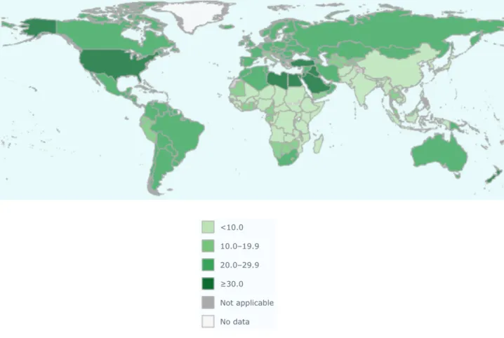
Research 2016; World health organization. Obesity: preventing and managing the global epidemic. Report of a WHO Consultation WHO Technical Report Series 2000). Global distribution of the prevalence of overweight and obesity is reported in Figures 1 and Figures 2. Figure 1: Graphical representation of overweight prevalence among male and female adult global population (http://www.who.int/en/).
Obesity, and other metabolic disease as Diabetes Mellitus (DM), has gained even more attention as a relevant and increasing public health burden, swiftly reaching global epidemic proportion, that embrace not only the economic sphere but also the social one. A recent study revealed disquieting evidences regarding the prevalence of obesity in the adult and young population (Ng M et al. Lancet 2014).
Figure 2: Graphic represents global prevalence of obesity among male and female adults
(http://www.who.int/en/).
Data collected showed that the prevalence of obesity has increased dramatically since 1980 involving both men and women (Ng M et al. Lancet 2014, Flegal KM et al. JAMA 2016). Despite in developed country such increasing trend seems to have reached the peak and evolved toward a plateau, in developing country obesity prevalence is still rising. Higher rates of obesity and overweight have been observed in the United States, Middle-East, Central America, Pacific and Caribbean Islands with proportions that in some cases broadly exceed the 50% for men or women. In
Western Europe as well as in Canada the prevalence of obesity reaches about 20% (Ng M et al. Lancet 2014).
The most dramatic evidence concern data regarding children and adolescents. In the last three decades, the incidence of overweight and obesity among younger population has dramatically increased (Bhupathiraju SN et al. Circ Res 2016, Ogden CL et al. JAMA 2016). Singer and Lumeng have recently reviewed the critical stage of obesity development during childhood emphasizing the note that obesity is a lifespan, chronic disease which have repercussions in adulthood health (Singer K et al. J Clin Invest rev 2017). The most recent data, describe that in the last 20 years the prevalence of obesity in young people has continued to rise. The more children become heavier, the more is the possibility to develop cardiovascular complications (Baker JL et al. N Engl J Med 2007) and it has been demonstrated that development of obesity in childhood has important repercussion in adulthood with earlier onset of cardiovascular disease (Agirbasli et al. Card Ther 2016). For these reasons, it is imperative the need of deep and massive efforts to counteract such global issue. Overall the main causes of the uncontrolled increase of obesity prevalence in both developed and developing country, could be ascribed to the progressive change in human habits towards a more sedentary lifestyle associated with higher caloric food consumption, contributed to the spreading of this systemic and crippling disorder (DeMarco VG et al Nat rev endocrinol 2014). Nevertheless, obesity is an entangled disease that cannot be ascribed solely to derangements in caloric intake and energy expenditure but different causal aspects, spanning from genetic to environmental ones, occur to determine this complex disease.
1.2 Obesity and Metainflammation.
The term "metabolic syndrome" summarizes a variety of different disease in which obesity is a central feature and responsible for the increasing risk for other pathologies development as insulin resistance and type 2 diabetes, respiratory diseases as Asthma, some type of cancer, neurodegenerative disease as dementia and importantly, cardiovascular diseases including atherosclerosis, stroke and hypertension.
In general, fat pad is the tissue designated to store energy that is released in response to metabolic need. Evolution, in fact, has positively selected mechanisms that allow accumulation of energetic molecules to deal with critical condition, as starvation or limited food availability, to preserve homeostasis (Hotamisligil GS et al. Nature 2017). On the other hand, however, no system or molecular pathway has been positively selected to face energetic overabundance, and its uncontrolled intake brings irreversibly to a pathological environment. Figure 3: Obesity is a pivotal risk factor for the development of a wide range of metabolic associated diseases. Schematic overview of metabolic disease cluster associated with obesity (Hotamisligil GS et al. Nature 2017).
Increase in size and structural alteration along with switch of cellular properties are critical features that characterize adipose tissue in the initial stages of obesity development (Lumeng CN Diabetes 2008).
Immune system assumes a critical and indispensable role presiding responses to a wide variety of stimuli that have to be activated to ensure a fast, effective and incisive reaction to protect host from enemy injury but, at the same time, that need to be turned off to prevent deleterious long-lasting immune reaction.
Classically, in order to counteract noxious challenges, immune system evokes a concerted and well-tuned response to restore a physiological environment. As a result, when the harmful agent has been eradicated, that vigorous but short-term immune activation returns to basal level, re-establishing the correct immune homeostasis. Differently, when the immune response is not able to overwhelm the damaging injury, the prolonged and not resolved immune activation could lead to worse outcome. In this view, metabolic disease, present features of inflammation that are completely different from the classic inflammatory paradigm lacking the presence of "dolor, rubor, calor and tumor". The “chronic” but “low grade” inflammation, has profound consequences in the critical derangement of metabolic homeostasis (Hotamisligil GS Nature 2006; Lumeng CN J Clin Invest 2011). Such pathological condition has been renamed as "meta-flammation" defining the metabolically triggered inflammation.
Importantly, prolonged and unresolved inflammation in metabolic tissue as fat pad, muscle and brain, is a key component that depict metabolic disorders especially obesity, type 2 diabetes and cardiovascular disease. The interest in unveiling the way in which metabolism affects immune system and conversely how immune system is involved in metabolic pathway regulation gave rise to a new field of investigation: immunometabolism (Cildir G et al. Trends Mol Med 2013).
Globally, fat pad can be divided in three categories: brown, beige and white that differ in body distribution, metabolic activities and function; each of these in turn can be subdivided in different subcategories. Among white adipose tissue (WAT),
increased visceral adipose tissue (VAT) has been identified to be a risk factor for insulin resistance and cardiovascular disease (Cildir G et al. Trends Mol Med 2013, Lim S et al. Int J Cardiol 2013).
Adipose tissue contains not only adipocyte, but also harbors pre-adipocytes, endothelial cells and, importantly, immune cells. Additionally, adipose tissue releases in the blood stream important molecules as chemokines, adipokines and chemokines that participate in maintaining homeostatic environment or promote pathological development through an endocrine or paracrine functions.
Both resident and infiltrating immune cells in the adipose tissue are conditioned by metabolic alteration and, in turn, can stimulate the worsening of metabolic derangements. For that reason, dysfunction in adipose tissue caused by infiltrating immune cells, is a critical aspect of hypertension or other cardiovascular disease. In 1993, the association between a component of the immune system (Tumor necrosis factor-alpha, TNF-α) and obesity-induced insulin resistance was demonstrated for the first time in a mouse model of obesity (Hotamisligis GS Science 1993), with increased proportion of TNF-α production during obesity development. Some years later, in a study conducted in humans, it was demonstrated that adipose tissue of obese individuals produce more TNF-α, and this occurrence was associated with insulin resistance (Hotamisligil GS J Clin Invest 1996) and increased BMI (McLaughlin T et al. J Clin Invest 2017).
In a short time, it has become even more clear the pivotal role that immune system assumes in the fine regulation of metabolic homeostasis.
During the onset of obesity, fat pad tissue undergoes profound alterations involving its structural and functional properties along with, and even more significantly, resident and recruited immune cell phenotype.
In healthy physiological condition, adipose tissue immune cells are all focused in maintaining the balanced environment. M2 macrophages and T lymphocyte regulatory cells, known as Treg cells, promote the anti-inflammatory environment with primary production of interleukin-10 (IL-10) and transforming growth
factor-beta (TGF-b), or other as IL-4, IL-5, IL-13 from Th2 aiming at constraining tissue inflammation (Winer S et al. Nat Medicine 2009, Lumeng CN et al. Diabetes 2008, Adamson SE et al J Clin Invest 2016). Fine regulation and production of anti-inflammatory mediators to maintain stable homeostasis is not ascribed only to macrophages or Treg cells but also to a proportion of IL-10 producing innate-like B cells and Adipose natural regulatory B cells (Breg) that populate visceral adipose tissue of healthy individuals (Nishimura S et al. Cell Metab 2013, Wu L et al. PNAS 2014).
On the other side, increased adipose tissue and development of obesity promote the recruitment of pro-inflammatory M1 macrophages overwhelming protective M2 macrophages and organizing in characteristic crown like structures that surround necrotic adipocytes (Lumeng CN et al. JCI 2007, Cildir G et al. Trends Mol Med 2013, Apovian CM et al. ATVB 2008, Kranendonk ME et al. Atherosclerosis 2015, Zheng C et al Cell Death Dis 2016). Pro-inflammatory macrophages sensing environmental inflammatory cytokines as IFN-g and in turn are stimulated to produce pro-inflammatory mediators as TNF-α, IL-1β, IL-6 and nitric oxide (NO) (Winer DA et al. Nat Med 2011). The sustained accumulation of macrophages, and consequential production of inflammatory molecules, promote the recruitment of other pro-inflammatory cells reinforcing inflammation and metabolic disease development. Proinflammatory CD8+ and CD4+ T cells along with Natural killer (NK) cells produce IFN-g participating to increase pathological inflammatory environment (Winer S et al. Nat Medicine 2009, Wensveen FM Nat Immunol 2015, Lee BC et al. Cell Metab 2016).
Moreover, evidences in DIO mouse models and human study highlighted the role played by Dendritic cells in presenting antigen peptide to T cells promoting their differentiation (Cildir G et al. Trends Mol Med 2013) and finally it has been reported in murine models that Neutrophils are recruited in the earliest phase of high-fat diet feeding (Elgazar-Carmon V et al. J Lipid Res 2008).
It is even more clear that both vascular and metabolic dysfunction with immune derangements are pivotal aspect that altogether occur in vascular diseases (Guzik TJ Cardiovas Res 2017) 1.3 Cardiovascular diseases: a focus on hypertension.
Important analyses defined blood pressure elevation, overweight and obesity, high cholesterol levels as some of the most critical causes of cardiovascular insults (Ezzati M et al. Lancet 2003). Cardiovascular disease are the most diffused chronic diseases that affect human population all over the world with higher prevalence among developing countries than the already developed ones. It has been estimated that one third of deaths are related to cardiovascular dysfunctions and high blood pressure is responsible in large proportion.
Even more evidences correlate blood pressure with the risk of incidence with a variety of comorbidities including kidney- and cardiovascular disease as stroke or hypertension. Hypertension is defined as a chronic increase in blood pressure, promoting end-organ damage and resulting in increased mortality. The latest categorization refers that normal blood pressure levels should be lower than 120 mmHg for systolic blood pressure (SBP) and 80 mmHg for diastolic blood pressure (DBP). Hypertension is defined with SBP higher than 130 mmHg and higher than 80 mmHg for DBP (Whelton PK et al. Hypertension 2017).
Great efforts have been spent to investigate the pathophysiological mechanisms underlying the pathogenesis of hypertension.
The causes of blood pressure increase, in the most part of cases, remain obscure. The term “secondary hypertension” refers to hypertension for which the causes have been established. For the other cases, in which no causes are known, that are the main percentage, are referred to “essential hypertension”. Arterial blood pressure is
subjected to continuous fluctuation and is constantly modulated by multiple factor (Figure 4): gene variants and interactions, the environment and at physiological level, neuronal and hormonal factors contribute to the control of blood volume, cardiac output and vascular resistance (Cowley Nat Rev Genet 2006).
Figure 4: Schematic summary of arterial blood pressure regulation pathway. (Arrows
represent function exerted, Dotted lines represent negative feedback control) (Cowley AW et al. Nat Rev Genet 2006) Renin-angiotensin–aldosterone system (RAAS) is probably one of the most essential blood pressure control system involving endocrine components. Since its pivotal role, already little derangements related to this pathway could lead to the development of hypertension. Firstly, inactive pro-renin is produced by kidney and released in the blood stream where is converted into renin, its active form (Danser AH et al. Nephrol Dial Transplant 2007). Now renin is responsible for the converting reaction from
Angiotensinogen to the inactive Angiotensin I (Ang I). At this point, Ang I can be converted by the Angiotensin–converting enzyme (ACE) in Angiotensin II (Ang II) (Figure 5). Ang II is the principal active molecule in RAAS pathway promoting important rise in blood pressure since its vasoconstriction effect and being involved in short-term but also long-term modulation of blood pressure (Munoz-Durango N et al. Int J Mol Sci 2016; Hall JE et al. Compr Physiol 2012). The more Ang II is produced, the more renal excretory activity is impaired with pressure natriuresis derangements. Angiotensin-(1-7) is an active product that derives from the cleavage of Ang I by specific endopeptidases or the cleavage of Ang II by angiotensin-converting-enzyme 2 (ACE-2). Angiotensin (1-7) gained important attention since its protective and opposite effect from Ang II on cardiovascular system (Santos RA Hypertension 2014). Aldosterone is another important effector involved in the RAAS pathway. Production and release of Aldosterone from the adrenal gland is stimulated by Ang II increase. Effects of this hormone are directed to modulate blood pressure mediating sodium and water retention and potassium excretion (Spat A and Hunyady L Physiol Rev 2004). Aldosterone, indeed, binds mineral corticoid receptors, activating transcription of genes involved in electrolyte and fluid homeostasis (Munoz-Durango N et al. Int J Mol Sci 2016).
Thus, it is clear the pivotal role exerted by RAAS system in blood pressure control. It is even more evident that such system needs to be finely balanced since expression impairments in any of these molecules could lead to damaging consequences including hypertension and alteration of organ structure and function.
Different therapeutic approaches have been developed to reduce RAAS activity targeting Ang II receptor, ACE, or mineralcorticoid-receptor (MR) promoting a correct sodium homeostasis aiming at reducing blood pressure level (Hall JE et al. Compr Physiol 2012). Alternative therapeutic strategies could involve Angiotensin-(1-7) enhancing its protective effects (Santor RA Hypertension 2014).
Figure 5: Schematic description of the renin-angiotensin-aldosterone-system pathway
(Munoz-Durango N et al. Int J Mol Sci 2016).
1.4 Obesity, Cardiovascular disease and Sympathetic nerve activity.
An epidemiological study focused on obesity, thoroughly reported the strong relationship with increased risk of heart failure or hypertension (Prospective study collaboration Lancet 2009), being an independent risk factor for cardiovascular disease affecting independently both men and women. Among obese patients, the prevalence of hypertension exceeds 60%. The incidence of hypertension is positively related to BMI and even just a little increase could be responsible of higher risk to develop hypertension. Long-term prospective study has highlighted that individuals
developing weight gain or obesity during early phase of mature age and middle-age were subjected to higher risk for the onset of hypertension. Overall, the study focuses on the fact that maintaining a normal controlled weight confers a lower risk to develop cardiovascular disease (Shihab HM et al. Circulation 2012). In this way, clinical trials have shown that reducing body weight through improved eating habits or physical activity, is a compelling behavior to prevent hypertension or reduce blood pressure level in hypertensive patients (Neter JE et al. Hypertension 2003).
The abnormal pathophysiological changes that endure adipose tissue in obesity, promoting an aberrant expression of a wide range of inflammatory mediators, firstly affect systemic metabolism and particularly contribute to the development of cardiovascular diseases. It is also clear that different fat depots have dissimilar consequences on the possibility to develop cardiovascular disease as confirmed by numerous studies conducted on humans and animal models, attributing to visceral adipose tissue a critical role in insulin resistance and cardiovascular disease development (Fuster jj et al. Circulation research 2016, Sironi AM et al. Hypertension 2004).
Adipose tissue not only harbors immune cells that secrete pro- or anti-inflammatory immune modulators, but it also produces and releases proteins called adipokines (Fuster JJ et al. Circ res. 2016, Lori A. et al. IJMS 2017) that are involved in the regulation of multiple functions including appetite, energy expenditure, glucose metabolism, blood pressure and inflammation. The majority of Adipokines, such as Leptin, TNF-α, IL-6 and resistin and others, are characterized by pro-inflammatory activity (Fuster JJ et al. Circ res. 2016, Ouchi N et al. Nat rev immunol 2011, Nakamura K et al J Cardiol 2014). On the contrary, adipose tissue produce a smaller quantity of anti-inflammatory adipokines as Adiponectin or Secreted frizzled-related protein 5 (Sfrp5) (Fuster JJ et al. Circ res. 2016, Ouchi N et al. Science 2010). Some of them, play their role reaching the Central Nervous System, influencing neuron activity in distinct brain areas. This is the case of Leptin. Leptin is a hormone highly and specifically expressed by adipose tissue. Its production is positively related to the
amount of fat pad, thus circulating levels are highly increased during obesity, developing, as a consequence, leptin resistance (Maffei M et al. PNAS 1995). The principal action of this adipokine is related to the control of energy balance influencing appetite: increased level of leptin suppresses appetite and enhances energy expenditure resulting in weight loss (Rahmouni K et al. Hypertension 2007). Crossing the Blood Brain Barrier, Leptin reaches the Central Nervous System exerting its function on feeding behavior and body weight (Zlokovic BV et al. Endocrinology 2000, Malpas SC et al. Physiol rev 2010). The regulation of this function is addressed in the mediobasal hypothalamic arcuate nucleus to the melanocortin system. Here two neuronal population: the orexigenic agouti-related peptide (AgRP) and neuropeptide (NPY) neurons and the anorexigenic proopiomelanocortin (POMC) neurons, modulate feeding behavior and energy expenditure in response to metabolic stimuli, with opposite function (Jais A et al. JCI 2017, Belgardt BF et al. Ann N Y Acad Sci 2010).
Besides this function, leptin has also pro-inflammatory action and its aberrant overproduction promotes cardiovascular disease development (Fuster JJ et al. Circ res. 2016). Despite during obesity the effects of leptin on energy balance is lost caused by leptin resistance, its ability in activating sympathetic nerve activity is preserved with detrimental consequences for the cardiovascular system increasing arterial pressure (Harlan SM et al. Circ res 2011, Rahmouni K et al Diabetes 2005, Malpas SC Physiol rev 2010). Such notion is corroborated by the evidence that acute infusion of Leptin in obese MICE induced renal sympathetic nerve activity increase but, conversely, the Leptin anorexigenic property was blunted (Mark AL. et al. J. Hypertension 2002).
A critical factor that is involved in blood pressure rise during obesity is the sympathetic nervous system overdrive and many interesting works have revealed the close relationship between weight increase and sympathetic nerves activation (Mark AL et al. Hypertension 2009, Hall JE Circ res 2015). Sympathetic nerves reach important organs as kidneys modulating its functions. Significantly, increased
sympathetic discharge to these organs has been related to the onset of hypertension (Rahmouni et al. Hypertension 2014). It has been widely demonstrated, with different animal model of obesity (i.e. diet-induced obesity on rabbits (Lim K et al. Hypertension 2016), mice (Chhabra H et al. Mol Metab 2017) or rats (Muntzel MS et al. Hypertension 2012), the involvement of Leptin in the over-activation of sympathetic tone (Grassi G et al. Circ Res 2015). Moreover, researchers highlighted the fact that obesity enhances sympathetic activity that plays a crucial role in hypertensive disease development (Rahmouni et al. hypertension 2014, Carnevale et al Nat Commun 2016, Simonds SE et al. Cell 2014).
Evidences from both diet induced obesity mouse models and human studies show that the melanocortin system is involved also in blood pressure regulation during obesity (Rahmouni K et al. Hypertension 2007, Purkayastha et al Nat Med 2011); abrogating Leptin-POMC neuron signaling pathway, in fact, protects against obesity-induced hypertension (Do Carmo et al. Hypertension 2011). The action of Leptin on renal sympathetic nerve activity in the central nervous system is not restricted to the arcuate nucleus, rather different research groups have characterized similar effects also in the brain subfornical organ (Young CN et al. Hypertension 2013) demonstrating that different brain areas modulate leptin metabolic and cardiovascular effects.
1.5 Placental growth factor: much more than an angiogenic factor.
Placental growth factor (PlGF) was firstly determined and cloned in 1991, from human placenta library of cDNA (Maglione D et al. Proc. Natl. Acad. Sci. 1991) and belongs to the family of the Vascular Endothelial Growth Factors (VEGFs). Besides PlGF, other structurally related members fall into the same family as VEGF-A, VEGF-B, VEGF-C, VEGF-D and VEGF-E (Dewerchin M et al. Cold Spring Harb Perspect Med 2012). Similarly to all VEGF family members, human PlGF proteins exist in different
isoforms (PlGF-1, PlGF-2, PlGF-3 and PlGF-4) due to alternative splicing processes. Differently, only the isoform PlGF 2 is encoded by murine PlGF gene (De Falco S et al. Exp Mol Med 2012). The importance of this family members is related to the main function of vasculogenesis and angiogenesis during pregnancy, tissue growth, wound healing but are also critically involved in the development of important diseases as cancer or amyotrophic lateral sclerosis. All members exert their activity assembling as dimer proteins but PlGF, along with VAGF-A and VEGF-B, can also generate heterodimeric structures (De Falco S et al Trends Cardiovasc Med 2002).
VEGF-A (or VEGF) was the first discovered member of VEGF family exerting its primary function in vascular growth. VEGF-A is an essential gene since its experimental deletion or blockade was lethal already at embryonic developmental stage (Carmeliet P et al. Nature 1996, Ferrara N et al. Nat Med 2003) promoting angiogenesis derangements. Its importance has been clarified also subsequently to the embryonic phase at neonatal development stage through severe kidney glomerular impairments (Ferrara N et al. Nat Med 2003). On the other side, VEGF antibodies administration in nude mice was effective in inhibiting progression of different hematological malignancies (Ferrara N et al. Nat Med 2003; Goel HL and Mercurio AM Nat Cancer rev 2013). Thus, the relevance of VEGF actions at different stages of individual development suggests that its expression must be regulated in spatial and temporal manner.
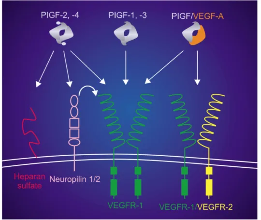
VEGF-A, VEGF-B and PlGF, exert their biological function binding specific type of related tyrosine kinase receptor: VEGF receptor-1 (VEGFR-1 also named as Flt1) and VEGF receptor-2 (VEGFR-2 or Flk1) (Autiero M et al. J Thromb Haemost 2003) (Figure 6). The dimeric form of VEGF-A binds VEGFR-1 and VEGFR-2 but the homodimeric form of PlGF and VEGF-B are able to bind only VEGFR-1 receptor (De Falco S et al. Exp Mol Med 2012). Finally, co-receptors, Neuropilin-1 and Neuropilin-2, have been described as important mediators of VEGF-A and PlGF receptor interaction (De Falco S et al. Trends Cardiovasc Med 2002; Ferrara N et al. Nat Med 2003) (Figure 1).
VEGFR-1 and Neuropilin-1 have been identified also as soluble receptors as a consequence of mRNA alternative splicing. The presence of soluble receptor is of fundamental importance for the spatial and temporal regulation of VEGF-A and PlGF, generating gradients necessary for the correct development of the organism, modulating their locally as well as systemically action.
Figure 6: Schematic model of PlGF isoforms homodimers and PlGF-VEGF heterodimer
binding affinity with Neuropilin 1 and 2, VEGFR-1 and VEGFR-1/VEGF-R2 receptors (De Falco S et al. Exp Mol Med 2012). Continuous insights in PlGF function, highlighted the fact that PlGF could play a role in VEGFR-2 activation through different modalities. First of all, Carmeliet et al. observed that the affinity of PlGF for VEGFR-1 could displace VEGF-A allowing its interaction with VEGFR-2 (Carmeliet P. et al. Nat Med 2001). In second instance, in studies
related to tumor development and angiogenesis, PlGF and VEGF-A could co-operate forming heterodimer. In such conformation, PlGF-VEGF interacts with both VEGFR-1 and the dimer VEGFR-1/VEGFR-2 (Tarallo V et al. Cancer res 2010; Autiero M et al. Nat Med 2003). Lastly, VEGFR-2 could be activated through transphosphorylation in response to PlGF-VEGFR-1 interaction with the consequence of further enhancing VEGF-induced angiogenesis (Autiero M et al. Nat Med 2003).
Since its discovery, PlGF was found expressed in several different cells and even more works concurred to increase knowledge about its functions. The first purpose in which PlGF was described was related to angiogenesis. Ziche et al., taking advantage of both “in vivo” and “in vitro” esperiments, demonstrated for the first time that PlGF was able to induce angiogenesis and moreover to enhance endothelial cells migration and proliferation (Ziche M et al. Lab Invest 1997). Some years later, genetic manipulation allowed to generate PlGF-deficient mice and it was observed that these mice were healthy and fertile with a normal vascular development (Carmeliet P et al. Nat Med 2001). Thus, since PlGF is expressed at early stage during embryonic development, its deficiency overall does not affect systemic development.
Other functions in which PlGF was found being involved concern the stimulation of mesenchymal fibroblast proliferation and migration; the derangemnts in dendritic cell differentiation along with antigen recognition; it is involved in neuronal devolpment promoting generation of Schwann cells and play important role in cancer growth (Dwerchin M and Carmeliet P Cold Spring Harb Perspect Med 2012). A schematic representation of PlGF expression and involvement in different aspect of cellular functions (Figure 7).
Figure 7: Schematic overview of PlGF pleiotropic action ranging from proliferative regulation to cellular migration and activation affecting both vascular and non-vascular cells (Dwerchin M and Carmeliet P Cold Spring Harb Perspect Med 2012).
In addition to these properties, PlGF has been also characterized as an endothelial cells and monocytes chemotactic factor, acquiring the property of “angiogenic cytokine” modulating both angiogenesis and inflammatory response during cardiovascular disease (Carnevale D and Lembo G Trends Cardiovasc Med 2012). In light of this, evidence from mouse model of cardiac remodeling induced by Toracic Aortic Coarctation (TAC) revealed that PlGF expression is increased and again PlGF is pivotal in modulating inflammatory activation providing the correct inflammatory environment to allow adaptive cardiac remodeling (Carnevale D et al Circulation 2011). Conversely, PlGF deficiency caused inflammatory derangements with aberrant
cardiac compensatory hypertrophy incurring irretrievably in heart failure (Carnevale D et al Circulation 2011).
Such evidence highlighted the importance of a fine balancing of immune response not only on metabolic syndrome but also in cardiovascular disease and PlGF gained even more importance, beyond its angiogenic properties, for a pivotal immune regulator.
1.6 Neuro-immune modulation of hypertension.
A great amount of studies unraveled the role carried out by immune cells for the onset of hypertension (Harrison DG J Am Soc Hypertens 2013, Madhur MS and Harrison DG Circ Res 2012, Guzik TJ et al. J. Exp. Med. 2007). Mouse model of hypertension induced by Angiotensin II infusion was used to decipher the role exerted by T cells. Mice lacking both T and B cells were used by Guzik et al. to demonstrated that T cells infiltration in target organs of hypertension as the aortic adventitia are essential to promote blood pressure rise (Guzik TJ et al. J. Exp. Med. 2007). More recently, valuable experiments based on chimeric mice with splenic transplantation clarified, for the first time, that renal and vascular infiltrating T lymphocytes induced by chronic Angiotensin II infusion, were of splenic origin (Carnevale et al. Immunity 2014). Otherwise, It was demonstrated from different works that Deoxycorticosterone acetate (DOCA)-salt stimulate the Renin Angiotensin system activating Angiotensin II receptors in the brain Subfornical organ (Hilzendeger et al Hypertension 2013) and that the same hypertensive stimulus is able to enhance sympathetic drive in the spleen (Carnevale et al. Nat Comm 2016). Conversely, the consequences on splenic immune activation evoked by different known hypertensive stimuli, such as high-fat diet, has not been fully clarified.
The interplay between immune- and nervous-system was further demonstrated with other evidence suggesting a role for the cholinergic signaling pathway in mediating immune response, showing that galantamine, an acetylcholinesterase inhibitor, was able to hamper the expression of both Tumor necrosis factor (TNF) and IL-6 with anti-inflammatory effects in endotoxemic mice (Pavlov VA et al. Brain Behav Immun. 2009) and, moreover, lowered inflammation, body weight, Insulin resistance along with other aspects related to metabolic syndrome in mouse model of high-fat diet induced obesity (Satapathy et al. Mol Med 2011). These aspects of neuro-immune interaction in metabolic syndrome deserve deepen study to unravel possible implication with cardiovascular diseases.
A possible link between metabolic syndrome, neuro-immune response and hypertension could be achieved by the Placental growth factor (PlGF).
First evidence of the importance of PlGF in modulating immune response in mouse model of cardiovascular disease were described by Carnevale et al. in 2011 studying PlGF in cardiac response to transverse aortic constricton (TAC). Since the peculiar features of PlGF, as on one hand an angiogenic factor and on the other acts as a cytokine, was demonstrated that it is responsible for correct and fundamental inflammatory response after TAC (Carnevale et al. Circulation 2011). PlGF-deficient mice, in fact, showed lower monocytes activation and inflammatory cytokine expression and consequential marked worsening of both structural and functional cardiac parameters (Carnevale et al. Circulation 2011).
In 2014, Carnevale et al. clearly described for the first time a new role for the splenic factor PlGF in modulating Sympathetic nervous system and immune cells in response to a hypertensive stimulus as chronic infusion of Angiotensin II. PlGF-deficiency protected mice from the development of hypertension. The importance of the spleen and the splenic PlGF is highlighted by experiments in which splenectomy or selective ablation of sympathetic innervation to the spleen through celiac ganglionectomy protected mice from blood pressure rise as well, realizing that the splenic PlGF is pivotal for hypertensive disease development (Carnevale et al. Immunity 2014).
PlGF was found highly expressed along the marginal zone of the spleen in response to Angiotensin II infusion and its activation is necessary for T cell stimulation and egression from the splenic reservoir toward the classical target organs of hypertension: kidneys and aorta (Carnevale et al. Immunity 2014).
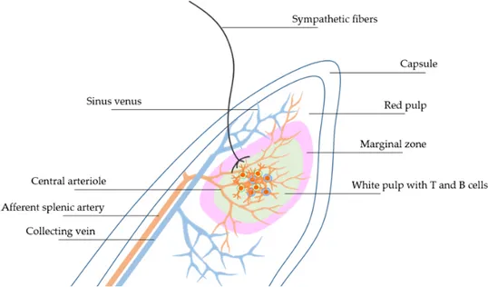
1.7 The Spleen: anatomical and functional properties for neuro-immune interaction.
Anatomically, the spleen is located into the abdomen, under the diaphragm and in contact with the stomach. Despite initially was depicted as a simple blood filter, the spleen has gained even more attention among the scientific community since it exerts a previously unrecognized role in immune system modulation. The spleen is encircled by a capsule made up of elastic and fibrous tissue. Starting from the capsule derive the trabecule, constituted by connective tissue, directed to the parenchyma of the spleen and that encompass nerves lymphs and blood vessels. The capsule together with trabeculae sustain the spleen conferring solidity to the structure and an organized environment (Cesta MF Toxicol Pathol 2006; Mebius RE and Kraal G Nat Rev Immunol 2005; Lori A et al. Int J Mol Sci 2017). Central arterioles originating from splenic artery spread through the trabeculae structure toward the splenic White Pulp (WP) (Lori A et al. Int J Mol Sci 2017). The WP is configured as a lymphoid structure in which both T-cells and B-cells are confined in specific areas near arterial vessels in response to chemoattractant molecules (Mebius RE and Kraal G Nat Rev Immunol 2005). Splenic marginal zone (MZ) is a critical area that is placed at the interface between WP and blood circulation. Along the MZ reside different population of immune cells that play active role in the surveillance against blood circulating antigens (Magri G et al. Nature immunology 2014). Blood that flows through the MZ reach the Red Pulp (RP) and venous sinuses where important function of blood filtration is exerted by specific macrophages subtracting old
erythrocytes, promoting reaction against bacterial infiltration and eventually iron recycling. Hereafter, blood flow through trabecular area directed toward the splenic vein (Lori A et al. Int J Mol Sci 2017) (Figure 8).
Another relevant immune activity that occurs in the RP is the production of antibodies. Plasmablast, firstly differentiate within the WP in response to specific antigen. Plasmablast overexpress CXC-chemokine receptor 4 that specifically binds the CXC-chemokine ligand 12 (CXCL12) highly produced in the RP and involved in mmigration and maintenance of cells in this specific zone of the spleen. In parallel, the repression of CXCR5 and CCR7 receptors on plasmablast along with the reduced response to the WP homing chemokine signals (Mebius RE et al Nat Rev Immunol 2005; Hargreaves et al. J exp Med 2001).
The splenic lymphoid area, residing in the WP, need a strictly and finely regulated system to preserve its cellular distribution. CXCR5 specific B-cell receptor, is fundamental for the B-cell follicles arrangement allowing B cell differentiation.
Likewise, CCL19 and CCL21 CC-chemokines ligands force both T-cells and Dendritic Cells (DCs), that specifically express CCR7 receptor on their membrane surface, into the WP T-cell zone (Lori A et al. Int J Mol Sci 2017). Derangements in the pattern expression involving these chemokine or related receptor, could result in aberrant immune cells localization from their appropriate splenic place (Mori S et al. J exp med 2001; Ramos-Perez WD et al Nat immunol 2015).
In the end, MZ is the connecting structure placed between circulating blood in the RP and the immune reservoir in the WP. Such configuration provides an efficient system to protect from blood-derived pathogens through relevant production of immunoglobulins (IgA, IgM, IgG) from B-cells producing B-cells (Puga I et al. Nat Immunol 2012). In addition to their action as Ig producers, B-cell could also act as an antigen presenting cell (APC) able to activate CD4+ T cells (Rubtsov AV et al. J Immunol 2015).
Overall, besides B-cells, splenic MZ is populated by a wide variety of immune cells: Neutrophils, Dendritic cells and Macrophages conferring a complete and effective immunosurveillance (Puga I et al. Nat Immunol 2012; Gatto D et al. Nat Immunol 2013; Ravishankar B et al. Proc Natl. Acad. Sci. U S A. 2014)
Since the last two decades, researchers made several steps forward in unveiling the basis of nervous-immune system communication. Neural mechanisms are pivotal regulators of several biological systems including the inflammatory reflex (Rosas-Ballina M Tracey KJ Neuron 2009; Carnevale D et al. Immunity 2014; Carnevale D et al. 2016 Nat comm 2016; Cole SW et al. Nature rev cancer 2015). Great efforts have been focused on the characterization of the splenic innervation; indeed, it has been described that fibers of the sympathetic nervous system innervate the spleen and that their synapses are in closely proximity with immune cells (Carnevale D et al. Nat comm 2016; Rosas-Ballina M et al. Science 2011; Andersson U Tracey KJ J Exp Med 2012; Lori A et al. Int J Mol Sci 2017) (Figure 8).
Neurotransmitters from the sympathetic nerves bind specific receptor on immune cell surface, generating a “synapse-like contact”, and influencing their activity in promoting or decreasing cytokine expression (Carnevale D et al. Immunity 2014; Straub RH et al. Trends Pharmacol Sci 2004; Lori A et al. Int J Mol Sci 2017). Evidences showed that immune cells, in turn, can influence Noradrenaline (NA) release from sympathetic synapses and also in that case enhancing or reducing its production. Immune cells release of adrenocorticotrophic hormone (ACTH) on one hand and IL-6, IL-2 or TNF on the other induce opposite consequence on NA production acting on pre-terminal autoreceptors on the neuronal cell surface (Straub RH et al. Trends Pharmacol Sci 2004). For these reasons, the neuro-immune interplay consists of a highly intricate but extremely well-tuned regulating processes.
Figure 8: Schematic description of the architectural organization of splenic areas containing various immune cells: White Pulp (WP) and Red Pulp (RP) with Sympathetic nerve fibers innervating Marginal Zones (MZ) (Lori A et al. Int J Mol Sci 2017). Overall, all these evidences support the fact that the spleen is an important lymphoid organ that hosts both component of the innate and adaptive immune system providing an essential role in immunosurveillance. Moreover, the neuro-immune interaction between sympathetic fibers and the splenic immune cells is of key relevance to understand novel mechanisms in the pathogenesis of hypertension (Carnevale D et al Immunity 2014; Carnevale D et al. Nat Comm 2016).
1.8 Animal model of diet-induced obesity.
In order to understand the pathophysiological aspect of human diseases, developing animal models are an unavoidable need. Moreover, the importance to dissect disease-associated complications and the possibility to deepen the molecular pathway that manage the pathological onset or progression of the disease are of critical relevance. Progressive and continuous developments enabled to critically discover biological features of a wide range of disease. The first goal, would be to be able to translate such new insight from the bench to the bedside for new and more powerful therapeutic treatment.
The significance to generate animal model for studying disease also resides in the opportunity to produce in vivo studies and deepen the complexity of many aspects in an entire biological system. Animal models, indeed, are chosen because they share extensive similarity with humans concerning physiological, anatomical and genetic aspects.
Rodents, and in particular mice and rats, are probably the most attractive animal model to study for metabolic and cardiovascular disease. The possibility to modify genome sequences through single nucleotide modification, or even the insertion or deletion of a DNA section, originating protein with loss of function, gain of function or completely abrogate their production, gave rise to the generation of genetic mutant rodent models of pivotal importance for several different studies and therapeutic approaches with the most advanced discoveries in molecular medicine.
Concerning metabolic disease and cardiovascular disease, mice and rats are probably the most appropriate, and studied, animal models.
A large amount of studies regarding metabolic syndrome focus on obesity being a central feature of this multifactorial disorder. Genetically modified mice, such as Leptin deficient mice (ob/ob mice) or mouse that carry a mutation in the leptin receptor gene (db/db mice) have been widely used to dissect obesity and its related comorbidities (Giesbertz et al. Diabetologia 2015; Drel VR et al. Diabetes 2006). Nevertheless, such genetic mutation, have been discovered only in a little part of
human population (O’Rahilly Nature 2009; Varga O. et al. Obesity Rev 2009; Simonds et al. Cell 2014). Thus, another model, that probably is more representative of the incidence in human population without involving genetic proceedings, is represented by diet-induced obesity. In general, most of the mice or rats fed with a high-fat diet progressively accumulate body mass becoming obese. However, it has been evidenced that different strains could manifest different responsiveness in term of weight gain or metabolic related dysfunction as glucose tolerance or insulin resistance (Rossmeisl et al. Diabetes 2003; Montgomery et al Diabetologia 2013). For example, C57BL/6J or AKR/J strains, despite different response in insulin resistance and glucose tolerance (Rossmeisl et al. Diabetes 2003) demonstrate more susceptibility to high-fat diet induced obesity. On the other hand, BALB/c background strain show great protection against glucose metabolism derangements induced by obesity (Montgomery et al Diabetologia 2013). Research based on these mouse model, and many others, have contributed and will provide even more insight to deepen metabolic disease, obesity and related cardiovascular complications (Calligaris SD et al Plos One 2013; Simonds SE et al Cell 2014; Asirvatham-Jeyaraj N et al Hypertension 2016; Weisbrod RM et al Hypertension 2013).
2. Aims of the study.
The aims of this study were focused on the following points: 1. To develop a murine model of diet-induced obesity. 2. To explore the relevance of Splenic Sympathetic Nervous System in the onset of obesity-induced hypertension.
3. To explore the involvement of Placentar Growth Factor in obesity development and onset of obesity-induced hypertension.
3. Methods.
3.1 Mice:
All the experimental procedures were performed according to European Communities guidelines and the Italian legislation on animal experimentation. The adopted protocol was approved by the Italian Ministry of Health and by our Institutional Committee.
Wild-type C57BL/6J mice were purchased from Charles River and used for sympathetic nerve recording, splenectomy and celiac ganglionectomy.
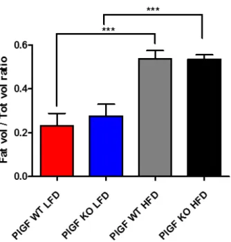
PlGF-deficient mice, from the original strain in 50% 129Sv/50% Swiss, were backcrossed for 11 generations in C57BL/6J mice as previously described (Carnevale et al. 2014, Gigante et al. 2006). As control, were used PlGF WT mice with the same genetic background (C57Bl/6J). 3.2 Diet-induced obesity paradigm:
At 6 weeks of age, all mice started the diet protocol for 16 weeks. Mice with free access to water, were fed a low-fat diet (LFD, 10% kcal fat; catalog #D12450J; Research Diets) or a high-fat diet (HFD, 60% kcal fat; catalog #D12492; Research Diets) ad libitum for 16 weeks (Figure 9). PlGF WT and knockout mice weight gain and diet intake were monitored weekly. Figure 9: Schematic overview of diet-induced obesity diet protocol. BW: body weight measurements, BP: blood pressure measurements.
3.3 Electrophysiological recording:
Electrophysiological recording was performed as previously described (Carnevale D et al. Nat. Comm. 2016). Mice were anaesthetized with 5% isoflurane and maintained with 1.5–2% (supplemented with 1 l min-1 oxygen). Blood pressure was continuously monitored with a single-pressure catheter (Millar, SPR-100) implanted in the left femoral artery and connected to a pressure transducer (Millar, MPVS ULTRA). Body temperature was monitored and maintained between 37 and 38 °C. After an abdominal incision, the intestinal tract was carefully moved exposing the splenic artery that was isolated, and the splenic nerve separated from tissue. A bipolar stainless-steel electrode (MLA1214 Spring Clip Electrodes, ADInstruments) was positioned on the splenic nerve. The ground wire was plugged into mouse soft tissues. Splenic SNA was recorded from the implanted electrode continuously for the next 2 h. Mice were then killed by isofluorane overdose, and Splenic SNA was recorded for a further 30 minutes to estimate post-mortem residual activity and background noise. The splenic nervous signal was amplified (gain 10,000) and successively sampled at 4 kHz with a digital amplifier (Animal Bio Amp; ADInstruments). The raw signal was filtered at 300–1,000 Hz and expressed as mV. Data were collected using a Power Lab data acquisition system and analyzed with Lab Chart 7 (Spike Analysis Module). The sympathetic burst was identified as a signal above the threshold determined by the background noise of the post-mortem recording. Firing frequency was calculated as the mean value of total number of spikes in a time window. Whereas the amplitude gain was evaluated as the mean spike amplitude and spike identification threshold ratio in the time window.
3.4 Blood pressure measurements:
Arterial blood pressure was measured by tail cuff system as previously described (Carnevale et al. 2014). Tail cuff plethysmography (BP-2000 Series II, Visitech Systems) was used to assess blood pressure between 9 am and 1 pm. Blood pressure
was measured for 5 consecutive days every 4 weeks since the beginning of diet procedure. 3.5 Celiac Ganglionectomy: Wild-type C57BL/6J mice at the age of 5 weeks, were anesthetized with Isoflurane (2-5 Vol %), supplemented with 1L/min oxygen. The abdominal cavity of mice was opened through a midline laparotomy, after Aorta and celiac artery isolation, the celiac ganglion was exposed and excised. As controls, sham wild-type C57BL/6J mice were equally anesthetized and celiac ganglion exposed but not removed.
3.6 Splenectomy:
Wild-type C57BL/6J mice at the age of 5 weeks, were maintained under isoflurane anesthesia (2-5 Vol%), supplemented with 1L/min oxygen. Abdominal cavity was opened and after accurate splenic vessel cauterization, the spleen was exposed and carefully removed. As control, was followed the same procedure in C57BL/6J mice at the same age, the spleen was exposed but not removed. 3.7 MicroCT: MicroCT scanning was performed with SKYSCAN 1178 (SKYSCAN, Kontich, Belgium), as previously described (Carnevale D et al. Immunity 2014). The tube parameters were set to 50 kV and 615 μA, exposure 480 ms, with a rotation step of 0.360°. Mice were anaesthetized with ketamine-xylazine and positioned on the bed. After image reconstruction the analysis was performed as follows: a ROI was placed from the top of the ribcage to the tail of the animal, the fat was segmented by thresholding in the range of [-300 ;-50] HU (Metzinger MN et al. Sensors (Basel) 2014), the global volume was segmented with an automated thresholding (Otsu) to discriminate background and foreground of an image. The numerical analyses were performed
using an in-house software based on OpenCV, the visual reconstruction was performed by Slicer3D (Fedorov A. et al. Imaging 2012). 3.8 Immunofluorescence analysis: Immunofluorescence analysis was performed as previously described (Carnevale D et al. Immunity 2014). Briefly, the spleen was collected and successively embedded in OCT. 25µm sections from spleen were post- fixed with 4% paraformaldehyde for 15 min and processed for staining with the following primary antibodies:
• Rat anti-CD169 (1:200; Serotec) specific for metallophilic marginal zone macrophages;
• Rat anti-ER-TR7 (1:200; Acris) specific for fibroblast reticular cells; • Hamster anti-CD3 (1:100; Serotec), specific for T cells labeling;
• rat anti-CD45R/B220 (1:50; BD Pharmigen) specific for splenic B cells labeling; • Rabbit anti-PlGF (1:250; Abcam) specific for PlGF labeling.
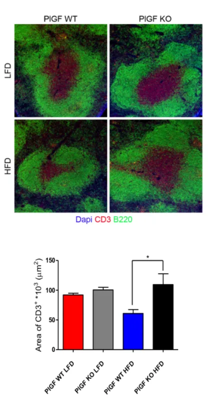
Sections were incubated with the appropriate secondary antibodies conjugated to Alexa Fluor 488 or Cy3. Thereafter, sections were coverslipped with DAPI-containing mounting medium (Vector laboratories) and then evaluated with Zeiss 780 confocal laser-scanning microscope. A 405 Diode laser was used to excite DAPI; a 488 nm argon laser to excite Alexa Fluor 488 and a 543 HeNe to excite Cy3. Quantitative analysis of white pulp area of the spleen was determined using Image J software (NIH), as previously described (Carnevale D et al. Nat Comm 2016). 3.9 Statistical analyses: Data are presented as mean ± SD. Statistical analyses were performed accordingly to the specific experimental design. P values were considered significant from p<0.05. All statistical analyses were conducted with Graphpad Prism software 7.0.
4. Results.
4.1 Generation of diet-induced obesity mouse model.
The first aim of this work was to generate a mouse model of diet-induced obesity. To achieve this goal, C57BL/6J mice starting from 6 weeks of age were fed with a high-fat diet (HFD) or a low-fat diet (LFD) as control for a time period of 8 weeks. At the beginning of the diet protocol, all mice weight ranged around 20 grams. Since the first weeks of HFD, mice became heavier and at the third week difference were already significant when compared to LFD controls mice, reaching around 30 grams at the eighth week of diet procedure. The difference between mice fed with the two diets were even more clear analyzing weekly body weight increment. The percentage increment in body weight was significant since the first week of diet (Figure 10). Figure 10: Body weight and Body weight increment in C57BL/6J mice fed with a HFD or a LFD for 8 weeks. Panel represent mean ± SD. (*** p<0.001 vs LFD controls), (n=8 per group).
In order to assess if HFD-induced obesity was able to promote the onset of hypertension, mice were analyzed for blood pressure increase with non-invasive tail cuff system.
Blood pressure was monitored every 4 weeks since the beginning of the diet protocol and we observed that at the eighth week C57BL/6J mice systolic blood pressure (SBP) was significantly increased compared to LFD-fed control mice. The same evidence was observed for diastolic blood pressure (DBP) (Figure 11). Figure 11: Blood pressure measurements: SBP and DBP in C57BL/6J mice fed with a HFD or a LFD for 8 weeks. Panel represent mean ± SD. (*** p<0.001 vs LFD controls), (n=8 per group).
Overall these data demonstrate that C57BL/6J mice upon HFD become significantly obese as compared to LFD-fed mice and importantly, mice develop blood pressure rise already after 8 weeks of HFD-feeding.
4.2 Diet-induced obesity induces Splenic Sympathetic Nervous System activation.
A great amount of studies has pointed out the relevance of Sympathetic Nervous System (SNS) activation in response to obesity (Hall JE et al. Circ Res 2015) and other hypertensive stimuli, as Angiotensin II and Deoxycorticosterone acetate (DOCA)-Salt (Carnevale et al. Immunity 2014, Carnevale et al. Nat Comm 2016), promoting the increase of blood pressure level.
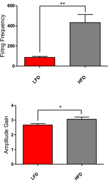
Once demonstrated the reliability of our mouse model of diet-induced obesity associated with hypertension development, the second aim was to explore the relevance of Sympathetic Nervous System at splenic district. To assess if HFD-feeding promotes increasing in Splenic Sympathetic Nerve Activity (SSNA), we performed nerve recording experiments directly on the splenic nerve in mice fed with HFD or LFD after 8 weeks of diet administration.
Sympathetic nerve discharges were recorded for a time period of 1 hour; meanwhile, blood pressure was monitored to ensure animal stability all over the nerve recording procedure.
In Figure 12, is shown a representative example of splenic nerve recording in both HFD-fed mice and LFD-fed controls.
We observed that HFD-fed mice had increased SSNA as compared to LFD-fed mice. Importantly, despite being under anesthesia, analysis of blood pressure monitoring in HFD-fed mice confirmed elevated levels as compared to LFD-fed mice.
Variation in SSNA was assessed with Firing Frequency and Amplitude Gain quantification in the same time window of 10 minutes for all mice. With the term Firing Frequency, we refer to the mean number of spikes that takes place in the
selected interval. Amplitude Gain, instead, refers to the mean amplitude increase of spikes occurring in the same time course. (Figure 13) Figure 12: Representative Electrophysiological Raw signal of SSNA (upper) and simultaneous blood pressure measurements and analysis (lower) in C57BL/6J mice after 8 weeks of HFD- or LFD-feeding (LFD mice n=7 and HFD mice n=7; independent samples Student’s t-test; Panels represent mean ± SD. **P<0.01, *P<0.05).
Figure 13: Electrophysiological raw analysis of Firing Frequency and Amplitude Gain in
C57BL/6J mice upon LFD and HFD (LFD mice n=7 and HFD mice n=7; independent samples Student’s t-test; Panels represent mean ± SD. **P<0.01, *P<0.05).
As a confirm of the increased sympathetic activity of the splenic nerve induced by HFD feeding, we performed immunostaining of tyrosine hydroxylase (TH). TH is involved in the pathway of Norepinephrine (NE) production (Carnevale et al Immunity 2014) that is the principal neurotransmitter released from celiac ganglion-originating nerve fibers that innervate the spleen (Zhai et al. Crit Med Care 2017). We observed, indeed, that TH was highly increased in the spleen of C57BL/6J mice upon HFD. The counterstaining with CD169 expressed on macrophages surface localize TH production along the splenic Marginal Zone (MZ) (Figure 14).
Figure 14: Immunostaining for TH in splenic MZ, showing increased expression in C57BL/6J mice upon HFD as compared to LFD-fed control mice. Overall, these data prove that HFD feeding for 8 weeks induced significant activation of SSNA expressed as increase in both firing frequency and amplitude gain. These results support the notion that multiple metabolites have been discovered having important effects on central neural pathways modulating the activity of peripheral nervous system (Lori et al Int J Mol Sci 2017) eliciting hypertension.
4.3 Sympathetic nerves activity and the splenic secondary lymphoid organ do not contribute to weight gain.
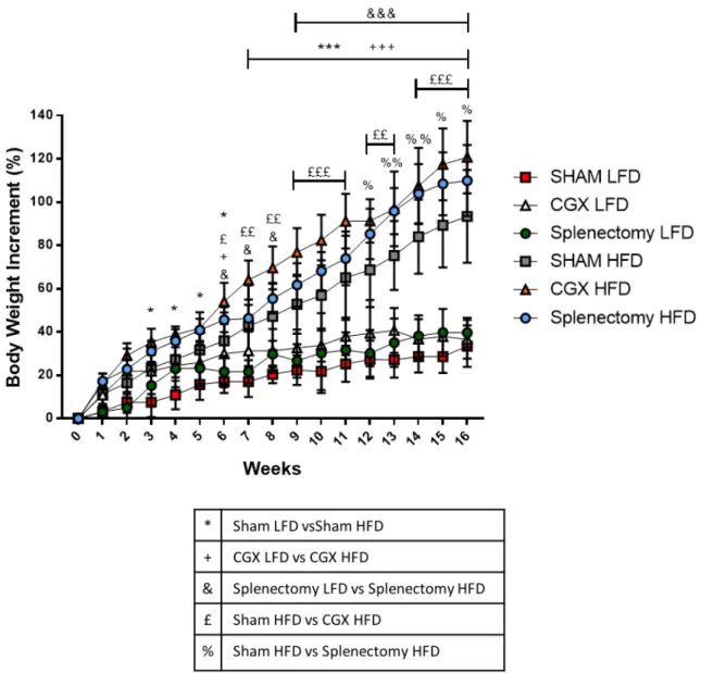
In light of what we have demonstrated up to this point, we tempted to explore the causal role of the enhanced sympathetic nerve discharge toward the spleen and the relevance of the splenic organ itself in the onset of obesity and obesity induced-hypertension. On this issue, we abrogated the sympathetic innervation through specific celiac ganglionectomy (CGX) in C57BL/6J mice and subsequently mice were fed with LFD or HFD. On the other side, to investigate the relevance of the spleen, we removed the splenic organ through splenectomy.
Sham mice fed with the low-fat or the high-fat diet were used as control and the diet protocol was extended to 16 weeks to assess long-term effect of different manipulation on body weight growth pace.
Monitoring body weights weekly, we observed that both Sham, CGX and splectomized mice upon HFD became obese reaching similar body weight at the end of the 16 weeks of diet, significantly higher than control mice upon LFD (Figure 15). To ensure a more accurate analysis of weight increase, data were additionally analyzed as body weight percentage of increment. Such analysis effectively demonstrated that CGX and Splenectomized mice upon HFD increased their weight significantly more as compared to Sham HFD mice (Figure 16).
In conclusion, these data provide important evidence that despite sympathetic activity is enhanced during the development of obesity, it does not play any role in weight accumulation processes since neither celiac ganglionectomy nor spleen surgical removal could prevent diet-induced obesity.
Figure 15: Body weight measurement of Sham, CGX and splenectomized mice upon HFD and
LFD. (Sham LFD n=7, CGX LFD n=4, Splenectomy LFD n=3, Sham HFD n=10, CGX HFD n=6, Splenectomy HFD n=6; two-way ANOVA followed by Tukey post-hoc test; Panels represent mean ± DS. *P<0.05, **P<0.01, ***P<0.001, ++P<0.01, +++P<0.001, &&P<0.01, &&&P<0.001).
Figure 16: Body weight increase of Sham, CGX and splenectomized mice upon HFD and LFD.
(Sham LFD n=7, CGX LFD n=4, Splenectomy LFD n=3, Sham HFD n=10, CGX HFD n=6, Splenectomy HFD n=6; two-way ANOVA followed by Tukey post-hoc test; Panels represent mean ± DS. *P<0.05, **P<0.01, ***P<0.001, +P<0.05, ++P<0.01, +++P<0.001, &P<0.05 &&P<0.01, &&&P<0.001; £P<0.05 ££P<0.01, £££P<0.001; %P<0.05 %%P<0.01). Progressively weight measurement is the simplest way to determine weight increase, but it does not allow to appreciate any difference in fat deposition all over the mouse body.
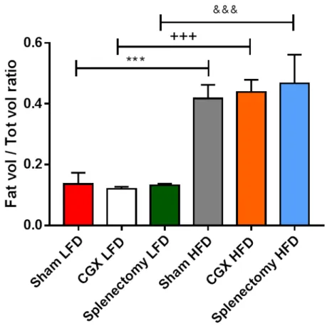
Despite no difference was found in final body weight among HFD-fed mice groups, to investigate if splenic sympathetic communication blockade or splenectomy could differentially influence fat accumulation, immediately after the sixteenth week of diet protocol, we performed a non-invasive quantitation of body fat mass composition in both obese and lean Sham, CGX and Splenectomized mice (Figure 17).
Figure 17: Representative whole-body micro-computed tomography (MicroCT) images of
Sham, CGX and Splenectomized mice upon LFD or HFD after 16 weeks of diet administration.
Whole-body micro-computed tomography (MicroCT) images do not revealed any differential accumulation of fat depots in lean or obese mice in response to surgical procedure as compared to sham controls.
Quantitative analysis of body fat increase, was based on total fat volume on total body volume ratio. Results clearly confirmed the similarity between obese Sham, CGX and Splenectomized mice and the significant difference when compared to lean
Figure 18: Quantitative analysis of fat pat increase in LFD- and HFD-fed Sham, CGX and
Splenectomized mice (Sham LFD n=7, CGX LFD n=4, Splenectomy LFD n=3, Sham HFD n=10, CGX HFD n=6, Splenectomy HFD n=6) (Panel represent mean ± SD) (***, +++, &&& P<0.001).
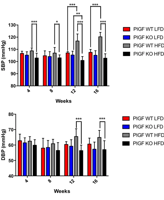
4.4 Despite developing obesity, CGX and Splenectomized mice are protected against hypertension.
In order to evaluate if sympathetic signal transmission pathway to the spleen or the spleen itself, could contribute to the onset of hypertension upon HFD, in parallel to weekly body weight assessment, non-invasive tail-cuff blood pressure measurements were performed every 4 weeks from the start of the diet protocol.
As expected from weight gain, Sham mice blood pressure increased significantly starting at the second month of high-fat feeding. Surprisingly, both CGX and splenectomized mice, despite becoming obese were protected against hypertension. Systolic blood pressure of CGX and splenectomized mice indeed, completely
overlapped that of all LFD-fed mice groups being significantly lower than that of Sham HFD mice (Figure 19). Figure 19: Non-invasive tail cuff blood pressure measurements assessed every 4 weeks from the beginning of the diet procedure. (Sham LFD n=7, CGX LFD n=4, Splenectomy LFD n=3,

![Subcutaneous insulin infusion (CSII) in italy: The third national survey. [La terapia insulinica sottocutanea continua (CSII) in Italia. Terza indagine nazionale]](data:image/gif;base64,R0lGODlhAQABAIAAAP///wAAACH5BAEAAAAALAAAAAABAAEAAAICRAEAOw==)