Cellular/Molecular
The NGF
R100W
Mutation Specifically Impairs Nociception
without Affecting Cognitive Performance in a Mouse
Model of Hereditary Sensory and Autonomic Neuropathy
Type V
Giovanna Testa,
1X
Marco Mainardi,
1Chiara Morelli,
2,3Francesco Olimpico,
1Laura Pancrazi,
1,4X
Carla Petrella,
5X
Cinzia Severini,
5Rita Florio,
6Francesca Malerba,
6Antonia Stefanov,
4Enrica Strettoi,
4Rossella Brandi,
6Ivan Arisi,
6Paul Heppenstall,
2Mario Costa,
4Simona Capsoni,
1,7and Antonino Cattaneo
1,61Bio@SNS, Scuola Normale Superiore, 56124 Pisa, Italy,2European Molecular Biology Laboratory (EMBL), 00015 Monterotondo (Rome), Italy,3EMBL
International PhD Programme, Faculty of Biosciences, Heidelberg University, 69120 Heidelberg, Germany,4Institute of Neuroscience, National Research
Council (CNR), 56124 Pisa, Italy,5Institute of Biochemistry and Cell Biology (IBBC), CNR, DOS Policlinico Umberto I, 00161 Rome, Italy,6Neurotrophins
and Neurodegenerative Diseases Laboratory, Rita Levi-Montalcini European Brain Research Institute (EBRI), 00161 Rome, Italy, and7Institute of
Physiology, Department of Biomedical and Specialty Surgical Sciences, University of Ferrara, 44121 Ferrara, Italy
Nerve growth factor (NGF) is a key mediator of nociception, acting during the development and differentiation of dorsal root
ganglion (DRG) neurons, and on adult DRG neuron sensitization to painful stimuli. NGF also has central actions in the brain, where
it regulates the phenotypic maintenance of cholinergic neurons. The physiological function of NGF as a pain mediator is altered in
patients with Hereditary Sensory and Autonomic Neuropathy type V (HSAN V), caused by the 661C
⬎T transition in the Ngf gene,
resulting in the R100W missense mutation in mature NGF. Homozygous HSAN V patients present with congenital pain
insensi-tivity, but are cognitively normal. This led us to hypothesize that the R100W mutation may differentially affect the central and
peripheral actions of NGF. To test this hypothesis and provide a mechanistic basis to the HSAN V phenotype, we generated
transgenic mice harboring the human 661C
⬎T mutation in the Ngf gene and studied both males and females. We demonstrate that
heterozygous NGF
R100W/wtmice display impaired nociception. DRG neurons of NGF
R100W/wtmice are morphologically normal,
with no alteration in the different DRG subpopulations, whereas skin innervation is reduced. The NGF
R100Wprotein has reduced
capability to activate pain-specific signaling, paralleling its reduced ability to induce mechanical allodynia. Surprisingly, however,
NGF
R100W/wtmice, unlike heterozygous mNGF
⫹/⫺mice, show no learning or memory deficits, despite a reduction in secretion and
brain levels of NGF. The results exclude haploinsufficiency of NGF as a mechanistic cause for heterozygous HSAN V mice and
demonstrate a specific effect of the R100W mutation on nociception.
Key words: allodynia; dorsal root ganglia; hereditary sensory and autonomic neuropathy type V; learning and memory; nerve growth
factor; skin innervation
Significance Statement
The R100W mutation in nerve growth factor (NGF) causes Hereditary Sensory and Autonomic Neuropathy type V, a rare
disease characterized by impaired nociception, even in apparently clinically silent heterozygotes. For the first time, we
generated and characterized heterozygous knock-in mice carrying the human R100W-mutated allele (NGF
R100W/wt). Mutant
mice have normal nociceptor populations, which, however, display decreased activation of pain transduction pathways.
NGF
R100Winterferes with peripheral and central NGF bioavailability, but this does not impact on CNS function, as
demon-strated by normal learning and memory, in contrast with heterozygous NGF knock-out mice. Thus, a point mutation allows
neurotrophic and pronociceptive functions of NGF to be split, with interesting implications for the treatment of chronic
pain.
Introduction
Nerve growth factor (NGF;
Levi-Montalcini, 1987
), in addition
to its classical neurotrophic actions, is an established mediator of
pain pathophysiology. Indeed, NGF is a key molecule in the pain
machinery, with powerful proinflammatory and sensitizing
actions (
Denk et al., 2017
). NGF regulates pain perception in
humans and pain-related behavioral responses in animals;
injec-tions of NGF in animals (
Lewin et al., 1993
) and humans (
Svens-son et al., 2003
) elicit rapid and long-lasting hyperalgesia, while
NGF-neutralizing molecules are effective analgesics in many
models of persistent pain (
McMahon et al., 1995
;
Ugolini et al.,
2007
). NGF is strategically positioned to regulate pain, acting on
both nociceptors (
Lewin et al., 2014
) and attraction of immune
cells at injury sites (
Levi-Montalcini et al., 1996
;
Skaper, 2017
).
NGF levels increase in affected tissues in various experimental or
pathological inflammatory conditions (
Minnone et al., 2017
).
Based on this, anti-NGF antibodies are being clinically evaluated
to alleviate chronic pain (e.g., due to osteoarthritis;
Lane et al.,
2010
). On the other hand, its robust neurotrophic properties
have led to testing NGF as a therapeutic candidate for different
conditions including diabetic polyneuropathy (
Apfel et al., 1994
;
Pittenger and Vinik, 2003
) and neurodegenerative diseases (
Mi-tra et al., 2019
). However, these trials failed due to significant
adverse effects, including NGF-induced pain (
Apfel, 2002
). Thus,
elucidating NGF actions on pain transmission, also taking into
account signaling through TrkA and p75
NTR, would be a step
toward overcoming these limitations.
In this regard, Hereditary Sensory Autonomic Neuropathies
(HSANs) are rare congenital pain insensitivity diseases that offer
an opportunity to dissect NGF roles in pain. Indeed, several
mu-tations leading to pain insensitivity have been described in the
genes encoding NGF and TrkA (tropomyosin receptor kinase A;
Indo, 2001
;
Capsoni, 2014
;
Nahorski et al., 2015
). For example,
recessive mutations in the TrkA gene cause HSAN IV,
character-ized by insensitivity to pain, autonomic defects, and mental
re-tardation (
Indo, 2001
). A Swedish family suffering from severe
loss of pain, leading to frequent bone fractures and Charcot
joints, was discovered to carry, instead, the 661C
⬎T transition in
the NGFb gene, resulting in the R100W mutation in mature NGF.
This “painlessness” disorder was called HSAN V (
Einarsdottir et
al., 2004
). Compared with HSAN IV patients, homozygous
HSAN V patients display a similar congenital indifference to
nox-ious stimuli, but no cognitive deficits (
Einarsdottir et al., 2004
).
In contrast, heterozygous carriers, despite reduced skin
innerva-tion and unmyelinated fiber number, along with altered
thermo-ception, do not present with readily detectable clinical signs and
have been identified only through pedigree and genetic screening
(
Axelsson et al., 2009
;
Minde et al., 2009
;
Perini et al., 2016
).
We and others have shown that the NGF
R100Wprotein
dis-plays reduced binding to, and signaling via, p75
NTR, whereas
interaction with TrkA is unaffected (
Covaceuszach et al., 2010
;
Capsoni et al., 2011
;
Sung et al., 2018
). Thus, we proposed that
NGF
R100W, with its biased TrkA agonist receptor profile (
Cova-ceuszach et al., 2010
;
Capsoni et al., 2011
), might help in
dissect-ing trophic and nociceptive actions of NGF. To elucidate how
these molecular features concur to determine the clinical HSAN V
phenotype, we describe here the characterization of a mouse knock-in
line harboring the NGF
R100Wmutation. We focused on
heterozy-gous NGF
R100W/wtmice, since homozygous NGF
R100W/R100Wmice
die by the first month of life (
Testa et al., 2019
).
We demonstrate that heterozygous NGF
R100W/wtmice display
impaired nociception, despite having normal dorsal root
gan-glion (DRG) neurons. The NGF
R100Wprotein has a reduced
ca-pability to activate pain-specific signaling, correlating with a
reduced ability to induce mechanical allodynia. Surprisingly,
however, NGF
R100W/wtmice, unlike heterozygous mNGF
⫹/⫺mice, show no learning or memory deficits, despite reduced NGF
secretion. Together, our results provide significant insights into
the molecular pathogenesis of the HSAN V phenotype and
dem-onstrate a specific effect of NGF
R100Won nociception, with no
im-pact on cognitive performance. These features make NGF
R100Wan
attractive tool to manipulate pain sensitivity and to exert
neu-rotrophic actions in the absence of pain sensitization effects.
Materials and Methods
Ethics statement on mouse experiments. All animal procedures were
ap-proved by the Italian Ministry of Health and were fully compliant with Italian (Ministry of Health guidelines, Legislative Decree n°26/2014) and European Union (Directive n°2010/63/UE) laws on animal research. The experiments were performed in strict accordance with the ARRIVE guidelines (Animal Research: Reporting in Vivo Experiments). In addi-tion, the principles of the Basel Declaraaddi-tion, including the “3R” concept, have been considered throughout the whole project. Both male and fe-male mice were used in this study.
Generation of knock-in human NGFR100W/wt mice.
pCMV6-XL5-human NGFwt plasmid was obtained from OriGene (catalog
#SC123827) and pCMV6-XL5-human NGFR100Wwas generated using
site-specific mutagenesis PCR. The targeting constructs were generated using classical cloning technologies. Briefly, a BAC (bacterial artificial chromosome; clone RP24-160F12) containing the entire regions of in-terest flanking the NGF sequence, was used to generate intermediate plasmids by cloning in pBluescript SK(⫺) the 5⬘ homology arm (from 89489 to 94076, restriction site MfeI) and 3⬘ homology arm (from 94803 to 99710, restriction site HindIII). The human NGFR100Wcoding
se-quence was cloned in the pKO2.1 targeting vector carrying the DTA (diptheria toxin A) negative selection cassette (provided by Dr. L. Ron-fani, San Raffaele Hospital, Milan, Italy). The targeting vector was trans-fected into R1p.15 cells (background SV129), and positive clones were selected using neomycin resistance.
Southern blot analysis. Genomic DNA was extracted by means of
phe-nol:chloroform:isoamyl alcohol from⬃350 cell clones electroporated with NGFR100Wtargeting vector. The purified DNA was first incubated
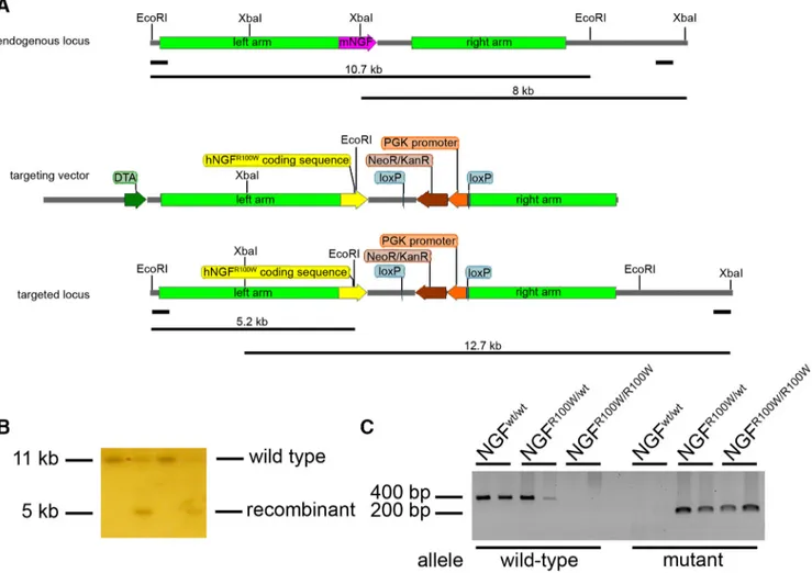
with EcoRI (for 5⬘ screening), then positive clones were confirmed by XbaI digestion (for 3⬘ screening). Digestions were run in a 0.8% agarose gel overnight (O/N) at 50 V. After a mild depurination and denaturation, gels were blotted on nitrocellulose and filters were incubated with an external 5⬘ or 3⬘ probe. The 5⬘ probe labels a 10.7 kb EcoRI band in the wild-type (WT) allele and a 5.2 kb EcoRI band in recombinant allele (Fig. 1A). The 3⬘ probe labels an 8 kb XbaI band in the WT allele and a 12.7 kb
XbaI band in the recombinant allele (Fig. 1B).
Received March 26, 2019; revised Oct. 1, 2019; accepted Oct. 27, 2019.
Author contributions: M.C., S.C., and A.C. designed research; G.T., M.M., C.M., F.O., L.P., C.P., C.S., A.S., E.S., R.B., P.H., and S.C. performed research; R.F. and F.M. contributed unpublished reagents/analytic tools; G.T., M.M., A.S., and I.A. analyzed data; G.T., M.M., S.C., and A.C. wrote the paper.
This work was supported by the EU FP7 PAINCAGE project (Grant 603191, to A.C.), by Fondazione Telethon (Grant GGP11179, to A.C.), by European Union’s Horizon 2020 research funds under the Marie Skłodowska-Curie (Grant 674901, to A.S. and E.S.), and by MIUR_PRIN17 project (Grant 2017HPTFFC_001, to A.C.). We thank Lorenza Ron-fani, Rosanna Rinaldi, and Ivana Benzoni (San Raffaele Hospital, Milan, Italy); Mara D’Onofrio (Rita Levi-Montalcini European Brain Research Institute); laboratory members Maria Antonietta Calvello, Vania Liverani, Nicola Maria Carucci, Francesco Gobbo, Caterina Rizzi, Alessandro Viegi, and Alexia Tiberi (BioSNS, Scuola Normale Superiore) for their help and support; Elena Novelli (Institute of Neuroscience, CNR) for valuable technical help; Nicola Origlia (Institute of Neuroscience, CNR) for support in electrophysiology experiments; Enrico Pracucci and Gian Michele Ratto [NEST (National Enterprise for nanoScience and nanoTechnology) Laboratory, Scuola Normale Superiore] for support in DRG sectioning; Irene Perini and India Morrison (Linko¨ping University, Linko¨ping, Sweden) or useful discussions; and Moses W. Chao (New York University, New York, NY) for useful discussion and critical reading of the manuscript.
The authors declare no competing financial interests.
Correspondence should be addressed to Antonino Cattaneo at [email protected] or Simona Capsoni at [email protected].
https://doi.org/10.1523/JNEUROSCI.0688-19.2019 Copyright © 2019 the authors
Positive clones were injected into C57BL/6 mouse blastocysts, and chimeric animals were obtained.
Mice were genotyped by PCR. The following PCR primers were used: fw_human: 5⬘-TTTAGCACCCAGCCTCCCCGTGAAG-3⬘; fw_mouse: 5⬘-CAGAAGGAGACTCTGTCCCTG-3⬘; and rev_human-mouse: 5⬘-CACCTCCTTGCCCTTGATGTCTG-3⬘.
Band sizes are as follows: wild-type, 400 bp; and mutant 200 bp (Fig. 1C).
Behavioral analyses. Experiments were performed on NGFwt/wt,
NGFR100W/wt, mNGF⫹/⫹, and mNGF⫹/⫺mice. Mice were kept under a
12 h light/dark cycle, with food and water available ad libitum.
Hot plate test. Mice were placed on a surface heated from 42°C to 54°C
with 3°C steps. Animals were sequentially tested, allowing a 10 min rest-ing period between each temperature step. The temperature threshold required to observe paw licking and the time required to observe this reaction at each temperature step were recorded.
Cold sensitivity test. Mice were put in a plastic cage and habituated for
30 min. Acetone (50l; Sigma-Aldrich) was sprayed onto the plantar surface of the hindpaw using a Gilson pipette, and the responses were reported as a 4-point score: 0⫽ no response; 1 ⫽ brisk withdrawal or flick of the paw; 2⫽ repeated flicking of paw; and 3 ⫽ repeated flicking of the hindpaw with licking. Acetone was applied six times, alternating between paws, with an interval of 5 min between each application. The frequency of response, expressed as a percentage (number of trial char-acterized by a response/total number of trials) was evaluated.
Capsaicin injection test. Mice were placed individually in a Plexiglas
box for 15 min before drug injection to allow habituation. Capsaicin
(catalog #141000, Abcam) was dissolved in dimethylsulfoxide (DMSO) and injected into the ventral surface of the right hindpaw using a Ham-ilton syringe at a concentration of 3g/l in saline solution (total injec-tion volume, 10l; DMSO final concentration, 0.1%). Control mice were injected with 10l of 0.1% DMSO in saline. Following the injec-tion, mice were observed for 15 min and the amount of time spent licking and/or lifting the injected paw was measured.
Object recognition test. The apparatus consisted of a PVC arena (60⫻
60⫻ 30 cm) with white floor and black walls. The test was performed in 3 d. On day 1, mice were subjected to a habituation phase in which they received two 5 min sessions in the empty arena, separated by a 30 min interval. On day 2, mice were exposed to two identical objects for 7 min to evaluate the total time of exploration. On day 3, mice were placed back in the arena and exposed to a familiar object and another novel object (memory phase). The time spent exploring each object was recorded.
Morris water maze. The test was performed in a water tank (120 cm
diameter) filled with white opaque water. The platform, placed in the center of the southwest quadrant, was submerged 1 cm below the water surface. The 6-month-old mice were trained with two trials per day, with a 40 min inter-val, for 9 consecutive days. Mice were allowed up to 2 min to locate the platform, and the latency to reach it was recorded. If the mouse failed, the experimenter guided it onto the platform. Data were acquired and analyzed using an automated tracking system (Ethovision XT, Noldus).
Tape response assay. Mice were habituated to a Plexiglas container for
5 min, and then a 3 cm piece of adhesive tape was applied to the back. Mice were observed for 5 min to measure the latency to the first tape removal attempt and the total number of attempts.
Figure 1. Molecular strategy for the generation of R100W knock-in mice. A, Top, Endogenous mouse NGF locus with 5⬘and3⬘Southernblotprobesandexpectedsizesofwild-typeSouthernblot bands. Middle, Targeting vector for site-specific recombination. Bottom, Targeted NGF locus with 5⬘and3⬘SouthernblotprobesandexpectedsizesofrecombinantSouthernBlotbands.Colorcodes are as follows: pink, mouse NGF coding sequence; yellow, human NGF coding sequence; brown, NeoR⫹selection cassette; orange, PGK promoter; blue, loxP sites; light green, left and right homology arms; dark green, DTA-negative selection marker. B, Representative image of embryonic stem cells Southern blot. C, PCR genotyping of NGFR100W/wt, NGFwt/wt, and NGFR100W/R100Wmice; wild-type band, 400 bp; mutant band, 200 bp.
Cotton swab assay. Mice were placed in an arena consisting of an
ele-vated chamber with a grid floor and were allowed to habituate for 1 h. A cotton swab was stroked through the floor along the plantar paw surface five times, alternating between paws with a 10 s interval. The number of withdrawals was counted and expressed as a percentage of the total num-ber of trials.
In vivo nociceptive assay. As reported in the study byCapsoni et al. (2011), CD1 male mice (Charles River, Italy) were subjected to a me-chanical allodynia behavioral test after the injection of either WT or R100W NGF in the hindpaw plantar surface at a 0.2g/l concentration (corresponding to 4g in a total injection volume of 20 l) in saline. Control mice were injected with 20l of saline. The von Frey test (Ugo Basile, Italy) was performed before treatment and 1, 3, 4, and 5 h postinjection.
NGF treatment. Mouse wild-type NGF was administered daily at the
dose of 1g/kg to pregnant dams by subcutaneous injection; treatment was protracted until 10 d after delivery. From postnatal day 10 (P10) to P60, pups received a daily subcutaneous injection (1g/kg) and intra-nasal administration (480 ng/kg) of NGF. A 21 d washout period was allowed before subsequent experiments.
Dorsal root ganglion neuron primary cultures. DRG neurons were
pre-pared from neonatal (5-d-old, P5) Wistar rats (Charles River, Italy) from both sexes, as reported in the studies byBonnington and McNaughton (2003)andTaneda et al. (2010). Briefly, DRGs were collected, incubated for 1 h at 37°C with 0.125% collagenase (Sigma-Aldrich), mechanically dissociated, and plated onto coverslips or Petri dishes pretreated with 10 g/ml poly-L-lysine (Sigma-Aldrich), at a density of 50,000 cells/well of a 48-well tissue culture plate. DRG cultures were maintained in serum-free medium, consisting of DMEM/F12 (Invitrogen) supplemented with 87.5 ng/ml 5-fluoro-2⬘-deoxyuridine, 37.5 ng/ml uridine, 50 U/ml penicillin, and 50g/ml streptomycin (all from Sigma-Aldrich) and 0.05% Invit-rogen N2 supplement (Thermo Fisher Scientific) at 37°C in 5% CO2. The
treatment with N2 supplement allows the presence of a physiological level of growth factors, thus preventing the occurrence of a neurotrophin withdrawal state. After 2–3 d in vitro, DRG cultures were stimulated for experimental procedures using either control human NGFwtor human
NGFR100W(100 ng/ml) for 5 d or were maintained in basal medium
conditions. At the end of this incubation period, B2R and phospho-transient receptor potential vanilloid-1 (pTRPV1) protein expression were measured by Western blot after 3 h of bradykinin (BK; 1M)
ap-plication (antibodies used were as follows: rabbit anti-B2R, 1:1000, Alo-mone Labs; and rabbit anti-pTRPV1 and anti-TRPV1, both 1:1000, Millipore). Substance P (SP) was quantified in the culture medium using commercial enzyme immunoassay, according to the manufacturer in-structions (Cayman Chemical).
To characterize neuronal viability, DRG cultures were fixed in 4% PFA for 10 min at room temperature (RT), incubated O/N at 4°C with mouse anti-NeuN (1:200; Sigma-Aldrich), and rabbit anti-Neurofilament 200 (1:200; Sigma-Aldrich) followed by goat anti-mouse secondary antibody (1:400; Sigma-Aldrich), goat anti-rabbit rhodamine-conjugated second-ary antibody (1:1000; Sigma-Aldrich), and Hoechst 33258 (0.25g/ml) for 1 h and 5 min, respectively, at RT. Fluorescence images were acquired using an Olympus BX51 microscope and a 60⫻ oil-immersion objective (numerical aperture, 1.4). The number of NeuN-immunoreactive cells was normalized on the total number of cells (i.e., Hoechst stained). At least 40 microscopic fields per coverslip, in four coverslips from three independent experiments, were quantified for each experimental group.
Human NGFR100Wpurification. Human NGFR100WcDNA was cloned
into the prokaryotic expression vector pET19b downstream the sequence of the human BDNF prodomain, to produce a chimeric human proBDNF/NGFR100Wconstruct, and expressed in the Escherichia coli strain Rosetta(DE3)PLysS. The corresponding chimeric protein was re-folded from inclusion bodies and purified using an adaptation of the protocol used for proNGF in the study byPaoletti et al. (2015). The purified proBDNF-NGFR100Wwas proteolytically processed with trypsin
to produce mature NGFR100W, as previously described (Paoletti et al.,
2015).
Immunohistochemistry. NGFwt/wt, NGFR100W/wt, mNGF⫹/⫹, and
mNGF⫹/⫺mice were transcardially perfused with 4% PFA in PBS, pH
7.4, and brains were dissected and postfixed O/N in the same solution, then cryoprotected in 30% sucrose in PBS for 36 h. The brains were sectioned with a sliding freezing microtome (Leica) to obtain 45- m-thick coronal sections that were washed three times in TBS with 0.3% Triton X-100, then treated with 3.5% H2O2in TBS to inactivate endog-enous peroxidases. Sections were blocked for 30 min with 10% FBS and 0.3% Triton X-100 in TBS, followed by an O/N incubation at 4°C with 1:500 goat anti-choline acetyltransferase (ChAT; catalog #AB144P, Mil-lipore). Biotinylated secondary antibodies (Vector Laboratories) were diluted in 10% FBS in PBS for 3 h at RT. Finally, sections were incubated in Vectastain ABC HRP Kit (Vector Laboratories) in PBS for 1 h, fol-lowed by another incubation in TBS solution containing diaminobenzi-dine (DAB; Sigma-Aldrich) and the enzyme Glucose Oxidase type VII (Sigma-Aldrich); the reaction was stopped after 10 min. Stained sections were mounted on glass slides using DPX Medium. Images were acquired with a Nikon Eclipse E600 Optical Microscope, and the density of immu-noreactive cells was calculated using ImageJ.
For analysis of superior cervical ganglia (SCGs), embryonic day 16.5 fetuses were extracted from pregnant dams, washed in PBS, and fixed by immersion in 4% PFA for 4 h, then cryoprotected in 30% sucrose in PBS and sectioned using a cryostat. The quantification of SCG cell number was performed on Nissl-stained sections, with the experimenter blind to the genotype of the animal, and representative images were obtained by staining for tyrosine hydroxylase (TH; 1:200; catalog #AB152, Millipore) O/N at 4°C (Crerar et al., 2019); subsequent steps were as described above.
Whole-mount staining was performed on internal organs dissected from P0.5 pups, O/N fixed in 4% PFA, then dehydrated in methanol series, followed by O/N quenching of endogenous peroxidases in 80% methanol, 17% DMSO, and 3% H2O2. After rehydration, samples were
blocked in 4% BSA, 1% Triton X-100 in PBS, and incubated with 1:200 anti-TH antibody diluted in the same blocking solution for 72 h at 4°C. The signal was revealed by incubation with HRP-conjugated anti-rabbit antibody (1:200; catalog #sc-2004, Santa Cruz Biotechnology) diluted in blocking solution O/N at 4°C, followed by DAB processing. Finally, sam-ples were cleared using a 2:1 solution of benzyl benzoate and benzyl alcohol (Crerar et al., 2019). Samples were imaged using a 4⫻ objective, and optical density of the signal was quantified with the experimenter blind to the genotype of the animal.
Skin and DRG immunofluorescence. DRGs from adult mice (2 and 6
months old) were collected in an Eppendorf tube containing cold PBS, then postfixed in 4% PFA for 30 min at RT, embedded in 2% agarose and sectioned at 50m thickness using a vibratome. Sections were washed twice with PBS-Triton 0.3%, then subjected to a 30 min blocking step in 5% NGS and 0.3% Triton X-100 in PBS, followed by an O/N incubation at 4°C with primary antibodies diluted as shown below. Alexa Fluor-conjugated secondary antibodies (Thermo Fisher Scientific) were diluted 1:1000 in 0.3% Triton X-100 and 5% NGS in PBS for 2 h at RT. Sections were mounted using Invitrogen Prolong Gold Medium (Thermo Fisher Scientific).
For immunofluorescence analysis, the hairy and glabrous skins were collected, allowed to dry, and postfixed in 4% PFA at 4°C O/N, then incubated in 30% sucrose in PBS and frozen in OCT (optimal cutting temperature) medium (Leica). Sections (50m thick for hairy skin, 20 m thick for glabrous skin) were obtained using a cryostat. Immuno-staining was performed as described above.
The antibodies and dilutions used were as follows: 1:500 mouse anti-NF200 (Sigma-Aldrich); 1:200 mouse anti-calcitonin gene-related pep-tide (CGRP; Rockland); 1:100 Invitrogen isolectin GS-B4-biotin conjugate (Thermo Fisher Scientific); 1:200 rabbit anti-B2R (Alomone Labs); 1:300 mouse anti-TRPV1 (Millipore); 1:200 Dako rabbit anti-PGP 9.5 (Agilent Technologies); and 1:300 rabbit anti-NGF M20 (Santa Cruz Biotechnology).
The M20 anti-NGF antibody was validated by measuring the immu-nofluorescence signal intensity obtained using different dilutions on gla-brous skin samples from NGF⫹/⫹and NGF⫹/⫺mice. A nonlimiting concentration of the primary antibody (0.67g/ml) was able to detect the lower skin NGF content in NGF⫹/⫺mice compared with NGF⫹/⫹ mice. Decreasing the antibody concentration (0.33g/ml) led to
incom-plete titration of NGF in the skin of NGF⫹/⫹mice, whereas the signal in samples from NGF⫹/⫺mice was unaffected. Further dilution of the an-tibody (0.17g/ml) resulted in inefficient detection of NGF in samples from both NGF⫹/⫹and NGF⫹/⫺mice. ANOVA-2 (antibody concentra-tion⫻ genotype interaction, F(2,34)⫽ 3.563, p ⫽ 0.039) followed by Holm–Sidak post hoc test (0.67g/ml; NGF⫹/⫹vs NGF⫹/⫺, p⬍ 0.001; 0.33g/ml; NGF⫹/⫹vs NGF⫹/⫺, p⬍ 0.001, 0.17g/ml; NGF⫹/⫹vs NGF⫹/⫺, p⫽ 0.314; NGF⫹/⫹: 0.67 vs 0.33g/ml, p ⫽ 0.303; 0.67 vs 0.17 g/ml, p ⬍ 0.001; 0.33 vs 0.17 g/ml, p ⬍ 0.001; NGF⫹/⫺: 0.67 vs 0.33
g/ml, p ⫽ 0.697; 0.67 vs 0.17 g/ml, p ⫽ 0.047; 0.33 vs 0.17 g/ml, p ⫽ 0.030; n⫽ 7 for each group).
All images were acquired with a Leica SP5 Confocal Microscope and analyzed with Fiji (NIH).
RNA preparation for microarray analysis. DRGs from wild-type and
NGFR100W/wtmice (6 months of age) were isolated and collected. The
total RNA was extracted with TRIzol reagent (Life Technologies) accord-ing to the manufacturer instructions, DNase treated, and purified usaccord-ing Qiagen columns. RNA content was determined on a NanoDrop UV-VIS Spectrophotometer. Only samples with an absorbance ratio of 1.8⬍ OD260/OD280⬍ 2.0 were further processed. Each sample was then qual-ity checked for integrqual-ity using a BioAnalyzer 2100 (RNA 6000 Nano Kit, catalog #G2938C, Agilent Technologies).
Whole-genome expression profiling. Gene expression profiling was
per-formed using the Agilent Technologies one-color microarray system. Two hundred nanograms of Poly A⫹RNA were retrotranscribed using oligo-dT primers linked to the T7 promoter, and the resulting cDNA was used as a template for cyanine 3-CTP (cytidine triphosphate)-labeled cRNA preparation, using the Agilent Technologies Low Input Linear Amplification Kit. The labeled cRNA was purified with RNeasy Mini Spin Columns (Qiagen). To monitor both the labeling reactions and the microarray performance, Agilent Technologies Spike-In Mix was added to the mRNA samples before labeling reactions according the RNA Spike-In protocol. Cyanine 3-labeled cRNA was hybridized to Agilent Technologies 8x60K whole-mouse genome oligonucleotide microarrays (Grid ID, 028005). Microarray hybridizations were performed in Sure-Hyb Sure-Hybridization Chambers (Agilent Technologies) containing 600 ng of cyanine 3-labeled cRNA per hybridization. The hybridization reac-tions were performed at 65°C for 17 h using the Gene Expression Hy-bridization Kit (Agilent Technologies). The hybridized microarrays were disassembled at RT in Gene Expression Wash Buffer 1 (Agilent Technol-ogies). After disassembly, the microarrays were washed in Gene Expres-sion Wash Buffer 1 for 1 min at RT, followed by washing with Gene Expression Wash Buffer 2 for 1 min at 37°C. Then, microarrays were treated with acetonitrile for 1 min at RT. Fluorescence signals of the hybridized Agilent Technologies Microarrays were detected using the Microarray Scanner System (catalog #G2564B, Agilent Technologies). The Feature Extraction Software (version 10.7.3.1, Agilent Technologies) was used to process the microarray image files.
Microarray data analysis. Data filtering, normalization, analysis, and
plotting were performed using R-Bionconductor ( https://www.biocon-ductor.org/). In particular, differential expression was analyzed with the R package limma version 3.5 (Ritchie et al., 2015). All the features with the flag gIsWellAboveBG⫽ 0 (too close to background) were filtered out and excluded from the following analysis. Filtered data were normalized by aligning samples to the 75th percentile. Differentially expressed genes were selected by a combination of fold-change and moderated t test thresholds (R limma test with FDR⬍ 0.05; 兩Log2 fold change ratio兩 ⬎ 1.0). Pathway analysis and network plotting of differential gene lists was performed using the on-line tool StringDB (https://string-db.org; (Szklarczyk et al., 2015).
Electron microscopy. Fixed nerves were washed in phosphate buffer at
RT (10⫻ 5 min), osmicated in 2% (w/v) OsO4in H2O (2 h at 4°C),
washed again (0.05Mmaleate buffer, pH 5.15–10⫻ 5 min), then dehy-drated in ethanol (70%, v/v) for 15 min; 80% (v/v) for 15 min; 90% (v/v) for 15 min; 100%, 4⫻ 15 min). Subsequently, nerves were infiltrated first with propylene-oxide (2⫻ 15 min) then with a mixture of 50% (v/v) propylene-oxide and 50% (v/v) resin catalyzed with 2% DMP30 over-night at RT. Embedding (100% resin catalyzed with 2% DMP30) was followed by 24 h heat treatment for proper polymerization of resin at
65°C. Myelinated axons were counted on semithin (1-m-thick) sec-tions stained with toluidine blue and imaged with a Zeiss Axioplan light microscope. Myelinated fibers were counted individually using Meta-Morph software (Molecular Devices). In cases of multiple nerve compo-nents (i.e., separate bundles of fibers, with individual connective sheets), fiber counts were performed for each component and the corresponding values were summed up. Unmyelinated axons were counted with the aid of transmission electron microscopy at 20,000⫻ magnification. Section-ing was performed usSection-ing a Leica Ultracut Ultramicrotome. Ultrathin sections (90 nm thick) were collected on single-hole copper grids (Form-var Support Film Slots, 2⫻ 1 mm Cu grids; FF2010-CU). Images were obtained using a Jeol 1200EX II Transmission Electron Microscope equipped with a charge-coupled device Olympus Veleta Megaview cam-era covering 5% of the total cross-sectional area of the nerve. Based on the cross-sectional area, a total of 211 fields/NGFwt/wt and 146 fields/
NGFR100W/wtsamples were obtained covering central as well as
periph-eral portions of each nerve systematically. Image files were saved as a TIFF and transferred to MetaMorph, where axons were counted manually.
Nerve conduction velocity measurements. Mice were killed using CO2
inhalation, and the saphenous nerve was dissected and placed in an organ bath (Wetzel et al., 2007). The chamber was perfused with a synthetic interstitial fluid buffer containing the following (in mM): NaCl 123, KCl 3.5, MgSO40.7, NaH2PO41.7, CaCl22.0, Na-gluconate 9.5, glucose 5.5,
sucrose 7.5, and HEPES 10, pH 7.4, at 3 ml/min at 32°C. The distal part of the nerve was placed in the organ bath, while the proximal part was placed in an adjacent chamber filled with mineral oil for recording. An electric probe was used to stimulate the nerve, and a compound action potential was recorded and analyzed using LabChart4 software (AD In-struments, Australia). Each electrical stimulus elicited a response consist-ing of three peaks, correspondconsist-ing to the A, A␦, and C fibers, respectively. To measure the conduction velocity of each fiber type, the distance between the electric probe and the recording electrode was di-vided by the time elapsed from the beginning of the stimulus to the appearance of the peak.
Electrophysiology on brain slices. Acute brain slices were prepared from
5- to 6-month-old mice, following the protocol described in the study by
Barone et al. (2018). Mice were killed by cervical dislocation, and the brain was quickly dissected in an ice-cold, O2-saturated cutting solution containing the following (in mM[SCAP]): NaCl 85, sucrose 75, glucose 25,
NaHCO324, KCl MgCl24, 2.5, NaHPO41.25, and CaCl20.5, pH 7.4. The
350-m-thick sections were prepared using a Leica VT-1200S vibratome while keeping the brain immersed in the same ice-cold cutting solution. Sections were transferred to a recovery chamber filled with artificial CSF (aCSF) containing the following (in mM): NaCl 119, glucose 10, HEPES 10, NaHCO36.2, KCl 2.5, CaCl22, MgCl21.2, and NaH2PO41, pH 7.4,
and was held at 32°C for 30 min, then recovery was completed for an additional 60 min at RT. For recording, sections were transferred to a submerged chamber continuously perfused with aCSF at 32°C saturated with O2, and a concentric bipolar stimulating electrode was used to
stim-ulate Schaffer collateral pathway fibers, while a glass pipette (1 M⍀ im-pedance) filled with aCSF was placed in the CA1 stratum radiatum. Field EPSPs (fEPSPs) were recorded using a MultiClamp 700A Amplifier plugged to a Digidata 1322A interface (Molecular Devices). Current stimuli were delivered using a stimulus isolator (WPI). By using a stim-ulus intensity eliciting a response that amounted to 30 –50% of the max-imum, a stable baseline, using a 0.05 Hz test stimulus frequency, was obtained before delivering high-frequency stimulation (HFS; four trains of 1 s at 100 Hz, with an intertrain interval of 10 s) to induce long-term potentiation. The post-HFS fEPSP was monitored for at least 60 min. Data were analyzed using Clampfit (Molecular Devices). The experi-menter was blind to the genotype of the animal.
Hek293 cells culture. Hek293 cells were maintained at 37°C, 5% CO2, in
Gibco DMEM/F-12 medium supplemented with 10% FBS, 1%
L-glutamine, and 1% penicillin/streptomycin (Thermo Fisher Scientific). Hek293 cells were transfected with pCMV6-XL5-human NGFwtand
pCMV6-XL5-human NGFR100Wplasmids following the manufacturer
instructions for Invitrogen Lipofectamine 2000 (Thermo Fisher Scien-tific). Forty-eight hours after transfection, the supernatants were
immu-noprecipitated and subjected to Western blotting as described below (see NGF immunoprecipitation and Western blot).
NGF immunoprecipitation and Western blot. Cerebral cortices were
isolated from adult mice and homogenized in lysis buffer (Tris-HCl 100 mM, NaCl 400 mM, SDS 0.1%, and Triton X-100 1%). The homogenates
were sonicated, incubated in ice for 30 min, and centrifuged at 15,000⫻
g for 30 min at 4°C. Protein concentration in the supernatant was
quan-tified using the Bradford method (Bio-Rad). Four milligrams of protein were immunoprecipitated with an excess of anti-NGF␣D11 antibody in NET Gel Buffer (Tris-HCl, pH 7.5, 50 mM; NaCl 150 mM; 0.1% Nonidet
P-40; EDTA, pH 8, 1 mM; 0.25% gelatin; and 0.02% NaN). After
immu-noprecipitation, total lysates were loaded on 12% acrylamide gels and blotted using nitrocellulose membranes. The primary antibody was anti-NGF M20 (1:500; Santa Cruz Biotechnology), the secondary antibody was goat anti-rabbit HRP-conjugated (1:500; Santa Cruz Biotechnol-ogy). Blot images were acquired using a ChemiDoc System (Bio-Rad), and the optical density was quantified using ImageJ (NIH).
Data analysis and statistics. Statistical significance was assessed using
SigmaStat 12 (Systat Software). Data are presented as the mean⫾ SEM; detailed statistics for every comparison are reported in the corresponding figure legend.
Results
Generation of NGF
R100W/wtmice and characterization of their
nociceptive phenotype
To shed light on the consequences of the HSAN V-associated
NGF
R100Wmutation, we generated a knock-in mouse line
har-boring the human NGF
R100Wsequence (
Fig. 1
). Homozygous
NGF
R100W/R100Wmice die by P30;
Testa et al., 2019
). On the other
hand, heterozygous mice thrive normally and show no visible
gross deficit. We analyzed the phenotype of heterozygous
NGF
R100W/wtmice in detail, during youth (2 months) and
adult-hood (6 months). Chemical nociception induced by capsaicin
injection in the hindpaw was impaired at both ages (
Fig. 2
A).
Thermal nociception was normal at 2 months of age and
de-creased at 6 months, with adult NGF
R100W/wtmice displaying a
higher latency to respond to a high-temperature stimulus (
Fig.
2
B). On the other hand, cold sensitivity, measured by topical
acetone application on the hindpaw was reduced at both ages
(
Fig. 2
C). In NGF
R100W/wtmice, a non-noxious stimulus
repre-sented by a small piece of tape applied to the back (i.e., to the
hairy skin) took more time to induce a removal reaction only at 6
months of age (
Fig. 2
D). On the other hand, the response to
gentle stroking of the glabrous skin was normal at both time
points (
Fig. 2
E).
These behavioral data show that NGF
R100W/wtmice have a
reduced responsiveness to chemical and thermal noxious stimuli,
are less sensitive to somatosensory inputs, but display normal
light touch.
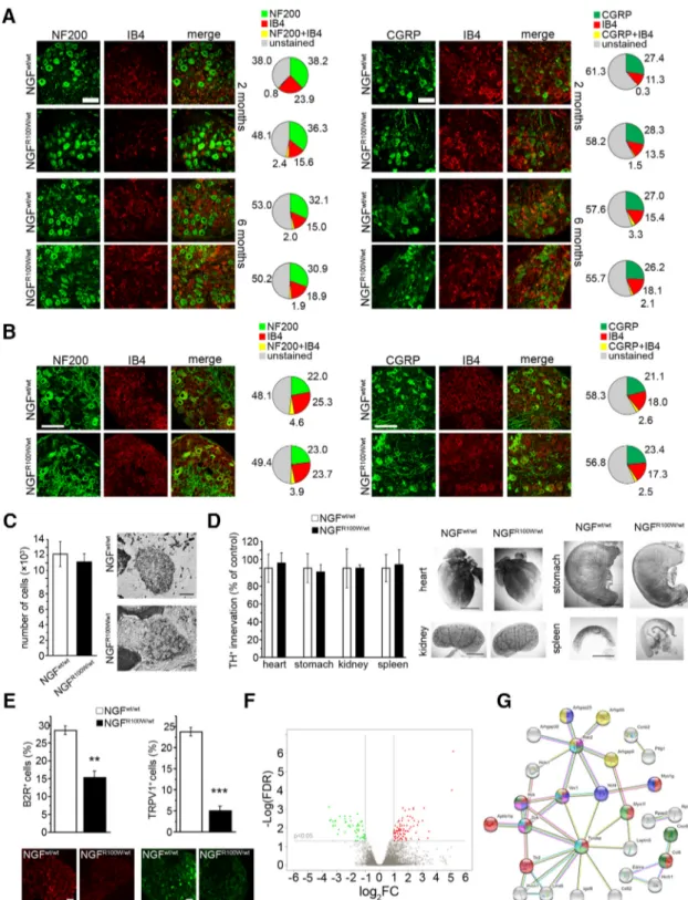
Phenotypic analysis of DRGs from NGF
R100W/wtmice
To look for a cellular-functional correlate of the impaired
noci-ceptive behavior of NGF
R100W/wtmice, we first focused on the
two main NGF-sensitive neuronal populations, defined by the
expression of IB4 and CGRP, respectively (
Harrison et al., 2004
).
We imaged DRGs from 2- and 6-month-old NGF
R100W/wtmice
and found no change in the total number of DRG neurons (2
months: NGF
R100W/wt, 1040.4
⫾ 37.9 cells/mm
2; NGF
wt/wt,
1003.3
⫾ 52.7 cells/mm
2; n
⫽ 3/group; Student’s t test, p ⫽ 0.598,
t
⫽ ⫺0.571; 6 months: NGF
R100W/wt, 1340.4
⫾ 36.8 cells/mm
2;
NGF
wt/wt, 1391.1
⫾ 29.5 cells/mm
2; Student’s t test, p
⫽ 0.324, t ⫽
1.044; NGF
R100W/wt, n
⫽ 6; NGF
wt/wt, n
⫽ 5) and in the
percent-ages of cells expressing the neurofilament marker NF200, the
nonpeptidergic nociceptor marker IB4, and the peptidergic
no-ciceptor marker CGRP, respectively (
Fig. 3
A). We also analyzed
DRGs from P5 mice, an early postnatal developmental stage
dur-ing which segregation between peptidergic and nonpeptidergic
neurons is in progress (
Molliver et al., 1997
) and found no
dif-ferences between NGF
R100W/wtmice and controls in the total cell
number (NGF
R100W/wt, 2134.3
⫾ 64.6 cells/mm
2; NGF
wt/wt,
2234.2
⫾ 116.9 cells/mm
2; n
⫽ 5 and n ⫽ 4, respectively;
Stu-dent’s t test, p
⫽ 0.454, t ⫽ 0.793) or in the percentages of either
NF200
⫹, IB4
⫹, or CGRP
⫹cells (
Fig. 3
B). This suggests that the
neurotrophic potency of NGF
R100Won DRG neurons is
compa-rable to NGF
wt. This was also demonstrated in an in vitro DRG
neuron survival assay (NGF
R100W, 158.2
⫾ 8.8%; NGF
wt,
162.3
⫾ 18.03%; control, 100.0 ⫾ 5.0% NeuN
⫹/total Hoechst
⫹cells; ANOVA-1, F
(2,17)⫽ 8.621, p ⫽ 0.003; followed by
Bonfer-roni post hoc test: NGF
R100Wvs NGF
wt, p
⫽ 1.000; NGF
wtvs
control, p
⫽ 0.006; NGF
R100Wvs control, p
⫽ 0.01; n ⫽ 6 for each
group). Given that the sympathetic nervous system is highly
de-pendent on an intact NGF function for proper development
(
Levi-Montalcini and Booker, 1960
;
Glebova and Ginty, 2004
),
the unaltered neurotrophic actions of NGF
R100Ware also
sup-ported by the normal cell number of the SCG (
Fig. 3
C) and by the
normal sympathetic innervation of key target organs (i.e., heart,
stomach, kidney, spleen), as revealed by TH
immunohistochem-istry (
Fig. 3
D).
However, when we analyzed BK receptor 2 (B2R), whose
ex-pression is upregulated by NGF (
Petersen et al., 1998
), and
TRPV1, an NGF-responsive detector of heat- and
capsaicin-induced pain (
Zhang et al., 2005
), we found a significantly
de-creased immunoreactivity for both receptors (
Fig. 3
E).
To detect subtler changes in DRGs from NGF
R100W/wtmice,
we performed a transcriptomic analysis to identify differentially
expressed genes with respect to DRGs from wild-type mice. The
data showed only a small number of differentially expressed
genes in DRGs from NGF
R100W/wtversus wild-type mice (
Fig. 3
F
and Fig. 3-1, available at
https://doi.org/10.1523/JNEUROSCI.
0688-19.2019.f3-1
), which is in line with the globally normal
ap-pearance of DRG neuronal subpopulations. Among the regulated
genes, TyroBP (DAP12) and toll-like receptor 2 (TLR2) are
note-worthy for their involvement in glial function (
Liu et al., 2012
;
Shboul et al., 2019
). Indeed, gene ontology and network analysis
of differentially expressed genes in DRGs revealed that the
R100W mutation significantly affects pathways involved in
im-mune response, phagocytosis and Rho GTPase cycle (
Fig. 3
G),
suggesting a major effect of the R100W mutation on DRG glial
and microglial cells, rather than on DRG neurons.
These findings demonstrate that DRG neurons from
NGF
R100W/wtmice show only specific molecular changes directly
linked to nociception, alongside an intriguing modulation of
glia-related genes.
Pain sensitization by NGF
R100Wprotein
To investigate whether the R100W mutation might also affect
DRG function, we explored the ability of the NGF
R100Wprotein
to activate and sensitize wild-type DRGs. Incubation of DRG
neuronal cultures with NGF
R100W, followed by acute
administra-tion of the inflammatory neuropeptide BK (
Chuang et al., 2001
),
led to reduced SP release (
Fig. 4
A), bradykinin B2R receptor
ex-pression (
Fig. 4
B), and TRPV1 phosphorylation (
Fig. 4
C),
re-spectively. The reduced ability of NGF
R100Wto sensitize neurons
in vitro was paralleled by in vivo experiments, using acute
injec-tion of NGF
R100Win the hindpaw of CD1 mice. This treatment
resulted in a significantly lower mechanical allodynia with respect
to mice injected with NGF
wt(
Fig. 4
D). Thus, NGF
R100Wdimin-ishes the propensity of DRGs to sensitize and to increase
trans-mission of noxious stimuli.
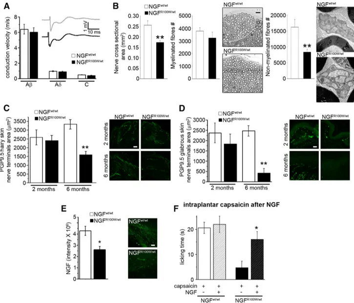
Exploring nociceptive information routes in
NGF
R100W/wtmice
The above-described in vitro and in vivo results prompted us
to analyze the nociceptive input path. We first ruled out that
the observed behavioral effects (
Fig. 2
) were due to a nerve
conduction deficit; indeed, the conduction velocities of the three
main sensory fiber populations, A
, A␦ and C fibers, were normal
(
Fig. 5
A).
We then analyzed the ultrastructure of the sciatic nerve
using transmission electron microscopy. Compared with
controls, the cross-sectional area of the whole nerve in
NGF
R100W/wtmice was significantly decreased (
Fig. 5
B). In
this regard, the number of myelinated axons contained in the
nerve was unaffected, whereas a significant decrease in the
number of nonmyelinated axons in NGF
R100W/wtmice was
observed (
Fig. 5
B), which might determine the smaller overall
nerve section. These neurophysiological and ultrastructural
data match analogous findings in HSAN V heterozygous
hu-man carriers (
Minde et al., 2009
).
Figure 2. The R100W mutation is associated with decreased sensitivity to noxious stimuli. A, Decreased hyperalgesic response to capsaicin in juveniles and adults. Two months, ANOVA-1 (F(2,12)
⫽ 12.869, p ⫽ 0.002), followed by Bonferroni post hoc test (NGFwt/wt
vs VEH, **p⫽ 0.002; NGFwt/wtvs NGFR100W/wt, *p⫽ 0.017; NGFR100W/wtvs VEH, n.s.); VEH, n⫽ 3; NGFwt/wt, n⫽ 5; NGFR100W/wt, n⫽5.6months,ANOVA-1(F(2,13)⫽49.995,p⬍0.001),followedbyBonferroniposthoctest(NGF
wt/wt
vs VEH, ***p⬍0.001;NGFwt/wtvs NGFR100W/wt, ***p⬍0.001;NGFR100W/wt vs VEH, p⫽ 0.017); VEH, n ⫽ 3; NGFwt/wt, n⫽ 5; NGFR100W/wt, n⫽ 6. B, Normal sensitivity to hot stimuli at 2 months of age and impairment in adult HSAN V mice, with increased latency in NGFR100W/wtanimals to respond to high temperatures. Left, 2 months, Student’s two-tailed t test (t⫽ 0.126, p ⫽ 0.901); NGFwt/wt, n⫽ 11; NGFR100W/wt, n⫽ 15. 6 months, Student’s two-tailed
t test (t⫽ 4.743, p ⬍ 0.001); NGFwt/wt, n⫽ 4; NGFR100W/wt, n⫽ 9. Right, ANOVA-2 (“genotype” ⫻ “temperature” interaction, F(4,79)⫽ 3.283, p ⫽ 0.017), followed by Bonferroni post hoc test,
***p⬍0.001,**p⫽0.003;NGFwt/wt, n⫽8;NGFR100W/wt, n⫽8.C,Impairedcoldsensitivityinbothjuvenilesandadults.2months,Student’stwo-tailedttest(t⫽2.445,*p⫽0.035);NGFwt/wt,
n⫽6;NGFR100W/wt, n⫽6.6months,Student’stwo-tailedttest(t⫽2.457,*p⫽0.026);NGFwt/wt, n⫽8;NGFR100W/wt, n⫽10.D,DecreasedhairyskinsensitivityinadultHSANVmice.2months, Student’s two-tailed t test (t⫽ 1.261, p ⫽ 0.226); NGFwt/wt, n⫽ 6; NGFR100W/wt, n⫽ 11. 6 months, Student’s two-tailed t test (t ⫽ 2.305, *p ⫽ 0.042); NGFwt/wt, n⫽ 5; NGFR100W/wt, n⫽ 8.
E, Normal light touch sensitivity. 2 months, Student’s two-tailed t test (t⫽0.155,p⫽0.879);NGFwt/wt, n⫽5;NGFR100W/wt, n⫽11.6months,Student’stwo-tailedttest(t⫽0.050,p⫽0.961); NGFwt/wt, n⫽ 8; NGFR100W/wt, n⫽ 7.
Figure 3. Analysis of DRG markers, sympathetic neurons and innervation, and DRG transcriptome of NGFR100W/wtmice. A, B, Normal expression of NF200, IB4, and CGRP in DRG neurons in both juveniles and adults (A) and in P5 mice (B). Scale bars, 100m. C, No significant difference in SCG cell numbers from NGFR100W/wtand NGFwt/wtmice; Student’s two-tailed t test: t⫽ 0.482, p ⫽ 0.650; NGFwt/wt, n⫽4;NGFR100W/wt, n⫽3.Scalebar,100m.D,NormalsympatheticinnervationoftargetinternalorgansinNGFR100W/wtmice with respect to NGFwt/wtcontrols; heart, Student’s two-tailed t test: t⫽⫺0.268,p⫽0.796;NGFwt/wt, n⫽6;NGFR100W/wt, n⫽4;stomach,Student’stwo-tailedttest:t⫽0.216,p⫽0.835;NGFwt/wt, n⫽5;NGFR100W/wt, n⫽4;kidney,Student’s two-tailed t test: t⫽ ⫺0.002, p ⫽ 0.999; NGFwt/wt, n⫽ 6; NGFR100W/wt, n⫽ 4; spleen, Student’s two-tailed t test: t ⫽ ⫺0.165, p ⫽ 0.874; NGFwt/wt, n⫽ 5; NGFR100W/wt, n⫽ 3. Scale bars, 1 mm. E, Reduced expression of B2R and TRPV1 in DRG neurons of adult mice. Scale bar, 50m.B2R,Student’stwo-tailedttest:t⫽6.219,**p⫽0.003;NGFwt/wt, n⫽3;NGFR100W/wt, n⫽3.TRPV1, Student’s two-tailed t test: t⫽ 12.455, ***p ⬍ 0.001; NGFwt/wt, n⫽ 4; NGFR100W/wt, n⫽ 4. F, Volcano plot for differentially expressed genes in DRGs. The x-axis corresponds to log2FC
(Log2FoldChange) differential expression, and the y-axis to⫺Log(FDR; ⫺Log false discovery rate corrected p value). Red and green regions show significantly upregulated and downregulated
genes, respectively. The log2FC and⫺Log(FDR) thresholds are shown as horizontal (⫺Log(FDR)⬍0.05) and vertical (兩log2FC兩⬎2.0) dashed lines (see Figure 3-1, available athttps://doi.org/
10.1523/JNEUROSCI.0688-19.2019.f3-1). G, Pathway analysis of differentially expressed genes in DRGs. The gene– gene network was obtained by StringDB tool (https://string-db.org). Colors indicate genes belonging to the main over-represented pathways and functional categories: immune response and chemokines (red and green), phagocytosis (blue and purple), killer cells mediated cytotoxicity (light blue), and rho GTPase (yellow).
Finally, we analyzed target tissues of sensory fibers, namely
hairy and glabrous skin (
Zimmerman et al., 2014
). The area of
PGP9.5-immunoreactive terminals was not significantly
differ-ent at 2 months of age, whereas, at 6 months of age, NGF
R100W/wtmice showed a significant reduction in both hairy and glabrous
skin sensory innervation (
Fig. 5
C,D). This is in keeping with the
reduction in the skin innervation of individuals with HSAN V
(
Axelsson et al., 2009
). Moreover, the age-dependent decrease in
PGP9.5-immunoreactive fibers in the hairy skin correlates with
the lower performance of NGF
R100W/wtmice in the tape removal
test (
Fig. 2
D).
Since target-derived NGF modulates innervation (
Davies et
al., 1987
;
Purves et al., 1988
), we analyzed NGF expression in the
glabrous skin by immunofluorescence and found a significantly
reduced signal intensity in NGF
R100W/wtmice, compared with
controls (
Fig. 5
E). To validate this measurement, we used the
same anti-NGF primary antibody to titrate the different levels of
NGF in the glabrous skin of NGF
⫹/⫹and NGF
⫹/⫺mice. Serial
dilutions were used to demonstrate that the dose of antibody used
for the experiment shown in
Figure 5
E can indeed detect the
relative differences in NGF levels corresponding to the two
geno-types, whereas, at a higher dilution, no significant difference was
observed in NGF signal intensity (see Materials and Methods).
Taking into account the NGF reduction in the peripheral
tar-get sites of sensory fibers, we reasoned that exogenous
adminis-tration of wild-type NGF could rescue the nociceptive deficit of
NGF
R100W/wtmice. To this aim, we performed a long-term NGF
treatment, from embryonal life until 2 months of age, and
ana-lyzed the sensitivity of treated mice to capsaicin. A 21 d washout
period after the last treatment was allowed (see Materials and
Methods) to exclude an acute sensitizing action of NGF. This
strategy proved successful in fully rescuing the nociceptive
im-pairment of NGF
R100W/wtmice (
Fig. 5
F ).
This evidence indicates that the R100W mutation is
responsi-ble for multiple alterations in the pathway carrying nociceptive
inputs, and that early treatment with wild-type NGF can restore
pain perception.
NGF
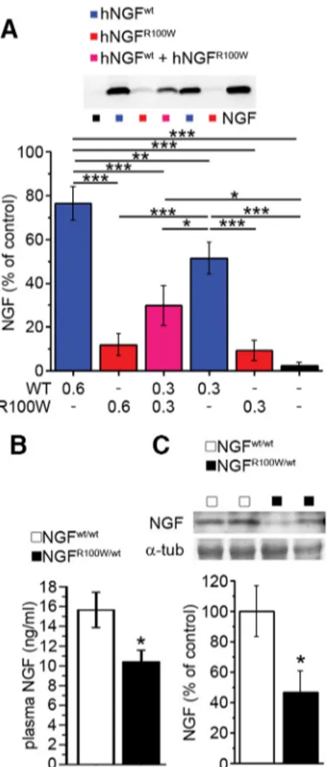
R100Waffects the secretion of wild-type NGF
The R100W mutation has been described to cause an impairment
in the secretion of mature NGF in PC12 and COS cells (
Larsson et
al., 2009
), but whether this is also true in human cells is not
known. NGF is secreted as a homodimer. It is not known,
how-ever, whether NGF
R100Waffects the secretion of wild-type NGF
when the two are coexpressed, as it happens in heterozygous
NGF
R100W/wtmice. We confirmed the secretion deficit of
NGF
R100Win human HEK cells (
Fig. 6
A). Strikingly, the secretion
of wild-type, mature NGF was also impaired when coexpressed
with NGF
R100W(
Fig. 6
A). This suggested that a similar
phenom-enon might occur in vivo in NGF
R100W/wtmice. Consistently, we
found reduced total NGF levels of in plasma and brain from
NGF
R100W/wtmice (
Fig. 6
B, C).
NGF
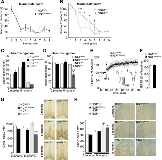
R100W/wtmice show no learning and memory deficits
NGF is not only involved in the development and survival of
sensory neurons, but is also a key regulator of brain development,
in addition to being required for learning and memory processes,
via its actions on CNS neuronal target cells such as forebrain
cholinergic neurons (
Chao, 2003
).
We studied the learning and memory phenotype of NGF
R100W/wtmice. Interestingly, NGF
R100W/wtmice, tested in the Morris water
maze (MWM), showed no difference in the learning
perfor-mance, compared with wild-type controls (
Fig. 7
A), despite the
lower brain NGF levels (
Fig. 6
B, C). A similar conclusion was
drawn when challenging NGF
R100W/wtmice and controls in the
object recognition test. Indeed, NGF
R100W/wtmice showed no
significant differences in the exploratory activity and in the
preference index (
Fig. 7
C,D), which indicate unaffected
visuospatial recognition memory. Moreover, Schaffer
collateral-CA1 long-term potentiation (LTP), a well established
electro-physiological correlate of learning and memory (
Davis et al.,
1992
), did not significantly differ in NGF
R100W/wtmice from
NG-F
wt/wtanimals, thus supporting an intact hippocampal function
(
Fig. 7
E, F ).
Heterozygous NGF knock-out mice (mNGF
⫹/) are also
char-acterized by lower brain levels of wild-type NGF (
Chen et al.,
1997
), similar to NGF
R100W/wtmice. In this case, as expected,
mNGF
⫹/⫺mice showed delayed learning in the MWM when
compared with their controls (
Fig. 7
B). No significant differences
in the latency to locate the platform where detected among the
four experimental groups on day 1 (NGF
wt/wt, 100.560
⫾ 11.742
s; NGF
R100W/wt, 98.438
⫾ 9.919 s; NGF
⫹/⫹, 117.714
⫾ 2.286 s;
Figure 4. Reduced in vitro and in vivo sensitization capability of NGFR100W. A, Bradykinin-induced SP release in DRG cultures is reduced by NGFR100Wcotreatment compared with NGFwt. ANOVA-1 (F(2,16)⫽10.501,p⬍0.002)followedbyStudent–Newman–Keulsposthoctest,***p⬍0.001,*p⫽0.03;hNGFwt
, n⫽5;hNGFR100W, n⫽6;control,n⫽6.B,DownregulationofB2Rexpression by NGFR100Win DRG cultures stimulated with bradykinin. ANOVA-2 (NGF⫻bradykinininteraction,F(2,45)⫽3.371,p⫽0.044)followedbyBonferroniposthoctest,***p⬍0.001;hNGF
wt
-vehicle,
n⫽7;hNGFR100W-vehicle, n⫽8;control-vehicle,n⫽8;hNGFwt-bradykinin, n⫽8;hNGFR100W- bradykinin, n⫽7;control-bradykinin,n⫽8.C,ReducedphosphorylationofTRPV1byNGFR100W in DRG cultures stimulated with bradykinin. ANOVA-2 (NGF⫻bradykinininteraction,F(2,47)⫽9.346,p⬍0.001)followedbyBonferroniposthoctest,***p⬍0.001,**p⫽0.008,*p⫽0.02;n⫽
8 for each group. D, Human NGFR100Wintraplantar injection causes reduced mechanical sensitization compared with human NGFwt. ANOVA-2 repeated-measures test (treatment⫻ time interaction, F(8,144)⫽ 5.785, p ⬍ 0.001) followed by Bonferroni post hoc test, hNGF
wt
versus saline, ***p⬍ 0.001; hNGFwtversus hNGFR100W, ###p⬍ 0.001, ##p ⫽ 0.002, hNGFR100Wvs saline, n.s. p⫽ 1.000; saline, n ⫽ 10; hNGFwt, n⫽ 11; hNGFR100W, n⫽ 9.
NGF
⫹/⫺, 113.286
⫾ 3.887 s; ANOVA-1: F
(3,26)⫽ 1.565, p ⫽
0.225). This indicates a uniform initial performance in this task
across different genotypes. In addition, mNGF
⫹/⫺mice had
im-paired object recognition memory, despite unaffected
explor-atory behavior (
Fig. 7
C,D).
Cholinergic neurons are an NGF target population with an
essential role in modulating learning and memory (
Li et al.,
1995
). Thus, we analyzed the density of ChAT-immunoreactive
neurons in the medial septum and striatum, and found them to
be normal in NGF
R100W/wtmice compared with controls (
Fig.
7
G,H ). On the other hand, the density of ChAT-immunoreactive
neurons in the same brain structures was decreased in mNGF
⫹/⫺mice (
Fig. 7
G,H ). These neuroanatomical data nicely correlate
with the corresponding learning and memory behavioral
pheno-types. Thus, the NGF
R100Wmutation, unlike ngf gene deletion,
does not affect spatial learning and memory processes, in keeping
with heterozygous R100W human carriers, who are reported to
be cognitively normal (
Einarsdottir et al., 2004
).
Discussion
Regulation of nociception and pain responses by NGF has long
been a key area of research on this neurotrophin (
Denk et al.,
2017
). This has spurred a significant translational interest for
treating chronic pain conditions (
Norman and McDermott,
2017
). However, current understanding of these processes is still
incomplete. Studying congenital insensitivity to pain disorders,
Figure 5. Reduced C-fiber density and skin innervation in NGFR100W/wtmice and rescue of nociceptive deficits by NGFwttreatment. A, No alteration in conduction velocity of A,A␦,andCfibers in adult mice. Afiberpeak,Student’stwo-tailedttest(t⫽0.435,p⫽0.669);A␦fiberpeak,Student’stwo-tailedttest(t⫽0.737,p⫽0.470);Cfiberpeak,Student’stwo-tailedttest(t⫽1.629,
p⫽0.120);NGFwt/wt, n⫽10;NGFR100W/wt, n⫽11nerves.B,Electronmicroscopyanalysisofadultsciaticnervereveals:significantreductionofcross-sectionarea,Student’stwo-tailedttest(t⫽ 3.810, **p⫽0.004);NGFwt/wt, n⫽5;NGFR100W/wt, n⫽6;unaffectednumberofmyelinatedaxons,Student’stwo-tailedttest(t⫽0.983,p⫽0.351);NGFwt/wt, n⫽5;NGFR100W/wt, n⫽6,and significant reduction of slow nonmyelinated C axons, Student’s two-tailed t test (t⫽ 3.295, **p ⫽ 0.009); NGFwt/wt, n⫽ 5; NGFR100W/wt, n⫽ 6. C, Representative images and quantification of PGP9.5 expression show normal innervation at 2 months and a significant reduction at 6 months of age. Scale bar, 50m. 2 months, Student’s two-tailed t test (t ⫽ 0.340, p ⫽ 0.751); NGFwt/wt,
n⫽ 3; NGFR100W/wt, n⫽ 3; 6 months, Student’s two-tailed t test (t ⫽ 4.779, **p ⫽ 0.004); NGFwt/wt, n⫽ 4; NGFR100W/wt, n⫽ 3. D, Age-dependent reduction in glabrous skin innervation. Scale bar, 50m. 2 months, Student’s two-tailed t test (t ⫽ 0.792, p ⫽ 0.473); n ⫽ 3 for both groups. 6 months, Student’s two-tailed t test (t ⫽ 5.800, **p ⫽ 0.002); NGFwt/wt, n⫽ 4; NGFR100W/wt,
n⫽3.E,DecreasedNGFlevelsintheglabrousskinofadultmice.Scalebar,50m.Student’stwo-tailedttest(t⫽3.169,*p⫽0.034);n⫽3forbothgroups.F,Rescueofthesensitivitytocapsaicin
after treatment with mouse NGFwtfrom gestation until 2 months of age. NGFwt/wt, Student’s two-tailed t test (t⫽0.323,p⫽0.754);saline,n⫽5;NGF,n⫽7;NGFR100W/wt, Student’s two-tailed
such as HSAN V, offers a unique opportunity to fill this gap,
through an alternative viewpoint on the NGF–TrkA axis
involve-ment in chronic pain. In this regard, HSAN V has intriguing
features, namely (1) the severe insensitivity to pain of
homozy-gous patients, as opposed to the often clinically silent phenotype
of heterozygous carriers; and (2) the absence of cognitive deficits,
as opposed to HSAN IV patients, carrying TrkA mutations
(
Minde et al., 2004
;
Minde et al., 2009
). We and others have
previously elucidated that NGF
R100W(1) is a TrkA biased agonist,
failing to effectively engage p75
NTRand activate the
nociception-specific PLC-
␥ pathway (
Covaceuszach et al., 2010
;
Capsoni et
al., 2011
;
Sung et al., 2018
); (2) retains full neurotrophic activity
via TrkA in a variety of cellular assays (
Capsoni et al., 2011
); and
(3) disrupts processing of proNGF in cultures, leading to
de-creased secretion of mature NGF (
Larsson et al., 2009
;
Fig. 6
A).
Moreover, topic injection of NGF
R100Xmutants does not induce
acute pain (
Capsoni et al., 2011
;
Sung et al., 2018
;
Fig. 4
D). We
have exploited these findings to obtain a “painless NGF”
mole-cule for therapeutic purposes (
Capsoni et al., 2017
;
Cattaneo and
Capsoni, 2019
), but how the features of NGF
R100Wcontribute to
the HSAN V phenotype is still far from clear. In particular, what
are the consequences of this mutation on the overall architecture
of pain-sensing structures and on nociceptive responses?
To shed light on these issues, we generated a new knock-in
mouse line carrying the sequence encoding human NGF
R100W.In
homozygosity, this mutation causes early postnatal lethality,
res-cued by NGF administration (
Testa et al., 2019
). This situation
matches the effect of complete deletion of both Ngf alleles (
Crow-ley et al., 1994
) and its rescue by overexpression of Ngf (
Harrison
et al., 2004
), thus pointing to reduced NGF bioavailability due to
haploinsufficiency as the prevalent mechanistic explanation for
the lethal phenotype of homozygous NGF
R100W/R100Wmice.
Heterozygosity might mitigate the reduction in NGF
bioavail-ability and reduce lethal developmental effects. Moreover, the
vitality of NGF
R100W/wtmice allows to look for further effects of
NGF
R100W, including those possibly arising from peculiar
signal-ing properties.
Heterozygous mice thrive normally and show no gross
defi-cits, but display a reduction in nociception, accompanied by
re-duced skin innervation and altered density of nonmyelinated
fibers, correlating with reduced NGF content in the skin.
The main DRG neuronal populations of NGF
R100W/wtmice
were unaffected and displayed a normal distribution in adults
and newborns, which is in line with the full neurotrophic power
of NGF
R100W. This rules out the possibility of an effect of the
mutation on DRG development. However, the expression of key
pain transduction-related molecules was altered. Triggered by
this finding, we analyzed the global gene expression profile of
DRGs by performing a transcriptomic study. NGF
R100W/wtmice
presented only subtle alterations, with a few hundred genes,
be-longing to gene categories involved in immune response,
phago-cytosis, and Rho-GTPase cycling, changed to a significant extent.
Among the most affected genes were TyroBP/DAP12 and TLR2,
which modulate production of proinflammatory cytokines in
neuropathic pain (
Liu et al., 2012
;
Kobayashi et al., 2016
). These
interesting points deserve further investigation in the future, also
considering the recent finding that microglia is a NGF target
(
Rizzi et al., 2018
).
Another well established target of NGF, critically requiring TrkA
signaling for proper development, is the sympathetic system (
Levi-Montalcini and Booker, 1960
;
Glebova and Ginty, 2004
;
Kuruvilla et
al., 2004
). The SCG of NGF
R100W/wtmice had a normal cell count,
and peripheral sympathetic innervation of internal organs was
sim-ilarly unaffected, further supporting the conclusion that NGF–TrkA
signaling is intact in NGF
R100W/wtmice.
The reactivity to algogens (capsaicin) and the sensitivity to
non-noxious stimuli were decreased in NGF
R100W/wtmice,
cor-relating with the reduction in glabrous and hairy skin
innerva-tion, and in nonmyelinated fibers. These alterations fit with the
lower NGF content in the target tissue (i.e., skin). Consistently,
classical data show that NGF is not involved in establishing skin
innervation during development, but is required for its
mainte-nance in the adult (
Davies et al., 1987
), and the treatment of
NGF
R100W/wtmice with NGF
wtwas able to rescue their
insensi-tivity to capsaicin-induced pain.
DRG neurons primed with NGF
R100Ware less prone to
sensi-tization and show lower nociception-related biochemical
re-sponses than NGF
wt. In addition, NGF
R100Wwas unable to
induce mechanical allodynia in vivo. This is in line with the failure
Figure 6. Impaired secretion and reduced NGF levels in the NGFR100Wcondition. A, Impaired secretion of human NGFR100Win HEK293 cells transfected with the corresponding plasmid. Cotransfection of HEK293 cells with human NGFwtand human NGFR100Wmimics the heterozy-gous condition, and shows a decrease in the secretion of human NGFwt. ANOVA-1 (F(5,24)⫽
23.529, p⬍ 0.001) followed by Student–Newman–Keuls post hoc test, ***p ⬍ 0.001, **p ⬍ 0.01, *p⬍ 0.05; NGF, n ⫽ 5; hNGFwt0.6g, n ⫽ 4; hNGFR100W0.6g, n ⫽ 5; hNGFwt⫹ hNGFR100W0.3g/each,n⫽3;hNGFwt0.3g,n⫽4;hNGFR100W0.3g,n⫽5;mock,n⫽ 4. B, Reduced plasma NGF levels in NGFR100W/wtmice. Student’s two-tailed t test (*p⫽0.031); NGFwt/wt, n⫽ 6; NGFR100W/wt, n⫽ 7. C, Lower abundance of NGF in brain extracts from NGFR100W/wtmice. Student’s two-tailed t test (t⫽ 2.465, *p ⫽ 0.031); NGFwt/wt, n⫽ 6; NGFR100W/wt, n⫽ 7.