i
Table of Contents
ABSTRACT ... 1
ABSTRACT ... 8
CHAPTER 1 ... 14
Metal complexes in medical chemistry ... 14
1.1 Classical antitumor metal agents ... 15
1.2 Non-classical antitumor metal agents ... 25
1.3 Liquid Crystals for/as DNA intercalating agents ... 40
1.4 Metal-based drug co-crystals ... 60
CHAPTER 2 ... 72
Synthesis and characterization of M(II) complexes (M(II)= Zn, Cu) containing aromatic nitrogen ligands ... 72
2.1 4,4’-dinonyl-2,2’-bipyridine (bpy-9) Zn(II) and Cu(II) complexes ... 73
2.2 Synthesis and characterization of curcumin-based complexes increasing the aromatic portion in the N,N chelating ligand ... 98
2.3 4,4’-bis(dimethoxy)-2,2’-bipyridine (bpy-OH) Zn(II) and Cu(II) complexes ... 105
2.4 Synthesis and characterization of tropolone-based complexes increasing the aromatic portion in N,N chelating ligand ... 124
CHAPTER 3 ... 130
Synthesis and characterization of tropolone-based metal containing co-crystals ... 130
3.1 Synthesis and characterization of [(bpy-OH)2Zn(Trop)][Sac], 11 ... 131
3.2 Synthesis and characterization of [(bpy-OH)Zn(Trop)(Sac)], 12 ... 136
ii
CHAPTER 4 ... 147
Synthesis and characterization of chromonic liquid crystals ... 147
4.1 Synthesis and characterization of [(bpy-OH)2Ag][CH3COO], 14 ... 149
4.2 Synthesis and characterization of [(bpy-OH)2Ag][CH3CH2COO], 15 ... 162
4.3 Synthesis and characterization of [(bpy-OH)2Ag][CH3(CH2)4COO], 16 .. 166
4.4 Synthesis and characterization of [(bpy-OH)3Ag2][(PF6)2], 17 ... 170
4.5 Synthesis and characterization of [(bpy-OH)2Ag][OTf], 18 ... 173
4.6 Synthesis and characterization of [(bpy-OH)2Ag][CF3COO], 19 ... 176
CHAPTER 5 ... 179
General equipment and procedures ... 179
X-Ray single crystal analysis ... 180
X-Ray powder diffraction analysis... 181
Solubility analysis ... 182
Phase diagram analysis ... 184
Cryo-TEM measurements... 184
Synthesis procedures ... 185
5.1 4,4’-dinonyl-2,2’-bipyridine (bpy-9) Zn(II) and Cu(II) complexes ... 185
5.2 Synthesis and characterization of curcumin-based complexes increasing the aromatic portion in the N,N chelating ligand ... 193
5.3 4,4’-bis(dimethoxy)-2,2’-bipyridine (bpy-OH) Zn(II) and Cu(II) complexes ... 197
5.4 Synthesis and characterization of tropolone-based complexes increasing the aromatic portion in N,N chelating ligand ... 203
5.5 Co-Crystals of 4,4’-bis(dimethoxy)-2,2’-bipyridine (bpy-OH) Zn (II) and Cu (II) complexes ... 207
iii
Working experience at the Institute of Medical Sciences of Aberdeen,
Scotland (UK) ... 220
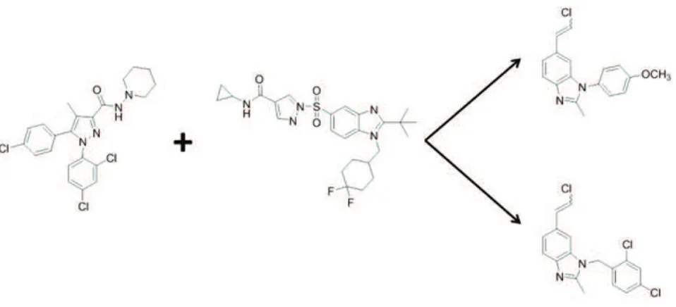
6.1 Synthesis and characterization of CB1 receptor ligands ... 222
6.2 General equipment and procedures ... 226
CONCLUSIONS ... 239 SUPPORTING INFORMATION ... 244 Complex 5 ... 244 Complex VII ... 260 Complex 8 ... 274 Complex 13 ... 293 Complex 13a ... 310
1
Abstract
Negli ultimi anni la sintesi di complessi metallo organici è diventata una importante area di ricerca nel campo della teragnostica, sfruttando le propietà di alcune molecole di agire contemporaneamente sia nell’ambito della diagnosi che della terapia. Il presente lavoro di tesi ha riguardato la sintesi di nuovi complessi di metalli di transizione e la loro completa caratterizzazione con possibili proprietà antitumorali mediante meccanismo di intercalazione.
Per ottenere tale scopo sono stati utilizzati gli ioni Zn(II) e Cu(II) come metalli centrali coordinati a due diverse tipologie di leganti chelanti con natura aromatica, allo scopo di ottenere sistemi eterolettici, ionici e neutri con proprietà intercalanti.
Il frammento metallo organico principale, sulla base del quale costruire per modificazioni successive i differenti complessi sintetizzati, è costituito dallo ione metallico centrale coordinato per chelazione ad una bipiridina ed ad un legante O,O chelante. Le varie funzionalizzazioni sul legante bipiridinico ed i differenti leganti O,O chelanti sono di seguito riportati nel Schema 1.
2
Schema 1.
Il complesso 1, sintetizzato precedentemente dal gruppo di ricerca con il quale è stato svolto il presente lavoro di tesi, presenta ottima attività citotossica e meccanismo di azione di tipo intercalativo, come dimostrato dagli studi condotti tra il complesso stesso ed frammenti di DNA liquido cristallini. Sulla base di questi risultati, e cercando di superare il limite relativo alla scarsa solubilità in solventi acquosi del complesso 1, modifiche strutturali, quali introduzione di ionicità e cambiamento dello ione metallico centrale sono state effettuate mediante le opportune strategie sintetiche. I complessi ionici 2 e 3 sono stati ottenuti con successo e ampiamente
3 caratterizzati, così come l’analogo complesso di Cu(II) 4.
Figura 1. Struttura molecolare dei complessi 1, 2, 3 e 4.
L’effetto citotossico dei complessi 2 e 3 è stato valutato per
investigare l’azione a diverse concentrazioni in cellule SHSY-5Y di neuroblastoma umano, in coltura su membrane di PCL. Si osserva inoltre che il composto 2 ha attività citotossica maggiore a concentrazione più bassa, rispetto a il complesso modelo 1 e il complesso 3. Il complesso 2 anche presenta una parziale solubilità in acqua. Per aumentare la solubilità in acqua dei complessi curcuminici si è deciso di sintetizare un complesso simile al complesso modelo 1 cambiando lo zinco (II) per un rame (II), ottenendo il complesso 4. Purtroppo, il cambio di metalo non ha aumentato la solubilità in acqua.
Sulla base di questi complessi ed al fine di continuare a migliorare le caratteristiche chimico fisiche, quali solubilità e quindi biodisponibilità in acqua, si è deciso di sintetizare complessi simili
4 contenenti il legante N,N-chelante funzionalizzato nella posizione 4, 4’ con un gruppo –CH2OH, il quale potrebbe aumentare la solubilità e
l’attività con i sistemi biologici tramite legami a ponte d’ idrogeno. Il complesso 1A, sintetizzato precedentemente dal gruppo di ricerca con il quale è stato svolto il presente lavoro di tesi, presenta ottima attività citotossica nonostante anche questo complesso abbia scarsa solubilità. Pertanto si è decisso di cambiare la curcumina con un diverso legante O,O-chelante come il tropolone (Fig. 2). Mantendo i gruppi sustituenti si è deciso di estender la parte aromatica (bichinolina e terpiridina) per facilitare le interazioni responsabili.
Figura 2. Struttura molecolare dei complessi 1A, 5, 6 e 7.
Il complesso 7 presenta solubilità in acqua mentre che i complessi
5 e 6 sono insolubili, per questo si è decisso di usare la técnica della
co-cristallizzazione per aumentare la solubilità di questi complessi e renderli più disponibili e meno tossici allo stesso tempo. Questa tecnica
5 si basa sulla formazione di un composto attraverso interazioni non covalenti tra una molecola biologicamente attiva ed una molecola biocompatibile. I complessi sono stati fatti reagire con saccarina in modo da permettere la co-cristallizzazione. I prodotti ottenuti sono stati cristallizzati in acqua e mediante analisi su cristallo singolo i composti mostrati in Figura 3 sono stati ottenuti:
Figura 3. Struttura molecolare dei complessi 8, 9 e 10.
Per testare la capacità dei nuovi complessi sintetizzati come potenziali agenti intercalanti, si è deciso di sintetizare complessi che abbiano propietà liquido cristalline cromoniche, in quanto é noto che il DNA forma strutture colonnari cromoniche mediante legami idrogeno. Questo, da un lato, ci permette di usarli come modelli simili al DNA (Figura 4) al fine di testare la capacità di intercalazione nei confronti di
6 altri complessi, e dall’altro ci consente di usarli come intercalatori aumentando l´affinità nei confronti del DNA.
Figura 4. Intercalazione nel DNA e nei cristalli liquidi chromonici.
I cristalli liquidi chromonici si organizano in strutture a colonna, hanno gruppi aromatici planari, gruppi ionici o idrofilici nella periferia ed infine interazioni devono presentare interazioni face to face. Sono stati sintetizzati i composti mostrati in Figura 5 con proprietà liquido-cristalline:
Figura 5. Struttura molecolare dei complessi chromonici.
Nel corso del triennio di dottorato ho svolto un periodo di 9 mesi al “Institute of Medical Sciences” dell’universitá di Aberdeen, dove ho sintetizzato nuove molecole interagenti con recettori CB1. Utilizzando come modello la molecola di Rimonabant (Figura 6) e una seconda
7 molecola (Figura 6) con migliore affinità, si è deciso di sintetizare delle nuove molecole che abbiano una struttura tale da avere maggiore affinità nei confronti dei recettori CB1 e che allo stesso tempo mostrino mimori effetti collaterali quali i rischi neurologici e psichiatrici mostrati dal Rimonabant. Le molecole sintetizzate dovranno essere dapprima testate, per studiare le interazioni con i recettori CB1 ed in seguito, marcandoli con 18F, verrá studiato il loro funzionamento attraverso la Tomografia ad Emissione di Positroni (PET).
8
Abstract
The goal of this thesis is the synthesis of a new generation of metal complexes containing planar, aromatic p-delocalised ligands and acting as possible metal-based antitumor drugs through intercalation and/or DNA G-quadruplex binding. To engineer these new complexes with antitumor activity, three metals have been used, respectively Zn(II), Cu(II) and Ag(I). These metals can be considered a good alternative to platinum because they exhibit an extensive coordination chemistry, they are low costs, widely used in medical chemistry and less toxic than Pt(II). Furthermore, several ligands have been used to fulfil the coordination sphere of these metal centres. In particular, N,N-chelating ligands with different aromatic regions and substituent groups, and O,O-chelating ligands. The latters were chosen as a function of their well known biological and antitumor activity.
Due to the physiological roles of Zinc(II) species in cells and organs,1 a large interest is growing in the use of Zn(II) coordination
complexes in medical therapeutic applications and biosensors. Zn(II) is also involved in the regulation of mitochondrial apoptosis of many mammalian cells.
1 (a) Vahrenkamp, H. 2007, Dalton Trans., pp. 4751-4759; (b) Anzellotti, A.I. Farrell, N. P. 2008, Chem. Soc. Rev., Vol. 37, pp. 1629-1651.
9 In this context, recently a low cost Zn(II) complex has been synthetized and characterized, showing promising cytotoxic activity against different human cancer cells (Fig. 1).2
Figure 1. Structures of the complex: [(bpy-9)Zn(curc)Cl].
Therefore, in order to continue this research work and aiming to increase the cytotoxic activity and the solubility of this starting derivative, new Zn(II) complexes have been synthesized and characterized. In these complexes, structure variations have been performed around the coordination sphere of the Zn(II) centre, in order to modulate both the geometry and the electrostatic nature of the resulting Zn(II) derivatives.
Subsequently, the metal centre has been changed to Cu(II). Copper is a transition element and in solution has two common oxidation states, +1 (preferred tetrahedral four-coordinated geometry) and +2 (typically square-planar or trigonal planar geometry). Cu(II) is an essential metal for the body, less toxic than Pt(II) and implicated in
2 Pucci, D. Bellini, T. Crispini, A. D’Agnano, I. Liguori, P. F. García Orduña, P. Pirillo, S. Valentini A. Zanchetta, G. 2012, Med. Chem. Commun., Vol. 3, pp.
10 cerebral activities, nervous and cardiovascular systems. A large number of Cu(II) complexes have been screened for antitumor activity.3,4
The structure variations around the metal centre (in both Zn(II) and Cu(II) complexes) was realized by changing the substituents at the periphery of the N,N chelating ligands and/or the type of the N,N- and respectively the O,O-chelating ligands. Initially, the substituent groups (-C9H19) on the N,N-chelating ligand present in the reference Zn(II)
complex (Fig. 1), have been replaced with hydrophilic (-CH2OH) groups,
giving rise to the 4,4’-dihydroxymethyl-2,2’-bipyridine ligand capable to interact via hydrogen bonds interactions to increase the solubility and the bioactivity (Fig. 2).
Nevertheless, the Zn(II) containing the the 4,4’-dihydroxymethyl-2,2’-bipyridine ligand presented low water solubility. To increase the solubility of these complexes co-crystallization with saccharin by liquid assistant grinding has been accomplished, yielding water soluble compounds.
Furthermore, an increase of the aromatic part of the N,N-chelating ligand was accomplished by changing the bipyridine ligands with biquinolines and terpyridine, in order to facilitate the possible intercalation and the G-quadruplex stabilization modes (Fig. 2b,c).
3
Chen, Q-Y. Fu, H-J. Zhu, W-H. Qi, Y. Ma Z-P. Gao, J. 2011, Dalton Trans., Vol. 40, pp. 4414.
4 Zhou, C. Y. Zhao, J. Wu, Y. B. Yin C. X. Yang, P. 2007, J. Inorg. Biochem.,
11
Figure 2. Model complexes for intercalation and/or G-quadruplex binding.
As O,O-chelating ligands, curcumin was used in continuation with the model Zn(II) complex.
Following the possible structural modifications, the O,O-chelating ligand was subsequently substituted with tropolone. These two ligands are well known for their good biological activities. Their chemical structure is reported in Figure 3.
Figure 3. (a) Curcumin and (b) Tropolone.
Curcumin is a yellow spice derived from the rhizome of Curcuma longa Linn. It is used in traditional medicine in China, India and Iran since a long time, in the treatment of many diseases like diabetes, liver disease, rheumatoid disease, atherosclerosis, infectious disease and cancers.5 Nevertheless, curcumin has a poor solubility in water and photosensitivity.
5 Ashkani-Esfahani, A. Noorafshan, S. 2013, Curr. Pharm.l Des., Vol. 19, pp.
12 Tropolone is a natural compound with a seven-member aromatic ring. This ligand presents powerful biological activity such as antibacterial, anticancer, antioxidant, antiviral, anti-inflammatory, insecticidal and antidiabetic.6 Its antitumor action is attributable to the
presence of the diketone structure, which facilitates the formation of metal chelates in the presence of various metal ions.7,8
In order to design metal containing chromonic liquid crystals, able to be used as intercalators (due to the miscibility of the chromonic phases) and/or DNA model to test intercalation agents, new ionic N,N- coordinated Ag(I) complexes having two 4,4’-dihydroxymethyl-2,2’-bipiyridine chelated ligands and carboxylate counterions, in which the length of the counterion has been varied, were synthesized and characterized (Fig. 4). These are indeed the first Ag(I) complexes that resulted to organize into chromonic mesophases.
The chemistry of Ag(I) complexes is in general extremely versatile in building supramolecular ‘soft’ ionic systems by coordination with oligopyridines and varying the counterions.Moreover, the choice of the hydroxyl substituents on the bipyridine ligand is related to their potential ability to help in the formation of solvate species and then to form networks of intermolecular interactions via hydrogen bonds.
6
Bentley, R. 2008, Nat. Prod. Rep., Vol. 25, pp. 118.
7 Zhao, J. 2007, Curr. Med. Chem., pp. 2597-2621.
8 Ononye, S. N. VanHeyst, M. D. Oblak, E. Z Zhou, W. Ammar, M. Anderson, A. C. Wright, D. L. 2013, ACS Med. Chem. Lett., Vol. 4, pp. 757-761.
13
Figure 4. First chromonic Ag (I) liquid crystals.
14
Chapter 1
Metal complexes in medical chemistry
Medical inorganic chemistry played an important role in the history as proved by several examples recover up to now. In Egypt, the water was sterilised by copper (3000 BC) and the zinc was used to healing wounds (1500 BC), gold was used in Arabia and China in a number of medicines 3500 years ago, during the Renaissance, mercurous chloride was used as diuretic and in the early twentieth century a treatment for Syphilis, based on arsenical, was development by Paul Ehrlich.9,10 Many metallic elements have a wide range of medical applications (Scheme 1.1), because of the attraction between the electron deficient metal ions and the electron rich molecules such as proteins and DNA.
Scheme 1.1. Areas of Medical Inorganic Chemistry.
9 Abrams, C. Orving and M. J. 1999, Chem. Rev., Vol. 99, pp. 2201-2204. 10
15 Although the metals have been used throughout our history, medical inorganic chemistry as a discipline started with the discovery of cisplatin, cis[Pt(NH3)2Cl2], in 1965 by Barnet Rosenberg.11,12 Pt-based
combination chemotherapy is still the most used treatment for specific type of cancer such as testicular and ovarian.
Nowadays, antitumor agents can be divided in two groups based on their mechanism of action. The first group is the classical antitumor metal agents, where the interaction with DNA is done through covalent bonds. The second group is defined as non-classical antitumor metal agents, in which the compounds interact with DNA by non-covalent interactions, such as p-p stacking or hydrogen bonds.
1.1 Classical antitumor metal agents
Since the serendipitous discovery of cisplatin medical properties over 40 years ago, today this metal drug is still considered one of the best-selling anticancer drug over the worldwide for the treatment of testicular and ovarian cancer.13
Cancer represents the major cause of death and disease worldwide. Although 50 years have passed since the discovery of cisplatin anticancer activity, its mechanism is still not completely clear.
11 Rosenberg, B. Vancamp, L. Trosko, J. E. Mansour, V. H. 1969, Nature, Vol.
222, pp. 385-386.
12 Rosenberg, R. Renshaw, E. Vancamp, L. Hartwick, J. Drobnick, J. 1967, J.
Bacteriol., pp. 716-721.
13
16 Briefly, the history of cisplatin as cancer therapy drug is summarised in Figure 1.1.
Figure 1.1. Milestones in the development of cisplatin for cancer therapy.
In particular, in 1965 Rosenberg and co-workers were interested to investigate whether cell division might be influenced by electric or magnetic dipole. They were using platinum electrodes (consider to be inert) in the growth chamber of Escherichia coli, when something unexpected happened. The bacteria become very long filaments, and did not replicate. Chemical analysis identified two active complexes, the cis and trans isomers of the diamminedichloroplatinum(II) complex reported in Figure 1.2. Extensive chemical analysis led them to the conclusion that cis[Pt(NH3)2Cl2], formed by electrolysis from the
platinum electrodes, was responsible to inhibit the bacterial replicative cycle.11 On the contrary, the trans isomer was much less active. The possibility to inhibit cell division generated great interest in the possible use of this metal complex in cancer therapy. Three years later, cisplatin was used for the treatment of a murine transplantable tumour in mice causing marked tumour regression. After in vivo tests, in 1971 the first patients were treated by cisplatin.11,12
17
Figure 1.2. Diamminedichloroplatinum(II) isomers.
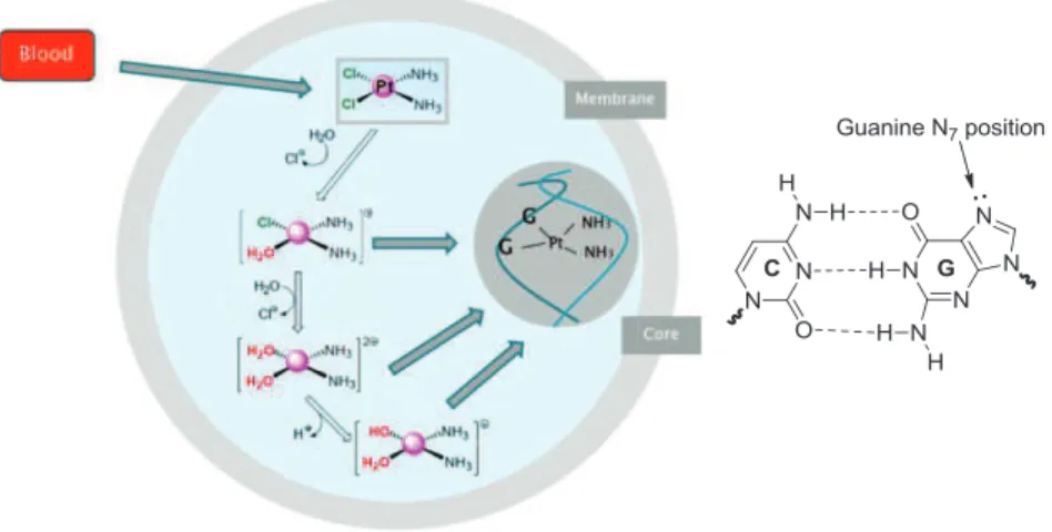
Several studies have shown that the mechanism of action of cisplatin, based on aquation and ligand exchange, is activated within the cell.14 Cisplatin is administered intravenously in a saline solution to prevent aquation. Furthermore, in the blood the concentration of chloride ions (ca. 100 mM) is sufficiently high to prevent hydrolysis. Cisplatin might enter cells using passive diffusion or transporters (copper transporter CTR1).15 Inside the cell (intracellular chloride
concentrations ca. 4-23 mM) the drug will transform by substitution of one or two chlorides (leaving group) with water molecules, to generate various reactive platinum species(Fig. 1.3).14 These new adducts make
covalent bonds with two adjacent guanines of the same strand of DNA or rarely with two guanines in opposite DNA strands. It is not surprising that cisplatin makes covalent bonds with the guanine nitrogen atoms (N7) since these sites are the most electron-dense and accessible sites
in DNA (Fig. 1.3). These new ionic Pt(II) species will change the secondary structure of DNA inhibiting transcription and replication preventing cancer cells growth.16
14 Siddik, Z. H. 2003, Oncogene, Vol. 22, pp. 7265-7279.
15 Sadler, A. M. Pizarro and P. J. 2009, Biochimie, Vol. 91, pp. 1198-1211. 16
18
Figure 1.3. Cisplatin mechanism inside cells and the guanine position for the covalent
bond with cisplatin.
Cisplatin remains the most used metal complex in the treatment of various cancerous malignancies, being very effective, particularly for patients with testicular, ovarian, cervical, bladder and head/neck tumours. Nevertheless, cisplatin administration is often limited because of its particularly toxicity (e.g. vomiting, nausea, ear damage, loss of sensation in hands and kidney toxicity). Another drawback is the intrinsic and acquired resistance possessed by various tumours, causing the lack of response to subsequent cycles of cisplatin treatment. Hence, the medical inorganic chemists have been working to discover less-toxic analogues retaining cisplatin anticancer activity. A large number of complexes have been developed and tested, as ‘second-generation’ and ‘third-generation’ platinum drugs for clinical practice to provide benefits to cancer patients.
19
1.1.1 Covalent binding mode: cisplatin analogues
A large number of platinum(II) based compounds structurally similar to cisplatin have been developed and tested. The platinum drug family tree is presented in Scheme 1.2.13
Scheme 1.2. Platin drug ‘family tree’.
These Pt(II) complexes, following the classical structure/properties relationship elaborated from cisplatin activity, should have two leaving ligands (which can become just one bidentate labile leaving ligand) and
20 ammine ligands coordinated to the Pt(II) ion, neutral nature and cis configuration.14,17
In order to reduce toxicity and improve activity, the type of ligands around the Pt(II) ion has been varied. One of these variation resulted in carboplatin, the first cisplatin analogue used in patients.18 The
structure is slightly different than cisplatin, where two chloride ligands have been substituted with a bidentate dicarboxylate. Carboplatin exhibits lower reactivity and slower DNA binding kinetics, although it forms the same reaction adducts. However, carboplatin has proved to be a chemotherapeutic drug used for ovarian, lung, head and neck tumours. Many years later, oxaliplatin (a cyclohexane-1,2-diamine Pt(II) complex) emerged in continuation with cisplatin. This metal complex has shown better efficacy against tumours than cisplatin and carboplatin, but also causes neurotoxicity at a severity level, limiting its use.19 More than 10 years later the first patient has been treated with
carboplatin, satraplatin was synthesized to be an orally active version of carboplatin. Satraplatin is an octahedral Pt(IV) compound, that is reduced inside cells loosing the two axial acetate ligands and giving rise to more active Pt(II) analogues. This antitumor agent presents a good activity in human cancer cells with acquired cisplatin resistance.20
17 Harper, B. W. Krause-Heuer, A. M. Grnat, M. P. Manohar, M.
Garbutcheon-Sigh, K. B. Aldrich-Wright, J. R. 2010, Chem. Eur. J., Vol. 16, pp.
7064-7077.
18
Harrap, K. R. 1985, Cancer Treat. Rev., Vol. 12, pp. 21-33.
19 Küng, A. Strickmann, D. B. Galanski, M. Keppler, B.K. 2001, J. Inorg.
Chem., Vol. 86, pp. 691-698.
20
21 Picoplatin was designed to provide steric bulky ligands around the Pt(II) centre, changing an ammine ligand with methyl-pyridine ligand. This structure was shown to lead to a relative reduction in inactivation by thiol containing species in comparison to cisplatin. Picoplatin retains activity against a wide range of cisplatin-resistant and oxaliplatin-resistant cells in vitro, which was independent of whether resistance was due to reduced transport, increased cytoplasmic detoxification or increased DNA repair. Moreover, picoplatin possesses antitumor activity in vivo by both the intravenous and oral routes, binding in a very similar manner to cisplatin. Clinical studies have revealed dose-limiting toxicities similar to carboplatin.21
It is well known that transplatin shows less anticancer activity than cisplatin. Since many other complexes in the trans configuration also were found to be ineffective, it was assumed that a cis configuration of the labile groups is required for the antitumor activity of such compounds. However, in the last 20 years, a good number of trans Pt(II) complexes have shown an interesting antitumor activity in vitro and in vivo. One of the last example is represented by the complex [Pt(CQ)2(Cl)2](CQ= choloroquine base) reported in Figure 1.4, where the
CQ ligand bound to the Pt(II) metal centre through the tertiary amine.22
This Pt(II) complex interacts with DNA by covalent binding mode and has shown, in six human tumour cell lines, better activity than cisplatin.
21 Holford, J. Sharp, S. Y. Abrams, M. Kelland, L. R. 1998, Br. J. Cancer, Vol. 77,
3, pp. 366-373.
22 Navarro, M. Castro, W. Higera Padilla, A. R. Sierraalta, A. Abad, M. J. Taylor, P. Sánchez Delgado, R. 2011, J. of Inorg. Chem., Vol. 105, pp.
22 It is interesting to note that the its cytotoxic activity is correlated with the strong interaction observed between this complex and DNA, similar to that shown by cisplatin.
Figure 1.4. Molecular structure of CQ, CQDP and optimized structure of [Pt(CQ)2(Cl)2].
An analogue complex, [Pt(CQDO)2Cl2] (Fig. 1.4) has been studied.
This Pt(II) complex bound DNA through electrostatic interactions and hydrogen bonds, showing less anticancer activity than complex [Pt(CQ)2(Cl)2].22
The different structural changes around the metal ion, to obtain platinum complexes with better properties than cisplatin, have induced in researchers the curiosity to find strategies for the development of new anticancer metal-based drugs. One strategy is the synthesis of new metal complexes with the same cisplatin binding mode, but containing different metal ions and different molecular geometries.
23
1.1.2 Covalent binding mode: non-platinum complexes
The limitation of platinum complexes (high toxicity and incidence of drug resistance) has provided the motivation to research alternative chemotherapeutic strategies for the isolation of effective antitumor compounds based on other metal centres.
The interest for discovery of more efficient metal-based drugs, has given rise to the synthesis of new Pd(II) complexes analogues to cisplatin. Pd(II) has a very similar chemistry to Pt(II), forming squared planar complexes. Two cisplatin analogues were synthesized, cis-[Pd(NH3)2Cl2] and cis-[Pd(DACH)Cl2](DACH =
(1R,2R)-(-)-1,2-diaminecyclohexane) (Fig. 1.5a).23 In contrast to cisplatin, these two
Pd(II) complexes do not show antitumor activity. They hydrolysed very fast interacting with several molecules contained in biological fluids, preventing Pd(II) complexes to reach DNA. To develop antitumor palladium drugs, the Pd(II) complexes must be stabilized by strongly coordinated nitrogen ligands and suitable leaving groups. An example is the new Pd(II) complex [(bipy)Pd(SeO3)] (bipy= 2,2’-bipyridine)
reported in Figure 1.5b, bearing two chelating (N,N) and (O,O) ligands. This Pd(II) complex presents better cytotoxicity than cisplatin making it as a good candidate for anticancer therapy.24 Numerous palladium
complexes with promising activity against some specific tumour cell
23
Butour, S. Wimmer, F. Wimmer, F. Castan, P. 1997, Chemico-Biological Interactions, Vol. 104, pp. 165-178.
24 Mansuri-Torshizi, H. Mital, R. Srivastava, T. S. Parekh H. Chitnis, M. P.
24 lines have been synthesized, applying structural changes around the metal ion such as the change of both leaving ligands and the amine groups or the isomerism. In general, research results indicated that most of the trans-palladium complexes showed a better activity than
cis-platinum and superior activity than that of cis-palladium isomers.
For example the complex reported in Figure 1.5c, [(MP)3Pd(Br)]Br (MP=
2-mercaptopyridine), has shown better cytotoxicity than cisplatin and than its Pt(II) analogues.25
Ruthenium complexes have been also studied as alternative to platinum compounds. One example is the Ru(II) complex [(PDTA)RuCl2]
(PDTA= 1,2-propylenediamine-N,N,N’,N’-tetraacetic acid) reported in Figure 1.5d. This Ru(II) complex has been tested in five different tumour cell lines, showing good activity. The complex is exceptionally well tolerated and shows an absolute lack of toxicity at all given doses.26
Figure 1.5. Metal complexes with antitumor activity. (a) cis-[Pd(NH3)2Cl2] and cis- [Pd(DACH)Cl2] , (b) [(bipy)Pd(SeO3)], (c) [(MP)3Pd(Br)]Br (d) [Ru(PDTA)Cl2].
25
Camarra, M. Berti, T. D’Ancona, S. Cherchi, V. Sindellari, L. 1996, Anticancer Res, Vol. 17, pp. 975-980.
26 Vilaplana, R. Romero, M. A. Quiros, M. Salas, J. M. Gonzalez-Vilchez, F.
25 Another strategy for the design of new anticancer metal-based drugs is the synthesis of new metal complexes (defined as non-classical antitumor metal agents) with an alternative DNA binding mode based on non-covalent interactions, such as electrostatic, p-p interactions or hydrogen bonds.
1.2 Non-classical antitumor metal agents
In the search of new metal complexes showing antitumor activity, a good option was the synthesis of antitumor agents able to interact with DNA by non-covalent interactions.
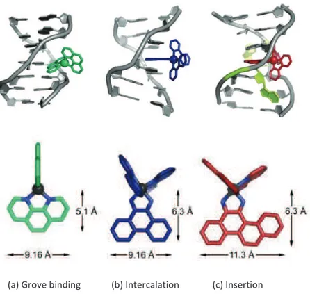
DNA offers many potential non-covalent binding sites due to its size and complexity. Metal complexes as well as pure organic molecules are able to bind DNA via three reversible interaction modes: groove binding (electrostatic interactions), intercalation (p-p stacking, hydrogen bonding and electrostatic interactions) and insertion (p-p stacking interactions) (Fig. 1.6).27 Certain small molecules bind DNA by groove binding, producing a small distortion in DNA. In a detailed analysis of DNA structure, there are two types of grooves that can be seen. The major groove has the nitrogen and oxygen atoms of the base pairs pointing inward toward the helical axis, whereas in the minor groove, the nitrogen and oxygen atoms point outwards. The grooves twist around the molecule on opposite sides. In the case of
27
26 groove binding, reversible intermolecular association such as electrostatic binding occurs due to the interaction between cations and the negatively charged phosphate backbone at the exterior surface of DNA (Fig. 1.6a).28 On the contrary, intercalators are compounds
interacting via p-p stacking interactions through their aromatic planar surface and two base pairs. For an optimal intercalation, the planar part of the molecule should have a minimum surface29 of about 28 Å2
(reached by three to four condensed five or six membered rings) (Fig. 1.6b).28 The intercalation produces a distortion in the DNA chain,
inhibiting cellular replication processes. The majority of non-covalent DNA binding compounds are groove binders or intercalating agents, but there are a small number of compounds interacting with DNA as insertors. Insertion is similar to intercalation, but the insertion agents eject the DNA bases with their planar ligands, acting as a p-stacking replacement in the DNA base stack. The insertion agents present bigger aromatic portion than the intercalating agents, and one example of metal ionic complex is [D-Rh(bpy)2(chrysy)]3+ acting as insertion agent have the coordinated ligand is chrysi (chrysi = 5,6-chrysene quinine diimine) (Fig. 1.6c).29
28 Zeglis, B. M. Pierre, V. C. Barton, J. K. 2007, Chem. Commun., pp.
4565-4579.
29
27
(a) Grove binding (b) Intercalation (c) Insertion
Figure 1.6. The binding modes of metal complexes with DNA and the candidates to
bind DNA.
Continuing the research of new metal-based drug with different cisplatin mechanism of action, intercalation has been shown to be a good strategy for anticancer treatment.
28
1.2.1 Intercalation
Intercalation was first proposed by Lerman30 to explain the strong
affinity for DNA of certain heterocyclic aromatic dyes such as acridines. In particular, Lerman observed a markedly enhanced viscosity and diminution of the sedimentation coefficient of solutions formed by DNA with small amounts of aromatic dyes, contrary to those expected on the basis of aggregation or simple electrostatic effects. These peculiar changes suggested considerable modification of the usual helical structure of DNA, being explained by an intercalation of the compounds between adjacent nucleotide-pair layers by extension and unwinding of the deoxyribose-phosphate backbone. The hydrodynamic changes were therefore the consequences of the diminished bending between layers, the lengthening of the molecule, and the diminished length-specific mass.
Furthermore, the intercalation of coloured fluorescent compounds into DNA has proved to be a rewarding method to detect and understand the intercalation process in itself. For example, ethidium bromide is an aromatic compound that, when exposed to ultraviolet light, shows fluorescence with an orange colour, intensifying almost 20-fold after binding to DNA.31 The reason for ethidium
bromide's intense fluorescence after binding with DNA is the hydrophobic environment found between the base pairs. By moving
30
Lerman, L. S. 1961, J. Mol. Biol., Vol. 3, pp. 18-30.
31
Vardevanyan, P.O. Antonyan, A.P. Manukyan G.A. Karapetyan, A.T. 2001,
29 into this environment and away from the solvent, the ethidium bromide cation is forced to shed any water associated molecules. As water is a highly efficient fluorescent quencher, the removal of water molecules allows the ethidium bromide to fluorescence. Major advances in research have been made with fluorescently labelled DNA.32
The structural modifications inducted by intercalating agents can lead to functional changes, often to inhibition of transcription and replication and DNA repair processes, which make intercalators potent mutagens. The intercalating molecule binds DNA by reversible interactions, causing structural modifications with consequent inhibition of the cell replication processes. Nowadays there is a noticeable interest in the use of intercalating agents to inhibit the cellular reproduction for cancer treatment.
Metal complexes showing DNA intercalative mechanism are built up by at least one intercalating ligand coordinated to the metal centre. These ligands have to be oriented parallel to the base pairs and protruding away from the metal centre (Fig. 1.7).33
32 Bauer, W. Vinogrand, J. 1970, J. Mol. Biol., Vol. 54, pp. 281-289. 33
30
Figure 1.7. Crystal structure of a metallo-intercalator demonstrating the intercalation
via major grove.
Several Pt(II) complexes with square-planar geometry can bind DNA by intercalation. For example in the complex [Pt(terpy)(HET)]+
(terpy= terpyridine, HET= 2-hydroxyethanethiolato), whose chemical structure is presented in Figure 1.8a, the presence of terpyridine aromatic rings provides a substantial degree of overlap with base pairs. Figure 1.8a shows also the Pt(II) complex intercalated between the two base pairs in the double helical fragment.34
Metal intercalation has been also extended to octahedral complexes using Ru(II) derivatives containing multi-heterocyclic aromatic ligands, such as phenantroline (phen) or 9,10-phenanthrenequinone diimine (phi) (Fig. 1.8b,c). The most studied example is the ionic system [Ru(phen)2(dppz)]Cl2 (Fig. 1.8b). The
34 Wang, A. H. Nathans, J. van der Marel, G. van Boom, J. H. X Rich, J. H.
31 complex excited states are highly deactivated by water, showing no luminescence in aqueous solution at room temperature. Nevertheless, intense photoluminescence appears when it intercalates into DNA, because of the local region of “non-water” provided by DNA around the metal complex.35 The chiral complex [Ru(phen)
3]Cl2 build up by
three phenantroline ligands reported in Figure 1.8c, bind to DNA also by intercalation.36 Hypochromicity representing a 17% decrease in intensity in the metal to ligand charge-transfer band and enhanced luminescence accompany binding to the duplex reflecting the decreased mobility of the complex when sandwiched into the helix.
Figure 1.8. (a) Molecular and crystal structure of [Pt(terpy)(HET]+, (b) [Rh(phen)3]Cl3, (c)[Rh(bpy)2phi]Cl and (d) [Ru(phen)2(dppz)]2+.
35
Friedman, A. E. Chambron, J. C. Sauvage, J. P. Turro N. J. Barton, J. K. 1990, J. Am. Chem. Soc., Vol. 112, pp. 4960-5962.
36 Barton, J. K. Danishefsky, A. T. Golberg, J. M. 1984, J. Am. Chem. Soc., Vol.
32 A new alternative intercalation mode is called intercalation by dual function (the intercalating complex can react with DNA by both covalent bond and aromatic intermolecular interactions). One example of a metal complex showing this type of DNA binding mode is [(tpy)Ru(dtdeg)PtCl]Cl3 (tpy=2,2’:6’,2’’-terpyridine, dtdeg=
bis[4’-(2,2’:6’,2’’-terpyridiyl)]-diethylenenglycol ether) (Fig. 1.9), containing a “Ru(tpy)” fragment with a flexible chain joined to a Pt(II) system at the end. This Pt(II)/Ru(II) complex, indeed, was designed to bind covalently with DNA, by mean of the Pt(II) moiety, and intercalating DNA through the molecular fragment “(Ru(tpy))”(Fig. 1.9).37
Figure 1.9. [(tpy)Ru(dtdeg)PtCl]Cl3.
Currently, investigations continue in the search of new metal-complexes with an improved affinity for DNA. Three examples of Cu(II) and Zn(II) DNA intercalating complexes recently published are shown in Figure 1.10.2,38,39
37
van der Schilden, K. Garcia, F. Kooijman, H. Spek, A. L. Haasnoot J. G.
Reedijk, J. 2004, Angew. Chem. Int. Ed., Vol. 43, pp. 5668-5670.
38 Raman, N. Mahalakshmi, R. Mitu, L. 2014, Spectrochimica Acta Part A, Vol.
33
Figure 1.10. New copper(II) and zinc(II) metallo-intercalators.
Several metallo-intercalator systems are based on Zn(II) and Cu(II) metal complexes, as the ones presented in Figure 1.10. Indeed, Zn(II) and Cu(II) metal ions are essential metals for the growth and development of biological systems40, are low cost metals, with extensive coordination chemistry and in the case of Zn(II) may yield luminescent complexes.
As seen in the above examples, most of the ligands used for the design of intercalating metal agents are based on the use of (N,N) aromatic chelating ligands, changing the substituent groups or the aromatic core.
39 Lauria, A. Bonsignore, R. Terenzi, A. Spinello, A. Giannici, F. Longo, A. Almerico, A. M. Barone, G. 2014, Dalton Trans., Vol. 43, pp. 6108.
40 (a) Armaroli, N. Accorsi, G. Cardinali, F. Listorti, A. 2007, Top. Curr. Chem.,
Vol. 280, pp. 69-115. (b) Stefanidou, M. Maravelias, C. Dona, A. Spiliopoulou,
34 All the above described metal-based anticancer agents are known to have DNA as main target. However, several studies are now devoted to the development of new compounds able to stabilize the secondary structure of DNA, called G-quadruplex (GQ).
1.2.2 G-quadruplex binding
As was stated up to now, platinum compounds are mostly used in clinical trials. Nevertheless, their high toxicity and resistance mechanisms, endeavoured researchers to concentrate their efforts world-wide in the discovery and the development of new agents able to be active against cancer by choosing novel selective therapeutic targets, such as G-quadruplex.41
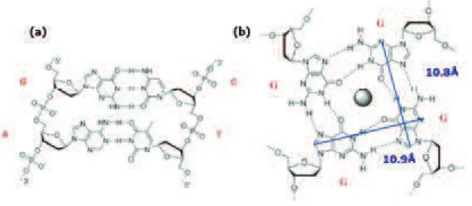
Genetic information is stored in specific sequences of nucleic acids forming the DNA structure. Watson and Crick in 1953 proposed an anti-parallel double helical structure for DNA (duplex DNA) in which the two strands are held together through canonical adenine/thymine and guanine/cytosine base pairing (Fig. 1.11a).42 Several studies on the DNA structure showed that DNA can adopt a wide variety of topologies. In particular, guanine rich sequences of DNA can form a secondary structure called G-quadruplex (GQ). This DNA secondary structure is formed by four guanine bases assembled into a planar structure via
41
Neidle, S. Balasubramanian and S. 2009, Curr. Opin. Chem.l Biol., Vol. 13,
pp. 345-353.
42
35 eight Hoogsteen hydrogen bonds. The guanines carbonyl groups create a strong negative electrostatic potential stabilized by the presence of alkali-metal cations (such as Na+ or K+) (Fig. 1.11b).43
Figure 1.11. (a) The canonical A/T and G/C base pairing. (b) The GQ structure.
Recent studies have shown that there are more than 350000 guanine-rich sequences in human genome with strong tendency to form G-quadruplex structures, in particular at chromosomal extremities (telomeres).44,45,46 Chromosome ends, or telomeres, are
composed of the tandem repeat sequences TTAGGG. In somatic cells, during the cell replication, the telomeres are gradually shortened until the cell death. On the contrary, in tumour cells the activity of telomerase inhibits this action. Telomerase is an enzyme over-expressed in approximately 85% of cancer cells, and it seems to be the
43 Murrat, P. Singht V. Defranq, E. 2011, Chem. Soc. Rev., Vol. 40, pp.
5293-5307.
44
Hupper, J. L. 2008, Biochimie, Vol. 90, pp. 1140-1148.
45 Balasubramanian, J. L. Huppert and S. 2007, Nucleid Acids Rev., Vol. 35, pp.
406-413.
46
36 cause of tumour cell immortality. The development of compounds capable to stabilize this type of DNA secondary structures is considered a new anticancer strategy.41,42,47,48,49
Eventually, the GQ stabilization occurs via p-p stacking and electrostatic interactions.50 Therefore, an effective agent for
G-quadruplex stabilisation should have several structural features, such as a large planar surface (for binding by p-stacking to the guanine quartet), positive charge area at the centre of the molecule (to interact with grooves and loops of the G-quadruplex and the phosphates backbone), similar dimensions to GQ (Fig. 1.11) and selectivity for GQ over duplex DNA (intercalation agent).51
G-quadruplex stabilization agents can bind to the GQ structures by end-stacking and intercalation.37 The dimension and shape of G-quadruplex stabilization agents are crucial for the binder mechanism (Fig. 1.12).
Figure 1.12. Different GQ binding modes.
47 Prommier, J. P. LEbeau, J. Ducray C. Sabatier, L. 1995, Biochimie, Vol. 77,
pp. 817-825.
48 Blasco, M. A. 2003, Eur. J. Cell Biol., Vol. 82, pp. 441-446.
49 Cuesta, J. Read M. A. Neidle, S. 2003, Mini-Rev. Med. Chem., Vol. 3, pp.
11-21.
50
Teulade-Fichou, D. Monchaud and M. P. 2008, Org. Biomol. Chem., Vol. 6, pp. 627-636.
51 Wang, J. T. Zheng, X. H. Xia, Q. Mao, Z. W. Ji, L. N. Wang, K. 2010, Dalton
37 A wide variety of techniques have been employed to study interactions of the designed stabilization agents with G-quadruplex DNA, aiming also to obtain data for structure–activity relationship studies. The key challenge in GQ stabilization agents design is to make them selective for G-quadruplex structures in comparison to duplex DNA or other secondary structures. Because of the high degree of structural polymorphism in GQ, it is important to determine the binding sites of the GQ stabilization agents and their interaction modes (Fig. 1.12).
Some of these techniques are X-Ray diffraction (revealing the dimensions of the complex), circular dichronism (the titration of G-quadruplex with GQ binders induces changes in the CD spectrum), or biological studies such as G-4 FID (based on the ability of a given ligand to displace the fluorescent probe thiazole orange from a G-quadruplex architecture) and FRET-melting (fluorescence resonance energy transfer is based on the monitoring of the stability imparted by a ligand to a fluorescently labelled G-quadruplex structure).
Up to now, several metal complexes with different shape and dimensions, able to act as GQ binders have appeared in the literature. It has been seen that a simple Pt(II) ionic complex as [Pt(phen)2](PF6)2
induces the stabilization of G-quadruplex (Fig. 1.13a). CD assays have shown that the CD spectrum of human telomeric DNA gave two bands at about 290 nm (positive band) and 250 nm (negative band). These two bands upon addition of the Pt(II) complex and in the presence of potassium cations, increase in intensity and reaching saturation. These data clearly suggest that the Pt(II) complex strongly stabilized
G-38 quadruplex conformation because CD offers a reliable means for following the structural conversion of the quadruplex to the unfolded conformation.52 Structural analysis have been done to reveal the structural affinity between the Pt(II) complex and the G-quadruplex. The dimensions of the Pt(II) complex are very similar to the guanine quartet dimensions (the maximal length of the complex is 10.4 Å very close to the 10.8 Å of the G-quadruplex) (Fig. 1.13a and Fig. 1.11b). The dimension and the structure of this Pt(II) complex are important for G-quadruplex stabilization.51
Recently, Pt(II) and Pd(II) complexes with metallo-rectangle shapes (rectangle structure with 12.90 Å of width), reported in Figure 1.13b, were synthesized and where shown to have a high affinity and selectivity for G-quadruplex over duplex DNA.53 Indeed, different studies have demonstrated their G-quadruplex affinity, such as ESI-MS (electrospray mass spectrometry) studies and biological assays such as G-4 FID and FRET studies. Upon addition of the Pt(II) and Pd(II) complexes to the corresponding DNA structure previously incubated with thiazole orange (TO), an increase in TO’s fluoresce intensity was observed. For FRET studies, the Pt(II) and Pd(II) complexes were mixed with different sequences of DNA, showing an increase in melting
52 (a) Monchaud, D. Yang, P. LAcroix, L. Teulade-Fichou, M. P. Mergny, J. L.
2008, Angew. Chem. Int. Ed., Vol. 47, pp. 4858. (b) Rahman, K. M. Reszka, A.
P. Gunaratnam, M. Haider, S. M. Howard, P. W. Fox, K. R. Neidle, S.
Thurston, D. E. 2009, Chem. Commun., pp. 4097. (c) Tan, J. H. Ou, T. M. Hou, J. Q. Lu, Y. J. Huang, S. L. Luo, H. B. Wu, J. Y. Huang, Z. S. Wong, K. Y. Gu, L. Q.
2009, J. Med. Chem., Vol. 52, pp. 2825.
53 Gosh, S. Mendoza, O. Cubo, L. Rosu, F. Gabelica, V. White, A. J. P. Vilar, R.
39 temperature induced by the high degree of G-quadruplex stabilization. Finally, the experimental and computational studies have indicated that these dimetallic rectangles interact with G-quadruplex DNA structures by end-stacking. Their structure is retained in solution (particularly in the case of platinum) and this is likely to be the reason for their high selectivity for quadruplex versus duplex DNA (Fig. 1.13b).
The first example of a G-quadruplex DNA binder based on gold(III) is the complex [Au(pzpy)2Cl] (pzpy= 2-(3’-Pyrazolyl)pyridine) (Figure
1.13c). The size of this [Au(pzpy)2]+ ion is about 9.8 Å x 8.4 Å similar to
G-quadruplex structure. Fluorescent intercalator displacement (FID), surface plasmon resonance (SPR) and circular dichroism (CD) studies clearly indicated that this Au(III) complex stabilizes the GQ DNA. Again, the geometry of the Au(III) complex and its size resulted to be fundamental to function as a good quadruplex DNA binder. 54
54 Suntharalingam, K. Guota, D. Sanz Miguel, P. J. Lippert, B. Vilar, R. 2010,
40
Figure 1.13. G-quadruplex binders: a) Pt(II) ionic complex, [Pt(phen)2](PF6)2 , b) metallo-rectangle shape Pd(II) and Pt(II) complexes, c) gold (III) complex, [Au(pzpy)2Cl].
1.3 Liquid Crystals for/as DNA intercalating agents
The ability of duplex DNA to form liquid crystalline phases was found in the late 1940s.55,56 Since that time, DNA studies have revealed
that linear DNA fragments in aqueous solutions have remarkable self-assembly capability forming multiple liquid crystalline phases (chiral
55
Luzzati, V. Nicolaieff, V. 1959, J. Mol. Biol., Vol. 1, pp. 127. 56
41 nematic and columnar phases) whose nature depends on the DNA concentration.
Initially all these forms of ordering and self-assembly were observed in solutions of long DNA molecules with more than 100 base pairs and were explained on the basis of their elongated shape, their partial flexibility, their acidic nature, and their helical structure. Furthermore, these molecules respect the Onsager theory57 postulating that in the case of cylindrical particles having length L and diameter d, an orientational ordering of the cylinders, favoured by a balance of orientational and translational entropy at volume fractions, above a critical value of 4d/L. According to this theory, no ordering is expected in sub-Onsager regime (L < 4d).
Subsequently, several research groups have presented evidences that solutions of very short duplex B-DNA, display the same type of liquid crystalline ordering, and specifically chiral nematic and columnar phases.58 The explanation of the mesophase formation in this case cannot be the same, since for these short strands L < 3d, and thus there is not enough shape anisotropy to satisfy the Onsager criterion. Therefore it was shown that the liquid crystalline ordering is a manifestation of the stacking forces acting between the paired terminal bases of different oligomeric duplexes.59 Indeed, paired nucleobases are planar polycyclic aromatic hydrocarbons (PAHs), thus
57 Onsager, L. 1949, Ann. New York Acad. Sci., Vol. 51, pp. 627-659. 58
Zanchetta, G. Nakata, M. Buscaglia, M. Clark, N. A. Bellini, T. 2008, J. Phys. Condens. Matter., Vol. 20, pp.494214.
59 Nakata, M. Zanchetta, G. Chapman, B. D. Jones, . C. D. Cross, J. O. Pindak, R. Bellini, T. Clark, N. A. 2007, Science, Vol. 318, pp.1276.
42 forming hydrophobic stacking in aqueous solution for avoiding exposure to water, leading to attractive interactions that acts to hold them together, forming aggregates. This lead to the suggestion that the DNA duplex is a kind of chromonic liquid cristal.60
It is not surprising that many biological systems exhibit liquid crystalline behaviour due to their intrinsic combination of fluidity and order. These two features are the definition of liquid crystals.
1.3.1 Liquid Crystals: general definition
The liquid crystalline state is a phase of matter which lies between the crystalline solid (order) and the isotropic liquid state (mobility and fluidity). The origin of the anisotropic properties of liquid crystals (LCs) is mainly due to the anisotropy in shape of the constituent molecular unit. Liquid crystal-forming molecules, termed mesogens, are generally divided into calamitic and columnar mesogens. The formation of mesophases (liquid crystalline phases) is due though the use of both temperature (thermotropic LCs ) and solvent (lyotropic LCs).61,62
60
Lydon, J. 1998, in Handbook of liquid crystals, ed. D. Demus, J. Goodby, G. W. Gray, H. W. Spiess and V. Vill, Wiley-VCH, Weinheim, Vol. 2B, pp. 98.
61 Atwood, J. W. Steed, J. L. 2009, Supramolecular Chemistry. 62
43
1.3.1.1 Thermotropic Liquid Crystals
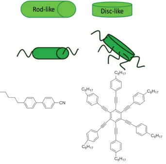
Thermotropic liquid crystals can be divided in two principal categories based on the building unit molecular shape in: calamitic (rod–like mesogens, molecules with a long and cylindrical core with aliphatic chains) and discotic (caused by disc-like molecules, containing a flat and circular core surrounded by aliphatic chains) (Fig. 1.14).
Figure 1.14. Rod-like and disc-like example molecules: 4’-n-pentyl-4-cyanobiphenyl as
rod-like and hexakis((4-octylphenyl)ethylnyl)benzene as disc-like.
There are three main classes of calamitic mesophases: nematic (N), smetic (Sm) and cholesteric.60 Nematic phase is characterized by a high degree of long-range orientational order of the molecules but no long-range translational order (Fig. 1.15a). In smetic phases, molecules
44 maintain the general order of the nematic phase but are aligned in layers. The two main smetic phases are smetic A (SmA) and smetic C
(SmC). In the smectic A phase, molecules can rotate about their long
axes within a given plane in which are orthogonally oriented, but they cannot readily slide past one another (Fig. 1.15b). Small differences between the space filling of the rigid cores and the pendant terminal chains can be compensated by tilting the aromatic cores with respect to the layer normal, giving rise to tilted smectic phases (e. g. SmC phase
reported in Figure 1.15c). A number of at least 12 different symmetries smetic phases have been characterized (labeled SA-SL).
In the cholesteric phase, molecules, instead of lining up in parallel way, are twisting in an ordered fashion. This phase shows very interesting optical properties (Fig. 1.15d).61
Figure 1.15. Calamitic phases: a) nematic phase, N, b) smectic A phase, SmA, c) smectic
C phase, SmC and d) cholesteric nematic phase, N*.
Discotic molecules self-assemble into columns like a pile of coins.62
Columnar LCs (or discotics) can be divided according to the reciprocal columnar organization in the 2D space. In particular, the symmetry of
45 the cell geometry arising from the organized columns can be distinct into different categories: columnar hexagonal (Colh) and columnar
rectangular (Colr) (Fig. 1.16), but also other lower phase symmetries
were reported like columnar oblique or tetragonal.
Figure 1.16. Discotic phases, columnar representation and schematic representation of
the 2D lattices.
Molecular shape, reduced symmetry, microphase separation and self-organisation are important factors that drive the formation of various liquid crystalline phases. The delicate interplay of the molecular interactions coupled with these factors can induce remarkable phase structures and behaviours.
Both organic and inorganic molecules may form liquid crystalline phases. Metal-containing liquid crystals are called metallomesogens. The discipline of metallomesogens has been actively investigated.63,64,65,66 Properties such as enhanced electronic
63 Donnio, B. Guillon, D. Deschenaux, R. Bruce, D. W. 2003, Compr. Coord.
Chem. II, Vol. 6, Metallomesogens. J. A. McCleverty and T. J. Meyer (Eds.), Elsevier: Oxford.
46 polarizability and hyperpolarizability, dichroism, paramagnetism and reactivity have been observed or are expected to result on inclusion of a metal centre in a liquid-crystal compound. Further, induced intermolecular interactions, e.g. metal-metal (metallophilic), ligand-ligand and metal-ligand-ligand, can have profound consequences on the properties and self-assembling abilities, and often lead to the emergence of new physical properties.
Moreover, metallomesogens allow easy access to non-conventional functional mesomorphic materials because of the huge diversity of organic structures that can be used in coordination complexes, as well as the different coordination geometries provided by the metals.64 This, in turn may lead to new types of molecular organization and potentially to new mesophase symmetries. The interest comprises both molecular and supramolecular level. An important variety of new structures has been described in the last few years, inconceivable with purely organic compounds.
Another important aspect of the incorporation of metal ions resides in the induction, modification or enhancement of the mesomorphism of the free parent ligands. New rational design methodologies to produce organized molecular assemblies with
64
Donnio, B. Duncan W. B. 1999, Liquid Crystals II, Structure and Bonding,
Vol. 95, pp. 193-247.
65
Serrano, J. L. 1996, Metallomesogens: Synthesis, Properties, and Applications, Ed. J. L. Serrano.
66 Giménez, R. Lydon, D. P. Serrano, J. L. 2002, Curr. Opin. Solid State Mater.
47 specific interactions (e.g. dipolar and metal-ligand interactions) are, therefore, not only important but also challenging.
Numerous metallomesogens were reported up to date, based on practically almost all available metals, like Ag(I)67, Au(I)68, Zn(II)69,
Pd(II)70, Pt(II)71, etc. Indeed, also high coordination number metal
complexes can exhibit mesomorphism, although initially it was believed that their bulky geometries were incompatible with mesophases formation. Indeed, with properly designed ligands, liquid crystalline materials are now reported with octahedral Ir(III)72, Ru(III)73
67(a) Pucci, D. Barberio, G. Bellusci, A. Crispini, A. La Deda, M. Ghedini, M. Szerb, E. I. 2005, Eur. J. Inorg. Chem., Vol. 12, pp. 2457 – 2463. (b) Pucci, D. Barberio, G. Bellusci, G. et al. 2005, Mol. Cryst. Liq. Cryst., Vol.441, pp. 251 – 260. (c) Pucci, D. Barberio, G. Bellusci, A. Crispini, A. Donnio, B. Giorgini, L.
Ghedini, M. La Deda, M. Szerb, E. I. 2006, Chem. Eur. J. Vol. 12, pp.
6738-3747.(d) Bellusci, A. Chedini, M. Giorgini, L. Gozzo, F. Szerb, E. I. Crispini, A.
Pucci, D. 2009, Dalton Trans., pp. 7381-7389. (e) Pucci, D. Crispini, A. Ghedini, M. Szerb, E. I. La Deda, M. 2011, Dalton Trans., Vol. 40, pp. 4614 – 4622.
2013. (f) Szerb, E. I. Pucci, D. Crispini, A. La Deda, M. 2013, Mol. Cryst. Liq. Cryst., Vol. 573, pp. 34 – 45.
68
Fujisawa, K. Kawakami, N, Onishi, Y. Izumi, Y. Tamai, S. Sugimoto, N.
Tsutsumi, O. 2013, J. Mater.Chem. C, Vol. 1, pp. 5359-5366.
69
Pucci, D. Crispini, A. Ghedini, M. La Deda, M. Liguori, P. F. Pettinari, C. Szerb, E. I. 2012, RSC Adv., Vol. 2, pp. 9071 – 9078.
70 Ghedini, M. Pucci, D. Crispini, A. Aiello, I. Barigelletti, F. Gessi, A. Frnacescangeli O. 1999, Appl. Organometal. Chem., Vol13, pp. 565–581. 71 Ghedini, M. Pucci, D. Crispini, A. Barberio, G. 1999, Organometallics, Vol.
18, pp. 2116-2124.
72 (a) Szerb, E. I. Talarico, A. M. Aiello, I. Crispini, A. Godbert, N. Pucci, D. Pugliese, T. Ghedini, M. 2010, Eur. J. Inorg. Chem., pp. 3270 – 3277. (b) Santoro, A. Prokhorov, A. M. Kozhevnikov, V. N. Whitwood, A. C. Donnio, B. Williamsand, J. A. G. Bruce, D. W. 2011, J. Am. Chem. Soc., Vol. 133, pp.
48 metal ions, or even with the higher coordination number complexes of lanthanide metal ions74.
As stated before, with properly designed ligands, a wide variety of mesophases and symmetries can be induced in metallomesogens. In Fig. 1.17 are presented examples of Ag(I) metallomesogens exhibiting columnar mesophases of different symmetries depending on the nature of the bipyridine ligand substituents and the counterions.
Figure 1.17. Chemical structure and mesomorphism for ionic Ag(I) complexes
containing two bipyridine-based ligands.67-75
73 Pucci, D. Bellusci, A. Crispini, A. Ghedini, M. Godbert, N. Szerb, E. I. Talarico, A. M. 2009, J. Mater.Chem., Vol 19, pp. 7643–7649.
74
(a) Szerb, E. I. Crispini, A. La Deda, M. Pucci, D. Liguori P. F. Petinari, C.
2011, Mol. Cryst. Liq. Cryst., Vol. 549, pp. 86–99. (b) Binemmans, K. 2009, J. Mater. Chem., Vol. 19, pp. 448–453.
75 Pucci, D. Barberio, G. Bellusci, A. Crispini, A. Ghedini, M. Szerb, E. I. 2005,
49
1.3.1.2 Lyotropic Liquid Crystals
Lyotropic liquid crystals form mesophases by the combined action of solvent and temperature. Conventional lyotropic liquid crystal molecules contain flexible rod-shape aliphatic chains ending with ionic groups. One example is the potassium palmitate reported in figure 1.18.76
Figure 1.18. Amphiphilic molecules.
Generally, mesogenic molecules able to form lyotropic mesophases in solvents (mostly water) are usually amphiphilic in nature (dual hydrophilic/hydrophobic character).59 The ambivalent nature of an amphiphile leads to a competition between the hydrophilic parts, attempting to increase their contact with water, and the hydrophobic ones trying to avoid it. Therefore, in high enough concentrations, beyond the so-called critical micelle concentration (cmc), the molecules arrange themselves into micellar aggregate,s with defined topologies, such as hollow spherical, rod-, and disc-like micelles, in which the segregation in space occurs, keeping unlike parts
76 Patel, P. J. Collins J. S. Handbook of liquid crystal research. New York :
50 isolated from unlike solvent (Fig. 1.18). With a further increase of concentration, the micelles begin to interact and to self-assemble into ordered arrays of supramolecular aggregates or lyotropic mesophases. The occurrence of the mesophase is thus driven by the hydrophilic/hydrophobic balance, packing constraints (chain and surface areas) and solvent effects. The nature of the mesophase is strongly connected to the type of aggregation, and the curvature between the polar/apolar interface. For instance, disc-like micelles will form nematic (discotic, ND), and lamellar La phases, cylindrical micelles
will give rise to columnar organisations such as columnar nematic (NCol), and columnar (hexagonal, H, and rectangular, R) phases, and
spherical micelles will arrange themselves into micellar cubic phases.77 Chromonic liquid crystals are considered to be a special sub-class of lyotropic liquid crystals, in which molecules with structures completely different than the conventional amphiphilic mesogens arrange themselves in solvent generating mesophases.
1.3.2 Chromonic Liquid Crystals
Chromonic liquid crystals (CLC) have recently attracted the attention of researchers because of their unique properties consisting in the combination of liquid crystalline properties (self-ordering, ease of alignment, sensitivity to changing conditions and additives) coupled with their optical and electro-optical properties. These characteristics
77
51 make possible the development of a range of sophisticated devices, including polarizers, optical compensators, light-harvesting devices and micropatterned materials.78,79,80,81,82,83,84,85 Furthermore, the fact that they are water-based makes them ideal candidates in biosensors for medical diagnosis.81,86 The most extensively studied chromonic
mesogen is the disodium cromoglycate (DSCG reported in Figure 1.19), an antiasthmatic drug studied initially by Woodard and co-workers in the 1970s and then investigated more extensively by Lydon and co-workers.60,61 Studying the DSCG it was discovered that this compound,
with V-shape, formed two novel liquid crystalline birefringent phases when dissolved in water. Several investigations have also shown that the structural units in both mesophases are columns of stacked molecules.
78 Tam-Chang, I. K. Iverson and S. 1999, J. Am. Chem. Soc., Vol. 121, pp.
5801-5802.
79 Matsunaga, D. Tamaki, T. Akiyama H. Ichimura, K. 2002, Adv. Mater., Vol.
14, pp. 1477.
80
Iverson, I. K. S. Casey, M. Seo, W. Tam-Chang, S. 2002, Lagmuir, Vol. 18, pp. 3510-3516.
81 Nastishin, Y. A. Liu, H. Schenider, T. Narazemko, V. Vasyuta, R. Shiyanovskii, S. V. Lavrentovich, O. D. 2005, Phys. Rev. E: Stat., Nonlinear,
Soc. Matter. Phys., Vol. 72, p. 041711.
82 Lavrentovich, O. D. et al. 7, 294, 370, 13 US Pat., November 2007.
83 Nazarenko, V. G. Boiko, O. P. Park, H. S. Brodyn, O. M. Melchenko, M. M. Tortora, L. Nastishin Y. A. Lavrentovich, O. D. 2010, Phys. Rev. Lett., Vol. 105,
pp. 017801.
84 Simon, K. A. Burton, E. A. Cheng, F. Varghese, N. Falcone, E. R. Wu L. Luk, Y. 2010, Chem. Mater., Vol. 22, pp. 2434-2441.
85 Lydon, J. 2010, Mater. Chem., Vol. 20, pp.10071-10099.
86 Helfinstine, S. L. Lavrentovich O. D. Woolverton, C. J. 2006, Lett. Appl.

![Figure 1.4. Molecular structure of CQ, CQDP and optimized structure of [Pt(CQ) 2 (Cl) 2 ]](https://thumb-eu.123doks.com/thumbv2/123dokorg/2867276.9105/29.892.214.655.373.623/figure-molecular-structure-cq-cqdp-optimized-structure-pt.webp)
 2 , b) metallo- metallo-rectangle shape Pd(II) and Pt(II) complexes, c) gold (III) complex, [Au(pzpy) 2 Cl]](https://thumb-eu.123doks.com/thumbv2/123dokorg/2867276.9105/47.892.217.650.240.589/figure-quadruplex-complex-metallo-metallo-rectangle-complexes-complex.webp)