UNIVERSITY OF NAPLES FEDERICO II
Department of Chemical Sciences
PhD Program in Chemical Sciences
XXXI cycle
2015-2018
TOWARDS DNA-TARGETING MAGIC BULLETS:
SEARCHING FOR POTENTIAL
CONFORMATION-SELECTIVE G-QUADRUPLEX LIGANDS
PhD student:
Dr. Chiara Platella
Tutor: Prof. Daniela Montesarchio
Supervisor: Prof. Luigi Petraccone
Coordinator: Prof. Luigi Paduano
Contents
Summary ... 1
CHAPTER 1 – INTRODUCTION ... 6
1.1 G-quadruplexes: an overview of their structural features ... 6
1.2 Biologically relevant G-quadruplex structures ... 14
1.2.1 Telomerase, telomeres and telomeric G-quadruplexes ... 17
1.2.2 Structural characterization of telomeric G-quadruplexes ... 21
1.2.3 G-quadruplexes in extra-telomeric regions ... 28
1.3 G-quadruplex ligands ... 32
1.4 Aims of the PhD project ... 39
CHAPTER 2 – PREPARATION OF A NOVEL SUPPORT FOR THE ON-LINE SYNTHESIS OF OLIGONUCLEOTIDES ... 41
2.1 Introduction ... 41
2.2 Functionalization of Long Chain AlkylAmine-CPG with 5’-O-DMT, 3’-O-acetylthymidine through a hexaethylene glycol spacer ... 43
2.3 Synthesis of oligonucleotide models on the CPG support ... 46
2.3.1 Assembly and deprotection of a hairpin duplex-forming oligonucleotide covalently attached to the CPG support ... 46
2.3.2 Assembly and deprotection of G4-forming oligonucleotides covalently attached to the CPG support ... 50
2.4 Experimental section ... 51
2.4.1 Synthesis of 4,4’-dimethoxytrityl-hexaethylene glycol-COOH (DMT-HEG-COOH) ... 51
2.4.2 Functionalization of LCAA-CPG with the hexaethylene glycol spacer DMT-HEG-COOH ... 51
2.4.3 Synthesis of 5’-O-DMT, 3’-O-acetyl-thymidine ... 52
2.4.4 Functionalization of support 1 with 5’-O-DMT, 3’-O-acetylthymidine ... 52
2.4.5 Assembly and deprotection of hairpin duplex- and G4-forming oligonucleotides on support 2 ... 53
CHAPTER 3 – G4-CPG ASSAY: AN AFFINITY CHROMATOGRAPHY-BASED METHOD FOR THE SCREENING OF CONFORMATION SELECTIVE G-QUADRUPLEX LIGANDS ... 54
3.1 General procedure for the binding assays on the novel functionalized CPG supports.. 54
3.3 Optimization and validation of the CPG-based method to identify G4-selective ligands
... 60
3.4 Characterization of the conformations adopted by the oligonucleotide models linked to the CPG support ... 67
3.5 Experimental section ... 75
3.5.1 General procedure adopted for the binding assays ... 75
3.5.2 Materials and general methods ... 75
3.5.3 Binding assays on nude CPG and nude OAS ... 76
3.5.4 Use of G4-and hairpin duplex-functionalized CPG and OAS supports for the binding assays ... 76
3.5.5 Confocal microscopy analyses ... 77
CHAPTER 4 – SCREENING AND EVALUATION OF A LIBRARY OF MOLECULES FEATURED BY A FUROBENZOXAZINE NAPHTHOQUINONE CORE ... 78
4.1 Introduction ... 78
4.2 Selection of compound 4 analogs ... 80
4.3 Experimental screenings by the G4-CPG assay ... 80
4.4 Solution studies on the interaction of the selected ligands with oligonucleotide models ... 84
4.4.1 CD and CD-melting experiments ... 84
4.4.2 NMR experiments ... 89
4.4.3 Microscale thermophoresis (MST) experiments ... 95
4.5 Molecular docking ... 96 4.6 Biological assays ... 98 4.7 Experimental section ... 102 4.7.1 Chemistry ... 102 4.7.2 G4-CPG assay ... 102 4.7.3 CD experiments ... 103 4.7.4 NMR experiments ... 103
4.7.5 Microscale thermophoresis (MST) experiments ... 104
4.7.6 Molecular docking ... 104
4.7.7 Biological experiments ... 105
CHAPTER 5 – SCREENING AND EVALUATION OF A LIBRARY OF NAPHTHALENE DIIMIDES ... 107
5.2 Experimental screenings by the G4-CPG assay ... 111
5.3 Biological assays ... 119
5.4 Solution studies on the interaction of NDI-8 with oligonucleotide models by CD and fluorescence experiments ... 122 5.5 Experimental section ... 127 5.5.1 Chemistry ... 127 5.5.2 G4-CPG assay ... 127 5.5.3 Biological experiments ... 128 5.5.4 CD experiments ... 129 5.5.5 Fluorescence experiments ... 129
CHAPTER 6 - STRUCTURAL STUDIES ON THE INTERACTION OF THE DIMERIC NAPHTHALENE DIIMIDE NDI-8 WITH G-QUADRUPLEX MODELS ... 131
6.1 Selection of the G-quadruplex-forming oligonucleotide models for NMR studies .... 131
6.2 NMR structures of the here studied DNA G-quadruplex models ... 131
6.3 Study on the interaction of NDI-8 with M2 and m-tel24 G-quadruplexes ... 135
6.4 PAGE experiments ... 153
6.5 Materials and methods ... 155
6.5.1 Sample preparation ... 155
6.5.2 NMR spectroscopy ... 155
6.5.3 Native PAGE ... 156
CHAPTER 7 – CONCLUSIONS AND PERSPECTIVES ... 157
Abbreviation ... 161
References... 164
List of publications and patents ... 175
1
Summary
In the search for effective and minimally toxic anticancer drugs, G-quadruplex (G4) structures emerged as appealing targets for their crucial roles in human telomeres and oncogene promoters. G4s are non-canonical nucleic acid secondary structures exhibiting marked structural polymorphism. To achieve an optimal recognition selectivity, thus reducing drug toxicity, a major challenge is to identify ligands which are not only structure-selective, i.e. able to discriminate G4 vs. duplex DNA, but also conformation-selective, i.e. able to specifically recognize different G4 conformations. Thus, considerable efforts are currently devoted to the design of molecules able to selectively target conformationally different G4s and discriminate duplex DNA. To be effective, the huge impulse to synthesize putative conformation-selective ligands has to be coupled with fast and reliable High Throughput Screening (HTS).
Fully inserted in this context is this PhD project, whose objectives were: i) the design and synthesis of oligonucleotide functionalized-solid supports for affinity chromatography; ii) the development of an affinity chromatography-based method for the screening of potential conformation-selective G4 ligands; iii) the identification of effective ligands from focused libraries by the here developed affinity chromatography-based method; iv) the evaluation of the recognition specificity of the selected ligands using proper controls, i.e. duplex structures-functionalized supports; v) the biophysical characterization in solution of the best ligands in their interaction with the target DNA; vi) in vitro biological tests to assess the anticancer activity of the selected ligands.
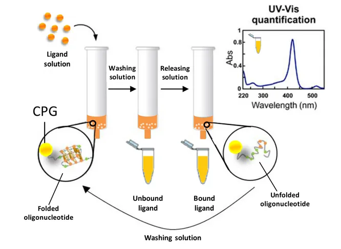
In the search for HTS method enabling a rapid and efficient identification of candidate anticancer drugs, our group recently described an affinity chromatography-based assay, named G-quadruplex on Oligo Affinity Support (G4-OAS), for the screening of libraries of putative G4-binders. It consists in flowing solutions of the potential ligands through a polystyrene resin functionalized with a G4-forming DNA sequence and quantifying the bound ligand by spectrophotometric measurements. Though rapid and simple, using this method we observed some problems with ligands featuring large aromatic cores and low hydrophilicity, which gave unspecific interactions with the polystyrene resin and could not therefore be analyzed.
With the aim of addressing this issue and particularly obtaining universal supports for effective screenings of putative conformation-selective G4-ligands, these studies were extended to Controlled Pore Glass (CPG). CPG is the support of choice for oligonucleotide synthesis; its success is essentially due to its chemical inertness, which in principle renders it more convenient than polystyrene also for affinity chromatography. Crucial in our design was the
2
choice of the linker, which must attach the first nucleoside to the solid support via a covalent bond chemically stable under basic conditions used in the final deprotection step of oligonucleotide synthesis, so to obtain the support-bound, fully deprotected oligonucleotides on which the affinity chromatography binding assays can be performed. Therefore, we designed a novel functionalization for CPG supports, involving a linker – made of a flexible spacer of hexaethylene glycol attached to the first nucleoside monomer (i.e., 5’-O-DMT-3’-O-acetylthymidine) through the nucleobase – suitable for the oligonucleotide elongation by standard protocols and chemically stable to the final deprotection procedure. Hence, using classical phosphoramidite chemistry, biologically relevant G4-forming DNA sequences, taken from human telomeres (tel26, tel46) and oncogene promoters (cmyc, ckit1, ckit2, hTERT1), were synthesized. Furthermore, to evaluate the G4 vs. duplex recognition specificity of the putative ligands, the novel synthesized CPG support was also functionalized with an oligonucleotide able to fold into a stable unimolecular hairpin duplex (ds27). Molecules having good or no affinity for G4s (distamycin, netropsin, resveratrol, RHPS4, TO, TMPyP4, 9-Acr-COOH), as well as molecules taken from a library of G4 ligands (7A, 5B, 10B, 1C, 3C, 7D, 7E, 7F) previously proved to have strong unspecific interactions with OAS, were then exploited to validate and optimize the affinity chromatography-based method with the novel functionalized CPG supports. The general procedure adopted for the binding assays was as follows: a weighed amount of each support was left in contact with a ligand solution in a column equipped with a frit. After incubation on a vibrating shaker, each support was eluted with defined volumes of a washing solution and all the eluted fractions were separately analyzed by spectrophotometric measurements. The composition of the washing solution was optimized so to reach the best compromise in terms of solubility of the tested ligands, minimization of undesired absorption of the tested ligands on the assay equipment and capacity of the oligonucleotide sequences to form stable secondary structures. From spectrophotometric measurements of the fractions eluted from the nude CPG, the amount of ligand unspecifically adsorbed on the support was evaluated. In turn, spectrophotometric analysis of the fractions recovered from G4- and hairpin duplex-functionalized supports allowed estimating the amount of ligand bound to the supports carrying the secondary structure-forming oligonucleotides. In all cases, the amount of bound ligand was calculated by subtracting the ligand eluted upon washing, derived by direct spectrophotometric measurements, from the ligand amount initially loaded on each support. Moreover, as further control, the direct measurement of the bound ligand was obtained by treating each support with a releasing solution, followed by spectrophotometric analysis of the eluted fractions. The amounts of bound ligands thus measured were in good agreement with
3
those previously determined as a difference with respect to the unbound amounts. The composition of the releasing solution was optimized on the basis of its ability to obtain a fast and quantitative recovery of the bound ligand. To allow the correct G4s and hairpin duplex refolding after this treatment, and thus reuse the same batch of support for subsequent binding assays, each support was resuspended in the washing solution and then subjected to an annealing procedure. This consisted in taking the functionalized support at 75 °C for 5 min, followed by slow cooling to room temperature. The reversibility of the process of folding/unfolding of the G4s and the hairpin duplex allowed effectively recycling the support; indeed, a large number of binding assays (typically more than 50) could be performed on the same batch of CPG without losing in efficiency and reliability of the experiments.
Notably, experimental results showed that the nude CPG has much lower unspecific interactions with all the tested model ligands than previously used OAS resin, allowing higher recovery of the ligands with smaller volumes of the washing solution. In addition, the affinity trend found for the tested ligands on the G4- and hairpin duplex-functionalized supports well reflects what observed for the same systems in solution, confirming the general reliability of the method.
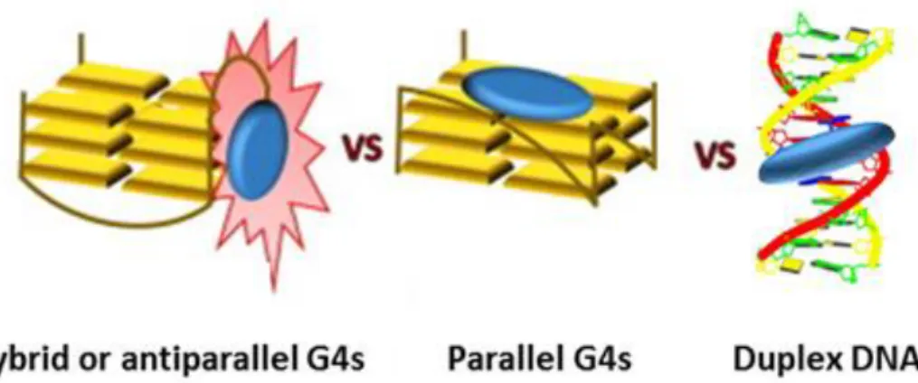
Additionally, to gain a deeper insight into the conformations effectively adopted by the G4s on the glass beads, we exploited a fluorescent core extended naphthalene diimide (cex-NDI)
recently designed to give different fluorescence responses upon interaction with different secondary structure-forming oligonucleotides, thus discriminating hybrid G4s, parallel G4s and duplex DNA. By confocal microscopy, we proved that the oligonucleotides, when anchored to CPG and left in contact with the selected buffer, adopt the same conformations they typically have in solution. This can be mainly attributed to the long and flexible hexaethylene glycol, chosen as the spacer in our synthetic protocol to guarantee proper flexibility and distance from the CPG support of the oligonucleotides, thus minimizing possible steric effects.
This result provided proof-of concept that our novel approach is a powerful tool to identify not only structure-selective G4-ligands, but even conformation-selective G4-ligands.
After full optimization of the G4-CPG assay, two different libraries of putative G4 selective ligands, based either on furobenzoxazine naphthoquinone or naphthalene diimide scaffold, were evaluated.
As far as the focused library of furobenzoxazine naphthoquinone derivatives is concerned, eleven molecules were selected as analogs of a lead-like G-quadruplex targeting compound (4), previously proved to be a strong G4 ligand, differing for the pendant groups on the N-atom of the oxazine ring. These molecules were tested vs. topologically different G4s by the G4-CPG
4
assay. The obtained results showed that all the compounds were able to bind several G4 structures, even though with peculiar preferences, and one of them (S4-5) fully discriminated G4 vs. duplex DNA. Biological assays proved that almost all the compounds produced effective DNA damage, also at the telomeric level, showing marked antiproliferative effects on tumour cells in the low µM range. Combined analysis of the G4-CPG binding assays and biological data led us to focus on compound S4-5, being less cytotoxic than the parent compound 4 on normal cells. An in-depth biophysical characterization of the binding of S4-5 to different G4s was carried out by CD, NMR and Microscale Thermophoresis, demonstrating that the here identified ligand had higher affinity for parallel-type G4s than hybrid-type G4s and duplex DNA. Molecular docking studies in agreement with the NMR data suggested that S4-5 interacted with the accessible grooves of the target G4 structures, giving clues for its increased binding selectivity. Considering that targeting the most variable regions of the G4 structures,
i.e. the grooves and the loops, could be a successful, even though still poorly explored, approach
for the specific recognition of different G4 conformations, and that very few G4-groove binders have been thus far characterized, these results are of great relevance to develop novel effective candidate anticancer drugs.
As far as the naphthalene diimides (NDIs) library is concerned, twelve novel functionalized monomeric and dimeric NDIs were designed, synthesized and evaluated by the G4-CPG assay. Overall, all the tested compounds proved to be effective G4 ligands, also more efficiently targeting intramolecular dimeric than monomeric G4s. In detail, 3, 4, 6, NDI-8 and NDI-9 emerged as the most promising ligands, due to their high G4s vs. duplex DNA selectivity. In addition, G4-CPG assay results provided clear evidence that mitigating the affinity of the NDI binding core for G4s allowed the core selectivity emerging.
In vitro biological assays unambiguously designated, among the five selected NDIs, NDI-8 as
the most promising candidate due to its strong activity against cancer cells (IC50 = 6.6 nM) and
high selectivity in killing cancer cells vs. normal cells. Therefore, NDI-8 was further investigated by CD, fluorescence, NMR and gel electrophoresis analyses. By combination of CD titrations and CD-melting experiments, NDI-8 was proved to preferentially affect the structure and thermal stability of G4s rather than duplex DNA. Moreover, the ability of NDI-8 to induce G4 structures formation in the absence of cations was proved by CD and native PAGE experiments. Furthermore, fluorescence experiments revealed binding stoichiometries of 1:1 and 3:2 for complexes of NDI-8 with a telomeric G4 monomer, while binding stoichiometries of 1:1 and 5:1 were found for NDI complexes with a telomeric G4 dimer. In-depth NMR analyses – performed during my three months-research stay at the Slovenian NMR Centre of
5
Ljubljana under the supervision of prof. Janez Plavec – showed the preferential binding of NDI-8 to the 5’-end spatially close residues, the upper quartet and half of the middle quartet of a modified G4 structure (m-tel24) taken from the human telomeric DNA. Finally, the remarkable ability of NDI-8 to promote the formation of dimeric G4 species was demonstrated by DOSY experiments.
Overall, the novel developed G4-CPG method based on our newly designed CPG support allowed us selecting two promising candidate drugs for in vivo studies, S4-5 and NDI-8, showing respectively binding preferences for parallel G4s and monomeric or higher order telomeric G-quadruplex structures, in addition to their high G4s vs. duplex DNA selectivity. Future work will be directed to extend the G4-CPG assay to biologically relevant human i-motif-forming and viral G4-forming DNA sequences, as well as automate our method.
6
CHAPTER 1 – INTRODUCTION
“DNA comes in many forms”. This review, written twenty-five years ago by Alexander Rich, well-summarized 40 years of molecular genetics supporting the polymorphic nature of DNA.[1] Only few years after the discovery of the three-dimensional structure of the DNA double helix in 1953,[2] it was found that polynucleotide strands are able to form also triple- and quadruple-stranded nucleic acids.[3] Nevertheless, only three decades ago, when also the biological relevance of these peculiar structures was recognized, non-canonical DNA architectures started to be the object of dedicated investigations.[4–7] The possibility to control genetic pathways responsible for a plethora of pathologies, especially cancer, is a still open and revolutionary challenge from both a medical and scientific point of view. Indeed, getting a deeper insight into the complex biological functions of peculiar DNA structures, as well as selectively targeting them by proper drugs, are among the hottest issues in the scientific research field, especially for the development of effective and minimally toxic targeted therapies.
Focus of this PhD thesis are the G-quadruplex (G4) structures of DNA, proved to have crucial roles in telomeres, replication origins and gene promoters, with G4-forming sequences found in regulatory regions of humans, as well as in virus genomes.[8–15]
1.1 G-quadruplexes: an overview of their structural features
A quadruple-stranded nucleic acid was first observed in 1958 when a fiber of polyinosinic acid was studied by X-ray diffraction.[3] Two different structures were proposed, a three- and a four-stranded one,[3] but only in 1974 the actual structure was determined to be quadruple-stranded.[16] Few years before, in 1962, Gellert and co-workers – likely inspired by Bang’s observations on the ability of guanylic acid to form gels, dated back to the beginning of 19th
century[17] – characterized the first guanine quartet from a gelatinous substance formed by
guanosine monophosphates (Figure 1).[18]
Figure 1. A) X-ray diffraction pattern of 5’-GMP; B) X-ray diffraction pattern of 3’-GMP. (Adapted
7
They suggested that four guanines formed a cyclic system and were bound through four pairs of hydrogen bonds. In detail, we know today that each G-quartet (also defined as G-tetrad) is stabilized by eight Hoogsteen-type hydrogen bonds involving, for each guanine, the N1 and the exocyclic NH2 on C2 as H-bond donors, and the O6 and N7 as H-bond acceptors (Figure 2).[15]
Figure 2. Structure of a G-quartet and a G-quadruplex.
Guanine arrangement in a G-tetrad determines the formation of a cavity in the centre of the planar structure, delimited by guanine carbonyl oxygens, which represents a specific binding site for metal ions,typically K+ or Na+.[15,19]
Two or more parallel planes of G-quartets can stack on each other resulting in a G-quadruplex structure (Figure 2), normally featured by a right-handed helical motif with a rise of 3.13-3.30 Å, right-handed twist of 30°, and a diameter of 25 Å.[15]
Moreover, G-quadruplexes exhibit a marked structural polymorphism,[15] which depends on: the number of strands involved in the structure;
the type of linking loops; the relative strands orientation;
the syn/anti conformation of the guanine residues; the nature of the associated metal cations.
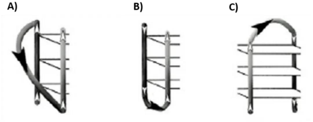
G-quadruplexes can be formed from one (unimolecular), two (bimolecular) or four (tetramolecular) polynucleotide strands (Figure 3).
Three different types of loops can be found in G-quadruplex structures: propeller loops, linking adjacent parallel strands on opposite G-quadruplex surfaces; lateral loops, linking adjacent antiparallel strands on the same G-quadruplex surface; and diagonal loops, linking opposite antiparallel strands on the same surface (Figure 4).[15]
8
Figure 3. A) Unimolecular, B) bimolecular and C) tetramolecular G-quadruplex structures.
Figure 4. Different types of linking loops in G-quadruplex structures: A) propeller, B) lateral and C)
diagonal. (Adapted from Musumeci et al.[20])
Moreover, based on the orientation of the four strands, G-quadruplexes are classified in the following topologies: i) parallel (all parallel strands), ii) antiparallel (all antiparallel) or iii) hybrid (3 parallel and 1 antiparallel) (Figure 5).[15,21–24] In particular, two different
antiparallel-type topologies can be observed: i) chair-antiparallel-type, in which all the three loops are lateral, and ii) basket-type, with two lateral loops and one diagonal (Figures 5C and 5D).[21,23]
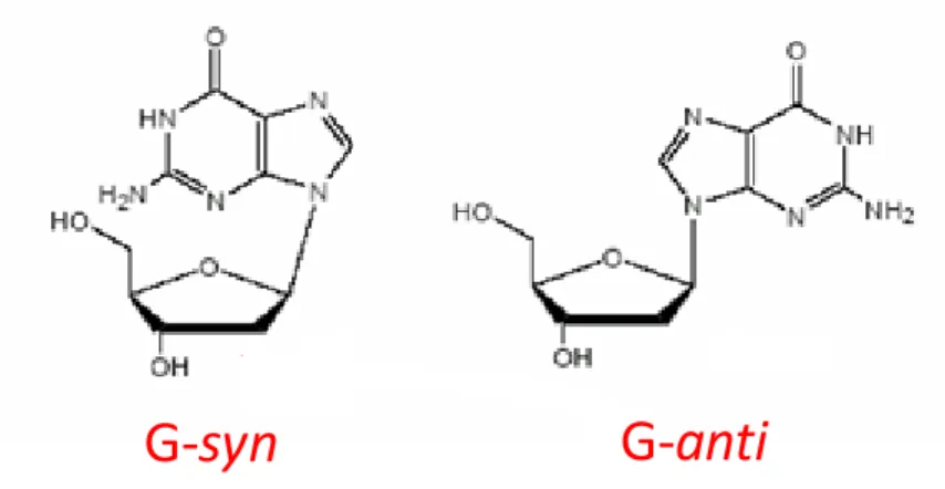
The G-quadruplexes can also be classified according to the conformation adopted by the guanines, which can be syn or anti along the N-glycosidic bond (Figure 6).[15,25] Interestingly,
while in B-DNA nucleobases adopt only the anti conformation, in G-quadruplex structures guanines can adopt either syn or anti conformation, thus giving rise to a variety of different arrangements within the G-tetrads, which sensibly contribute to increase the topological diversity of G-quadruplexes.[26]
9
Differently from B-DNA, in which there are only two different grooves, a major and a minor one, the sugar-phosphate backbone of a G-quadruplex generates four grooves that can accommodate a well-defined network of ordered water molecules.[15] Groove dimension is a consequence of the variation in the glycosidic torsion angles (Figure 7).[27,28]
Figure 5. Different G-quadruplex topologies classified on the basis of strands orientation: A) parallel,
B) hybrid, C) antiparallel (chair-type), D) antiparallel (basket-type). (Adapted from Musumeci et al.[20])
Figure 6. Syn and anti conformations of the guanines along the N-glycosidic bond.
10
Figure 7. G-quadruplex groove dimensions as a consequence of the different glycosidic torsion angles.
Indeed, if a guanosine in the syn conformation points its H-bond donor groups towards a guanosine in the anti conformation, the groove formed between them is narrow.[27,28] Conversely, if a guanosine in the anti conformation directs the N1 and the exocyclic NH2
towards a guanosine in the syn conformation, the groove is wide.[27,28] Finally, if neighbouring guanosines in the G-tetrad adopt the same glycosidic conformation, the groove is medium.[27,28] In particular, in a parallel four-stranded G-quadruplex, where all the guanosines adopt the anti conformation, all the four grooves are of medium width (Figure 7A). Different groove widths alter the hydration network of the resulting structure and determine a different accessibility to hydrogen-bond donors and acceptors of proteins, nucleic acids and small ligands.[27,28]
Intrinsically related to glycosidic conformation of the bases are also the different stacking geometries that two adjacent tetrads can adopt.[29] Considering that, within a G-tetrad, the hydrogen bonds polarity is defined in the direction of hydrogen bond donor to acceptor, from
A)
anti
anti
anti
anti
Medium
M
e
d
iu
m
M
e
d
iu
m
Medium
B)
anti
anti
syn
syn
C)
Medium
Medium
anti
anti
syn
syn
11
NH2 on C2 to N7 and from N1 to O6 (Figure 8), three different stacking geometries in a single
G-quadruplex have been experimentally found: i) “partial 5/6-ring” stacking of the anti/anti step, formed by same-polarity stacked tetrads with a partial overlap of the 5-membered ring of one guanine with the 6-membered ring of another; ii) “5-ring” stacking of the syn/anti step, formed by opposite-polarity stacked tetrads with an overlap of the 5-membered rings of stacked guanines; iii) “partial 6-ring” stacking of the anti/syn step, formed by opposite-polarity stacked tetrads with a partial overlap of the 6-membered rings of stacked guanines (Figure 9).[29]
Figure 8. A) Hydrogen bonds polarity is defined in the direction of hydrogen bond donor to acceptor;
B) Stacking between two tetrads with opposite polarities; C) Stacking between two tetrads with the same polarity. (Adapted from Lech et al.[29])
12
Figure 9. Different stacking geometries experimentally found for two adjacent tetrads in a single
G-quadruplex. (Adapted from Lech et al.[29])
Furthermore, two or more G-quadruplexes can stack on each other forming dimeric and higher-order structures. By analysing all the NMR and crystallographic dimeric G-quadruplex structures in the Protein Data Bank archive, the following stacking geometries were found at the interface of stacked G-quadruplexes: “partial 6-ring”, “6-ring”, “5/6-ring” and “5-ring” (Figure 10).[29]
Figure 10. Different stacking geometries experimentally found at the interface of two stacked
G-quadruplexes. (Adapted from Lech et al.[29])
By computational studies, tetrad stacking energies for experimental geometries were calculated and ranked as follows, core stacking modes: partial 5/6-ring < 5-ring < partial 6-ring, interface stacking modes: 5/6-ring < 6-ring < 5-ring < partial 6-ring.
Finally, investigation of PDB structures and computational studies suggested that 5’-5’ ends stacking with 5/6-ring geometry is the favourite arrangement for two stacked G-quadruplexes and probably is a good model also for higher-order structures.[29]
Additional key elements in the modulation of G-quadruplex topology and stability are the associated metal cations. Several metal cations with different radii can be hosted in the central cavity of a G-quadruplex. Circular dichroism (CD) studies revealed that cations with ionic radii between 1.3 and 1.5 Å, such as K+, Rb+, NH4+, Sr2+ and Ba2+, stabilize the G-quadruplex better
13
than other ions, such as Li+, Na+, Cs+, Mg2+ and Ca2+, due to their optimal fit between two adjacent G-tetrads coordinating eight guanines carbonyl oxygens.[30] Smaller cations, such as Li+, or larger cations, such as Cs+, do not fit well within two consecutive G-tetrads.[30] The general ranking of G4 stabilization by cations is Sr2+ > Ba2+ > K+ > Ca2+ > Na+, NH4+, Rb+ >
Mg2+ > Li+ ≥ Cs+.[31] Notably, if G-quadruplex and monovalent cation concentrations are low enough, some divalent cations, such as Ca2+, Co2+, Mn2+, Zn2+, Ni2+ and Mg2+ can instead induce G4 instability and dissociation.[32] Presumably, the divalent ions interact with the G-quadruplex structures in two different ways: they can bind to the phosphate groups, reducing the charge repulsion, and, at higher concentrations, also coordinate in a bidentate manner the 6-keto and 7-imine groups of guanines involved in hydrogen bonding, thus disrupting the G-quadruplex structure.[33] Therefore, even cations with similar ionic radii, e.g. Ca2+ (0.99 Å) and Na+ (0.97 Å), can have different effects on G-quadruplex stability.[34] Indeed, not only the ionic
radius but also other properties of the cations, including the dehydration energy and the coordination number, are essential to determine their overall effect on folding and stability of G-quadruplex structures.[34,35]
Figure 11. Side view (A, D), top view (B, E) and schematic representations (C, F) of G4 structures
obtained from Oxytricha nova telomeric DNA d(GGGGTTTTGGGG) in the presence of K+ (A, B, C)
or Na+ (D, E, F). K+ ions are located between two adjacent G-tetrads (PDB entry: 1JPQ), while Na+
14
Finally, the precise location of the cations in the G-quadruplex structures is dependent on the nature of the ion: Na+ ions can be located between two G-tetrads in some structures, whereas they lie within the G-tetrad planes in others; K+ ions always form sandwich complexes between two adjacent G-tetrads, coordinating eight carbonyl oxygen atoms (Figure 11).[36]
Structural polymorphism of G-quadruplexes, just outlined, is probably the main reason why nature chose them as key elements for the fine regulation of specific biological mechanisms.
1.2 Biologically relevant G-quadruplex structures
By sequencing many genomes it readily came out that they are rich in sequence motifs containing more consecutive guanines. In particular, bioinformatic analyses, exploiting the Quadparser algorithm based on the consensus sequence d(G3+N1–7G3+N1–7G3+N1–7G3+),
suggested that there could potentially be about 376,000 sequences in the human genome able to fold into G-quadruplex structures.[37] Besides, the above algorithm represents a simplification significantly underestimating the actual number of G4-forming sequences in the genome for several reasons.[37] First of all, since G4s composed of two stacked tetrads are generally less stable than single-stranded or duplex DNA, only sequences able to form three or more G-tetrads are considered physiologically relevant; secondly, loops with lengths from 1 to 7 bases were found to form G4s, with stability decreasing on length increasing; finally, sequences with discontinuities in the G-tracts are not very stable.[37] Indeed, many experimental studies proved
that Quadparser algorithm generates both false positives and negatives. To overcome these limitations, Mergny and co-workers recently developed a different algorithm called G4Hunter.[38] It considers the G-richness of a sequence, as well as the C-richness of its complementary strand able to form a stable duplex competing with the potential G-quadruplex structure, and, on this basis, gives a quadruplex propensity score as the output. After validation of the model by analysis of a large dataset of sequences, it was applied to the human genome proving that the number of sequences able to fold into G-quadruplex structures is higher than what previously estimated by a factor of 2-10.[38] Moreover, high-resolution sequencing-based methods to detect G4s in the human genome also confirmed the higher number of G4s that can be formed in the genome (> 700,000) than previously predicted by computational methods.[39] Notably, G4 location is non-random, with putative G4-forming sequences found in functional and highly conserved human, as well as viral, genomic regions. Indeed, G4-forming sequences
15
have been found in telomeres, oncogene promoters, around transcription start sites regions, introns, immunoglobulin switch regions and 5’ untranslated regions.[9,10,14]
This PhD thesis will focus on DNA G-quadruplex structures found in oncogene promoters and telomeres.[40]
A proto-oncogene is a normal gene coding for a protein responsible for cell growth and division.[41] If a mutation occurs in this gene, it can turn into an oncogene.[41]An oncogene is a gene that encodes for a protein capable of transforming normal cells and inducing cancer.[41] The formation of G-quadruplex structures in oncogene promoters is one of the regulation systems of their transcription.[13] In this context, ligands able to interact with oncogene promoter G-quadruplexes can inhibit a specific mutated proto-oncogene (Figure 12, left).[9]
Telomeres are the ends of eukaryotic chromosomes protecting chromosomal termini from unwanted recombination and degradation, thus guaranteeing proper replication.[42,43] They are
made up of double-stranded DNA of 2-20 kb, and of a 3′ single-stranded overhang of approximately 200 nucleotides containing repetitive nucleotide sequences.[44,45]
Telomerase is the enzyme that adds copies of these repetitive sequences to the end of the single-stranded overhang, providing the main mechanism of telomere length maintenance.[46] This enzyme is transcriptionally repressed in human somatic cells, while it is overexpressed in about 85% of cancer cells. In the remaining 15% of human tumours, telomere lengthening is provided by a different mechanism, known as alternative lengthening of telomere (ALT), based on homologous recombination between telomeric sequences.[44,47]
Thus, while in normal cells telomeres get shorter and shorter over time, leading to irreversible cell growth arrest, also known as cellular senescence, in cancer cells telomeres are maintained to a stable length, making cancer cells immortal.[40] Based on this mechanism, it is now widely accepted that telomere maintenance has a key role in the development of tumours.[42] Hence, interfering with telomere homeostasis is an attractive strategy in the search of new anticancer therapies. The 3′ single-stranded overhang of the telomeric DNA, being a G-rich region with repetitive sequences, can fold into G-quadruplex structures.[48] Ligands able to stabilize these peculiar conformations can alter the single-stranded overhang structure, which consequently is no more recognized and elongated by telomerase or by ALT, thus inducing cancer cells senescence and apoptosis (Figure 12, right).[14,47]
16
Figure 12. Inhibition of oncogene transcription and telomerase activity through G-quadruplexes
formation mediated by ligands.
Hence, there are many advantages in using telomerase activity inhibition mediated by G4 ligands as anticancer therapeutic strategy.[47,49] First of all, telomerase is a specific enzyme for most cancer cells and the most widely expressed tumour marker. Secondly, the risk for development of therapy resistance is very limited because telomeres elongation is the most efficient mechanism for cell immortalization. Thirdly, the very low expression of telomerase in normal cells, along with the longer telomeres in normal stem and germ cells than cancer cells, in principle guarantee specificity towards cancer cells, low toxicity in normal cells, and limited risks in stem and germ cells if the treatment is restricted in time.[47]
The presence of G-quadruplex structures has been proved in the genetic material of human cells by using specific antibodies,[50–52] thus validating both their biological relevance and their role as targets for anticancer strategies (Figure 13).[53–56] In addition, several experiments have fully ascertained that G-quadruplex formation in DNA is modulated during the cell cycle (Figure 13B) and that DNA G-quadruplex structures can be stabilized by small molecule ligands (Figure 13C).[51]
These findings account for the growing interest in the synthesis of small organic molecules as G4 ligands able to interfere with the G-quadruplex functions, essentially aimed at the development of antitumor targeted therapies.
17
Figure 13. A) Visualization of G-quadruplex structures in telomeres and extra-telomeric regions of
metaphase chromosomes by BG4 specific antibody; B) Increase in BG4 foci number during cell cycle, from G0/G1 to S phase; C) Increase in BG4 foci number after treatment with G4 ligand pyridostatin (PDS). Chromosomes and nuclei are counterstained with DAPI (blue). Scale bars correspond to 2.5 μm in A) and 20 μm in B) and C). (Adapted from Biffi et al.[51])
1.2.1 Telomerase, telomeres and telomeric G-quadruplexes
The DNA polymerase is not able to replicate the chromosome up to its termination. This problem is referred to as “end replication problem”.[57] In the absence of an effective mechanism
to solve it, at each cell duplication, a stretch of DNA is not replicated, thus resulting into a loss of genetic information. The solution to this problem is provided by telomerase.[57]
18
Telomerase is an RNA-dependent ribonucleoprotein consisting of two subunits:
hTERT (Telomerase Reverse Transcriptase), which has reverse transcriptase enzymatic activity;
hTR (Telomerase RNA Component), the RNA template used for the synthesis of new telomeric repeats.[58,59]
Telomerase adds repetitive units of oligonucleotide sequences to the 3' end of the chromosomes (Figure 14).[58,59]
Figure 14. Mechanism of action of telomerase.
CCCCAACCCCAACCC-5’ GGGGTTGGGGTTGGGGTTGGGGTTGGGGTTGGGGTTG-3’CCCAACCCCAAC CCCCAACCCCAACCC-5’ GGGGTTGGGGTTGGGGTTGGGGTTGGGG-3’AACCCCAAC CCCCAACCCCAACCC-5’ GGGGTTGGGGTTGGGGTTGGGGTTGGGGTTG-3’AACCCCAAC A) Elongation B) Translocation C) Elongation Telomerase Primase DNA polymerase D) Primer synthesis E) DNA replication F) Primer removal CCCCAACCCCAACCC-5’ GGGGTTGGGGTTGGGGTTGGGGTTGGGGTTG-3’AACCCCAAC CCCCAACCCCAACCC-5’ GGGGTTGGGGTTGGGGTTGGGGTTGGGGTTGGGGTTG-3’AACCCCAAC CCCCAACCCCAACCC GGGGTTGGGGTTGGGGTTGGGGTTGGGGTTGGGGTTG-3’CAACCCCAACCCCAACCCCAAC CCCCAACCCCAACCC GGGGTTGGGGTTGGGGTTGGGGTTGGGGTTGGGGTTG-3’CAACCCCAACCC-5’
19
The RNA template hybridizes to the strand at the 3' end of the target DNA; the nucleotides not complementary to this strand are used as a template for the addition of a defined number of nucleotides; the telomerase moves to the new 3’-end and the elongation step is repeated. When the telomerase has added a high number of repetitions, a primase synthesizes RNA primers and DNA polymerase copies the second strand of the telomere. The primer is then removed, leaving the 3' end as a single strand (Figure 14).[60]
Telomeric repeat sequence is species-specific, but in general is a guanine-rich oligonucleotide. In particular, human telomeres comprise tandem repeats of the short DNA motif d(TTAGGG).[58,59,61]
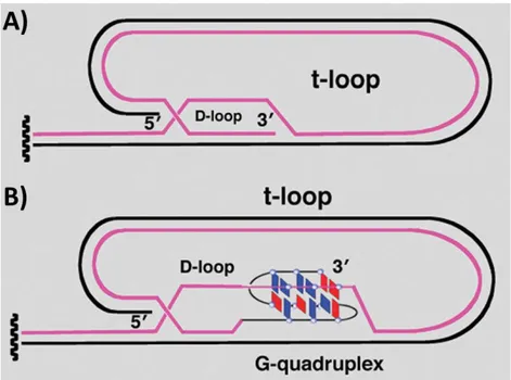
Electron microscopy studies proved that the 3’-overhang of the telomere invades the double stranded region of telomeric DNA, making a structure defined displacement loop or D-loop.[62] Probably, the G-rich 3’-overhang interacts with the double stranded telomeric region by base pairing with the C-rich strand. Consequence of strand invasion is the formation of a duplex lariat structure, defined t-loop (Figure 15A).[45] Recently, a structural study suggested that the
stabilization of the t-loop is mediated by the formation of G-quadruplexes at the level of the D-loop (Figure 15B).[63]
Figure 15. Structure of the 3’ telomeric overhang: A) D- and t-loop; B) stabilization of D-loop mediated
20
Moreover, a six-protein complex, called shelterin, is associated to the human telomere (Figure 16).[62] Three shelterin proteins, TRF1, TRF2, and POT1, directly recognize telomere DNA sequences: TRF1 and TRF2 bind the double-stranded telomeric DNA while POT1 binds the 3’-single stranded overhanging of the chromosome end.[64,65] POT1 is involved in the telomerase maintenance mechanism since it disrupts telomeric G-quadruplexes.[66] Conversely, another shelterin protein, Rap1, interacts with TRF2 and promote G-quadruplex formation.[67] The other two shelterin proteins are TIN2 and TPP1. TIN2 interconnects TRF1 and TRF2 with the TPP1-POT1 heterodimer, while TPP1 interacts with TPP1-POT1 and TIN2 and has the fundamental role of recruiting POT1 at the level of telomeres.[64,65] In conclusion, the shelterin proteins have the complex role of protecting telomeres and regulating their length together with telomerase.[62]
Figure 16. Six protein-complex shelterin.
As mentioned above, telomerase is transcriptionally repressed in healthy somatic cells, therefore telomeres get shorter and shorter over time, and when their length is about 4-6 kb, replicative senescence, also known as mortality stage 1 (M1), is triggered.[68] Hence, normal cells have a defined number of possible cell divisions, the so-called Hayflick limit; once reached this limit, cells undergo to apoptosis. However, some cells can escape M1 and continue to shorten their telomeres, eventually entering mortality stage 2 (M2), featured by genomic instability, fusion/breakage mutagenic events and huge cell death.[68] Furthermore, some cells
can reactivate and overexpress telomerase during M1 or M2, leading to immortalization. Even if immortalization is not enough to induce malignant transformation, immortalization acquired from reactivated telomerase in combination with genome instability and mutation from telomere shortening promote cancer onset.[68]
Small molecules, able to stabilize telomeric single strand into G-quadruplex structures can interfere with telomere lengthening by telomerase, leading to cancer cell death.
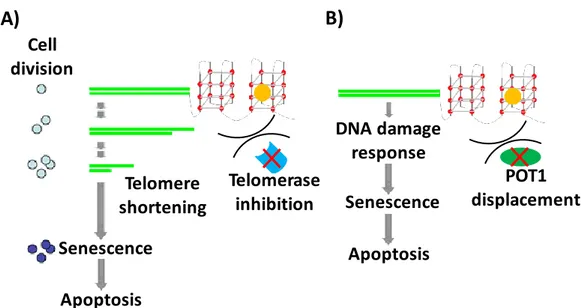
Two mechanisms of action have been proposed for telomeric G4 binding ligands as anticancer agents:[11,14] i) the classical, slow one, according to which inhibition of telomerase activity due
21
to ligand-G4 interaction results in telomere shortening, and, after a defined number of cell divisions, cancer cells enter in senescence and finally in apoptosis (Figure 17A); and ii) the fast one, according to which telomeric G4 binding ligands compete with POT1 protein; loss of POT1 deprotects telomeres, and initiates DNA damage-response mediated cancer cell death (Figure 17B).[11,14]
If G4 ligands followed the slow mechanism, senescence would be reached in about 40-50 days after first treatment with drug. Contrarily, experimental evidence proved that several G4 ligands induce senescence after few days of exposure, validating the fast mechanism as the effective one.[11]
Figure 17. Two different mechanisms of action proposed for telomeric G4 binding ligands as anticancer
agents: A) slowly triggered apoptosis by telomere shortening after several cell divisions; B) fast triggered apoptosis by immediate DNA damage response after POT1 protein displacement. G4 ligands are represented as yellow circles.
1.2.2 Structural characterization of telomeric G-quadruplexes
G-quadruplexes formed at telomeric level have been characterized in detail by NMR spectroscopy, X-ray crystallography and circular dichroism.
By NMR studies, the structure of the G-quadruplex telomeric sequence d[AGGG(TTAGGG)3]
(tel22) has been obtained in solution containing Na+ ions (Figure 18A).[27,69] In these conditions, the oligonucleotide tel22 adopts a basket-type topology formed by three planes of G-tetrads connected through three TTA loops, one diagonal and two lateral ones, and featured by the following N-glycosidic conformations: syn:syn:anti:anti in the upper and lower tetrads, and
anti:syn:syn:anti in the middle one.[27,70] On the other hand, crystallographic studies have revealed a different conformation for tel22 in the presence of K+ ions (Figure 18B).[71] Indeed,
Cell division Telomere shortening Senescence Apoptosis Senescence Apoptosis Telomerase inhibition POT1 displacement DNA damage response
A)
B)
22
in the solid state it adopts a parallel G-quadruplex structure with three propeller loops TTA, with the adenine intercalating between the two thymines in each loop, and all the guanosines in
anti conformation.[70,71]
Figure 18. Schematic drawings of the folding topologies of G-quadruplex telomeric sequence
d[AGGG(TTAGGG)3] (tel22): A) Basket-type unimolecular G-quadruplex in Na+ solution, as
determined by NMR studies; B) Propeller-type parallel-stranded unimolecular G-quadruplex in the presence of K+, obtained by X-ray crystallography. Yellow box = (anti) guanine, red box = (syn)
guanine. (Adapted from Dai et al.[70])
More recent studies, carried out by NMR and CD, have shown that in solutions containing K+ ions, telomeric sequences do not adopt a single G-quadruplex conformation, but present multiple conformations.[72] In particular, two are the major ones. These structures, indicated as hybrid 1 and hybrid 2 (Figure 19), differ significantly from the basket-type or parallel conformations, respectively adopted by telomeric DNA in Na+ solutions or found in the presence of K+ in crystals.
Hybrid structures, both constituted by three planes of G-tetrads, differ in the arrangement of the loops, in the relative orientation of the strands and in the conformation of the N-glycosidic bonds.[72,73] In both structures, there are three parallel strands and one antiparallel, and for this
reason they are also known as (3 + 1) structures: the antiparallel strand is the third in hybrid 1, while the second in hybrid 2.[72,73] As far as the conformation of the N-glycosidic bonds is
concerned, for hybrid 1 the upper tetrad is syn:syn:anti:syn, and the other two are
23
anti:syn:anti:anti.[72,73] Another feature that distinguishes the two hybrid conformations is the size of the grooves, which is obviously different for the two structures.[72,73]
Figure 19. Schematic drawings of: A) hybrid 1 and B) hybrid 2 folding topologies of unimolecular
telomeric G-quadruplexes in K+ solution, determined by NMR using the following sequences: A)
d[AAA(GGGTTA)3GGGAA] and B) d[(TTAGGG)4TT]. Yellow box = (anti) guanine, red box = (syn)
guanine. (Adapted from Dai et al.[72])
It was also demonstrated that the folding of telomeric sequences in one of two possible hybrid conformations is sensibly affected by the flanking sequences.[74] Indeed, while the hybrid 2 conformation seems to be the predominant form in the case of sequences with TT stretches at the 3' end (for example, the 26-mer native sequence d[(TTAGGG)4TT], tel26), hybrid 1 is the
conformation adopted by telomeric sequences which lack the 3’-flanking segment, as in the case of the 24-mer d[(TTAGGG)4] (tel24).[72,75]
This is explained considering that the 3’-flanking TT nucleotides are important for the formation of a stable T:A:T triple capping structure below the lower quartet, that selectively stabilizes the hybrid-2 topology (Figure 20A).[72] Instead, an adenine triple capping structure above the upper quartet was found to form in the hybrid-1 structure, which provides additional stabilization specific to the hybrid-1 topology (Figure 20B).[75]
Another technique extremely useful in structural studies in solution of nucleic acids, and, in particular, for the characterization of the conformations adopted by G-quadruplex-forming oligonucleotides, is circular dichroism spectroscopy.[15,76]
HYBRID 1 HYBRID 2
24
Figure 20. A) Bottom view of the T:A:T triple structure capping the bottom G-tetrad (blue) of hybrid 2
structure (Adapted from Dai et al.[72]); B) Top view of the adenine triple structure capping the top
G-tetrad (light blue) of hybrid 1 structure. The potential hydrogen bonds are depicted with yellow dash lines. (Adapted from Dai et al.[75])
CD spectral signature of G-quadruplex structures is mainly affected by the relative strands orientation and G-tetrads stacking geometries.[77,78] Parallel-stranded G-quadruplexes, in which
all guanines have the anti glycosidic conformation, are characterized by CD spectra with a positive peak at ~265 nm and a small negative peak at 240 nm (Figure 21A).[21] Antiparallel-stranded G-quadruplexes, in which guanines have alternating anti and syn glycosidic conformations along each DNA strand, have CD spectra exhibiting a positive peak at 295 nm, a small negative peak at 265 nm, and a small positive peak at 245 nm (Figure 21B).[21] Conversely, the CD spectrum of a hybrid-type G-quadruplex shows a strong positive peak around 290 nm with a shoulder peak around 270 nm, and a small negative peak at 240 nm (Figure 21C). The positive peak around 290 nm is due to the alternating anti and syn glycosidic conformations along the G-strands between the top and middle G-tetrads, and its shifting from 295 nm is probably due to the presence of the positive peak at 260 nm. The positive peak around 270 nm and the small negative peak around 240 nm are due to the non-alternating anti and syn glycosidic conformations between the middle and bottom G-tetrads, as elucidated by Ambrus
et al.[21]
25
Figure 21. Average CD spectra, obtained from analysis of several G4-forming oligonucleotide
sequences, showing CD spectral features for: A) parallel-type G-quadruplex, B) antiparallel, C) hybrid. (Adapted from del Villar-Guerra et al.[79])
Using CD spectroscopy, the ability of telomeric sequences to interconvert among different G-quadruplex conformations in the presence of different cations has been investigated by means of titration experiments.[21] Stepwise additions of K+ ions to preformed tel26 G-quadruplex in Na+ produces clear spectral changes (Figure 22A), indicating the conversion from a basket-type to a hybrid-type topology. The inverse titration, i.e. adding Na+ ions to preformed tel26 G-quadruplex in K+, does not induce changes in the CD spectrum (Figure 22B), indicating that the presence of Na+, even in large excess, does not affect the conformation induced by K+.[21] These results indicate that the hybrid-type telomeric G-quadruplex is the predominant form in the presence of K+. The lower stability of the basket-type conformation is generally attributed to the steric interference of the flanking sequences with the diagonal loop, both positioned on the same side of the structure.[21]
Furthermore, considering that the intracellular environment is featured by higher concentrations of K+ ions than Na+,[80] the above study also suggests that hybrid-type is the predominant
conformation adopted by telomeric G-quadruplexes in cellulo.
In addition, a peculiar structural feature of human telomeric DNA is the ability of forming higher-order structures, known as multimers.[70,78] Indeed, since telomeric DNA is ca. 100-200
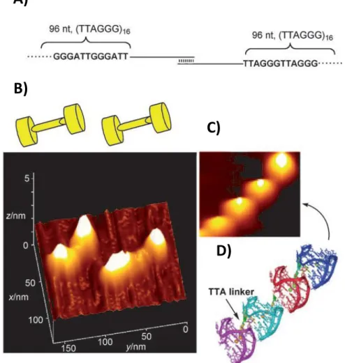
nucleotides long, it can potentially fold into about 8 consecutive G-quadruplexes.[81,82] However, unfavourable coupling free energies could limit complete folding of the 3’-overhang.[83]
An elongated telomeric sequence, depicted in Figure 23A, has been visualized by atomic force microscopy (AFM).[84] The AFM image clearly shows that the higher-order G-quadruplex
26
consists of blob-shaped protrusions arranged end-to-end and formed by adjacent G-quadruplex units (Figures 23B, 23C and 23D).
Further studies proved that higher-order telomeric G-quadruplexes preferentially adopt structures in which two adjacent quadruplex units have different folding motifs, thus forming a characteristic hybrid1-hybrid2 interface stabilized by stacking interactions (Figure 24).[85,86] Therefore, it can be concluded that monomeric and multimeric G-quadruplexes folded into hybrid topologies can be considered as biologically relevant targets with unique binding sites for ligands,[87,88] and used for the design of novel classes of telomeric-specific DNA drugs.
Figure 22. A) CD titration experiments of tel26 by adding K+ in the presence of 150 mM Na+; B) CD
titration experiments of tel26 by adding Na+ in the presence of 100 mM K+. (Adapted from Ambrus et
al.[21])
A)
27
Figure 23. A) Schematic drawing of a DNA model used for AFM visualization. The model has a
TTAGGG repeat sequence (96 nt) at both ends and a single-stranded DNA segment (240 nt) linked by a 15 nt duplex; B) AFM image of a DNA model and schematic drawing of its dumbbell-shaped structure; C) High resolution AFM image of higher-order telomeric DNA structures; D) Putative model for the higher-order telomeric DNA structure: four G-quadruplex units linked by TTA linkers. (Adapted from Xu et al.[84])
Figure 24. Schematic model of G-quadruplex multimers in human telomeres alternating hybrid 1 and 2
topologies. (Adapted from Dai et al.[70])
A)
B)
C)
D)
28
1.2.3 G-quadruplexes in extra-telomeric regions
Interestingly, even if the highest abundance of G-rich sequences is at telomeres, the majority (~75%) of G4s are found in extra-telomeric regions, such as in the tumour-related gene promoters c-myc, c-kit and hTERT.[9]
c-Myc oncoprotein regulates the expression of 15% of all human genes and its main roles are to promote cell proliferation and arrest cell differentiation.[89] Regarding the c-myc oncogene, many studies have proved that its dysregulation is closely related with tumour initiation and progression. Indeed, 20% of human cancers can be associated with the overexpression of c-myc.[90] Furthermore, c-myc activation increases the expression of hTERT, the gene coding for
the telomerase catalytic subunit, therefore involved in cell immortalization.[91] Noteworthy, the
first G4 ligand which entered phase II clinical trials is quarfloxin (ClinicalTrials.gov Identifier: NCT00780663), selectively targeting the c-myc G4.[92] Overall, these findings validate c-myc G4 as an attractive anticancer target. Although the c-myc transcription is regulated by multiple promoters, the nuclease hypersensitivity element III1 (NHE III1), consisting of 33 nucleotides, controls 80-90% of the transcriptional activity of c-myc.[93–96] The 33-mer sequence d(TGGGGAGGGTGGGGAGGGTGGGGAAGGTGGGGA) (cmyc)in NHE III1 contains six G-tracts of unequal length and is potentially able to fold into different G4 conformations, involving different guanine runs.[6,97,98] Shorter sequences extracted from the 33-mer involving, respectively, the G-tracts 2, 3, 4, and 5 (Myc2345, Figure 25A) and 1, 2, 4 and 5 (Myc1245, Figure 25B) have been investigated by NMR and proved to fold into parallel-type topologies, with Myc2345 G4 resulting as the most stable one.[22,97,99,100] Interestingly, in contrast to stability data obtained by melting experiments, only Myc1234 G-quadruplex (Figure 25C) was observed in supercoiled plasmid models, thus pointing out the importance of supercoiling in quadruplex formation and re-evaluating Myc1234 as the potential biologically relevant G-quadruplex in the c-myc promoter.[101]
The c-kit protein regulates several signal transduction cascades important for the control of cell growth and proliferation.[102–104] Mutations and overexpression of the c-kit oncogene play central roles in oncogenic transformation and are especially related to gastrointestinal stromal tumours (GIST).[105] Despite c-kit kinase inhibitors have been recently approved for clinical use in the treatment of GIST, resistance related to protein mutations remains to be overcome.[106–
108] Therefore, the development of small molecules effective as c-kit G-quadruplex ligands can
29
Figure 25. Schematic drawings of: A) Myc2345 d(TGAGGGTGGGGAGGGTGGGGAA) structure in
K+ solution, obtained by NMR; B) Myc1245 d(TGGGGAGGGTTTTTAGGGTGGGGA) structure in
K+ solution, determined by NMR; C) Myc1234 structure proposed by in vitro plasmid footprinting
experiments. (Adapted from Phan et al.[97] and Sun et al.[101])
c-kit promoter contains two different G-quadruplexes (ckit1 and ckit2) separated by about three turns of DNA.[109] In detail, one is located between -87 and -109 bp and the other between -140 and -160 bp upstream of the transcription initiation site.[109] The molecular structure of the 22-mer d(AGGGAGGGCGCTGGGAGGAGGG)ckit1 G4 has been determined by NMR (Figure 26A).[110] Having a unique topology with respect to the other known G4s, it results a specific target for drug design.[110,111] In addition, the crystal structure determined for ckit1 G4 is in good agreement with the one solved by NMR.[112] ckit1 G4 adopts a parallel conformation with two single-nucleotide propeller loops and a long five-nucleotide lateral stem loop; moreover, one non-G-tract guanine is involved in the core of the G-quartets (Figure 26A).[110,112,113] On the other hand, the 21-mer ckit2 of sequence d(CGGGCGGGCGCGAGGGAGGGG), depending
C)
Myc1234
Myc2345
30
on the K+ concentration in solution, adopts two distinct parallel conformations in slow exchange: i) a monomeric one with two single-nucleotide and a long five-nucleotide propeller loops (Figure 26B) and ii) an unusual dimeric structure, in which stacking interactions between the two G-quadruplexes are mediated by a sandwiched A-A non-canonical pair (Figure 26C).[114]
Figure 26. Schematic drawings of: A) ckit1, B) ckit2 monomeric form and C) ckit2 dimeric form
determined by NMR respectively in 70 mM KCl, 20 mM potassium phosphate buffer, 20 mM KCl, 5 mM potassium phosphate buffer and 100 mM KCl, 5 mM potassium phosphate buffer. (Adapted from Phan et al.[110] and Kuryavyi et al.[114])
hTERT is the gene coding for the catalytic subunit of telomerase.[43,115] Its overexpression is associated with over 85% of human cancers, and its transcriptional repression accounts for the lack of telomerase activity in normal somatic cells.[43,115,116] Expression of hTERT is mainly regulated by the core promoter containing five Sp1 (Specificity protein 1) binding sites and one c-Myc protein binding site.[91,115] In particular, the middle three Sp1 binding sites (-167÷-100) have proved to be crucial for hTERT overexpression.[91] There are two different structural models for this G-rich 68-mer sequence, both obtained by low resolution techniques: one consisting of a hairpin structure, a hybrid G-quadruplex and a parallel one (Figure 27A),[117] and the other consisting of three contiguous stacked parallel G4s (Figure 27B).[118] A detailed NMR study was performed for the shorter sequence d(AGGGGAGGGGCTGGGAGGGC) taken from hTERT core promoter, proving the coexistence of a hybrid (> 40%) and a propeller-type parallel-stranded (< 40%) G-quadruplexes in equilibrium (Figure 27C).[119]
A)
B)
31
Figure 27. Schematic drawing of two different structural models for the hTERT 68-mer sequence
obtained by low resolution techniques: A) model consisting of a hairpin structure, a hybrid G-quadruplex and a parallel one, and B) model consisting of three contiguous stacked parallel G4s. C) Schematic drawing of the structural models obtained by NMR for the shorter sequence taken from hTERT d(AGGGGAGGGGCTGGGAGGGC) in K+ solutions: on the left, the hybrid one, on the right the
parallel one. Light blue box = (anti) guanine, violet box = (syn) guanine. (Adapted from Palumbo et
al.[117], Chaires et al.[118] and Lim et al.[119])
A)
B)
32
1.3 G-quadruplex ligands
Small organic molecules can interact with G-quadruplex structures through non-covalent interactions, i.e.: i) stacking with terminal G-tetrads and/or nucleobases in loops or in the flanking segments, and/or ii) hydrogen bonds or electrostatic interactions with the grooves or loops (Figure 28).[120]
Figure 28. Different binding modes for G-quadruplex ligands: A) stacking with terminal G-tetrads; B)
hydrogen bonds or electrostatic interactions with grooves.
Therefore, in order to bind with high affinity G-quadruplex structures, in the design of potential small molecule-based ligands three common features are required:[15]
a large aromatic core, to realize π-stacking interactions with the G-quartet surface; H-bond donors and/or acceptors, to respectively bind acceptors and donors of the
oligonucleotide;
positive charges, to interact with the negative phosphate groups of the DNA backbone through electrostatic interactions.
Since G-quadruplex binding ligands features are also those generally required to interact with duplex DNA, typically ligands recognizing G-quadruplexes are able to interact also with double-stranded DNA.[48] Furthermore, considering that DNA in duplex form is present in the chromosomes in large excess with respect to G-quadruplex structures, a significant increase in selective recognition of G-quadruplexes vs. duplex DNA is one of the main requirements to allow the use of G4 ligands as potential anticancer drugs.[48] Essentially, the structural requirements to guarantee G-quadruplex/duplex selectivity involve steric features of ligands and ability to discriminate between quadruplex and duplex grooves, representing the main diversity elements between the two structures.
Several small molecules were extensively studied by NMR and X-rays to obtain detailed information on their interaction with G-quadruplex DNA.
33
For the pentacyclic acridinium RHPS4, G-quartets end-stacking binding mode was observed in the NMR structure of the complex between the ligand and the parallel human telomere d[(TTAGGGT)]4 G-quadruplex (Figure 29).[121] Several studies showed that the ligand inhibits
telomerase and induces telomere uncapping and damage. Furthermore, in vivo anticancer activity of RHPS4 proved to be very rapid in xenograft models and well-correlated with its telomerase-inhibitory properties.[122]
Figure 29. A) NMR structure of the parallel-stranded DNA quadruplex d[(TTAGGGT)4] complexed
with RHPS4 (PDB entry: 1NZM). RHPS4 is shown as a space-filled model coloured by atom type, the two potassium ions as violet balls and the four strands in different colours. B) Chemical structure of RHPS4.
For the 3,6,9-trisubstituted acridine BRACO-19 (Figure 30), a crystal structure of its complex with two bimolecular human telomeric G-quadruplexes of sequence d(TAGGGTTAGGGT), arranged in a 5’-3’ stacking, was reported.[123] This ligand showed satisfactory
G-quadruplex/duplex selectivity and antitelomerase activity (IC50 = 115 nM).[124] In vivo
anticancer activity reported for BRACO-19 in xenograft models is consistent with its ability of inhibiting the capping and catalytic functions of telomerase.[125]
For the tetra-N-methyl-4-pyridyl porphyrin TMPyP4 (Figure 30), high affinity for G-quadruplex structures and ability to inhibit telomerase activity (IC50 = 6 μM) were proved.[126]
By X-ray crystallography studies, the structure of TMPyP4 bound to the bimolecular human telomere G-quadruplex d(TAGGGTTAGGG) was determined.[127] The observed binding mode indicated that two porphyrin molecules stack on the TTA nucleotide tract, either of the external
A)
B)
34
loop or at the 5' region, without direct contacts with the G-quartets.[127] Contrarily to what observed for the telomeric sequence, TMPyP4 in the complex with a modified sequence taken from c-myc promoter d(TGAGGGTGGIGAGGGTGGGGAAGG) directly stacks on the upper quartet, as determined by NMR.[128] Interestingly, TMPyP4 showed promising anticancer activities in vivo, down-regulating the transcription of both c-myc oncogene and human telomerase reverse transcriptase.[129]
Figure 30. Chemical structures of BRACO-19 and TMPyP4.
Telomestatin is a natural metabolite extracted from Streptomyces anulatus bacteria.[130] It showed a high level of G-quadruplex stabilization, quadruplex/duplex selectivity and antitelomerase activity (IC50 = 5 nM).[131,132] Even if it is one of the most promising
G-quadruplex ligands as anticancer drug candidate, no structure has been solved yet for G4 complexes with telomestatin. However, a high-resolution structure of the complex between the telomestatin derivative and the intramolecular human telomeric G-quadruplex d(TTGGGTTAGGGTTAGGGTTAGGGA) was recently determined by NMR (Figure 31).[133] This compound interacts with the upper quartet through π-stacking and with the loops by electrostatic interactions. Moreover, telomestatin anticancer activity was proved in xenograft mouse model,without displaying any relevant toxicity.[134]
Quinacridine-based molecules also proved to be effective G-quadruplex stabilizing ligands (Figure 32).[135] They interact with the tetramolecular telomeric G4 d[(TTAGGGT)4] by
stacking with two G-quartets and by electrostatic interactions with the grooves.[135] These compounds showed remarkable in vitro anticancer activity with IC50 values in the 100-500 nM
range.[135]
Perylene diimides are characterized by a broad hydrophobic core with two external amine pendant groups. This family of compounds was shown to be active as telomerase inhibitors (IC50 =20 µM).[136] In particular, the perylene derivative PIPER (Figure 32) proved to induce
TMPyP4 BRACO-19
35
G-quadruplex formation in vitro from both telomeric DNA and the hTERT promoter region and inhibit telomerase in a dose-dependent manner.[137,138] In these experiments, the telomerase activity well-correlated with the level of hTERT mRNA, suggesting that the reduction in telomerase activity by PIPER is due to the down-regulation of hTERT expression.[137]
Figure 31. A) NMR structure of the unimolecular DNA quadruplex d(TTGGGTTAGGGTTAGGGTTAGGGA) complexed with the telomestatin derivative here depicted (PDB entry: 2MB3). The backbone is represented as a ribbon. The top G-tetrad and the ligand are highlighted: the potassium ion is coloured in orange, the nitrogen atoms are coloured in blue, the oxygen atoms are coloured in yellow, the ligand is coloured in magenta and the guanine residues are coloured in cyan. B) Chemical structure of the telomestatin derivative used in NMR study. C) Chemical structure of telomestatin. (Adapted from Chung et al.[133])
Figure 32. Chemical structures of a quinacridine-based molecules and PIPER.
Telomestatin Telomestatin derivative A) B) C)
PIPER
meta-Quinacridine
n-propylamine
36
Other interesting molecules as high affinity G-quadruplex ligands are naphthalene diimides (NDIs).[139,140] It has been shown that the substitution pattern of the NDI core as well as the chemical nature of the substituents play a crucial role in the G4 binding and selectivity toward G4 rather than duplex DNA.[141,142] Neidle et al. have shown that in a crystal structure π-π stacking of the NDI naphthalene core with the G-tetrad of telomeric G4 d[AGGG(TTAGGG)3]
occurs at the 3’-end, while the 5’-end is stacked on the 5’-end of a second G4, resulting in the formation of a 5’-5’ dimer (Figure 33).[143] In addition, the NDI positively charged substituents
protrude into the G4 grooves and interact with the phosphates of the DNA backbone (Figure 33).[143] Recently, some NDIs have been shown to be very cytotoxic on selected cancer cell lines,[144] and proved to have antitumor activity in vivo in human pancreatic ductal adenocarcinoma (PDAC) animal models.[145]
Figure 33. A) Chemical structures of naphthalene diimide based molecules. B) and C) Cartoon representations of the naphthalene diimide-based molecules complexed with telomeric G4 d[AGGG(TTAGGG)3]. The end-to-end quadruplex dimers are shown, highlighting packing, topology
and ligand binding orientation. The potassium ions are shown as blue spheres. (Adapted from Collie et
al.[143])