doi: 10.3389/fncel.2019.00524
Edited by: Annalisa Scimemi, University at Albany, United States Reviewed by: Andrea Tamas, University of Pécs, Hungary Carmelo Sgobio, German Center for Neurodegenerative Diseases (DZNE), Germany *Correspondence: Lucia Ciranna [email protected]
Specialty section: This article was submitted to Cellular Neurophysiology, a section of the journal Frontiers in Cellular Neuroscience Received: 03 September 2019 Accepted: 08 November 2019 Published: 27 November 2019 Citation: Ciranna L and Costa L (2019) Pituitary Adenylate Cyclase-Activating Polypeptide Modulates Hippocampal Synaptic Transmission and Plasticity: New Therapeutic Suggestions for Fragile X Syndrome. Front. Cell. Neurosci. 13:524. doi: 10.3389/fncel.2019.00524
Pituitary Adenylate
Cyclase-Activating Polypeptide
Modulates Hippocampal Synaptic
Transmission and Plasticity: New
Therapeutic Suggestions for Fragile
X Syndrome
Lucia Ciranna
1* and Lara Costa
21Department of Biomedical and Biotechnological Sciences, University of Catania, Catania, Italy,2Department of Clinical
and Experimental Medicine, University of Messina, Messina, Italy
Pituitary adenylate cyclase-activating polypeptide (PACAP) modulates glutamatergic
synaptic transmission and plasticity in the hippocampus, a brain area with a key role
in learning and memory. In agreement, several studies have demonstrated that PACAP
modulates learning in physiological conditions. Recent publications show reduced
PACAP levels and/or alterations in PACAP receptor expression in different conditions
associated with cognitive disability. It is noteworthy that PACAP administration rescued
impaired synaptic plasticity and learning in animal models of aging, Alzheimer’s disease,
Parkinson’s disease, and Huntington’s chorea. In this context, results from our laboratory
demonstrate that PACAP rescued metabotropic glutamate receptor-mediated synaptic
plasticity in the hippocampus of a mouse model of fragile X syndrome (FXS), a genetic
form of intellectual disability. PACAP is actively transported through the blood–brain
barrier and reaches the brain following intranasal or intravenous administration. Besides,
new studies have identified synthetic PACAP analog peptides with improved selectivity
and pharmacokinetic properties with respect to the native peptide. Our review supports
the shared idea that pharmacological activation of PACAP receptors might be beneficial
for brain pathologies with cognitive disability. In addition, we suggest that the effects of
PACAP treatment might be further studied as a possible therapy in FXS.
Keywords: pituitary adenylate cyclase-activating polypeptide, fragile X syndrome, cyclic adenosine monophosphate, long-term depression induced by metabotropic glutamate receptor, hippocampus, learning
INTRODUCTION
Pituitary adenylate cyclase-activating polypeptide (PACAP) was initially discovered in ovine
hypothalamus as an endocrine regulator (
Miyata et al., 1989
). PACAP is highly expressed in the
brain and in peripheral tissues in two forms of 38 and 27 amino acid residues, PACAP-38 and
PACAP-27 (
Arimura et al., 1991
;
Koves et al., 1991
). In the present review, “PACAP” refers to
PACAP-38, the most abundant form in the brain (
Arimura et al.,
1991
;
Vaudry et al., 2009
). We will specifically indicate PACAP-38
and PACAP-27 to underline differences between the two forms.
Pituitary adenylate cyclase-activating polypeptide activates
two main classes of G protein-coupled receptors: PAC1 and
VPAC (
Vaudry et al., 2009
). PAC1 is PACAP specific, with
a high nanomolar affinity for PACAP and a 1,000-fold lower
affinity for the structurally related vasoactive intestinal peptide
(VIP). VPAC receptors include VPAC1 and VPAC2 subtypes,
with equally high nanomolar affinity for PACAP and VIP.
PAC1 and VPAC receptors are expressed in peripheral tissues
and in the central nervous system (CNS), where PAC1 is the
most abundant (
Jolivel et al., 2009
). All PACAP/VIP receptors
are positively coupled to adenylate cyclase; PAC1 receptors
can also activate phospholipase C and Ca
2+release. The
brain localization, pharmacological features, signal transduction
mechanisms, and biological effects of PACAP/VIP receptors
are described in details in excellent reviews (
Dickson and
Finlayson, 2009
;
Vaudry et al., 2009
;
Harmar et al., 2012
;
Hirabayashi et al., 2018
).
In the CNS, PACAP is a neurotrophic and a neuroprotective
factor regulating differentiation of neuronal precursors,
promoting neuronal survival, and exerting neuroprotective
effects after brain damage (
Arimura et al., 1994
;
Shioda and
Nakamachi, 2015
;
Reglodi et al., 2018c
). Acting on the brain,
PACAP also regulates important physiological functions,
among which are feeding (
Sekar et al., 2017
), circadian
rhythm (
Holland et al., 2018
), body temperature (
Tan et al.,
2016
), learning, and memory (see below). We will initially
describe the physiological role of PACAP on learning and then
highlight recent findings showing an involvement of PACAP
in cognitive disability. Finally, we will discuss the possibility
to use PACAP as a pharmacological tool in conditions of
learning impairment.
PITUITARY ADENYLATE
CYCLASE-ACTIVATING POLYPEPTIDE
MODULATES HIPPOCAMPAL SYNAPTIC
TRANSMISSION, SYNAPTIC
PLASTICITY, AND LEARNING IN
PHYSIOLOGICAL CONDITIONS
Pituitary adenylate cyclase-activating polypeptide receptors are
highly expressed in rat hippocampus (
Shioda et al., 1997
;
Jaworski
and Proctor, 2000
;
Joo et al., 2004
). On cultured hippocampal
neurons, PACAP stimulated axon outgrowth (
Ogata et al.,
2015
) and increased the size and density of dendritic spines
(
Hayata-Takano et al., 2019
). Accordingly, PACAP-deficient mice
displayed a reduced hippocampal spine density with respect to
wild type (
Hayata-Takano et al., 2019
), indicating an important
role of PACAP on synapse formation.
Pituitary adenylate cyclase-activating polypeptide increases
the firing rate of hippocampal neurons (
Di Mauro et al.,
2003
;
Liu et al., 2003
) and inhibits potassium currents
responsible for membrane repolarization (
Taylor et al., 2014
;
Gupte et al., 2015
), which likely accounts for PACAP-induced
increase in intrinsic excitability. Importantly, PACAP modulates
hippocampal synaptic transmission and plasticity (
Yang et al.,
2010
) and hippocampus-dependent learning (
Borbely et al.,
2013
). In CA1 neurons, PACAP dose dependently modulates
glutamate-mediated transmission (
Kondo et al., 1997
;
Roberto
et al., 2001
;
Ciranna and Cavallaro, 2003
;
Ster et al., 2009
),
exerting different effects on AMPA (
Costa et al., 2009
;
Toda
and Huganir, 2015
) and NMDA receptor-mediated synaptic
responses (
Yaka et al., 2003
;
Macdonald et al., 2005
). PACAP
also stimulates acetylcholine release in the rodent hippocampus
(
Masuo et al., 1993
), which in turn affects glutamate-mediated
transmission (
Roberto and Brunelli, 2000
;
Roberto et al., 2001
;
Pecoraro et al., 2017
).
Pituitary adenylate cyclase-activating polypeptide-deficient
mice show reduced long-term depression (LTP) in the dentate
gyrus (
Matsuyama et al., 2003
) and impaired memory (
Ago
et al., 2013
;
Takuma et al., 2014
). Impaired hippocampal
LTP was also observed in PAC1 receptor-deficient mice (
Otto
et al., 2001
;
Matsuyama et al., 2003
), together with a specific
deficit in contextual fear conditioning, an index of associative
memory, but not in Morris water maze test performance,
indicative of spatial discrimination (
Sauvage et al., 2000
;
Otto et al., 2001
). In wild-type rats, intravenous injection of
PACAP improved spatial memory (
Ladjimi et al., 2019
); direct
infusion of PACAP in the hippocampus and amygdala improved
learning in contextual fear conditioning (
Schmidt et al., 2015
);
intracerebroventricular administration of PACAP exerted
bi-directional effects (initial impairment and later improvement)
on fear conditioning memory (
Meloni et al., 2016, 2018
) and, at
low doses, improved learning in passive avoidance response test
(
Sacchetti et al., 2001
).
DYSREGULATION OF PITUITARY
ADENYLATE CYCLASE-ACTIVATING
POLYPEPTIDE FUNCTIONS IN
CONDITIONS OF COGNITIVE
IMPAIRMENT
Recent studies show reduced brain levels of PACAP and/or
altered PACAP receptor expression in cognitive deficit (Table 1).
PACAP-KO mice display neuronal apoptosis, oxidative damage,
and neuroinflammation similar to those observed at old age; thus,
PACAP deficiency has been proposed as a model of premature
aging (
Reglodi et al., 2018b
). Accordingly, age-related cognition
impairment was associated with reduced levels of PACAP in
rhesus macaque brain (
Han et al., 2017
) and in an invertebrate
model of aging, in which memory loss was rescued by activation
of PAC1 receptors (
Pirger et al., 2014
).
Defects in PACAP-mediated functions are well documented
in
Alzheimer’s
disease
(AD),
the
most
devastating
neurodegenerative disease leading to memory loss and dementia.
Reduced PACAP levels were observed in the brain of different
transgenic AD mouse models (
Wu et al., 2006
;
Han et al., 2014b,
2017
) and in cortical brain samples from AD patients, where
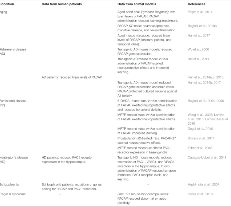
TABLE 1 | Involvement of PACAP in conditions involving cognitive impairment.
Condition Data from human patients Data from animal models References
Aging – Aged pond snail (Lymnaea stagnalis): low
brain levels of PACAP. PACAP
administration rescued learning impairment.
Pirger et al., 2014
PACAP-KO mice: neuronal apoptosis, oxidative damage, and neuroinflammation.
Reglodi et al., 2018b
Aged rhesus macaque: reduced brain levels of PACAP (striatum, parietal, and temporal lobes).
Han et al., 2017
Alzheimer’s disease (AD)
Transgenic AD mouse models: reduced PACAP gene expression.
Wu et al., 2006
Transgenic AD mouse model: in vivo administration of PACAP exerted neuroprotective effects and improved learning.
Rat et al., 2011
AD patients: reduced brain levels of PACAP. Han et al., 2014a,b, 2015 Transgenic AD mouse model: reduced
PACAP gene expression and brain levels. PACAP protected cultured neurons against Aβ toxicity.
Han et al., 2014b, 2017
Parkinson’s disease (PD)
– 6-OHDA-treated rats: in vivo administration of PACAP exerted neuroprotective effects and reduced behavioral deficits.
Reglodi et al., 2004, 2006
MPTP-treated mice: in vivo administration of PACAP exerted neuroprotective effects.
Wang et al., 2008;Lamine et al., 2016;Lamine-Ajili et al., 2016
MPTP-treated mice: in vivo administration of PACAP improved learning.
Deguil et al., 2010
Prostaglandin J2-treated mice: PACAP-27 exerted neuroprotective effects.
Shivers et al., 2014
MPTP-treated macaque: altered PAC1 receptor expression in basal ganglia
Feher et al., 2018
Huntington’s disease (HD)
HD patients: reduced PAC1 receptor expression in the hippocampus.
Transgenic HD mouse models: reduced expression of PAC1, VPAC1, and VPAC2 receptors in the hippocampus. In vivo administration of PACAP rescued synapse formation, PAC1 receptor levels, and learning.
Cabezas-Llobet et al., 2018
Schizophrenia Schizophrenia patients: mutations of genes coding for PACAP and PAC1 receptors.
– Hashimoto et al., 2007
Fragile X syndrome – Fmr1 KO mouse hippocampal slices:
PACAP rescued abnormal synaptic plasticity.
Costa et al., 2018
PACAP, pituitary adenylate cyclase-activating polypeptide; AD, Alzheimer’s disease; 6-OHDA, 6-hydroxydopamine; MPTP, 1-methyl-4-phenyl-1,2,3,6-tetrahydropyridine.
PACAP levels were inversely related to the amount of amyloid
plaques and neurofibrillary tangle, as well as to dementia rating
scores (
Han et al., 2014a
). Reduced PACAP levels and altered
PAC1 receptor expression in the brain of AD patients were
detected since early stages of the progressive neurodegeneration
characterized by mild cognitive impairment (
Han et al., 2015
).
Interestingly, besides exerting a neuroprotective role, PACAP
stimulates a non-amyloidogenic processing pathway of amyloid
precursor protein (APP) (
Kojro et al., 2006
), suggesting that
PACAP might be used in AD therapy. Administration of
PACAP has proven to be effective against A
β-induced toxicity in
different AD mouse models (
Rat et al., 2011
;
Han et al., 2014b
).
Intranasal administration of PACAP to APP-transgenic mice
increased brain expression of PACAP and PACAP receptors,
stimulated the production of neurotrophic and antiapoptotic
factors [brain-derived neurotrophic factor (BDNF) and Bcl-2],
enhanced the expression of the Aβ-degrading enzyme neprilysin,
and improved learning (
Rat et al., 2011
). Accordingly, VIP
decreased amyloid plaques and prevented brain atrophy in
the 5xFAD mouse model of AD (
Korkmaz et al., 2018
). The
same authors suggest that VIP-mediated neuroprotective effects
might also be used for therapy of Parkinson’s disease (PD)
(
Korkmaz and Tuncel, 2018
).
Parkinson’s
disease,
a
neurodegenerative
disorder
characterized by loss of dopaminergic neurons in the substantia
nigra, primarily affects motor control and also involves cognition
deficit (
Aarsland et al., 2017
). A new study shows a decreased
PAC1 receptor expression in basal ganglia in a macaque model
of PD (
Feher et al., 2018
), suggesting that reduced PACAP
function contributes to neurodegeneration and PACAP might
become a promising tool for PD therapy (
Reglodi et al., 2017
).
Administration of PACAP to rats treated with the neurotoxin
6-hydroxydopamine (6-OHDA), a model of PD, prevented
degeneration of nigral dopaminergic neurons and rescued
behavioral deficits (
Reglodi et al., 2004, 2006
). Likewise, in
a different murine PD model [mice treated with
1-methyl-4-phenyl-1,2,3,6-tetrahydropyridine
(MPTP)],
intravenous
injection of PACAP-27 prevented neuronal loss in the substantia
nigra (
Wang et al., 2008
) and rescued learning deficit (
Deguil
et al., 2010
). Still in MPTP-treated mice, PACAP treatment
exerted neuroprotective effects in the substantia nigra (
Lamine
et al., 2016
) and reduced abnormal autophagy, a mechanism that
might contribute to neuronal death (
Lamine-Ajili et al., 2016
).
PACAP-27 also prevented dopaminergic neuronal loss and
motor deficit in prostaglandin J2-treated mice, another proposed
PD model (
Shivers et al., 2014
).
A reduced expression of all PACAP receptor subtypes
was observed in two different mouse models of Huntington’s
disease (HD), an inherited degenerating motor and cognitive
disease, and PAC1 receptors were downregulated in postmortem
hippocampal samples from HD patients (
Cabezas-Llobet et al.,
2018
). Remarkably, intranasal administration of PACAP to HD
mice increased PAC1 receptor expression, stimulated BDNF
production, reduced the formation of huntingtin aggregates,
prevented the loss of hippocampal glutamatergic synapses, and
improved memory (
Cabezas-Llobet et al., 2018
).
Finally, mutations of the genes coding for PACAP and
for PAC1 receptors were found in schizophrenic patients
together with reduced hippocampal volume and impaired
memory (
Hashimoto et al., 2007
). Schizophrenia involves
dysregulation of brain dopaminergic system (
Weinstein et al.,
2017
). Interestingly, genetic ablation of D3 receptors increased
the expression of PACAP and PACAP receptors in mouse
hippocampus and enhanced memory (
Marzagalli et al., 2016
),
suggesting a close interplay between PACAP and dopamine on
learning and memory.
PITUITARY ADENYLATE
CYCLASE-ACTIVATING POLYPEPTIDE
RESCUES SYNAPTIC PLASTICITY IN A
MOUSE MODEL OF FRAGILE X
SYNDROME
Fragile X syndrome (FXS) is a genetic form of intellectual
disability affecting 1/4,000 males and 1/6,000 females. FXS
patients show cognitive and language deficits; subgroups of
patients also display autistic features, epilepsy, attention deficit
and hyperactivity disorder (ADHD), and mood disorders
(
Bardoni et al., 2006
;
Maurin et al., 2014
;
Gross et al., 2015
;
Hagerman et al., 2017
). FXS is caused by transcriptional silencing
of the FMR1 gene coding for fragile X mental retardation
protein (FMRP) (
Verkerk et al., 1991
), an mRNA-binding protein
mostly functioning as a repressor (
Garber et al., 2006
) and in
some cases as an enhancer (
Bechara et al., 2009
) of protein
translation (
Darnell and Klann, 2013
). Hundreds of mRNAs have
been identified as FMRP targets, particularly mRNAs coding for
proteins involved in synapse development and function (
Bassell
and Warren, 2008
;
Bear et al., 2008
). Cortical neurons from
Fmr1
knock out (Fmr1 KO) animal models of FXS (
Comery et al.,
1997
) and FXS patients (
Irwin et al., 2000
) display an increased
density of dendritic spines, with a long and thin morphology
reminiscent of immature filopodia. Abnormal dendritic spine
morphology has crucial consequences on synaptic function.
Many alterations of glutamate-mediated synaptic transmission
and plasticity were found in the brain of
Fmr1 KO mice.
Among the first discovered, hippocampal LTP induced by
metabotropic glutamate receptors (mGluR-LTD) is abnormally
enhanced (
Huber et al., 2002
). Exaggerated mGluR-LTD led
to formulation of the “mGluR theory” of FXS, pointing out
excessive signaling downstream activation of mGluRs (
Bear et al.,
2004
). In
Fmr1 KO neurons, mGluRs also show altered
cell-surface mobility, abnormal coupling to NMDA receptors, and
impaired mGluR-LTD of NMDA-mediated synaptic currents
(
Aloisi et al., 2017
). Other malfunctions of glutamatergic
synapses in
Fmr1 KO mouse brain include a reduced coupling
of mGluRs to Homer proteins (
Giuffrida et al., 2005
), a
reduced NMDA/AMPA ratio (
Yun and Trommer, 2011
;
Gocel
and Larson, 2012
;
Aloisi et al., 2017
), and altered
NMDA-dependent plasticity (
Uzunova et al., 2014
;
Bostrom et al., 2015
).
An increased expression of Ca
2+-permeable AMPA receptors
was recently found in human neural precursors derived from
FXS patients (
Achuta et al., 2018
). Inhibitory synapses are
also affected in the brain of FXS animal models, with a
deficit of GABAergic inhibition (
Martin et al., 2014
;
Braat and
Kooy, 2015
) and abnormal functioning of GABA
Areceptors
(
He et al., 2014
).
At a cellular level, FMRP absence is associated with
dysregulation of many signaling pathways, among which
upregulation of PI3K/Akt/mTOR pathway (
Sharma et al., 2010
;
Huber et al., 2015
), overactivation of GSK3 (
Min et al., 2009
), and
altered MAPK/ERK signaling (
Kim et al., 2008
;
Osterweil et al.,
2010
). The large amount of data now available on the molecular
basis of FXS provides several cues for a possible therapy of FXS,
currently under investigation (
Santoro et al., 2012
;
Sethna et al.,
2014
;
Gross et al., 2015
;
Castagnola et al., 2017
). Each proposed
strategy might be useful in subsets of FXS patients, owing to
a large individual heterogeneity with respect to the type and
severity of symptoms (
Jacquemont et al., 2014
).
Interestingly, early observations on FXS patients and
latest findings on FXS animal models have pointed out a
downregulation of the cyclic adenosine monophosphate (cAMP)
pathway, originating a “cAMP theory” of FXS (
Kelley et al.,
2008
). A recent study shows that the mRNA coding for
phosphodiesterase 2A (PDE2A), a cAMP-degrading enzyme,
is among the most prominent targets of FMRP (
Maurin
et al., 2018a
). In the brain of
Fmr1 KO mice, PDE2A is
overexpressed and overactive, causing reduced cAMP formation
and dysregulation of cAMP downstream signaling (
Maurin
et al., 2018b
). In line with this, synaptic plasticity, learning,
and behavior in
Fmr1 KO mice are rescued by agonists of
serotonin 5-HT
7receptors, positively coupled to adenylate
cyclase (
Costa et al., 2012, 2015, 2018
;
Ciranna and Catania,
2014
), by PDE4 inhibitors (
Choi et al., 2015, 2016
), and by
a selective PDE2A inhibitor (
Maurin et al., 2018b
). Of note,
inhibition of PDE2A also corrected abnormal dendritic spine
morphology of cortical neurons from
Fmr1 KO mice (
Maurin
et al., 2018b
). All these data confirm a deficit in
cAMP-mediated signaling in
Fmr1 KO neurons and demonstrate that
pharmacological manipulations increasing cAMP levels can
rescue synaptic morphology and function, learning, and behavior
in animal models of FXS.
Pituitary adenylate cyclase activating polypeptide is a potent
stimulator of adenylate cyclase activity (
Miyata et al., 1989
;
Vaudry et al., 2009
). Consistent with the cAMP hypothesis
of FXS, we found that PACAP reversed mGluR-LTD in the
CA3-CA1 hippocampal synapse in wild-type mice and reduced
exaggerated mGluR-LTD in
Fmr1 KO, thus correcting a synaptic
defect typically observed in FXS mouse models (
Costa et al.,
2018
). This result offers novel suggestions for a possible therapy
of FXS, for which no specific cure is presently available. In
future studies, it would be interesting to test if PACAP can
correct other abnormal features in
Fmr1 KO neurons (dendrite
development, synapse formation and function, ion channel
expression, membrane excitability, and intracellular signaling)
and rescue learning and behavior when administered
in vivo to
Fmr1 KO mice.
Dysregulation of cyclic nucleotide pathways was found at
different levels (synthesis, functioning, and/or degradation by
PDE) in aging and age-related cognitive decline (
Kelly, 2018
).
Accordingly, inhibition of PDE activity improved memory in
animal models of AD (
Gulisano et al., 2018
) and HD (
Saavedra
et al., 2013
) and seems to prevent memory loss in elderly
humans and in AD patients (
Prickaerts et al., 2017
). As indicated
above, in FXS, the cAMP pathway is also dysregulated, and
increasing cAMP rescues several phenotypes. Therefore, altered
cAMP signaling might be a common feature in cognitive deficits
of very different origin, in which administration of PACAP
might be beneficial.
SYSTEMIC ADMINISTRATION ROUTES
FOR EFFECTIVE BRAIN DELIVERY OF
PITUITARY ADENYLATE
CYCLASE-ACTIVATING POLYPEPTIDE
Pituitary adenylate cyclase activating polypeptide-38 is actively
transported into the brain across the blood–brain barrier (BBB)
by a saturable carrier, whereas PACAP-27 passes the BBB by
transmembrane diffusion (
Banks et al., 1993
). After intravenous
administration, the amount of PACAP-38 brain uptake was high
enough to exert neuroprotective effects (
Banks et al., 1996
).
The rate of PACAP-38 transport varies largely in different brain
areas, with maximal uptake in the hypothalamus and in the
hippocampus (
Nonaka et al., 2002
), and can be altered in
pathological conditions (
Banks et al., 1998
;
Nonaka et al., 2002
;
Rhea et al., 2018
).
On the other side, PACAP-38 and PACAP-27 are transported
out of the brain by a common efflux mechanism, reducing
their brain levels (
Banks et al., 1993
). It is noteworthy that
antisense inhibition of peptide transport system-6 (PTS-6)
improved brain uptake and neuroprotective effects of
PACAP-27 in murine models of AD and stroke (
Dogrukol-Ak et al.,
2009
), suggesting that inhibition of PTS-6 efflux component
might become a therapeutic strategy to enhance central
effects of PACAP.
Some issues were raised about intravenous administration
of PACAP to humans: PACAP-38 induced headache in healthy
and migraine-suffering subjects (
Schytz et al., 2009
). Headache
was also reported after intravenous infusion of PACAP-27 to
healthy subjects (
Ghanizada et al., 2019a
) and migraine patients
(
Ghanizada et al., 2019b
).
Other issues against intravenous administration of PACAP
concern metabolic stability and selectivity. In fact,
PACAP-38 showed a very short half-life (
<5 min) in human plasma
in vitro, being converted by the blood enzyme dipeptidyl
peptidase IV into shorter peptides that behave as PACAP
receptor antagonists, whereas PACAP-27 was relatively stable
(
Bourgault et al., 2008
).
Besides, parenteral administration of PACAP can induce
undesired peripheral actions, among which are cardiovascular
(
Runcie et al., 1995
;
Farnham et al., 2012
) and hormonal effects
(
Tsutsumi et al., 2002
). To overcome these limitations, new
synthetic agonists of PACAP receptors have been developed with
improved metabolic stability, higher brain uptake, selectivity for
PAC1 receptors (the predominant PACAP receptors in the CNS),
and reduced side effects (
Bourgault et al., 2008
;
Dejda et al., 2011
;
Doan et al., 2011
;
Lamine et al., 2016
).
Another strategy exploits conjugation of PACAP with a TAT
peptide, improving passage through the BBB (
Yu et al., 2012a,b
).
Carrier vesicles can also be used to protect peptides from
blood-degrading enzymes (
Dufes et al., 2004
).
Administration routes for brain delivery of PACAP are
illustrated in details in a recent review (
Reglodi et al., 2018a
).
A promising non-invasive and easy route is intranasal
application, by which PACAP reaches the brain fast and
effectively in rodents, exerting neuroprotective effects in mouse
models of AD (
Rat et al., 2011
;
Nonaka et al., 2012
) and HD
(
Cabezas-Llobet et al., 2018
). Intranasal administration of
PACAP was also tested on human volunteers, showing good
safety and tolerability (
Doberer et al., 2007
;
Reglodi et al.,
2018a
) with only mild local adverse reactions (
Kinhult
et al., 2003
). Interestingly, headache was not reported
after intranasal application of PACAP-38 (
Doberer et al.,
2007
), suggesting this route as a valuable alternative to
intravenous administration.
CONCLUDING REMARKS
Pituitary adenylate cyclase activating polypeptide plays an
important role in learning and has a therapeutic potential in
cognition deficits associated with disruption of cyclic nucleotide
signaling. We suggest that the effects of PACAP might also be
studied for a possible therapy of FXS, in which a deficit in
cAMP formation and downstream signaling were evidenced. For
future translational applications, it would be interesting to test
in vivo effects of PACAP on learning and behavioral deficits in
FXS animal models.
Pituitary adenylate cyclase activating polypeptide is brain
permeant and has already proved to be safe on healthy
humans and thus might be tested in clinical trials. To
improve PACAP brain uptake, it would be worth focusing
on new selective PAC1 receptor agonists with enhanced
metabolic stability, on delivery carriers, and/or on suitable
administration routes.
AUTHOR CONTRIBUTIONS
LCi designed and directed the research projects and wrote the
manuscript. LCo performed the experiments and data analysis,
and contributed to the manuscript preparation.
FUNDING
The present work was financed by Telethon Foundation
(grant GGP13145) and by Università di Catania, Italy
(grant Chance 2017).
REFERENCES
Aarsland, D., Creese, B., Politis, M., Chaudhuri, K. R., Ffytche, D. H., Weintraub, D., et al. (2017). Cognitive decline in Parkinson disease.Nat. Rev. Neurol. 13, 217–231. doi: 10.1038/nrneurol.2017.27
Achuta, V. S., Moykkynen, T., Peteri, U. K., Turconi, G., Rivera, C., Keinanen, K., et al. (2018). Functional changes of AMPA responses in human induced
pluripotent stem cell-derived neural progenitors in fragile X syndrome.Sci.
Signal 11:eaan8784. doi: 10.1126/scisignal.aan8784
Ago, Y., Hiramatsu, N., Ishihama, T., Hazama, K., Hayata-Takano, A., Shibasaki, Y., et al. (2013). The selective metabotropic glutamate 2/3 receptor agonist MGS0028 reverses psychomotor abnormalities and recognition memory deficits in mice lacking the pituitary adenylate cyclase-activating polypeptide. Behav. Pharmacol. 24, 74–77. doi: 10.1097/FBP.0b013e32835cf3e5
Aloisi, E., Le Corf, K., Dupuis, J., Zhang, P., Ginger, M., Labrousse, V., et al. (2017). Altered surface mGluR5 dynamics provoke synaptic NMDAR dysfunction and
cognitive defects in Fmr1 knockout mice.Nat. Commun. 8:1103. doi: 10.1038/
s41467-017-01191-2
Arimura, A., Somogyvari-Vigh, A., Miyata, A., Mizuno, K., Coy, D. H., and Kitada, C. (1991). Tissue distribution of PACAP as determined by RIA: highly abundant in the rat brain and testes.Endocrinology 129, 2787–2789. doi: 10.1210/endo-129-5-2787
Arimura, A., Somogyvari-Vigh, A., Weill, C., Fiore, R. C., Tatsuno, I., Bay, V., et al.
(1994). PACAP functions as a neurotrophic factor.Ann. N. Y. Acad. Sci. 739,
228–243. doi: 10.1111/j.1749-6632.1994.tb19825.x
Banks, W. A., Kastin, A. J., and Arimura, A. (1998). Effect of spinal cord injury on the permeability of the blood-brain and blood-spinal cord barriers to the
neurotropin PACAP.Exp. Neurol. 151, 116–123. doi: 10.1006/exnr.1998.6786
Banks, W. A., Kastin, A. J., Komaki, G., and Arimura, A. (1993). Passage of pituitary adenylate cyclase activating polypeptide1-27 and pituitary adenylate cyclase activating polypeptide1-38 across the blood-brain barrier.J. Pharmacol. Exp. Ther. 267, 690–696.
Banks, W. A., Uchida, D., Arimura, A., Somogyvari-Vigh, A., and Shioda, S. (1996). Transport of pituitary adenylate cyclase-activating polypeptide across the blood-brain barrier and the prevention of ischemia-induced death of hippocampal neurons.Ann. N. Y. Acad. Sci. 805, 270–277. doi: 10.1111/j.1749-6632.1996.tb17489.x
Bardoni, B., Davidovic, L., Bensaid, M., and Khandjian, E. W. (2006). The fragile X syndrome: exploring its molecular basis and seeking a treatment.Expert Rev. Mol. Med. 8, 1–16. doi: 10.1017/s1462399406010751
Bassell, G. J., and Warren, S. T. (2008). Fragile X syndrome: loss of local mRNA regulation alters synaptic development and function.Neuron 60, 201–214. doi: 10.1016/j.neuron.2008.10.004
Bear, M. F., Dolen, G., Osterweil, E., and Nagarajan, N. (2008). Fragile X: translation
in action. Neuropsychopharmacology 33, 84–87. doi: 10.1038/sj.npp.130
1610
Bear, M. F., Huber, K. M., and Warren, S. T. (2004). The mGluR theory of fragile X mental retardation.Trends Neurosci. 27, 370–377. doi: 10.1016/j.tins.2004.04. 009
Bechara, E. G., Didiot, M. C., Melko, M., Davidovic, L., Bensaid, M., Martin, P., et al. (2009). A novel function for fragile X mental retardation protein
in translational activation. PLoS Biol. 7:e16. doi: 10.1371/journal.pbio.100
0016
Borbely, E., Scheich, B., and Helyes, Z. (2013). Neuropeptides in learning
and memory. Neuropeptides 47, 439–450. doi: 10.1016/j.npep.2013.
10.012
Bostrom, C. A., Majaess, N. M., Morch, K., White, E., Eadie, B. D., and Christie, B. R. (2015). Rescue of NMDAR-dependent synaptic plasticity
in fmr1 knock-out mice. Cereb. Cortex 25, 271–279. doi: 10.1093/cercor/
bht237
Bourgault, S., Vaudry, D., Botia, B., Couvineau, A., Laburthe, M., Vaudry, H., et al. (2008). Novel stable PACAP analogs with potent activity towards the PAC1 receptor.Peptides 29, 919–932. doi: 10.1016/j.peptides.2008.01.022
Braat, S., and Kooy, R. F. (2015). Insights into GABAAergic system deficits in fragile X syndrome lead to clinical trials.Neuropharmacology 88, 48–54. doi: 10.1016/j.neuropharm.2014.06.028
Cabezas-Llobet, N., Vidal-Sancho, L., Masana, M., Fournier, A., Alberch, J., Vaudry, D., et al. (2018). Pituitary adenylate cyclase-activating polypeptide (PACAP) enhances hippocampal synaptic plasticity and improves memory performance
in Huntington’s Disease.Mol. Neurobiol. 55, 8263–8277. doi:
10.1007/s12035-018-0972-5
Castagnola, S., Bardoni, B., and Maurin, T. (2017). The search for an effective therapy to treat fragile X syndrome: dream or reality?Front. Synaptic Neurosci. 9:15. doi: 10.3389/fnsyn.2017.00015
Choi, C. H., Schoenfeld, B. P., Bell, A. J., Hinchey, J., Rosenfelt, C., Gertner, M. J., et al. (2016). Multiple drug treatments that increase cAMP signaling restore long-term memory and aberrant signaling in fragile X
syndrome models.Front. Behav. Neurosci. 10:136. doi: 10.3389/fnbeh.2016.
00136
Choi, C. H., Schoenfeld, B. P., Weisz, E. D., Bell, A. J., Chambers, D. B., Hinchey, J., et al. (2015). PDE-4 inhibition rescues aberrant synaptic plasticity in drosophila and mouse models of fragile X syndrome.J. Neurosci. 35, 396–408. doi: 10.1523/ JNEUROSCI.1356-12.2015
Ciranna, L., and Catania, M. V. (2014). 5-HT7 receptors as modulators of neuronal excitability, synaptic transmission and plasticity: physiological role and possible
implications in autism spectrum disorders.Front. Cell Neurosci. 8:250. doi:
10.3389/fncel.2014.00250
Ciranna, L., and Cavallaro, S. (2003). Opposing effects by pituitary adenylate cyclase-activating polypeptide and vasoactive intestinal peptide
on hippocampal synaptic transmission. Exp. Neurol. 184, 778–784.
doi: 10.1016/s0014-4886(03)00300-5
Comery, T. A., Harris, J. B., Willems, P. J., Oostra, B. A., Irwin, S. A., Weiler, I. J., et al. (1997). Abnormal dendritic spines in fragile X knockout mice: maturation and pruning deficits.Proc. Natl. Acad. Sci. U.S.A. 94, 5401–5404. doi: 10.1073/pnas.94.10.5401
Costa, L., Santangelo, F., Li Volsi, G., and Ciranna, L. (2009). Modulation of AMPA receptor-mediated ion current by pituitary adenylate cyclase-activating polypeptide (PACAP) in CA1 pyramidal neurons from rat hippocampus. Hippocampus 19, 99–109. doi: 10.1002/hipo.20488
Costa, L., Sardone, L. M., Bonaccorso, C. M., D’Antoni, S., Spatuzza, M., Gulisano, W., et al. (2018). Activation of serotonin 5-HT7 receptors modulates hippocampal synaptic plasticity by stimulation of adenylate cyclases and rescues
learning and behavior in a mouse model of fragile X syndrome.Front. Mol. Neurosci. 11:353. doi: 10.3389/fnmol.2018.00353
Costa, L., Sardone, L. M., Lacivita, E., Leopoldo, M., and Ciranna, L. (2015). Novel agonists for serotonin 5-HT7 receptors reverse metabotropic glutamate receptor-mediated long-term depression in the hippocampus of wild-type and
Fmr1 KO mice, a model of Fragile X Syndrome.Front. Behav. Neurosci. 9:65.
doi: 10.3389/fnbeh.2015.00065
Costa, L., Spatuzza, M., D’Antoni, S., Bonaccorso, C. M., Trovato, C., Musumeci, S. A., et al. (2012). Activation of 5-HT7 serotonin receptors reverses metabotropic glutamate receptor-mediated synaptic plasticity in wild-type and
Fmr1 knockout mice, a model of Fragile X syndrome. Biol. Psychiatry 72,
924–933. doi: 10.1016/j.biopsych.2012.06.008
Darnell, J. C., and Klann, E. (2013). The translation of translational control by FMRP: therapeutic targets for FXS.Nat. Neurosci. 16, 1530–1536. doi: 10.1038/ nn.3379
Deguil, J., Chavant, F., Lafay-Chebassier, C., Perault-Pochat, M. C., Fauconneau, B., and Pain, S. (2010). Neuroprotective effect of PACAP on translational control
alteration and cognitive decline in MPTP parkinsonian mice.Neuro. Res. 17,
142–155. doi: 10.1007/s12640-009-9091-4
Dejda, A., Seaborn, T., Bourgault, S., Touzani, O., Fournier, A., Vaudry, H., et al. (2011). PACAP and a novel stable analog protect rat brain from ischemia:
insight into the mechanisms of action.Peptides 32, 1207–1216. doi: 10.1016/
j.peptides.2011.04.003
Di Mauro, M., Cavallaro, S., and Ciranna, L. (2003). Pituitary adenylate cyclase-activating polypeptide modifies the electrical activity of CA1 hippocampal
neurons in the rat.Neurosci. Lett. 337, 97–100. doi: 10.1016/s0304-3940(02)
01316-2
Dickson, L., and Finlayson, K. (2009). VPAC and PAC receptors: from ligands to
function.Pharmacol. Ther. 121, 294–316. doi: 10.1016/j.pharmthera.2008.11.
006
Doan, N. D., Bourgault, S., Dejda, A., Letourneau, M., Detheux, M., Vaudry, D., et al. (2011). Design and in vitro characterization of PAC1/VPAC1-selective agonists with potent neuroprotective effects.Biochem. Pharmacol. 81, 552–561. doi: 10.1016/j.bcp.2010.11.015
Doberer, D., Gschwandtner, M., Mosgoeller, W., Bieglmayer, C., Heinzl, H., and Petkov, V. (2007). Pulmonary and systemic effects of inhaled PACAP38 in healthy male subjects.Eur. J. Clin. Invest 37, 665–672. doi: 10.1111/j.1365-2362. 2007.01832.x
Dogrukol-Ak, D., Kumar, V. B., Ryerse, J. S., Farr, S. A., Verma, S., Nonaka, N., et al. (2009). Isolation of peptide transport system-6 from brain endothelial cells: therapeutic effects with antisense inhibition in Alzheimer and stroke
models. J. Cereb. Blood Flow Metab. 29, 411–422. doi: 10.1038/jcbfm.
2008.131
Dufes, C., Gaillard, F., Uchegbu, I. F., Schatzlein, A. G., Olivier, J. C., and Muller, J. M. (2004). Glucose-targeted niosomes deliver vasoactive intestinal peptide (VIP) to the brain.Int. J. Pharm. 285, 77–85. doi: 10.1016/j.ijpharm.2004.07.020 Farnham, M. M., Lung, M. S., Tallapragada, V. J., and Pilowsky, P. M. (2012). PACAP causes PAC1/VPAC2 receptor mediated hypertension and sympathoexcitation in normal and hypertensive rats.Am. J. Physiol. Heart Circ. Physiol. 303, H910–H917. doi: 10.1152/ajpheart.00464.2012
Feher, M., Gaszner, B., Tamas, A., Gil-Martinez, A. L., Fernandez-Villalba, E., Herrero, M. T., et al. (2018). Alteration of the PAC1 receptor expression in the
basal ganglia of MPTP-induced parkinsonian macaque monkeys.Neuro. Res.
33, 702–715. doi: 10.1007/s12640-017-9841-7
Garber, K., Smith, K. T., Reines, D., and Warren, S. T. (2006). Transcription, translation and fragile X syndrome.Curr. Opin. Genet. Dev. 16, 270–275. doi: 10.1016/j.gde.2006.04.010
Ghanizada, H., Al-Karagholi, M. A., Arngrim, N., Ghanizada, M., Larsson, H. B. W., Amin, F. M., et al. (2019a). Effect of pituitary adenylate cyclase-activating polypeptide-27 on cerebral hemodynamics in healthy volunteers: a 3T MRI study.Peptides 121:170134. doi: 10.1016/j.peptides.2019. 170134
Ghanizada, H., Al-Karagholi, M. A., Arngrim, N., Olesen, J., and Ashina, M. (2019b). PACAP27 induces migraine-like attacks in migraine patients. Cephalalgia 12:333102419864507. doi: 10.1177/0333102419864507
Giuffrida, R., Musumeci, S., D’Antoni, S., Bonaccorso, C. M., Giuffrida-Stella, A. M., Oostra, B. A., et al. (2005). A reduced number of metabotropic glutamate subtype 5 receptors are associated with constitutive homer proteins in a mouse
model of fragile X syndrome.J. Neurosci. 25, 8908–8916. doi: 10.1523/jneurosci. 0932-05.2005
Gocel, J., and Larson, J. (2012). Synaptic NMDA receptor-mediated currents in anterior piriform cortex are reduced in the adult fragile X mouse.Neuroscience 221, 170–181. doi: 10.1016/j.neuroscience.2012.06.052
Gross, C., Hoffmann, A., Bassell, G. J., and Berry-Kravis, E. M. (2015). Therapeutic strategies in fragile X syndrome: from bench to bedside and back. Neurotherapeutics 12, 584–608. doi: 10.1007/s13311-015-0355-9
Gulisano, W., Tropea, M. R., Arancio, O., Palmeri, A., and Puzzo, D. (2018). Sub-efficacious doses of phosphodiesterase 4 and 5 inhibitors improve memory
in a mouse model of Alzheimer’s disease.Neuropharmacology 138, 151–159.
doi: 10.1016/j.neuropharm.2018.06.002
Gupte, R. P., Kadunganattil, S., Shepherd, A. J., Merrill, R., Planer, W., Bruchas, M. R., et al. (2015). Convergent phosphomodulation of the major neuronal dendritic potassium channel Kv4.2 by pituitary adenylate cyclase-activating
polypeptide. Neuropharmacology 101, 291–308. doi: 10.1016/j.neuropharm.
2015.10.006
Hagerman, R. J., Berry-Kravis, E., Hazlett, H. C., Bailey, DB Jr, Moine, H., Kooy, R. F., et al. (2017). Fragile X syndrome.Nat. Rev. Dis. Primers 3:17065. doi: 10.1038/nrdp.2017.65
Han, P., Caselli, R. J., Baxter, L., Serrano, G., Yin, J., Beach, T. G., et al. (2015). Association of pituitary adenylate cyclase-activating polypeptide with cognitive
decline in mild cognitive impairment due to Alzheimer disease.JAMA Neurol.
72, 333–339. doi: 10.1001/jamaneurol.2014.3625
Han, P., Liang, W., Baxter, L. C., Yin, J., Tang, Z., Beach, T. G., et al. (2014a). Pituitary adenylate cyclase-activating polypeptide is reduced in Alzheimer
disease.Neurology 82, 1724–1728.
Han, P., Tang, Z., Yin, J., Maalouf, M., Beach, T. G., Reiman, E. M., et al. (2014b). Pituitary adenylate cyclase-activating polypeptide protects against beta-amyloid toxicity.Neurobiol. Aging 35, 2064–2071. doi: 10.1016/j.neurobiolaging.2014. 03.022
Han, P., Nielsen, M., Song, M., Yin, J., Permenter, M. R., Vogt, J. A., et al. (2017). The impact of aging on brain pituitary adenylate cyclase activating polypeptide,
pathology and cognition in mice and rhesus macaques.Front. Aging Neurosci.
9:180. doi: 10.3389/fnagi.2017.00180
Harmar, A. J., Fahrenkrug, J., Gozes, I., Laburthe, M., May, V., Pisegna, J. R., et al. (2012). Pharmacology and functions of receptors for vasoactive intestinal peptide and pituitary adenylate cyclase-activating polypeptide: IUPHAR review 1.Br. J. Pharmacol. 166, 4–17. doi: 10.1111/j.1476-5381.2012.01871.x Hashimoto, R., Hashimoto, H., Shintani, N., Chiba, S., Hattori, S., Okada, T., et al.
(2007). Pituitary adenylate cyclase-activating polypeptide is associated with schizophrenia.Mol. Psychiatry 12, 1026–1032.
Hayata-Takano, A., Kamo, T., Kijima, H., Seiriki, K., Ogata, K., Ago, Y., et al. (2019). Pituitary adenylate cyclase-activating polypeptide modulates dendritic spine maturation and morphogenesis via microRNA-132 upregulation. J. Neurosci. 39, 4208–4220. doi: 10.1523/JNEUROSCI.2468-18.2019
He, Q., Nomura, T., Xu, J., and Contractor, A. (2014). The developmental switch in GABA polarity is delayed in fragile X mice.J. Neurosci. 34, 446–450. doi: 10.1523/JNEUROSCI.4447-13.2014
Hirabayashi, T., Nakamachi, T., and Shioda, S. (2018). Discovery of PACAP and its receptors in the brain.J. Headache Pain 19:28. doi: 10.1186/s10194-018-0855-1 Holland, P. R., Barloese, M., and Fahrenkrug, J. (2018). PACAP in hypothalamic regulation of sleep and circadian rhythm: importance for headache.J. Headache Pain 19:20. doi: 10.1186/s10194-018-0844-4
Huber, K. M., Gallagher, S. M., Warren, S. T., and Bear, M. F. (2002). Altered synaptic plasticity in a mouse model of fragile X mental retardation.Proc. Natl. Acad. Sci. U.S.A. 99, 7746–7750. doi: 10.1073/pnas.122205699
Huber, K. M., Klann, E., Costa-Mattioli, M., and Zukin, R. S. (2015). Dysregulation of mammalian target of rapamycin signaling in mouse models of autism. J. Neurosci. 35, 13836–13842. doi: 10.1523/JNEUROSCI.2656-15.2015 Irwin, S. A., Galvez, R., and Greenough, W. T. (2000). Dendritic spine structural
anomalies in fragile-X mental retardation syndrome.Cereb. Cortex 10, 1038–
1044. doi: 10.1093/cercor/10.10.1038
Jacquemont, S., Berry-Kravis, E., Hagerman, R., von Raison, F., Gasparini, F., Apostol, G., et al. (2014). The challenges of clinical trials in fragile X syndrome. Psychopharmacology 231, 1237–1250. doi: 10.1007/s00213-013-3289-0 Jaworski, D. M., and Proctor, M. D. (2000). Developmental regulation of
expression in the rat central nervous system.Brain Res. Dev. Brain Res. 120, 27–39. doi: 10.1016/s0165-3806(99)00192-3
Jolivel, V., Basille, M., Aubert, N., de Jouffrey, S., Ancian, P., Le Bigot, J. F., et al. (2009). Distribution and functional characterization of pituitary adenylate cyclase-activating polypeptide receptors in the brain of non-human primates. Neuroscience 160, 434–451. doi: 10.1016/j.neuroscience.2009.02.028
Joo, K. M., Chung, Y. H., Kim, M. K., Nam, R. H., Lee, B. L., Lee, K. H., et al. (2004). Distribution of vasoactive intestinal peptide and pituitary adenylate cyclase-activating polypeptide receptors (VPAC1, VPAC2, and PAC1 receptor) in the rat brain.J. Comp. Neurol. 476, 388–413. doi: 10.1002/cne.20231
Kelley, D. J., Bhattacharyya, A., Lahvis, G. P., Yin, J. C., Malter, J., and Davidson,
R. J. (2008). The cyclic AMP phenotype of fragile X and autism.Neurosci.
Biobehav. Rev. 32, 1533–1543. doi: 10.1016/j.neubiorev.2008.06.005
Kelly, M. P. (2018). Cyclic nucleotide signaling changes associated with normal aging and age-related diseases of the brain.Cell Signal 42, 281–291. doi: 10.1016/ j.cellsig.2017.11.004
Kim, S. H., Markham, J. A., Weiler, I. J., and Greenough, W. T. (2008). Aberrant early-phase ERK inactivation impedes neuronal function in fragile X syndrome. Proc. Natl. Acad. Sci. U.S.A. 105, 4429–4434. doi: 10.1073/pnas.0800257105 Kinhult, J., Adner, M., Uddman, R., and Cardell, L. O. (2003). Pituitary adenylate
cyclase-activating polypeptide, effects in the human nose.Clin. Exp. Allergy 33, 942–949.
Kojro, E., Postina, R., Buro, C., Meiringer, C., Gehrig-Burger, K., and Fahrenholz, F. (2006). The neuropeptide PACAP promotes the alpha-secretase pathway for
processing the Alzheimer amyloid precursor protein.FASEB J. 20, 512–514.
doi: 10.1096/fj.05-4812fje
Kondo, T., Tominaga, T., Ichikawa, M., and Iijima, T. (1997). Differential alteration of hippocampal synaptic strength induced by pituitary adenylate cyclase
activating polypeptide-38 (PACAP-38).Neurosci. Lett. 221, 189–192. doi: 10.
1016/s0304-3940(96)13323-1
Korkmaz, O. T., Ay, H., Aytan, N., Carreras, I., Kowall, N. W., Dedeoglu, A., et al. (2018). Vasoactive intestinal peptide decreases beta-amyloid accumulation and prevents brain atrophy in the 5xFAD mouse model of Alzheimer’s Disease. J. Mol. Neurosci. 68, 389–396. doi: 10.1007/s12031-018-1226-8
Korkmaz, O. T., and Tuncel, N. (2018). Advantages of vasoactive intestinal peptide
for the future treatment of Parkinson’s Disease.Curr. Pharm. Des. 24, 4693–
4701. doi: 10.2174/1381612825666190111150953
Koves, K., Arimura, A., Gorcs, T. G., and Somogyvari-Vigh, A. (1991). Comparative distribution of immunoreactive pituitary adenylate cyclase activating polypeptide and vasoactive intestinal polypeptide in rat forebrain. Neuroendocrinology 54, 159–169. doi: 10.1159/000125864
Ladjimi, M. H., Barbouche, R., Ben Barka, Z., Vaudry, D., Lefranc, B., Leprince, J., et al. (2019). Comparison of the effects of PACAP-38 and its analog, acetyl-[Ala(15), Ala(20)] PACAP-38-propylamide, on spatial memory, post-learning
BDNF expression and oxidative stress in rat.Behav. Brain Res. 359, 247–257.
doi: 10.1016/j.bbr.2018.10.023
Lamine, A., Letourneau, M., Doan, N. D., Maucotel, J., Couvineau, A., Vaudry, H., et al. (2016). Characterizations of a synthetic pituitary adenylate cyclase-activating polypeptide analog displaying potent neuroprotective activity and
reduced in vivo cardiovascular side effects in a Parkinson’s disease model.
Neuropharmacology 108, 440–450. doi: 10.1016/j.neuropharm.2015.05.014 Lamine-Ajili, A., Fahmy, A. M., Letourneau, M., Chatenet, D., Labonte, P.,
Vaudry, D., et al. (2016). Effect of the pituitary adenylate cyclase-activating polypeptide on the autophagic activation observed inin vitro and in vivo models of Parkinson’s disease.Biochim. Biophys. Acta 1862, 688–695. doi: 10.1016/j. bbadis.2016.01.005
Liu, Z., Geng, L., Li, R., He, X., Zheng, J. Q., and Xie, Z. (2003). Frequency modulation of synchronized Ca2+ spikes in cultured hippocampal networks through G-protein-coupled receptors.J. Neurosci. 23, 4156–4163. doi: 10.1523/ jneurosci.23-10-04156.2003
Macdonald, D. S., Weerapura, M., Beazely, M. A., Martin, L., Czerwinski, W., Roder, J. C., et al. (2005). Modulation of NMDA receptors by pituitary adenylate cyclase activating peptide in CA1 neurons requires G alpha q, protein kinase C, and activation of Src.J. Neurosci. 25, 11374–11384. doi: 10.1523/jneurosci. 3871-05.2005
Martin, B. S., Corbin, J. G., and Huntsman, M. M. (2014). Deficient tonic GABAergic conductance and synaptic balance in the fragile X syndrome amygdala.J. Neurophysiol. 112, 890–902. doi: 10.1152/jn.00597.2013
Marzagalli, R., Leggio, G. M., Bucolo, C., Pricoco, E., Keay, K. A., Cardile, V., et al. (2016). Genetic blockade of the dopamine D3 receptor enhances hippocampal expression of PACAP and receptors and alters their cortical distribution. Neuroscience 316, 279–295. doi: 10.1016/j.neuroscience.2015.12.034
Masuo, Y., Matsumoto, Y., Tokito, F., Tsuda, M., and Fujino, M. (1993). Effects of vasoactive intestinal polypeptide (VIP) and pituitary adenylate cyclase activating polypeptide (PACAP) on the spontaneous release of acetylcholine
from the rat hippocampus by brain microdialysis.Brain Res. 611, 207–215.
doi: 10.1016/0006-8993(93)90504-g
Matsuyama, S., Matsumoto, A., Hashimoto, H., Shintani, N., and Baba, A. (2003). Impaired long-term potentiation in vivo in the dentate gyrus of pituitary adenylate cyclase-activating polypeptide (PACAP) or PACAP type 1
receptor-mutant mice.Neuroreport 14, 2095–2098. doi:
10.1097/00001756-200311140-00017
Maurin, T., Lebrigand, K., Castagnola, S., Paquet, A., Jarjat, M., Popa, A., et al. (2018a). HITS-CLIP in various brain areas reveals new targets and new
modalities of RNA binding by fragile X mental retardation protein.Nucleic
Acids Res. 46, 6344–6355. doi: 10.1093/nar/gky267
Maurin, T., Melancia, F., Jarjat, M., Castro, L., Costa, L., Delhaye, S., et al. (2018b). Involvement of phosphodiesterase 2A activity in the pathophysiology of fragile
X syndrome.Cereb. Cortex [Epub ahead of print].
Maurin, T., Zongaro, S., and Bardoni, B. (2014). Fragile X syndrome: from
molecular pathology to therapy.Neurosci. Biobehav. Rev. 46(Pt 2), 242–255.
doi: 10.1016/j.neubiorev.2014.01.006
Meloni, E. G., Kaye, K. T., Venkataraman, A., and Carlezon, W. A. Jr. (2018). PACAP increases Arc/Arg 3.1 expression within the extended amygdala after fear conditioning in rats.Neurobiol. Learn Mem. 157, 24–34. doi: 10.1016/j.nlm. 2018.11.011
Meloni, E. G., Venkataraman, A., Donahue, R. J., and Carlezon, W. A. Jr. (2016). Bi-directional effects of pituitary adenylate cyclase-activating polypeptide (PACAP) on fear-related behavior and c-Fos expression after fear conditioning in rats.Psychoneuroendocrinology 64, 12–21. doi: 10.1016/j.psyneuen.2015.11. 003
Min, W. W., Yuskaitis, C. J., Yan, Q., Sikorski, C., Chen, S., Jope, R. S., et al. (2009). Elevated glycogen synthase kinase-3 activity in Fragile X mice: key
metabolic regulator with evidence for treatment potential.Neuropharmacology
56, 463–472. doi: 10.1016/j.neuropharm.2008.09.017
Miyata, A., Arimura, A., Dahl, R. R., Minamino, N., Uehara, A., Jiang, L., et al. (1989). Isolation of a novel 38 residue-hypothalamic polypeptide which stimulates adenylate cyclase in pituitary cells.Biochem. Biophys. Res. Commun. 164, 567–574. doi: 10.1016/0006-291x(89)91757-9
Nonaka, N., Banks, W. A., Mizushima, H., Shioda, S., and Morley, J. E. (2002). Regional differences in PACAP transport across the blood-brain barrier in mice: a possible influence of strain, amyloid beta protein, and age.Peptides 23, 2197–2202. doi: 10.1016/s0196-9781(02)00248-6
Nonaka, N., Farr, S. A., Nakamachi, T., Morley, J. E., Nakamura, M., Shioda, S., et al. (2012). Intranasal administration of PACAP: uptake by brain and regional brain targeting with cyclodextrins.Peptides 36, 168–175. doi: 10.1016/j.peptides.2012. 05.021
Ogata, K., Shintani, N., Hayata-Takano, A., Kamo, T., Higashi, S., Seiriki, K., et al. (2015). PACAP enhances axon outgrowth in cultured hippocampal neurons to a
comparable extent as BDNF.PLoS One 10:e0120526. doi: 10.1371/journal.pone.
0120526
Osterweil, E. K., Krueger, D. D., Reinhold, K., and Bear, M. F. (2010). Hypersensitivity to mGluR5 and ERK1/2 leads to excessive protein synthesis in the hippocampus of a mouse model of fragile X syndrome.
J. Neurosci. 30, 15616–15627. doi: 10.1523/JNEUROSCI.3888-10.
2010
Otto, C., Kovalchuk, Y., Wolfer, D. P., Gass, P., Martin, M., Zuschratter, W., et al. (2001). Impairment of mossy fiber long-term potentiation and associative learning in pituitary adenylate cyclase activating polypeptide type I receptor-deficient mice.J. Neurosci. 21, 5520–5527. doi: 10.1523/jneurosci.21-15-05520. 2001
Pecoraro, V., Sardone, L. M., Chisari, M., Licata, F., Li Volsi, G., Perciavalle, V., et al. (2017). A subnanomolar concentration of pituitary adenylate cyclase-activating polypeptide (PACAP) pre-synaptically modulates glutamatergic transmission in the rat hippocampus acting through acetylcholine.Neuroscience 340, 551–562. doi: 10.1016/j.neuroscience.2016.10.061
Pirger, Z., Naskar, S., Laszlo, Z., Kemenes, G., Reglodi, D., and Kemenes, I. (2014). Reversal of age-related learning deficiency by the vertebrate PACAP and IGF-1 in a novel invertebrate model of aging: the pond snail (Lymnaea stagnalis).J. Gerontol. A Biol. Sci. Med. Sci. 69, 1331–1338. doi: 10.1093/gerona/ glu068
Prickaerts, J., Heckman, P. R. A., and Blokland, A. (2017). Investigational phosphodiesterase inhibitors in phase I and phase II clinical trials for
Alzheimer’s disease. Exp. Opin. Invest. Drugs 26, 1033–1048. doi: 10.1080/
13543784.2017.1364360
Rat, D., Schmitt, U., Tippmann, F., Dewachter, I., Theunis, C., Wieczerzak, E., et al. (2011). Neuropeptide pituitary adenylate cyclase-activating polypeptide (PACAP) slows down Alzheimer’s disease-like pathology in amyloid precursor protein-transgenic mice.FASEB J. 25, 3208–3218. doi: 10.1096/fj.10-180133 Reglodi, D., Atlasz, T., Jungling, A., Szabo, E., Kovari, P., Manavalan, S., et al.
(2018a). Alternative routes of administration of the neuroprotective pituitary adenylate cyclase activating polypeptide.Curr. Pharm Des. 24, 3892–3904. doi: 10.2174/1381612824666181112110934
Reglodi, D., Atlasz, T., Szabo, E., Jungling, A., Tamas, A., Juhasz, T., et al. (2018b).
PACAP deficiency as a model of aging.Geroscience 40, 437–452. doi: 10.1007/
s11357-018-0045-8
Reglodi, D., Vaczy, A., Rubio-Beltran, E., and MaassenVanDenBrink, A. (2018c). Protective effects of PACAP in ischemia.J. Headache Pain 19:19. doi: 10.1186/ s10194-018-0845-3
Reglodi, D., Lubics, A., Tamas, A., Szalontay, L., and Lengvari, I. (2004). Pituitary adenylate cyclase activating polypeptide protects dopaminergic neurons and improves behavioral deficits in a rat model of Parkinson’s disease.Behav. Brain Res. 151, 303–312. doi: 10.1016/j.bbr.2003.09.007
Reglodi, D., Renaud, J., Tamas, A., Tizabi, Y., Socias, S. B., Del-Bel, E., et al. (2017). Novel tactics for neuroprotection in Parkinson’s disease: role of antibiotics,
polyphenols and neuropeptides.Prog. Neurobiol. 155, 120–148. doi: 10.1016/
j.pneurobio.2015.10.004
Reglodi, D., Tamas, A., Lengvari, I., Toth, G., Szalontay, L., and Lubics, A. (2006). Comparative study of the effects of PACAP in young, aging, and castrated males
in a rat model of Parkinson’s disease.Ann. N. Y. Acad. Sci. 1070, 518–524.
doi: 10.1196/annals.1317.072
Rhea, E. M., Bullock, K. M., and Banks, W. A. (2018). Effect of controlled cortical impact on the passage of pituitary adenylate cyclase activating polypeptide
(PACAP) across the blood-brain barrier. Peptides 99, 8–13. doi: 10.1016/j.
peptides.2017.10.013
Roberto, M., and Brunelli, M. (2000). PACAP-38 enhances excitatory synaptic
transmission in the rat hippocampal CA1 region. Learn Mem. 7, 303–311.
doi: 10.1101/lm.34200
Roberto, M., Scuri, R., and Brunelli, M. (2001). Differential effects of PACAP-38
on synaptic responses in rat hippocampal CA1 region.Learn Mem. 8, 265–271.
doi: 10.1101/lm.40501
Runcie, M. J., Ulman, L. G., and Potter, E. K. (1995). Effects of pituitary adenylate cyclase-activating polypeptide on cardiovascular and respiratory responses in
anaesthetised dogs. Regul. Pept. 60, 193–200. doi: 10.1016/0167-0115(95)
00131-x
Saavedra, A., Giralt, A., Arumi, H., Alberch, J., and Perez-Navarro, E. (2013). Regulation of hippocampal cGMP levels as a candidate to treat cognitive deficits in Huntington’s disease.PloS One 8:e73664. doi: 10.1371/journal.pone.0073664 Sacchetti, B., Lorenzini, C. A., Baldi, E., Bucherelli, C., Roberto, M., Tassoni, G., et al. (2001). Pituitary adenylate cyclase-activating polypeptide hormone
(PACAP) at very low dosages improves memory in the rat.Neurobiol. Learn.
Mem. 76, 1–6. doi: 10.1006/nlme.2001.4014
Santoro, M. R., Bray, S. M., and Warren, S. T. (2012). Molecular mechanisms of fragile X syndrome: a twenty-year perspective.Annu. Rev. Pathol. 7, 219–245. doi: 10.1146/annurev-pathol-011811-132457
Sauvage, M., Brabet, P., Holsboer, F., Bockaert, J., and Steckler, T. (2000). Mild deficits in mice lacking pituitary adenylate cyclase-activating polypeptide receptor type 1 (PAC1) performing on memory tasks.Brain Res. Mol. Brain Res. 84, 79–89. doi: 10.1016/s0169-328x(00)00219-9
Schmidt, S. D., Myskiw, J. C., Furini, C. R., Schmidt, B. E., Cavalcante, L. E., and Izquierdo, I. (2015). PACAP modulates the consolidation and extinction of the
contextual fear conditioning through NMDA receptors.Neurobiol. Learn. Mem.
118, 120–124. doi: 10.1016/j.nlm.2014.11.014
Schytz, H. W., Birk, S., Wienecke, T., Kruuse, C., Olesen, J., and Ashina, M. (2009). PACAP38 induces migraine-like attacks in patients with migraine without aura. Brain 132, 16–25. doi: 10.1093/brain/awn307
Sekar, R., Wang, L., and Chow, B. K. (2017). Central control of feeding behavior by the secretin, PACAP, and glucagon family of peptides.Front. Endocrinol. 8:18. doi: 10.3389/fendo.2017.00018
Sethna, F., Moon, C., and Wang, H. (2014). From FMRP function to potential therapies for fragile X syndrome.Neurochem. Res. 39, 1016–1031. doi: 10.1007/ s11064-013-1229-3
Sharma, A., Hoeffer, C. A., Takayasu, Y., Miyawaki, T., McBride, S. M., Klann, E., et al. (2010). Dysregulation of mTOR signaling in fragile X syndrome. J. Neurosci. 30, 694–702. doi: 10.1523/JNEUROSCI.3696-09.2010
Shioda, S., and Nakamachi, T. (2015). PACAP as a neuroprotective factor in ischemic neuronal injuries.Peptides 72, 202–207. doi: 10.1016/j.peptides.2015. 08.006
Shioda, S., Shuto, Y., Somogyvari-Vigh, A., Legradi, G., Onda, H., Coy, D. H., et al. (1997). Localization and gene expression of the receptor for pituitary adenylate cyclase-activating polypeptide in the rat brain.Neurosci. Res. 28, 345–354. Shivers, K. Y., Nikolopoulou, A., Machlovi, S. I., Vallabhajosula, S., and
Figueiredo-Pereira, M. E. (2014). PACAP27 prevents Parkinson-like neuronal loss and motor deficits but not microglia activation induced by prostaglandin J2. Biochim. Biophys. Acta 1842, 1707–1719. doi: 10.1016/j.bbadis.2014.06.020 Ster, J., de Bock, F., Bertaso, F., Abitbol, K., Daniel, H., Bockaert, J., et al. (2009).
Epac mediates PACAP-dependent long-term depression in the hippocampus. J. Physiol. 587, 101–113. doi: 10.1113/jphysiol.2008.157461
Takuma, K., Maeda, Y., Ago, Y., Ishihama, T., Takemoto, K., Nakagawa, A., et al. (2014). An enriched environment ameliorates memory impairments in PACAP-deficient mice.Behav. Brain Res. 272, 269–278. doi: 10.1016/j.bbr.2014.07.005 Tan, C. L., Cooke, E. K., Leib, D. E., Lin, Y. C., Daly, G. E., Zimmerman, C. A., et al.
(2016). Warm-sensitive neurons that control body temperature.Cell 167:e15.
doi: 10.1016/j.cell.2016.08.028
Taylor, R. D., Madsen, M. G., Krause, M., Sampedro-Castaneda, M., Stocker, M., and Pedarzani, P. (2014). Pituitary adenylate cyclase-activating polypeptide (PACAP) inhibits the slow afterhyperpolarizing current sIAHP in CA1
pyramidal neurons by activating multiple signaling pathways.Hippocampus 24,
32–43. doi: 10.1002/hipo.22201
Toda, A. M., and Huganir, R. L. (2015). Regulation of AMPA receptor
phosphorylation by the neuropeptide PACAP38.Proc. Natl. Acad. Sci. U.S.A.
112, 6712–6717. doi: 10.1073/pnas.1507229112
Tsutsumi, M., Claus, T. H., Liang, Y., Li, Y., Yang, L., Zhu, J., et al. (2002). A potent and highly selective VPAC2 agonist enhances glucose-induced insulin release and glucose disposal: a potential therapy for type 2 diabetes.Diabetes 51, 1453–1460. doi: 10.2337/diabetes.51.5.1453
Uzunova, G., Hollander, E., and Shepherd, J. (2014). The role of ionotropic glutamate receptors in childhood neurodevelopmental disorders: autism
spectrum disorders and fragile x syndrome.Curr. Neuropharmacol. 12, 71–98.
doi: 10.2174/1570159X113116660046
Vaudry, D., Falluel-Morel, A., Bourgault, S., Basille, M., Burel, D., Wurtz, O., et al. (2009). Pituitary adenylate cyclase-activating polypeptide and its receptors: 20
years after the discovery.Pharmacol. Rev. 61, 283–357. doi: 10.1124/pr.109.
001370
Verkerk, A. J., Pieretti, M., Sutcliffe, J. S., Fu, Y. H., Kuhl, D. P., Pizzuti, A., et al. (1991). Identification of a gene (FMR-1) containing a CGG repeat coincident with a breakpoint cluster region exhibiting length variation in fragile
X syndrome.Cell 65, 905–914. doi: 10.1016/0092-8674(91)90397-h
Wang, G., Pan, J., Tan, Y. Y., Sun, X. K., Zhang, Y. F., Zhou, H. Y., et al. (2008). Neuroprotective effects of PACAP27 in mice model of Parkinson’s disease involved in the modulation of K(ATP) subunits and D2 receptors in the striatum.Neuropeptides 42, 267–276. doi: 10.1016/j.npep.2008.03.002 Weinstein, J. J., Chohan, M. O., Slifstein, M., Kegeles, L. S., Moore, H.,
and Abi-Dargham, A. (2017). Pathway-specific dopamine abnormalities in
schizophrenia.Biol. Psychiatry 81, 31–42. doi: 10.1016/j.biopsych.2016.03.
2104
Wu, Z. L., Ciallella, J. R., Flood, D. G., O’Kane, T. M., Bozyczko-Coyne, D., and Savage, M. J. (2006). Comparative analysis of cortical gene expression in mouse
models of Alzheimer’s disease.Neurobiol. Aging 27, 377–386. doi: 10.1016/j.
Yaka, R., He, D. Y., Phamluong, K., and Ron, D. (2003). Pituitary adenylate cyclase-activating polypeptide (PACAP(1-38)) enhances N-methyl-D-aspartate receptor function and brain-derived neurotrophic factor expression via
RACK1.J. Biol. Chem. 278, 9630–9638. doi: 10.1074/jbc.m209141200
Yang, K., Lei, G., Jackson, M. F., and Macdonald, J. F. (2010). The involvement of
PACAP/VIP system in the synaptic transmission in the hippocampus.J. Mol.
Neurosci. 42, 319–326. doi: 10.1007/s12031-010-9372-7
Yu, R., Guo, X., Huang, L., Zeng, Z., and Zhang, H. (2012a). The novel peptide PACAP-TAT with enhanced traversing ability attenuates the severe lung injury
induced by repeated smoke inhalation.Peptides 38, 142–149. doi: 10.1016/j.
peptides.2012.09.005
Yu, R., Zeng, Z., Guo, X., Zhang, H., Liu, X., Ding, Y., et al. (2012b). The TAT peptide endows PACAP with an enhanced ability to traverse bio-barriers. Neurosci. Lett. 527, 1–5. doi: 10.1016/j.neulet.2012.08.005
Yun, S. H., and Trommer, B. L. (2011). Fragile X mice: reduced long-term potentiation and N-Methyl-D-Aspartate receptor-mediated neurotransmission in dentate gyrus.J. Neurosci. Res. 89, 176–182. doi: 10.1002/jnr.22546
Conflict of Interest:The authors declare that the research was conducted in the absence of any commercial or financial relationships that could be construed as a potential conflict of interest.
Copyright © 2019 Ciranna and Costa. This is an open-access article distributed under the terms of the Creative Commons Attribution License (CC BY). The use, distribution or reproduction in other forums is permitted, provided the original author(s) and the copyright owner(s) are credited and that the original publication in this journal is cited, in accordance with accepted academic practice. No use, distribution or reproduction is permitted which does not comply with these terms.