UNIVERSITY OF CATANIA
DEPARTMENT OF BIO-MEDICAL SCIENCESSECTION PHYSYOLOGY
Ph.D. in
NEUROSCIENCES
Coordinator: Prof. Vincenzo Perciavalle
XXVI Cycle
D
R.
A
DRIANAC
AROLE
LEONORAG
RAZIANOMOLECULAR MECHANISMS
INVOLVED IN KRABBE’S DISEASE
THESIS
Tutor: Prof. Venera Cardile
To my parents, Antonina and Giovanni Graziano,
my sister, Maria Cristina, my nephew, Giulia,
for their never-ending love, encouragement and support.
To my extraordinary Tutor Prof. Venera Cardile
for revealing to me the endless appeal of
i
ACKNOWLEDGEMENTS
The many people who have participated in my education formation over the years made possible this work.
First, I would like to acknowledge my tutor, Prof. Venera Cardile, for introducing me in “cell physiology world” with her superb mentorship and constant support. She has spent every opportunity to patiently guide me through my mistakes and my successes. She has always worked to improve my scientific skills required to obtain these scientific results. Her advice was invaluable at both personal and scientific levels. She is the “cell’s mother” in Bio-medical Department, really!
I show my gratitude to all members of Venera Cardile’s lab, Dr. Debora Lo Furno, Dr. Silvia Caggia, Dr. Giuseppina Frasca and Giovanna Pannuzzo for giving me a friendly and cooperative scientific environment where working was a challenging as well as pleasant adventure for me.
I thank all Professors and all people who work in Physiology Section of Bio-medical Sciences Department at University of Catania for their excellent kindness and sensibility, especially Prof. Rosario Giuffrida, Prof. Rosalba Parenti, Prof. Guido Li Volsi, Prof. Flora Licata, Dr. Lucy Privitera, Dr. Maria Grazia D’Antona, Mr. Giuseppe Mio, Mr. Giuseppe D’Urso and Dr. Salvatore Zafarana. Special thanks are reserved to Dr. Marinella Coco for her ongoing support, friendship and Spartan view of life.
I also thank Dr. Marco Abate and Elisa Giuffrida for their admirable and perfect skill in animals care and processing.
During these three years of Ph.D school, I always felt good and my laboratory became my second sweet home thanks to their companionship.
A special gratitude is reserved to Dr. Alice Luddi and Prof. Elvira Costantino-Ceccarini for their excellent teaching, scientific collaboration, hospitality and friendship during my stay at University of Siena.
I thank all my friends, in particular Vito La Bruna, Cristina Ventura and Giovanni Puglisi for their continuous encouragement and faithful friendship.
Finally, I would like to express my gratitude to Prof. Vincenzo Perciavalle for his scientific guidance, mentorship, uplifting spirit, kindness and humor.
Last, but not least, I want to thank my family; no words could satisfy my gratefulness for their support in every decision and their unconditioned love. They were constantly attentive and always they tried to understand my work, even if so far from their daily life.
Briefly, I show my gratitude to all people have contributed to my scientific career and growing.
ii
PREFACE
Usually, when common peoples talk about nervous system the first thought is directly addressed to brain and neurons, forgetting that glial cells constitute the large majority of cells in the nervous system (Pfrieger and Barres, 1995). Moreover, for long time neuroscientist attention was often concentrated almost exclusively on impulses and synapses.
The “neuronal doctrine” penalized and maintained in shadows oligodendrocytes and astrocytes by considering neurons and synaptic contacts as the only centre of nervous system and by assigning to glia a simple supporting role (Verkhratsky, 2007).
Since its identification, glia was described as “cells other than neurons” by Rudolph Virchow who called them “nervenkitt” (nerve glue), i.e., “neuroglia” (Virchow, 1846), considering them as the connective tissue of the brain.
Today, even if the name survives, the original concept radically is changed and the glia role during development, the active participation in the physiology of the brain and the consequences of their dysfunction on the pathology of the nervous system are objects of interest and emphasis.
It is clear glial cells are necessary for correct neuronal development and the functions of mature neurons. Their ability to respond to changes in the cellular and extracellular environment is essential to the function of the nervous system. Furthermore, there is now growing recognition that glia, possibly through a glial network, may have communication skills that complement those of the neurons themselves. For example, it seems likely that oligodendrocytes have functions other than those related to myelin formation and maintenance, but the number of studies on it appears like a small light in a very dark night. It can be quickly demonstrated by using each glial cell type as a keyword in Pubmed (Fig.1).
Astrocytes 6,27% Microglia 2,71% Neurons 87,16% Oligodendrocytes 3,86%
iii
In 2013, more of 17,000 published articles focus on neuron; a large number despite the 800 works about oligodendrocytes in the same year.
The lack of research and the absence of a consistent background in literature about oligodendrocytes role and response in physiological conditions make more difficult the understanding of their behavior in pathology, and it opens new unexplored field. A number of typical oligodendrocytes disorder represents the major challenge, like the dysmyelinating and demyelinating genetic diseases that involve genes in critical pathways for myelin formation, such as for defects in lipid metabolism. Among them Krabbe disease has been recently emphasized, as shown by Pubmed search (Fig. 2), but, still today, the involved molecular mechanisms are not fully investigated.
0 5 10 15 20 25 30 35 40 45 2013 2012 2011 2010 2009 2008 2007 2006 2005 2004 2003 2002 2001 2000 1999 1998 1997 1996 1995 1994 1993 1992 1991 1990 1989 1988 1987 1986 1985 1984 1983 1982 1981 1980 1979 1978 1977 1976 1975 1974 1973 1973 1971 1971 1970 1968 1967 1966 1966 1963 1960 1959 1957 1956 1955 1954 1953 1951 1950 N u m b er s of it em s Year
Fig. 2. Published articles regarding Krabbe disease
Since 1950, the total number of investigations is 1059 [Adapted from PubMed]
The principal cause of this situation could be due to poor interest in investing time and money for a rare pathology, even if extremely disabling and lethal.
Designed and developed in the described background, this study is a contribution to the knowledge of Krabbe disease. It was inspired and motivated by the history of Grazia, a little angel.
TABLE OF CONTENTS
ACKNOWLEDGEMENTS ... i PREFACE ...II ABSTRACT ... I SOMMARIO ... III INTRODUCTION... 1 OLIGODENDROCYTES ... 2 Functions of oligodendrocytes ... 3 Differentiation of oligodendrocytes ... 7Oligodendrocytes and neurodegeneration ... 13
Myelinating cells and diseases ... 16
KRABBE DISEASE ... 18
Lysosomal storage diseases ... 18
Krabbe disease description ... 23
Krabbe disease in humans ... 24
Human GALC: gene and protein ... 27
Animal models ... 32
Therapy ... 34
MECHANISM OF KRABBE DISEASE PATHOGENESIS ... 36
Galactosylceramide ... 37
Psychosine ... 39
EXPERIMENTAL DESIGN ... 44
SPECIFIC AIMS ... 45
MATERIALS AND METHODS ... 48
Animals ... 48
Oligodendrocytes precursor cells (OPCs) isolation ... 48
OPCs growing condiction ... 49
OPCs differentiating medium ... 49
Fibroblasts ... 49
Psychosine treatment ... 49
Cell viability test using MTT assay ... 50
Cell growth curve ... 50
Apoptosis assays... 50
E.L.I.S.A. ... 50
Western blot analysis ... 51
Immunocytochemistry ... 51
Patients ... 52
Cultures of human MSCs from adipose tissue ... 52
Determination of MSCs markers ... 52
OECs cultures and preparation of OECs-CM ... 53
B104 cultures and preparation of B104-CM ... 54
MSCs treatments ... 54
Effects of OECs-CM or B104-CM treatment ... 54
STATISTICAL ANALYSIS ... 55
RESULTS ... 56
DISCUSSION ... 84
CONCLUSIONS ... 93
"Stay Hungry. Stay Foolish!"
The Whole Earth Catalog, 1970 Stewart Brand
ABSTRACT
Krabbe disease is an autosomal recessive disorder resulting in the deficiency of galactosilcerebrosidase (GALC), a lysosomal enzyme involved in the catabolism of two lipids extremely represented during formation of myelin sheet: galactosylceramide and psychosine. Despite the GALC activity deficiency, galactosylceramide does not increase in the brain of patients, while psychosine accumulation is reported as cause of myelin-generating cells death and of severe neurodegeneration. The present work outlined a molecular explanation for how psychosine mediates a dose-dependent cell death in oligodendrocytes precursor and fibroblast cells with and without GALC mutation. In addition, it was analyzed how this mechanism may interfere in oligodendrocytes maturation. The connexins play essential roles in cell homeostasis, growth, differentiation and death. Among connexin family members, Connexin43 is recently reported as an active element in apoptosis by gap junction-dependent or -independent mechanisms. Still today, there are no scientific researches evaluating Connexin43 expression and localization in normal and in GALC mutated oligodendrocytes precursor cells. The results of this study established that the different effects of exogenous psychosine addition could be linked to different sensitivity of cell membrane. Furthermore, it was determined a primary involvement of cell proliferation and a relationship between caspase-3 activation and disturbance of membrane-activated protein involved in cellular growth and survival. Psychosine treatments resulted in proteolysis of procaspase-3 proportioned to PTEN up-expression and PI3K activity down-regulation, confirmed by Bad release, p53 enhanced levels and decrease of NF-kB. The intrinsic apoptosis activation was established. Moreover, psychosine–treated oligodendrocyte precursors increased Connexin43, demonstrating preferential intracytoplasmatic localization and indicating that no gap- junction was involved. These in vitro findings were supported by examination of Connexin 43 levels in mouse brains: wild-type mouse brain proteins were compared to those ones of twitcher mouse, a natural occurring model of Krabbe disease. Reminding that Connexin43 never was found in mature oligodendrocytes, the second part of this work was pioneer in evaluating Connexin43 expression during wild-type- and GALC-mutated oligodendrocytes maturation steps without psychosine treatment. In experimental condition applied, it was verified that wild-type oligodendrocyte progenitors differentiate into a mature phenotype with loss of Connexin43. Under the same condition, GALC-mutated oligodendrocyte precursor did not express marker of maturity, they became multinuclear and Connexin43 levels remained higher than control, suggesting that psychosine is accumulating in early steps of oligodendrocyte precursor cell maturation. Taken together, these findings led to the general idea that psychosine may functionally impact cells through interference on the growth system and alteration in maturation programme, strongly related to Connexin 43 expression. Thus, the third part of this work probed the likelihood of using adipose tissue-derived mesenchymal stem cells (AT-MSCs) as source of oligodendrocytes. Two different conditioned media were
used and the results demonstrated that AT-MSCs can be influenced by the environment versus a neural phenotype. For this reason, stem cells can furnish potential material for cell replacement therapy. In summary, this thesis provides a new and unexplored molecular-mechanism for understanding the appearance and maintaining Krabbe disease hallmark. This work is the first description of a potential Connexin43 involvement in oligodendrocytes maturation and psychosine-inducted toxicity.
SOMMARIO
La leucodistrofia di Krabbe o a cellule globoidi è un disturbo autosomico recessivo a carico del gene codificante per galactosilcerebrosidase (GALC). GALC è coinvolto nel catabolismo di due lipidi molto prodotti nel processo di formazione della guaina mielinica: galactosilceramide e psicosina. A differenza delle classiche lipidosi, galactosilceramide non si accumula nel cervello di pazienti, mentre ciò non accade per la psicosina, che determina la scomparsa degli oligodendrociti cui fa seguito una severa demielinizzazione e neurodegenerazione invalidante. Questo studio è stato rivolto all’approfondimento di alcuni meccanismi molecolari tramite i quali la psicosina determina l’induzione dell’apoptosi in precursori di oligodendrociti e in fibroblasti con e senza mutazione a carico del gene GALC. Poiché, fino ad oggi, non sono mai state condotte ricerche mirate a valutare l’espressione e la localizzazione di proteine che svolgono ruoli essenziali nell’omeostasi e nella comunicazione cellulare mediata dalle gap-junctions di membrana in precursori di oligodendrociti privi o dotati della deficienza di GALC, l’eventuale coinvolgimento della Connessina43 è stato considerato. I meccanismi che interferiscono nel processo di maturazione degli oligodendrociti e la possibilità di impiegare cellule mesenchimali staminali ottenute da tessuto adiposo in malattie neurodegenerative sono state, inoltre, esaminati. I risultati hanno dimostrato che la psicosina esogena determina effetti differenti sui modelli utilizzati che dipendono dalla diversa sensibilità delle singole tipologie di cellule alle varie concentrazioni di lipide usato. La psicosina ha influenzato primariamente la proliferazione e la crescita cellulare, determinando l’attivazione di un programma apoptotico mediando l'attivazione della caspase-3 e alterando i livelli di PTEN e PI3K, coinvolti nella crescita e sopravvivenza cellulare. In parallelo, la presenza della psicosina ha determinato nei modelli cellulari impiegati la proteolisi della procaspase-3, l’aumento di espressione di Bad e di p53 e l’attenuazione dell’attivazione di NF-kB. Gli effetti più severi sono stati registrati nei precursori degli oligodendrociti piuttosto che nei fibroblasti. La presente ricerca ha dimostrato, per la prima volta, che i precursori di oligodendrociti esprimono Connessina43, normalmente espressa negli astrociti, ma non negli oligodendrociti maturi. Inoltre, gli alti livelli intracitoplasmatici di Connessina43 giustificano le differenze dose-dipendenti del danno indotto dalla psicosina e il mancato sviluppo del fenotipo maturo dei precursori di oligodendrociti. Da quest’ultima considerazione è nata la curiosità di indagare sulla probabilità di rimpiazzare gli oligodendrociti non maturati con cellule mesenchimali staminali ottenute da tessuto adiposo (AT-MSCs). Gli studi in vitro, descritti in questo lavoro, hanno dimostrano che le AT-MSCs possono essere indirizzate dal “milieu” extracellulare verso un fenotipo neurale e, nello specifico, oligodendrocitario. Benché i risultati evidenzino che le AT-MSCs possano fornire un potenziale materiale per la “cell replacement therapy”, nuovi esperimenti sono necessari per confermare questa ipotesi. In conclusione, i risultati presentati in questa tesi offrono un meccanismo molecolare nuovo e inesplorato per comprendere gli aspetti
patognomonici della leucodistrofia di Krabbe. Inoltre, suggeriscono per la prima volta un potenziale coinvolgimento della Connessina43 nella mancata maturazione degli oligodendrociti e nella tossicità indotta dalla psicosina e forniscono i preliminari per una “cell replacement theraphy” applicata a patologie neurodegenerative.
1
INTRODUCTION
“J’ai la conviction que quand la physiologie sera
assez avancée, le poète, le philosophe et le
psysiologiste s’entendront tous”
1Claude Bernard (1865)
1Conclusive statement of a short, popular article entitle “Etude sur la physiologie du Coeur”, published in the “Revue des Deux Mondes”.
2
OLIGODENDROCYTES
The cells of the nervous system have been divided into two main categories: neurons and glia (Kettenmann and Verkhratsky, 2008).
Glial cells are divided into two major categories: macroglia and microglia. The macroglia (also known as neuroglia) are made up of oligodendrocytes, astrocytes, NG2+ cells (polydendrocytes) and ependymal cells. The microglia are derived from myeloid-monocytic cells and are the resident macrophages of the CNS (Tambuyzer et al., 2009).
Even if these scholastic description is clear today, glial cells identification and characterization required a very long, confusing and often controversial process (Somjen, 1988).
In 1856, the German physician Rudolph Virchow, also known as the “Pope of pathology” (Kettenmann and Ransom, 2005; Magner, 2002), introduced the term
neuroglia for describing a “connective substance ... in which nervous system elements are embedded” and referred to it as “nervenkitt” (or nerve putty), as the
word etymology suggest (from archaic Greek, meaning something sticky or clammy). Long time, this description was a kind of dogma and led to consider neuroglia an acellular connective tissue rather than made up of cells as it is really. The first appearance in literature of cells that later were known as oligodendrocytes was in 1900, when W. Ford Robertson described a group of small cells with few processes that he believed to be mesodermal in origin and subsequently named them mesoglia.
Despite this initial description, the oligodendrocytes were not considered, or at least ill defined, for the next two decades, when, in 1911, the eminent neuropathologist Ramòn y Cajal defined a group of cells distinct from both neurons and astrocytes with the name of “the third element of Cajal”.
The differentially stain of the cell types of the third element of Cajal was made by Rio Hortega, in 1918 and the introduction of the terms microglia and oligodendroglia was made possible (Gill and Binder, 2007).Synthetically, the techniques of metallic impregnation were essential for characterizing the major glial cell types:
gold impregnation by Ramon y Cajal (Ramon y Cajal S,1913) identified the astrocyte among neuronal cells, as well as the third element which was not impregnated by this technique;
silver carbonate impregnation, developed by Rio Hortega, found two other cell types, the oligodendrocyte (Rio Hortega DP, 1928), first called interfascicular glia, and another cell type distinguished from the macroglial cells (i.e., macroglia), and called microglia (Rio Hortega DP, 1921).
3
Rio Hortega reported that these cells were present in large numbers in all regions of the CNS but predominantly in the white matter, frequently located near neurons, blood vessels and in series along nerve fibers. Furthermore, he suggested that the membrane around central myelinated fibers, identified by Cajal, was a derivative of oligodendroglia cells with functions similar to those of Schwann cells of peripheral myelinated fibers.
The electron microscopy confirmed this assertion in the 1960s.
Rio Hortega later went on to define perineuronal or interfascicular oligodendrocytes of four classes (Rio Hortega DP, 1928) according to their number of processes and morphology (also described in Bunge et al., 1961), a classification which remains largely unchanged.
According to their morphology and the size or thickness of the myelin sheath, Butt et al. (1995.) distinguished four types of myelinating oligodendrocytes: from small cells supporting the short, thin myelin sheaths of 15–30 small diameter axons (type I), through intermediate types (II and III), to the largest cells forming the long, thick myelin sheaths of 1–3 large diameter axons. At the electron microscopic, the cytoplasmic densities and the clumping of nuclear chromatin gave a spectrum of morphological variations, thus light, medium, and dark oligodendrocytes were described (Mori and Leblond, 1970). Based on labelling with tritiated thymidine of the corpus callosum of young rats, light oligodendrocytes seem to be the most actively dividing cells, medium progressively in maturation, and dark with the densest cytoplasm.
Functions of oligodendrocytes
The main function of oligodendrocytes is the formation of a myelin sheath around most of axons in the central nervous system (CNS), while Schwann cells do it in periferal nervous system (PNS). Traditionally, oligodendrocytes support myelinated axons and promote action potential conduction by insulating effects of the multilayered myelin sheath. Among the three main types of glial cells in the CNS, oligodendrocytes have the most special and intimate interactions with the axons: the myelin sheet is an extension of their plasma membrane which, enriched in lipids, ensheats the axon (Fig. 3).
4 Fig. 3. The intimate structural interaction between oligodendrocytes and neurons
Oligodendrocytes can wrap neighboring axons through multilayered myelin sheath. [from Liu and Zhou, 2013]
In contrast to other membranes, composed by 30–50% lipid, oligodendrocytes-producted myelin is approximately formed by 70% lipid and 30% protein (Norton and Cammer, 1984). This peculiarity gives to myelin-forming cell an important function in maintaining low capacitance, enveloping high resistance membrane and insulating electrically the axon; thus the rapid and efficient propagation of impulses is allowed. In addition, myelin provides trophic support to the axon and maintains its long-term integrity.
Therefore, oligedendrocytes membranes extend their function beyond providing insulation: they facilitate tri-directional communication between the neuron, the myelinating cell, and the environment (Nave and Trapp, 2008).
Recent findings obtained by genetic ablation of oligodendrocytes in adult mice strongly suggest that oligodendrocytes are necessary for axonal integrity under physiological conditions (Ghosh et al., 2011; Pohl et al., 2011; Oluich et al., 2012). These studies were carried out by three independent research groups, using distinct methods, and the data obtained seem in agreement with the hypothesis that oligodendrocytes can support their associated axons and maintain long-term functional integrity through mechanisms other than myelination (Nave and Trapp, 2008; Nave, 2010a and 2010b).
These scientific approaches could be useful for explaining the observed relationship between the intact myelin sheath, oligodendrocytes loss and the axon damages in several neurological diseases such as multiple sclerosis (MS) and leukodystrophies (Feigenbaum et al., 2000; Schiffmann and van der Knaap, 2004; Trapp and Nave, 2008). The exact molecular processes underlying these damages are still in debate.
5
Mugnaini E. suggested another remarkable link in 1986. In a review on cell junctions of the CNS supporting cells, the author comments: “Cell junctions require careful analysis because they reflect not only the biology of individual cells, but also their sociology; that is, the cooperativity with other cells, and the relation to the environment.” Morphological studies in situ have shown that astrocyte makes gap junctions localized between cell bodies, between processes and cell bodies, and between astrocyte end-feet that surround brain blood vessels (Yamamoto et al., 1990). In vitro, junctions between astrocytes were observed too (Fischer and Kettenmann, 1985; Venance et al., 1997). Gap junctions occur also between oligodendrocytes, as observed in situ (Butt and Ransom. 1989; Robinson et al., 1993; Sotelo and Angaut, 1973) and in vitro (Venance et al., 1995.), but less frequently. Moreover, astrocyte-to-oligodendrocyte gap junctions were identified between cell bodies, cell bodies and processes, and astrocyte processes and the outer myelin sheath (Waxman and Black, 1984; reviewed in Giaume and Venance, 1995). Thus, glial cells form a generalized glial syncytium (Mugnaini, 1986) also called “panglial syncytium,” that is a large glial network, radially extended (Rash et al., 1997) (Fig. 4).
Fig. 4. Myelinating glial cells, myelin structure, and composition in the PNS and in CNS In the PNS, the myelinating Schwann cell myelinates only one segment of axon (top left corner), whereas in the CNS (top right corner), the oligodendrocyte is able to myelinate several axons. The myelin proteins are schematically described; they differ between PNS and CNS.
6
Gap junctions are channels that link the cytoplasm of adjacent cells and permit the intercellular exchange of small molecules with a molecular mass of 1–1.4 kDa, including ions, metabolites, and second messengers (Bruzzone and Ressot, 1997; Giaume and Venance, 1995).
Homologous coupling could serve to synchronize the activities of neighboring cells that serve the same functions. This type of junction acts as a functional network: the different channels groups vary in their permeability, voltage sensitivities, and potential for modulation by intracellular factors (Giaume and Venance, 1995).
Heterotypic coupling, instead, may also serve to coordinate the activities of the coupled cells by providing a pathway for the selective exchange of molecules below a certain size. Heterocoupling between astrocytes and oligodendrocytes has been proposed to serve K+ buffering around myelinated axons (Li, Hertzberg and Nagy, 1997; Zahs et al, 1998).
In this way, the glial network could have new and unexplored functions, far away the early and simple role as “nerve glue”.
7
Differentiation of oligodendrocytes
Oligodendrocytes derived from neuroectodermal cells within the subventricular zone and their process for differentiating is extremely fascinating.
Oligodendrocytes respond to axonal signals (Kidd et al., 1990; Scherer et al., 1992) for their maturation program, although they can also differentiate in culture and in
vivo in the absence of neurons and viable axons, respectively (Dubois-Dalcq et al.,
1986; Mirsky et al., 1980; Ueda et al., 1999).
Before their final maturation involving myelin formation, the oligodendrocytes go through a default pathway by an intrinsic program controlling the timing of development (Temple and Raff, 1986) and external signals promoting or inhibiting oligodendrocyte differentiation and CNS myelination (Coman et al., 2005; Nave and Trapp, 2008; Simons and Trajkovic, 2006).
The many stages of development can be identified by different methods: the observation of morphology;
the analysis of specific components expressed on plasma membrane surface, at distinct time points. Several of those are glycosphingolipids (GSLs) (Jackman et al, 2010);
the visualization of spectrum with electron microscopy.
For characterizing cell development, morphological criteria alone are often insufficient both in vivo and in vitro, while the identification based on the expression of various specific components (antigenic markers) and the mitotic and migratory status of these cells seems to be more selective and efficient for distinguishing all phenotypic stages, both in vivo and in vitro.
Based on it, the different developmental stages of the oligodendrocytes lineage have been well-characterized (Pfeiffer et al., 1993):
Early oligodendrocytes progenitors are bipolar, migratory, proliferative cells recognized by monoclonal antibody (mAb) A2B5, against surface gangliosides, GD3, GT3, and O-acetylated GT3, that are subsequently downregulated as the progenitors differentiate into an immature oligodenrocytes. These latter proliferate in vitro, in response to growth factors such as fibroblast growth factor (FGF) (Bansal, 2002) and platelet derived growth factor (PDGF) (Yan and Rivkees, 2002; Gard and Pfeiffer, 1993; Milner, 1997; Hardy and Reynolds, 1993; Raff et al., 1988; Richardson et al, 1988).
Late progenitors (Prooligodendroblasts,Pro-OL) elaborate more processes. In addition to A2B5 immunoreactivity, they express POA, an unidentified sulfated glycolipid recognized by the monoclonal antibody O4 (Bansal et al., 1992; Bansal et al.,1989; Bansal et al., 1999; Gard and Pfeiffer, 1990; Sommer and Schachner, 1982). At this stage, cells are less motile (Orentas
8
and Miller,1996), or even post migratory (Pfeiffer et al., 1993), and lose their mitogenic response to PDGF (Gao et al., 1998; Hart et al., 1989; Pringle and Richardson,1993). Probably platelet derived growth factor receptor alpha (PDGF-Rα) is recruited into lipid rafts as oligodendrocytes shift from a proliferative phenotype in a differentiation program, changing the role of PDGF from a mitogen to a survival factor (Baron et al., 2003).
Transient “Pre-GalC” stage ologodendrocytes, when the cells stop proliferation and are on the verge of entering terminal differentiation marked by immunoreactivity against R-mAb (a monoclonal antibody that recognizes GalC and sulfatide) (Bansal and Pfeiffer, 1992; Bansal and Pfeiffer, 1989; Bansal et al., 1989; Ranscht, 1982).
Mature oligodendrocytes, that form ‘myelin-like’ membranes in vitro and myelin sheaths that enwrap axons in vivo. They represent the last differentiation stage, developing a more complex morphology and begin to abundantly synthesize galactosylceramidase (GalC) (recognized by O1 mAb in addition to R-mAb) and sulfatide (O4 at this stage recognizes sulfatide in addition to pro-oligodendroblast antigen; POA).
Myelin proteins, such as 2′-3′-cyclic nucleotide 3′ phosphohydrolase (CNP),
myelin-associated glycoprotein (MAG), myelin basic protein (MBP),
myelin/oligodendrocyte glycoprotein (MOG), and proteolipid protein (PLP) are also sequentially expressed during the progressive morphological maturation of cells. Several studies suggest that glicosphingolipids can interfere in terminal differentiation of the oligodendrocytes: the treatment with R-mAb, that recognizes both GalC and sulfatide, inhibits terminal differentiation of late progenitors to GalC+ oligodendrocytes (Fig. 5), suggesting that these glicosphingolipids can act as sensors and transmitters of environmental information (Bansal and Pfeiffer, 1989).
9 Fig. 5. Representation of oligodendrocyte differentiation pathway
Expression of specific lipids, protein markers, change in morphology, migratory and proliferative capacity of cells are hallmarks of each stage of oligodendrocyte maturation. Lineage progression can be reversibly blocked in culture by treatment with R-mAb antibody.
OL: oligodendrocyte; POA: pro-oligodendroblast antigen; GalC: galactocerebroside; SUL: sulfatide; GD3: ganglioside. A2B5, O4, O1, R-mAb: names of antibodies. [Adapted by Taylor et al. 2004 from Pfeiffer et al., 1993;]
Specifically, antibodies against sulfatide, but not GalC, produced arrest in terminal differentiation, and upon removal of the antibody, differentiation proceeded normally (Bansal et al., 1999).
Different studies analyzed the interference of lipid-mediated signaling in oligodendrocytes maturation suggesting that lipids are involved in maintaining the myelin integrity (Hirahara et al., 2004; Marcus et al., 2000; Shroff, 2009; Dyer and Benjamins, 1988; Dyer and Benjamins 1990).
The differentiation stages of olidodendrocytes are regulated by intrinsic and extrinsic factors that orchestrate the myelination phenomena, even if the signaling pathways are far from complete.
Differentiation is predominantly regulated on the level of gene transcription and protein translation and much progress has been made in the characterization of these intrinsic factors. On the level of gene transcription, the transcription factors promoting maturation are: Olig1, Olig2 and Nkx2.2 and Sox10 (Liu and Casaccia, 2010; Miron et al., 2011). Especially relevant for the initiation of differentiation is the concomitant expression of Olig2 and Nkx2.2 (Zhou et al., 2001), while the others transcription factors are expressed in all stages of oligodendrocytes development. Indeed, Hes5, Id2, Id4, Sox5 and Sox6, strongly inhibit the maturation program (Liu and Casaccia, 2010; Miron et al., 2011). In addition, expression of other proteins can be regulated by histone deacetylation. Thus, the expression of stathmin is repressed
10
by this mechanism during oligodendrocytes differentiation (Liu et al., 2003) thereby promoting microtubule polymerization. Interesting studies investigated intrinsic mechanism by miRNAs that are small non-coding RNAs processed from larger precursor RNAs (Dugas et al., 2010; Zhao et al., 2010).
Two main classes of extrinsic factors act on oligodendrocytes maturation program: long-range signals such as growth factors; short-range signals such as extracellular matrix (ECM) and cell adhesion molecules.
A schematic representation of extracellular factors is given in Table 1, focusing on the principal source of stimulus.
Predominant source Effect on oligodendrocyte lineage F ac tor s Soluble factors
Adenosine Neurons promotes differentiation Glutamate Neurons promotes differentiation PDGF-AA Astrocytes
promotes migration and proliferation; inhibits
differentiation FGF-2 Astrocytes
promotes migration and proliferation; inhibits
differentiation IGF-1 Astrocytes promotes differentiation CNTF Astrocytes promotes differentiation LIF Astrocytes promotes differentiation BMP Astrocytes inhibits differentiation Membrane
proteins
LINGO-1 Neurons inhibits differentiation Jagged Neurons inhibits differentiation PSA-NCAM Neurons inhibits interaction with axon Neuregulin-1 Neurons promotes myelination
ECM molecules
Laminin-2 Neurons promotes differentiation Fibronectin Astrocytes
promotes migration and proliferation; inhibits
differentiation Tenascin C Astrocytes inhibits migration and
differentiation Other factors Electrical activity Neurons promotes differentiation
Table 1 Neuronal and astrocytic factors regulate oligodendrocytes behavior and differentiation
As it is possible understanding in Table 1, many growth factors are involved in the proliferation, differentiation, and maturation of the oligodendrocyte lineage (Barres et al., 1994; Canoll et al, 1999; Canoll et al., 1996; Hardy and Reynolds, 1991; Hardy and Reynolds, 1993;McKinnon et al., 1990; McMorris et al., 1988). Most of these studies have been performed in vitro, because in vivo multiple factors may act in
11
concert to achieve the regulation of the complex process of oligodendrocyte development and myelination.
Among the growth factors:
PDGF is synthesized during development by both astrocytes and neurons (Mudhar et al., 1993, Yeh et al., 1991). In vitro, PDGF is a survival factor for oligodendrocyte precursors (Grinspan and Franceschini, 1995), and a potent mitogen. The blockade of the intracellular signaling pathways from the PDGF receptor to the nucleus (Hart et al., 1989) controls the number of cellular divisions (Gao et al., 1998). The PDGF- receptors disappear at the O4+ stage of oligodendrocyte maturation (Ellison and de Vellis, 1994; Nishiyama et al., 1996). PDGF also stimulates the motility of oligodendrocyte progenitors in vitro and is chemoattractive.
Basic FGF (bFGF), also called FGF 2, is a mitogen for neonatal oligodendrocyte progenitors (Eccleston, 1984). It upregulates the expression of PDGFR- and therefore increases the developmental period during which oligodendrocyte progenitors or preoligodendrocytes are able to respond to PDGF (McKinnon, 1990). Preoligodendrocytes can even revert to the oligodendrocyte progenitor stage when cultured with both PDGF and bFGF (Bogler et al., 1990).
Insulin-like growth factor-I (IGF-I) stimulates proliferation of both oligodendrocyte progenitors and preoligodendrocyte O4+ cells, and IGF receptors is present on cells of the oligodendrocyte lineage (McMorris et al, 1988).
Glial growth factor (GGF), a member of the neuregulin family of growth factors generated by alternative splicing, including Neu, heregulin, and the acetylcholine receptor-inducing activity (ARIA), is a neuronal factor, mitogenic on oligodendrocyte precursors (Canoll et al., 1996, Milner et al., 1997); it is also a survival factor for these cells. It delays differentiation into mature oligodendrocytes (Canoll et al., 1999). Neuregulin is a strong candidate as an axon-derived promoter of myelinating cell development (Barres and Raff, 1999).
Ciliary neurotrophic factor (CNTF) can act as comitogen with PDGF; it also promotes oligodendrocyte survival in vivo (Barres et al., 1993).
12 Fig. 6. Scheme of oligodendrocytes maturation under neuronal and astrocytic factors
Stage-specific markers are boxed. RNAs are in italics.
[From Lubetzki et al., 1993; Hardy and Reynolds,1993; Pfeiffer et al.,1993]
In order to give an exhaustive description of this finely regulated differentiation program, another factor influences glial cell maturation leading to myelin formation: 3,3’,5-triiodothyronine (T3). In T3-enriched culture medium, oligodendrocyte progenitors stop the cell division and differentiation, although this process may occur even in absence of this signal. Thyroid hormone may be involved in the timing of differentiation of this cell population (Barres et al., 1994) and it promotes morphological and functional maturation of post-mitotic oligodendrocytes, thus determining the number of mature oligodendrocytes (Baas et al., 1997; Ibarrola and Rodriguez-Pena, 1997).
Recent advances in evaluation of parameter interfering during oligodendrocytes development indicate different neurotransmitter receptors involved in processes other than nerve conduction, as trophic functions in glial and neuronal differentiation Oligodendrocyte progenitors are equipped with a variety of ligand- and voltage-gated ionic channels. It is known that glutamate is the most abundant excitatory neurotransmitter in the mammalian brain, acting on its ionotropic receptors. Glutamate is involved in the shaping of the oligodendrocyte population. The main ionotropic glutamate receptors expressed by oligodendrocytes belong to the dl-a-amino-3-hydroxy-5-methylisoxazole- 4-propionic acid (AMPA) and kainate classes (Matute, 1998). It has been shown that non-NMDA glutamate receptor agonists are able to inhibit oligodendrocytes progenitor proliferation in cell cultures (Gallo et al., 1996). In cerebellar slice cultures, glutamate is an antimitotic signal at all proliferative phases of the oligodendrocyte lineage whereas in the same context,
13
astrocyte proliferation and number are not affected. The glutamate effects are receptor and cell specific, because they are selectively mediated through AMPA receptors (Yuan et al., 1998).
The dopamine D3 receptor (D3R) has been found to be expressed by precursors and immature oligodendrocytes, but absent in mature oligodendrocytes (Bongarzone et al., 1998). Another dopamine receptor, D2R, is present in a subset of mature interfascicular oligodendrocytes in the rat corpus callosum (Howard et al., 1998). GABA-A receptors have also been reported in oligodendrocytes (Berger et al., 1992).
Oligodendrocytes express opioid receptors, -receptors are apparent at the earliest stages of oligodendrocyte development, while -receptors are detected later at the time that MBP is expressed (Kolodny, 1993). There is a proliferative response to -receptor stimulation.
These changes in neurotransmitter receptors expression could be linked to additional function of their specific ligands during orchestration of oligodendrocytes differentiation, but furthermore studies are necessary.
Oligodendrocytes and neurodegeneration
The significance of oligodendrocyte under pathological conditions especially in neurodegenerative diseases has not been fully recognized, even if they are an important player in normal brain functions.
Emerging evidence demonstrates that dysfunction of oligodendrocytes can induce pronounced neurodegeneration, although the precise molecular and cellular mechanisms still need to be unraveled. Thus the evaluation of this potential role of oligodendrocytes became more and more interesting and considered in last years. For example, roles of oligodendrocytes in protein aggregation related to neurodegenerative diseases, such as tauopathies and synucleinopathy, have been documented. Oligodendrocytes and myelin disturbance and their possible roles in the initiation and progression of Alzheimer disease (AD) and Huntington’s disease (HD) have attracted more attention from the scientific community, recently (Desai et al., 2010; Mitew et al., 2010; Valenza et al., 2010; Desai et al., 2011; Valenza and Cattaneo, 2011).
In summary, oligodendrocytes are now considered as multi-functional glial cells in the central nervous system and they actively play important roles under both physiologic and pathological conditions (Fig. 7).
14 Fig. 7. Mechanisms of neurodegeneration induced by oligodendrocyte dysfunction
Series of insults can cause dysfunctions or death of oligodendrocytes that in turn results in axon neurodegeneration through myelin breakdown, neuroinflammation, lactate uncoupling, decreased neurotrophic factors secretion and other unknown mechanisms. [From Liu and Zhou, 2013]
Oligodendrocyte disturbances are associated with major diseases of the nervous system.
The knowledge of basic principles occurring in oligodendrocyte apoptotic loss could offer new avenues for therapeutic interventions. In a review, Bradl and Lassmann (2010) clarified the goals obtained in the last decades and offered a well-detailed work on oligodendrocytes biology, with a little accent to the most researched signals involved in oligodendrocytes suffering and death.
Due to the combination of a high metabolic rate with its toxic byproducts, high intracellular iron, and low concentrations of the antioxidative glutathione, oligodendrocytes are particularly vulnerable to oxidative damage (Thorburne and Juurlink, 1996; Juurlink, 1997). Hence, oxidative damage is a common contributor to oligodendrocyte loss under many pathological conditions like multiple sclerosis (MS) and ischemia. It can act in concert with the sphingomyelinase/ceramide pathway: ceramide is the core component of sphingolipids, the major lipid components of myelin sheaths (Baumann and Pham-Dinh, 2001). Ceramide is released by the action of sphingomyelinase that is normally inactive, but becomes activated in response to oxidative stress (Jana and Pahan, 2007; Singh et al., 1998),
15
inflammatory mediators (Singh et al., 1998; Brogi et al., 1997; Testai et al., 2004), injury or infection (Schenck et al., 2007). Once released within oligodendrocytes, ceramide can activate pro-apoptotic signaling cascades eventually culminating in oligodendrocyte loss (Brogi et al., 1997; McTigue and Tripathi, 2008).
Oligodendrocytes express an arsenal of molecules rendering them susceptible to excitotoxic cell death (Domercq et al., 2007; Li and Stys, 2000; Matute et al., 1997;McDonald et al., 1998; Sanchez-Gomez and Matute, 1999). They possess AMPA (Tanaka et al., 2000), kainate (Sanchez-Gomez and Matute, 1999; Alberdi et al., 2006), and NMDA (Karadottir et al., 2005; Micu et al., 2006; Salter and Fern,2005) receptors which make them vulnerable to glutamate toxicity. Moreover, they have the ATP receptor P2X7 (Matute et al., 2007) that predisposes them to the damaging action of sustained levels of extracellular ATP (Matute et al., 2007).
Oligodendrocyte loss can occur by exposure to inflammatory cytokines. For example, tumor necrosis factor- (TNF-) can induce apoptosis of oligodendrocytes by binding to their p55 TNF receptor (Jurewicz et al., 2005).
The situation is more complex in the case of interferon gamma (IFN). This cytokine is highly toxic for actively proliferating, much less for immature, and not at all for mature oligodendrocytes (Horiuchi et al., 2006). Besides these direct actions, inflammatory mediators may damage oligodendrocytes indirectly through stimulation of radical production from microglia and possibly astrocytes.
Oxygen- and nitric oxide-radicals are particularly toxic for mitochondria through interaction and blockade of various proteins of the respiratory chain (Mahad et al., 2008; Smith and Lassmann, 2002). Others studies on changes of gene expression in glia cells showed that many different pro-inflammatory cytokines can induce mitochondrial injury (Lisak et al., 2009). As mentioned above, oligodendrocytes are particularly vulnerable to oxidative damage and mitochondrial injury. Oligodendrocytes are damaged in certain toxic states, which interfere with mitochondrial function. Examples for this are the selective oligodendrocyte apoptosis and demyelination induced by cuprizone, a copper chelator interfering with complex IV of the mitochondrial respiratory chain (Torkildsen et al., 2008), and by the intoxication with cyanides blocking the respiratory chain at the level of complex IV too (Carelli et al., 2002; Hirner,1969).
All these mechanisms described above do not destroy oligodendrocytes specifically, but may impair function and viability of other cells, such as neurons and astrocytes. However, oligodendrocytes and their myelin sheaths are in general more susceptible to damage than other cellular components of the nervous system. This explains the so-called ‘‘bystander damage’’ of myelin and oligodendrocytes observed in many inflammatory disease states, in which the immune reaction is not specifically directed against these cells (Wisniewski and Bloom, 1975). In fact, demyelination and oligodendrocyte death is a common feature of inflammatory white matter
16
lesions, both in humans and experimental models. A particularly illustrative example is Devic’s neuromyelitis optica (NMO). This disease has been originally classified as an inflammatory demyelinating disease due to the presence of widespread primary demyelination in the spinal cord and optic nerves (Lucchinetti et al., 2002). But, in this case, immunological studies provide clear evidence that in NMO the primary targets of the pathogenic immune (autoantibody) response are not oligodendrocytes, but astrocytes (Lennon et al., 2004; Lennon et al., 2005). Time course studies on lesion development in NMO patients revealed that astrocytes are destroyed first, but that this is followed by profound demyelination and oligodendrocyte, axons and nerve cells destruction (Misu et al., 2007; Misu et al., 2008). It is currently unresolved whether, in this disease, oligodendrocyte injury is only a bystander reaction of the inflammatory process or whether a specific disturbance of the homeostatic interaction between astrocytes and oligodendrocytes plays an additional role.
Besides that through non-specific bystander mechanisms, oligodendrocytes can be destroyed by specific, cell selective immune mechanisms. Autoantibodies directed against an epitope on the extracellular surface of myelin or oligodendrocytes can induce demyelination through activation of complement or through their recognition by Fc-receptors of activated macrophages. The most compelling examples for such autoantibodies are those directed against myelin oligodendrocyte glycoprotein (MOG), (Linington et al., 1988) and galactocerebroside (Dubois-Dalcq et al., 1970). Antibody-mediated demyelination is an important mechanism in models of autoimmune encephalomyelitis and seems to play a role also in a subset of patients with MS-like inflammatory demyelinating diseases (Lucchinetti et al., 2000, O’Connor et al., 2007). Similarly, cytotoxic T-lymphocytes, directed against a myelin or oligodendrocyte antigen, or a foreign (e.g., virus) antigen expressed in oligodendrocytes, can induce oligodendrocyte apoptosis, followed by selective demyelination (Huseby et al., 2001; Na et al., 2008; Saxena et al., 2008).
Myelinating cells and diseases
The most frequent disease involving oligodendrocytes is multiple sclerosis. It is characterized by a loss of myelin in defined areas of brain and spinal cord that leads to an impairment of axonal conductance. Recovery can occur due to re-myelination, but the continuous occurring relapses could be due to continuous neurodegeneration. The primary cause for the loss of oligodendrocytes is yet unknown. It is evident that the demyelinated region contains inflammatory cells such as infiltrating lymphocytes and macrophages and activated microglia. These cells might potentiate or even initiate the damage cascade.
Other inherited myelin disorders of the central nervous system are Pelizaeus-Merzbacher and Pelizaeus-Pelizaeus-Merzbacher-like diseases and other forms of leukodystrophies. Most of the genetically determined pathologies are associated with mutations in myelin proteins or connexins, the molecular entities forming gap junctions. Similarly, mutations in Schwann cell myelin or gap junction proteins lead
17
to neuropathies such as the Charcot–Marie–Tooth disease. Therefore the peripheral myelin formation is essential for the survival of vertebrates (Kettenmann and Verkhratsky, 2011).
The complete sequencing of genomes has opened up new and powerful approaches to study oligodendrocytes. Only a handful of genes have been identified that, when mutated, cause myelin diseases in human (Table 2).
Gene Gene locus
(humans) Human disease Mouse model Reference
Proteolipid protein 1 (Warshawsky et al.,2005) Xq22 Pelizaeus_Merzbacher disease (PMD) jimpy, jimpymsd, rumpshaker, plp1knockout (Garbern, 2007)
Myelin basic protein
(MBP) 18q22-qter 18q- syndrome (hypomyelination detected by MRI) shiverer mbp knockout (Gay et al., 1997; Loevner et al., 1996) Connexin 47 (Uhlenberg et al., 2004) 1q41_42 Pelizaeus-Merzbacher-like disease, autosomal recessive cx47 knockout (Henneke et al., 2008; Salviati et al., 2007; Uhlenberg et al., 2004) Connexin 32 Xq13.1 Charcot-Marie-Tooth CMT1X with central conduction slowing cx32 knockout (Murru et al., 2006; Seeman et al., 2001) Eukaryotic initiation factor 2B (eIF2B) 12q24.3 14q24 1p34.20 2p23.3 3q27
Vanishing white matter; childhood ataxia with CNS
hypomyelination(Labauge et al., 2007) (Labauge et al., 2007; Leegwater et al.,2001; Maletkovic et al., 2008) Aspartoacylase (ASPA) 17pter _
p13 Canavan disease tremor
(rat) aspa knockout (Moffett et al., 2007; Namboodiri et al., 2006) Galactocerebrosidase (Gard et al., 1995) 14q31 Globoid cell
leukodystrophy (Krabbe’s ) twitcher (Suzuki, 2003)
ABCD1 Xq28 X-linked adrenoleukodystropy (X-ALD) abcd1 knockout (Moser et al., 2007) Quaking (Qk1) 6q25_27 Susceptibility to schizophrenia quaking (Aberg et al., 2006) Nogo receptor (NGR) eticulon 4 receptor 22q11 Susceptibility to schizophrenia ngr knockout (Sinibaldi et al., 2004)
Table 2. Oligodendrocyte gene mutations associated with CNS myelin diseases
18
KRABBE DISEASE
Lysosomal storage diseases
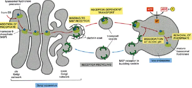
In 1955, De Duve discovered the lysosome, degradative organelle present inside cell cytoplasm. The inner lysosomal milieu has an acid pH and it contains a wide array of enzyme, at least 50 type of hydrolases that explain an extremely selective catalytic activity on different substrate (Fig. 8). Lysosomes have an important role in cell metabolism and consequently in cell function.
Fig. 8. Mechanism of lysosomal digestion
Nascent lysosomal enzymes (green balls) from the endoplasmic reticulum shuttle in the Golgi apparatus where they bind the mannose 6-phosphate receptor (M6PR). Most of the enzymes are then trafficked to the mature lysosome. [From The Art of MBoC3, 1995]
Every moments several types of endogenous macromolecules, like proteins, neurotransmitter, signal transduction components, lipids, and/or cell ingested materials are delivered to the lysosome for organizing their degradation and their components recycling (Frandson et al., 2003), when admitted.
When this machinery is wrong and/or a genetic error occurs, cell integrity and viability are compromised, because an unbalance between anabolism and catabolism make collapse in physiological cellular homeostasis.
This elementary described situation is the theoretical basis of lysosomal storage diseases (LSDs) that are a group of genetic inherited disorders occurring in humans and animals when there is lowered activity of a lysosomal enzyme (Platt and Walkley, 2004). The result is the accumulation or storage of non-catabolised products, due to a defect in a hydrolytic enzyme, activator protein, transport protein, or enzyme required for the correct processing of other lysosomal proteins. This storage initiates a cascade of pathological dysfunction typically observed in the nervous system (Platt and Walkley, 2004).
19
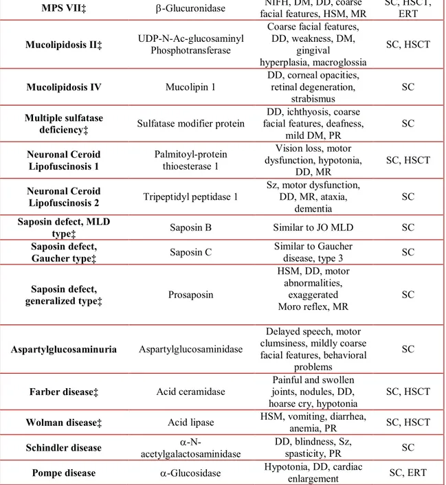
The majority of LSDs have autosomal recessive inheritance and they are rare, occurring approximately 1 in 5000 to 8000 live births in the United States, Europe, and Australia (Meikle et al., 1999). Despite the rarity of these diseases, some of these disorders result in purely non-neurologic manifestations (e.g., Gaucher disease type 1), but many others are characterized by a wide range of neurologic symptoms, with or without somatic features, presenting from birth to adulthood and have devastating effects, imposing a significant burden on families and the community (Table 3). Still today, about 40 different genes have been identified as sites for mutations resulting in a LSD with neurologic involvement.
Disease Defective Protein Presenting Signs and Symptoms Treatment Options†
GM1 gangliosidosis‡ Acid -galactosidase
IO: hypotonia, DD, coarse facial features
HM, CRS (±) LO: DD, ataxia, dysarthria, PR, dystonia SC,§ HSCT GM2 gangliosidosis, B variant, Tay-Sachs disease‡ Hexosaminidase A IO: hypotonia, hyperacusis, DD, CRS
LO: ataxia, dystonia, psychoses, PR
SC, HSCT
GM2 gangliosidosis,O
variant, Sandhoff disease‡ Hexosaminidase A & B Similar to Tay-Sachs disease SC
GM2 gangliosidosis, AB
variant‡ GM2 activator protein Similar to Tay-Sachs disease SC
Fabry disease‡ -Galactosidase crises, corneal opacities, Acroparesthesia, pain fatigue,angiokeratomas
SC, ERT HSCT
Gaucher disease, types
2 and 3‡ Glucocerebrosidase HSM, DD, strabismus, Sz, myoclonus, horizontal supranuclear gaze palsy SC, ERT,HSCT
Niemann-Pick type A‡
F Sphingomyelinase HSM, hypotonia, DD, CRS (±) SC
Niemann-Pick type C1‡ NPC1
Neonatal onset: jaundice, HSM, hypotonia LO: emotional lability,
ataxia, dystonia, HSM (±), VSO
SC, HSCT
Niemann-Pick type C2‡ NPC2 Similar to Niemann-Pick type C1 SC
Metachromatic
leukodystrophy‡ Arylsulfatase A
Late IO: weakness, hypotonia, DD, genu
recurvatum JO: weakness, PR, ataxia,
behavior changes AO: pyramidal or cerebellar signs, behavior
changes, psychoses, dementia
20 Krabbe disease‡ Galactocerebrosidase IO: spasticity, irritability,
hypotonia, fisting, DD LO: spastic paraparesis,
weakness, burning paresthesia, ataxia, weakness, vision loss
SC, HSCT
-Mannosidosis‡ -Mannosidase DD, hearing loss, mildly coarse facial features
(large jaw), mild DM SC, HSCT -Mannosidosis‡ -Mannosidase mild facial coarsening, DD, MR, hearing loss,
angiokeratomas
SC
Sialidosis,Mucolipidosis
I‡ Sialidase
IO: NIFH, DD, coarse facial features, DM, HSM, PR,renal disease
LO: myoclonus, CRS, ataxia, visual defects
SC
Sialic acid storage
disease, Salla disease‡ Transport protein
IO: severe DD, fair hair and skin, HSM, coarse
facial features LO: hypotonia, MR, ataxia, DD, speech delay,
coarse facial features
SC
Galactosialidosis‡ Protective protein, cathepsin A
Neonatal onset: NIFH, HSM, severe DD IO and late IO: coarse
facial features, HSM, kidney and heart defects,
DD, DM, MR LO: coarse facial features, DM, corneal clouding, MR, ataxia,Sz,
CRS(±)
SC
Fucosidosis‡ -L-fucosidase facial features, DM, MR, Spasticity, DD, coarse angiokeratomas
SC, HSCT
MPS I (Hurler and
Hurler-Scheie)‡ -L-iduronidase
Coarse facial features, DD, DM, MR, hearing loss, corneal clouding,
hernias
SC, HSCT, ERT
MPS II (Hunter) Iduronate-2-sulfatase DD, DM, hearing loss, coarse facial features, joint stiffness
SC, HSCT, ERT
MPS III A (Sanfilippo) Glucosamine-N-sulfatase
Aggressive behavior, DD, mildly coarse facial
features, hirsute, coarse hair, mild DM
SC, HSCT
MPS III B‡ -N-Ac-glucosaminidase Similar to MPS III A SC MPS III C -glucosaminide-AcCoA:
Nacetyltransferase
Similar to MPS III A SC
MPS III D N-acetylglucosamine-6- sulfatase
21 MPS VII‡ -Glucuronidase facial features, HSM, MR NIFH, DM, DD, coarse SC, HSCT, ERT
Mucolipidosis II‡ UDP-N-Ac-glucosaminyl Phosphotransferase
Coarse facial features, DD, weakness, DM, gingival hyperplasia, macroglossia SC, HSCT Mucolipidosis IV Mucolipin 1 DD, corneal opacities, retinal degeneration, strabismus SC Multiple sulfatase
deficiency‡ Sulfatase modifier protein
DD, ichthyosis, coarse facial features, deafness,
mild DM, PR SC
Neuronal Ceroid
Lipofuscinosis 1 Palmitoyl-protein thioesterase 1
Vision loss, motor dysfunction, hypotonia,
DD, MR SC, HSCT
Neuronal Ceroid
Lipofuscinosis 2 Tripeptidyl peptidase 1
Sz, motor dysfunction, DD, MR, ataxia,
dementia
SC
Saposin defect, MLD
type‡ Saposin B Similar to JO MLD SC Saposin defect,
Gaucher type‡ Saposin C
Similar to Gaucher
disease, type 3 SC
Saposin defect,
generalized type‡ Prosaposin
HSM, DD, motor abnormalities,
exaggerated
Moro reflex, MR SC
Aspartylglucosaminuria Aspartylglucosaminidase
Delayed speech, motor clumsiness, mildly coarse facial features, behavioral
problems
SC
Farber disease‡ Acid ceramidase joints, nodules, DD, Painful and swollen
hoarse cry, hypotonia SC, HSCT
Wolman disease‡ Acid lipase HSM, vomiting, diarrhea, anemia, PR SC, HSCT
Schindler disease acetylgalactosaminidase-N- DD, blindness, Sz, spasticity, PR SC
Pompe disease -Glucosidase Hypotonia, DD, cardiac enlargement SC, ERT
Table 3. Presenting features in Lysosomal Storage Diseases with prominent neurologic findings
Abbreviations: AO, adult onset; CRS, cherry-red spots; DD, developmental delay; DM, dysostosis multiplex; ERT, enzyme replacement therapy; HSCT, hematopoietic stem cell transplantation; HM, hepatomegaly; HSM, hepatosplenomegaly; IO, infantile onset; JO, juvenile onset; LO, late onset; L, leukocytes; MLD, metachromatic leukodystrophy; MPS, mucopolysaccharidosis; MR, mental retardation; NIFH, nonimmune fetal hydrops; PR, psychomotor regression; Sz, seizures; SC, supportive care; VSO, vertical supranuclear ophthalmoplegia. †HSCT is not available for all patients with a given diagnosis, and ERT may be in use or only at the stage of preclinical trials. The use of these treatments in a few patients does not necessarily indicate a successful outcome.‡Diseases diagnosed in author’s laboratory.§SC indicates any procedure performed to alleviate pain, discomfort, and seizures and may include splenectomy and kidney transplantation when indicated.
22
The differences in symptoms demonstrate that even if most lysosomal enzymes are ubiquitous, the effect of undigested-substrate accumulation depends on the cell or tissue type in which the substrate is synthesized with a high turnover. For example, accumulation of glycogen in Pompe involves myopathy for its role in muscle metabolism, while several sphingolipidosis are characterized by severe neuropathology, because of their high concentration in the brain.
Thus, owing to the complexity of the storage products (Table 4) and differences in their tissue distribution and rates of accumulation, the disease can cause pathologic changes in multiple organ systems or can be confined to the nervous system (Wenger et al, 2003).
Classification Accumulated
Substrate Disease Examples Species
Lipidoses
Lipids. Includes sphingolipids, cholesterol esters and
triglycerides Krabbe disease Gaucher disease Wolman disease Humans, mice, dogs, sheep, monkey Humans, mice, dogs, sheep Humans, mice Mucopolysaccharidoses (MPS) Mucopolysaccharides (glycoaminoglycans) MPS I MPS II MPS III Humans, mice, cats, dogs Humans, mice, dogs Humans, dogs, cattle
Glycogenosis Glycogen Pompe disease Humans, mice, cattle, dogs, cats
Glycoproteinoses Glycoproteins and/or
oligosaccharides Fucosidosis α- mannosidoses Sialidosis Humans, dogs Humans, cattle, mice, guinea pig,
cats Humans, mice Table 4 Categories of LSDs based on accumulated substrate
[Modified from Hopwood et al., 2004]
The treatment options for LSDs, like enzyme replacement therapy, hematopoietic stem cell transplantation are not completely effective; so the exit is unfavorable. Supportive care became necessary and a definitive protocol of treatment remains limited despite ongoing research (Kolter and Sandhoff, 2006; Platt and Walkley, 2004; Wenger et al., 2001).
23
Therefore, it is critical that the patients be diagnosed as early as possible, by recognizing the symptoms from the beginning. Several structures perform newborn screening for LSDs to identify presymptomatic individuals that may be candidates for early therapeutic intervention. Bio-banks institution could be important in the next future, due to the augmented efficiency of scientific searching methods.
Krabbe disease description
Krabbe disease (OMIM #245200, OMIA #1140/000578) is one of the classic genetic LSDs with autosomal recessive inheritance that affects both CNS and PNS in several species including humans, rhesus macaques, dogs, mice (Suzuki and Suzuki, 1985; Suzuki, 2003) and sheep (Pritchard et al., 1980). In 1916, the Danish neurologist Knud Haraldsen Krabbe described the presence of globoid cells in the brain (Krabbe, 1916). So the disease was named after him and later globoid cell leukodystrophy, due to the presence of multinuclear (globoid) macrophages in the white matter. This was the unique microscopic pathological characteristic of Krabbe disease and reliable diagnostic method until the beginning of the 70s, when Suzuki and his colleagues (Suzuki et al., 1970a; Suzuki et al., 1971) identified the genetic deficiency of enzyme galactosylceramidase (galactocerebroside β-galactosidase) (GALC) as the cause of human and canine globoid cell leukodystrophy.
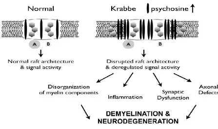
GALC is a lysosomal enzyme that catalyzes the degradation of galactose from galactosylceramide (Gal-cer) and galactosylsphingosine (psychosine, Psy). It acts in myelin turnover to catabolize the myelin lipid (Lefebvre and Vartanian, 2002; Wenger et al., 2001). In the normal nervous system, these substances are processed by the lysosome (Kolter and Sandhoff, 2006) and recycled components are able to enter the remyelination pathway (Lefebvre and Vartnanian, 2002; Suzuki, 2003). Occurring mutations in the GALC gene (Luzi et al., 1997; Rafi et al., 1995; Sakai et al., 1996; Victoria et al., 1996) result in a lower (Wenger et al., 2001) or loss activity of GALC and lipid degradation during myelin turnover is impaired (Suzuki, 2003; Wenger et al., 2001). Remyelination does not occur effectively in Krabbe disease (Suzuki, 2003; Wenger et al., 2001), but myelin turnover continues (Suzuki, 2003). For this reasons, Krabbe disease is a sphingolipidosis with the peculiarity that, despite the GALC deficiency, galactosyceramide does not increase in the brain of patients. However, psychosine accumulates in the brain causing death of myelin-generating cells.
The galactosylceramidase deficiency results in degeneration of oligodendrocyte, astrocytic gliosis, and appearence of globoid cells, and demyelination that. Alteration of neuronal conduction and neurological dysfunctions are hallmark of pathology.
24
Krabbe disease in humans
Krabbe disease in humans is typically a neurodegenerative disease of infancy, but there are rare examples in which Krabbe disease has been diagnosed in older children and adults (Wenger et al., 2001).
Symptoms of Krabbe disease usually indicate CNS and PNS involvement, particularly the cerebellum (Jacob et al., 1973; Suzuki, 2003) which controls the refinement and execution of movement and the co-ordination of muscle activity (Starr and Taggart, 2001), as region of the brain responsible for the initiation of movement and conscious sensation. Clinical sign of disease progress is the loss of higher cerebral functions (Suzuki, 2003; Wenger et al., 2001).
The symptoms of the infantile form (95% of known cases) usually onset at 3-6 months and initially are characterized by generalized hyperirritability, hypersensitive to the external environment, stiffness of the limbs (Suzuki and Suzuki, 1985; Suzuki, 2003; Wenger et al., 2001), and episodic fever of unknown origin. The cerebrospinal fluid protein level is already highly elevated and rapid and severe motor and mental deterioration develops: psychomotor functions deteriorate with marked hypertonicity, extended and crossed legs, flexed arms, and the backward-bent head. At the end stage of the disease, sometimes reached within a few weeks or months, the infant is decerebrate and blind and become unresponsive and unaware of their surroundings. In this case, rarely patients survive beyond two years of age (Suzuki, 2003; Wenger et al., 2001) and often die of respiratory infections.The parents of an affected child are obligate heterozygotes and therefore each parent carries one normal and one mutated GALC allele, the measured GALC enzyme activity can range widely in carriers because of polymorphisms in the normal copy of the gene. Although some parents have quite low GALC enzyme activity measured in vitro, none has clinical disease. At conception, each sib of an affected individual has a 25% chance of being affected, a 50% chance of being an asymptomatic carrier, and a 25% chance of being unaffected and not a carrier. Once an at-risk sib is known to be unaffected, the chance of his/her being a carrier is 2/3.
The clinical phenotype of the later-onset forms is much more variable and it is often divided into two groups: the late infantile (onset 6 months to 3 years), and the juvenile (onset 3-8 years). In the late infantile group, irritability, psychomotor regression, stiffness, ataxia, and loss of vision are frequent initial symptoms. The course is progressive, resulting in death in approximately 2 to 3 years after the onset. In the juvenile group, patients commonly develop loss of vision, together with hemiparesis, ataxia, and psychomotor regression.
Sometimes adult patients have a milder phenotype and a slower rate of progression. Some adult patients could have a normal life span. While some individuals remain stable for long periods of life, others show a steady decline in vegetative state and death. The cerebrospinal fluid protein is normal or only mildly elevated in juvenile or adult patients.