Contents
Abstracts 2
Introduction
6
1. Mechanism of pain 7
2. Veterinary Pain Evaluation 17
3. Veterinary Pain Management 28
4. Loco-Regional Analgesia 34
Materials and Methods
36
Results 45
Discussion 47
Conclusions 54
Appendix 55
Sommario
Scopo del lavoro
L’obiettivo di questo studio è quello di valutare la durata dei blocchi motorio e sensitivo, la qualità dell’anestesia e dell’analgesia dopo il blocco neuro-stimolato dei nervi sciatico e femorale, utilizzando solo bupivacaina o combinandola con la dexmedetomidina, in cani sottoposti a procedure chirurgiche interessanti l’articolazione del ginocchio.
Materiali e metodi
Trentuno cani, di 4.4 anni (1-11) e di peso 25.2 kg (3-68), assegnati in maniera random a tre gruppi: A, B e C. Il Gruppo A riceveva bupivacaina 0,25 mgkg-1 per ogni nervo; il Gruppo B riceveva bupivacaina 0,25 mgkg-1 con dexmedetomidina 0,25 µgkg-1 per ogni nervo; il Gruppo C riceveva bupivacaina 0,5 mgkg-1 per ogni nervo.
I dati raccolti durante l’anestesia sono stati: frequenza cardiaca (HR), frequenza respiratoria (RR), pressione media (MAP), anidride carbonica a fine espirazione (EtCO2), end-tidal isofluorano (EtISO), temperature esofagea (T°).
Sono stati registrati anche i tempi di durata di chirurgia, anestesia ed estubazione. La Short Form della Glasgow Composite Pain Scale (GCPS-SF score) è stata usata per valutare il dolore post-operatorio a 1, 2, 4, 6, 8, 12, 16 e 20 dall’estubazione La capacità di camminare, la propriocezione e il pinch test sono stati usati per valutare la durata del blocco motorio e sensitivo ad ogni ora dopo l’estubazione e fino alla fine del blocco.
Risultati
Il test della normalità di Shapiro-Wilk mostra che i dati non sono normalmente distribuiti.
Differenze significative sono state trovate tra i gruppi in relazione al peso corporeo, ma non per BCS ed età.
Le frequenze cardiache e respiratorie restano nei ranges fisiologici in tutti i gruppi. La pressione arteriale è stata variabile nei differenti punti della procedura; è stato necessario somministrare una fluidoterapia di supporto in quasi tutti gli individui del Gruppo C, per compensare la moderata ipotensione perianestetica.
L’analisi statistica dei punteggi del dolore mostra che c’è una differenza significativa alla 16° ora rispetto alla 1° nel Gruppo B, e alla 16° e 20° ora nel Gruppo C; nessuna differenza è stata trovata nel Gruppo A.
Nessuna differenza significativa è stata trovata tra i gruppi per quanto concerne la durata dei blocchi motorio e sensitivo.
Nel Gruppo B sono state trovate correlazioni tra la durata dei blocchi motorio e sensitivo e il peso corporeo dei soggetti.
Conclusioni
Sulla base dei risultati preliminari di questo studio, la combinazione di bupivacaina e dexmedetomidina usata per il blocco perineurale dei nervi sciatico e femorale (0,25 mgkg-1 and 0,25 µgkg-1 rispettivamente per ogni nervo) potrebbe significativamente aumentare la durata del blocco e apportare dei benefici sulla stabilità della pressione arteriale. Per confermare statisticamente la tendenza trovata con i nostri risultati, è necessario un campione di studio più grande e omogeneo.
Abstract
Objective
The aim of this study is to evaluate the motor and sensitive block duration, related to quality of anaesthesia and analgesia after sciatic/femoral electrolocation nerve block with bupivacaine alone and in combination with dexmedetomidine in dogs undergoing unilateral stifle joint surgery.
Materials and methods
Thirty-one dogs, median age 4.4 years (1 – 11) and median weight 25.2 kg (3 - 68), randomly assigned to three groups: A, B and C. Group A received bupivacaine 0,25 mgkg-1 to each nerve; Group B received bupivacaine 0,25 mgkg-1 mixed with dexmedetomidine 0,25 µgkg-1 to each nerve; Group C received bupivacaine 0,5 mgkg-1 to each nerve.
Data recorded over all the anaesthesia procedure were: heart rate (HR), respiratory rate (RR), median blood pressure (MAP), carbon dioxide end-tidal (EtCO2), end-tidal
isoflurane (EtISO), esophageal temperature (T°).
Surgery, anaesthesia and extubation times were also collected.
Short Form of the Glasgow Composite Pain Scale (GCPS-SF score) was used to assess post-operative pain at 1, 2, 4, 6, 8, 12, 16 e 20 hours after extubation.
Ability to walk, proprioception and pinch test were used to assess motor and sensory block duration every one hour after the extubation and until blockade end.
Results
Shapiro-Wilk normality test shows that data were not normally distributed.
Significant differences were found among groups in relation of body weight, but not for BCS and age.
Heart and respiratory rates remained in physiological ranges in all three groups. Arterial pressure was variable along the different points of the procedure; fluid therapy support was necessary in almost all individuals for Group C to compensate a mild perianaesthetic hypotension.
Pain score was increased at 16th hour compared to the 1st hour in Group B, and at 16th and 20th hours compared to the 1st in Group C; no differences are found in Group A. No significant difference were found between groups concerning motor and sensory blocks duration. Between motor and sensory block scores and dogs weights there were some correlations in Group B.
Conclusions
On the basis of the preliminary results of this study, the mixture of bupivacaine and dexmedetomidine used for perineural blockage of the sciatic and femoral nerves (0,25 mgkg-1 and 0,25 µgkg-1 respectively for each nerve) can significantly increase the blockade duration and improving some benefits on arterial pressure stability. To statistically confirm the tendence found by our results a larger and homogenous sample will be indispensable.
Introduction
Animal welfare is nowadays considered an essential point for the correct management of the patient, for various needs, as surgical, oncology, obstetric or medical.services.
For this reason, the efforts in findings anaesthetic and analgesic protocols, able to adapt to different needs and situations is mandatory.
Pain is defined in 1979 by the International Association for the Study of Pain (IASP) as “Unpleasant sensory and emotional experience associated with actual or potential
tissue damage, or describe in terms of such damage”.(1)
Recently, in 2016, Williams and Craig proposed another definition of pain as “a
distressing experience associated with actual or potential tissue damage, with sensory, emotional, cognitive and social components”. It effectively describes the
subjectivity of pain experience, differentiating it from physiological processes, although biological mechanisms govern that experience.(2)
An unpleasant emotional experience (eg, fear) can be the trigger of homeostatic responses similar to those induced by noxious stimuli, characterized by reflex withdrawal, behavioral, autonomic nervous system, neuroendocrine, and immune system responses.(3)
1. Mechanism of pain
Pain sensation involves different physiologic processes as stimuli transduction, transmission, modulation, projection, and perception.
The transduction occurs when a stimulus activates nociceptors and high-threshold sensory nerve fibres, warning the organism of a potentially damaging event.(3)
(from: Muir WW, Woolf CJ. Mechanisms of pain and their therapeutic implications. J Am Vet Med Assoc 219:1346, 2001)
Nociceptors can be classified in several ways:
1) on the basis of their responsiveness to the various stimulation modalities in:
- mechanical, - thermal, - chemical;
2) on the basis of the structure of their axons in:
- myelinated (A-fiber), usually characterized by thin axons with fast
conduction velocities (2-25 m/s). There is a small percentage of A-fiber nociceptors that have faster conduction velocities, up to 50m/s;
- unmyelinated (C-fiber), with a slow conduction velocities ranging from
Not all A and C-fibres sensory receptors are nociceptive.(4)
Nociceptors encode the intensity, duration, location, and quality of the noxious stimulus and, once activated, transduce noxious stimuli into depolarizing electrical potentials; after that moment, the transmission of the stimulus to the spinal cord can occurs.(3)
(from: Muir WW, Woolf CJ. Mechanisms of pain and their therapeutic implications. J Am Vet Med Assoc 219:1346, 2001)
Many of the C and finely A fibers contain the excitatory amino acids aspartate (NMDA) and glutamate and a wide variety of neuropeptides, including substance P, calcitonin gene-related peptide, cholecystokinin (CCK), galanin, somatostatin, and others, that are selectively released in the spinal dorsal horn and in peripheral tissue.(5)
The dorsal horn of the spinal cord is divided into laminae based on the types of neurons and their organization:
- lamina II (substantia gelatinosa) - laminae III –VI (nucleus proprius)
- lamina X (the area around the central canal)
The target of somatic C nociceptors is ipsilateral lamina II, whereas Aδ nociceptors terminate in ipsilateral lamina I and, to a lesser extent, lamina V; the large Aβ sensory nerve fibres terminate on neurons predominantly located in laminae III, IV, and V.(3,5)
Visceral C nociceptors have a more diffuse projection to lamina II that is spread out over several segments, as well as bilateral projections to laminae V and X. The widespread distribution of the visceral C fibers may explain the diffuse quality of visceral pain.(5)
In the laminae can be distinguished two kind of neurons:
- nociceptive specific neurons (NS): are so-named because they receive
peripheral input exclusively from nociceptive afferents; they are excited by both A-δ and C fibers. NS neurons are located mainly in lamina I and II of the dorsal horn; they are present in lesser number in lamina V;
- wide dynamic range neurons (WDR): are so-named because they receive
input from large diameter, non-nociceptive afferents, as well as both A-δ and C fibers. They are concentrated in deeper layers of the dorsal horn (lamina V and VI) but are also found in smaller numbers in I, II, and IV.(6)
In spinal cord, modulation of sensory informations takes place, before these are relayed to the brain. All afferent sensory nerve fibers that enter the spinal cord are separated in order to innervate second-order neurons in the different zones or laminae of the gray matter of the dorsal horn of the spinal cord. The dorsal horn laminae comprise layers of functionally distinct cells that form columns extending the length of the spinal cord. The columns contain a large number of second-order excitatory and inhibitory interneurons that receive multiple inputs from surrounding columns and send outputs to the brain and to the ventral (motor) horn. The ventral horn contains interneurons and motor neurons that control the muscles of the trunk and limbs, whereas the transition zone between the dorsal and ventral horn (in
thoracic and sacral cord segments) contains autonomic preganglionic neurons that mediate involuntary (visceral) functions and transmit some sensory afferent information via multiple parallel circuits to the brain.(3)
The Aδ- and C fibres input to the dorsal horn releases glutamate that preferentially binds to α-amino-3 hydroxy-5-methyl-4- isoxazolepropionic acid (AMPA), kainite, and Nmethyl-D-aspartate (NMDA) ligand-gated sodium and calcium channels. Under normal conditions, NMDA ion channels are blocked by magnesium ion (Mg2+). Activation of AMPA receptor ion channels depolarizes the membrane, producing fast excitatory postsynaptic potentials that are focused by local and descending inhibitory neurons, many of which release the inhibitory substances glycine and γ-aminobutyric acid (GABA). Fast excitatory potentials carried predominantly by A-δ sensory nerve fibres signal the onset, duration, and location of the noxious stimulus and are responsible for the well localized and relatively transient sharp, pricking, stinging, and stabbing pain that serves to warn and protect the animal from additional tissue damage. Intense thermal or mechanical stimulation increases the quantity of glutamate and substance P released from afferent sensory nerve terminals, activating postsynaptic neurokinin-1 and metabotropic glutamate receptors (mGluR), facilitating and prolonging the release of intracellular calcium and generating plateau potentials by activation of VGCC (voltage-gated calcium channels
)
and nonspecific cation channels. The magnitude of these events is proportional to stimulus intensity and is responsible for the removal of the magnesium blocking of NMDA receptors and the generation of a greater and longer (> 10 seconds) postsynaptic depolarizing response, such that pain remains long after the stimulus is removed. (3)Once sensory informations are processed, the projection phase starts. WDR and NS axons cross to the opposite side of the spinal cord, ascend in the anterolateral quadrant and terminate primarily in the thalamus. This pathway is called the lateral spinothalamic tract. Neurons in the thalamus receiving messages along this tract subsequently project their axons upward to a number of cerebral cortex locations including the primary somatosensory cortex. There are numerous other terminations of spinal cord pain projection neurons throughout the midbrain, pons and medulla.
Spinal cord pain projection neurons do not project only to those areas of the thalamus which relay pain messages to primary somatosensory cortex. In addition, the anterolateral quadrant which contains the lateral spinothalamic tract is not the sole pathway for transmission of pain messages to levels above the spinal cord.(6)
The ascending pain pathways are summarized in the table below.
Ascending Pain Pathways
( from: Robinson AJ. Central nervous system pathways for pain transmission and pain control. J Hand Ther 10:64-77, 1997)
The perception of sensory informations occurs in:
- rostroventromedial medulla: an important region for the integration and
processing of ascending nociceptive information and modulation of descending output from the brain. This region contains excitatory and inhibitory cells that either facilitate or inhibit nociceptive reflexes and nociresponsive behaviours;
- thalamus: integrates and relays information to the somatosensory cortex,
which in turn projects to adjacent cortical association areas, such as the limbic system;
- limbic system: includes the cingulate gyrus (involved with behaviour and
emotion), amygdala (conditioned fear and anxiety), hippocampus (memory), hypothalamus (sympathetic autonomic activity), locus
coeruleus (arousal, vigilance, behaviour), and portions of the periaqueductal gray matter (fight or flight response, stress-induced analgesia);
- somatosensory cortex.(3)
Afferent nerve input, both somatic and autonomic, from an area of trauma or injury, activates hypothalamic–pituitary axis and sympathetic nervous system. There is a failure of the normal feedback mechanisms of hormone secretion control that causes immunological and haematological changes. There is generally release of catabolic hormones such as the catecholamines and pituitary hormones whereas anabolic hormones such as insulin and testosterone are suppressed. (7)
Changes occurring during the stress response
(from: Burton D, Nicholson G, Hall G. Endocrine and metabolic response to surgery. In Continuing Education in Anaesthesia, Critical Care & Pain | Volume 4 Number 5, 2004: 144-147)
1) Sympathetic nervous system:
Hypothalamic activation of the sympathetic nervous system causes an increased secretion of catecholamines from the adrenal medulla and release of norepinephrine from presynaptic nerve terminals. The increased of sympathetic activity acts on:
- cardiovascular system, with consequently tachycardia and hypertension - renal and adrenal gland functions, with releasing of renin that causes the
conversion of angiotensin I to angiotensin II; this stimulates the secretion of aldosterone from the adrenal cortex, which in turn increases sodium reabsorption from the distal convoluted tubule in the kidney;
- pancreatic and hepatic functions: with increasing in glucagon secretion,
that stimulates the breakdown of glycogen in the liver and muscle leading to increased glucose and lactate concentrations as well as mobilization of free fatty acids (FFAs) from available lipid stores. (7,8)
2) Hypothalamic-pituitary-adrenal axis
- Anterior pituitary: under stimulation by hypothalamic releasing factors,
the pituitary synthesizes pro-opiomelanocortin, that can be metabolized within the pituitary into ACTH (adrenocorticotrophic hormone), β-endorphin and an N-terminal precursor.
The ACTH stimulates the adrenal cortical secretion of glucocorticoids with consequently increasing of circulating concentration of cortisol. Surgical stimulus also increases the GH (growth hormone or somatotrophin) and PRL (prolactin) secretion. GH stimulates protein synthesis and inhibits protein breakdown, promotes lipolysis and has an anti-insulin effect, with inhibition of glucose uptake and use by cells, which spares glucose for its use by neurons in situation of glucose scarcity; GH may also stimulate glycogenolysis in the liver. Concentrations of TSH, FSH and LH remain unchanged.
- Posterior pituitary: produces the ADH (antidiuretic hormone) and also
stimulates, with corticotrophin-releasing factor, the secretion of pro-opiomelanocortin from the anterior pituitary. ADH is an important vasopressor and enhances haemostasis; ACTH release is enhanced by ADH.
- Adrenal cortex: cortisol secretion from adrenal cortex increases rapidly
following the start of surgery; the cortisol response can be modified by anaesthetic intervention. Usually, the increased concentrations of circulating cortisol inhibit further secretion of ACTH, but this control is not effective after surgery, so the concentrations of both hormones remain high. Cortisol promotes proteolysis, lipolysis and gluconeogenesis; it inhibits glucose use by cells, so that blood glucose concentrations are increased. Cortisol has also an anti-inflammatory activity, with the inhibition of accumulation of macrophages and neutrophils into areas of inflammation and can interfere with the synthesis of inflammatory mediators, above all the prostaglandins.
- Pancreas: in response to trauma, the failure of the body to secrete insulin is partly caused by the inhibition of the β-cells in the pancreas by the a2-adrenergic inhibitory effects of catecholamines. Thus, the perioperative period is characterized by a state of functional insulin deficiency. In contrast to insulin, glucagon release promotes hepatic glycogenolysis and gluconeogenesis, but insulin effects predominate. Glucagon secretion increases briefly during surgery but it is not thought to make a major contribution to the hyperglycaemia.
- Thyroid: thyroids hormones stimulate oxygen consumption in many
organs, increase the metabolic rate and heat production. Their circulating concentrations are inversely correlated with sympathetic activity and after surgery there is a reduction in thyroid hormone production, which returns to normal over a few day.(7,8)
Hormonal changes during surgery
Endocrine Hormones Change in secretion glands
Anterior pituitary ACTH Increases GH Increases
PRL Increases
TSH May Increase or decrease FSH/LH May Increase or decrease Posterior pituitary ADH Increases
Adrenal cortex Cortisol Increases Aldosterone Increases Pancreas Insulin Often decreases Glucagon Usually small increases Thyroid Thyroxine
Tri- iodothyronine
(From: J. P. Desborough. The stress response to trauma and surgery. BJA: British Journal of Anaesthesia, Volume 85, Issue 1, 1 July 2000, Pages 109–117)
ACTH: adrenocorticotrophic hormone; GH: growth hormone; PRL: prolactin; TSH: thyroid-stimulating hormone; FSH: follicle-thyroid-stimulating hormone; LH: luteinizing hormone; ADH: arginine vasopressin or antidiuretic hormone
3) Metabolic consequences of the endocrine response: Consist in the increasing of catabolic process:
- carbohydrate metabolism: hyperglycaemia is a major feature of the
metabolic response to surgery and results from an increase in glucose production and a reduction in glucose utilization. This is facilitated by catecholamines and cortisol, which promote glycogenolysis and gluconeogenesis. The mechanisms, which regulate glucose homeostasis, are ineffective because of initial failure of insulin secretion followed by insulin resistance. Potential risks of prolonged perioperative hyperglycemia include wound infection and impaired wound healing.
There is also an increased risk of ischemic damage to the nervous system and myocardium.
- protein metabolism: initially there is inhibition of protein anabolism, followed later, if the stress response is severe, by enhanced catabolism, stimulated by increased cortisol and cytokine concentrations. The amino-acids, derived from catabolism, form new proteins in the liver known as acute phase proteins, but albumin production is reduced interfering with the maintenance of the extracellular volume. The liver also converts amino-acids into other substrates as glucose, fatty acids or ketone bodies. The loss of protein can be measured indirectly by increased nitrogen excretion (urea) in the urine.
- lipid metabolism: increased catecholamine, cortisol and glucagon secretion, in combination with insulin deficiency, promotes lipolysis and ketone body production. Triglycerids are converted by lipolysis to fatty acids and glycerol, and this latter become the substrate for gluconeogenesis; fatty acids may be oxidized and converted to ketone bodies or re-esterified.
- salt and water metabolism: Arginine vasopressin secretion results in water retention, concentrated urine, and potassium loss and may continue for 3–5 days after surgery. Renin is secreted from the juxtaglomerular cells of the kidney secondary to sympathetic efferent activation. It
converts angiotensin to angiotensin II, which in turn releases aldosterone from the adrenal cortex promoting sodium and water retention from the distal convoluted tubule. (7,8)
4) Immunological and haematological changes:
The immune system and neuroendocrine system are closely related. In response to tissue injury from surgery or trauma, cytokines are synthesized by activated macrophages, fibroblasts, endothelial and glial cells. Cytokines are low molecular weight, heterogeneous glycoproteins that include interleukins (IL) 1–17, interferons, and tumour necrosis factor. Although they exert most of their effects locally (paracrine), they can also act systemically (endocrine). Cytokines play an important role in mediating immunity and inflammation by acting on surface receptors of target cells. The most important cytokine associated with surgery is IL-6 and peak circulating values are found 12–24 h after surgery. The size of IL-6 response reflects the degree of tissue damage which has occurred. IL-6, and other cytokines, cause the acute phase response, which includes the production of acute phase proteins such as fibrinogen, C reactive protein, complement proteins, a2-macroglobulin, amyloid A and ceruloplasmin. Other effects of cytokines include fever, granulocytosis, haemostasis, tissue damage limitation and promotion of healing. Cytokines may increase the release of cortisol and this latter limits their production via a negative feedback system. Thus, the cortisol response to surgery limits the severity of the inflammatory response.(7)
2. Veterinary pain evaluation
Differences in pain tolerance have been demonstrated experimentally in people and animals and these play an important role in pain management; in fact, it is important
to know that there are individuals with a lower threshold for pain and others with particularly stoic nature, that may be sex-related (9,10), age-related (10,11) or
breed-related. (12,13)
The American College of Veterinary Anaesthesiologists (ACVA) in the 2006 has written a position paper where it said that “animal pain and suffering are clinically important conditions that adversely affect an animal's quality of life, either in the short or long term”. It also said that “an important part of determining whether an animal is in pain is the ability to recognize departures from normal behaviour and appearance of that animal”. In this paper a list of behavioural changes to be considered for the evaluation of the presence or absence of pain in an animal is drawn up:
Changes in personality or attitude. A normally quiet and docile animal becomes suddenly aggressive, or an aggressive animal becomes quiet. An animal may attempt to bite, especially when a painful area is palpated. The animal may not interact with the clinician in a normal manner, but may seem to be unresponsive or withdrawn.
Abnormal vocalization, especially when a painful area is palpated or the animal is forced to move. For example, dogs whine or whimper, cats hiss or growl, pigs grunt and squeal excessively, primates grunt or scream, rats squeak at an unusual pitch, mice chatter. Vocalization tends to be an insensitive and nonspecific indicator of pain and should not be relied on as the sole criterion for determining whether an animal requires treatment for pain.
Licking, biting, scratching or shaking of a painful area. If excessive, these behaviours can lead to self-mutilation.
Changes in the appearance of the haircoat. Ruffled fur, a greasy appearance indicative of a lack of grooming, and piloerection may be indicative of pain.
Changes in posture or ambulation. Limping or carrying of a painful appendage; tensing of abdominal and back muscles to produce a tucked up appearance is especially noticeable in dogs, cats, and rodents.
Changes in activity level. An animal may become restless and pace or repetitively lie down, get up, and lie down again. In contrast, an animal may be recumbent and lethargic or reluctant to move with guarding of the painful area.
Changes in appetite, such as a decrease in food and water consumption leading to weight loss and dehydration.
Changes in facial expression. Eyes become dull and pupils may be dilated. Pinning of the ears, grimacing, and a sleepy or photophobic appearance may be evident.
Excessive sweating or salivation. Horses frequently sweat in response to pain; however, cattle do not. Stressed rodents often salivate excessively.
Oculonasal discharge. Rats when stressed often shed porphyrin pigment in their tears and appear to be bleeding from their eyes and nose.
Teeth grinding is frequently heard in rabbits, cattle, sheep, and goats experiencing pain.
Changes in bowel movements or urination, such as diarrhea with soiling of the perineum, dysuria, and tenesmus.
Other parameters that are indicative of pain include increasing in heart rate, respiratory rate, arterial pressure and body temperature. Blood samples can be evaluated for elevations in glucose, corticosteroid, and catecholamine concentrations.(14)
The detection of pain is one of the most challenging problems in veterinary medicine. In people, the self-reporting of pain is the gold standard for the assessment of pain; in veterinary medicine, the recognition of pain is based on the interpretation of animal’s behaviour and cardiovascular variations by the veterinary surgeon/nurse in the case of acute pain and by the owner in the case of chronic pain.(15, 16)
Before analgesic drugs can be administered to reduce pain, the signs of pain must be assessed quantitatively, with using pain scales.
In the past the scales used to assess pain in animals and human beings have been based on assessments of its intensity alone, the so-called unidimensional scales, as:
- Simple Descriptive Scale (SDS), which uses 4 or 5 points based on verbal
description (nil, mild, moderate, severe, very severe). The use of this scale for comparative purposes is limited by its lack of sensitivity for detecting relatively small changes.
- Numerical Rating Scale (NRS), marked 0-10 or 0-20
- Visual Analogue Scale (VAS), utilises a straight line, conventionally 10 cm long, whose extreme limits are marked by perpendicular lines. At the ends of the scale there is a verbal description of each extreme of the symptom to be evaluated. (17)
( from: Downie WW, Leatham PA, Rhind VM, Wright V, Branco JA, Anderson JA. Studies with pain rating scales.Ann Rheum Dis. 1978 Aug;37(4):378-81)
These scales have been shown to be unreliable in the assessment of pain in patients who cannot communicate, for example patients suffering of dementia, children and animals.
This limitation led to the development of multidimensional scales. Melzack and Torgerson (1971) developed a 'language of pain', which considered other qualities of
pain in addition to its intensity, based on the interpretation of behaviours such as facial expressions, postures and activities. This language formed the basis of the McGill pain questionnaire (Melzack 1975) which was designed to provide quantitative assessments of clinical pain that could be treated statistically.
Formats of the numerical rating (NRS) and
It is only recently that the utility of such methods has been recognised by veterinarians.
- Morton and Griffiths (1985) first proposed a composite scale for
assessing pain in animals, in the form of 'Guidelines for the recognition of pain, distress, and discomfort in experimental animals and a hypothesis for assessment'. They suggested that specific behaviours, including posture, vocalisation, temperament, food and water intake and locomotion might be used as indicators of pain. Scores from 0 to 3 were allocated for specific changes in eight characteristics, including the animal's bodyweight, its appearance, any relevant clinical signs, unprovoked behaviour and responses to stimuli. The total pain score was the sum of these scores and could range from 0 to 24. They also defined what interpretations should be applied to certain ranges of total scores; 0 to 4 was regarded as normal; 5 to 9 the animal should be monitored carefully; 10 to 14 the animal should be treated with analgesics or euthanased, or the experiment should be terminated. They did not distinguish between species. The guidelines were designed to ensure that practice was consistent between laboratories and to provide an objective set of criteria for safeguarding animals' welfare. No criteria for the development of the guidelines were provided, and it is therefore questionable whether they can do more than ensure consistent practice between laboratories.
- Sanford and al. (1986) published further 'Guidelines for the recognition
and assessment of pain in animals'. Their aim was to provide an effective and uniform set of criteria for controlling the severity of the pain. The behaviours associated with pain were the animal’s posture, facial expression, gait, acceptance of handling, vocalisation and overall mental status. These guidelines also suggested that a clinical examination should be carried out, paying particular attention to physiological signs such as pupil dilation, and changes in blood pressure, heart rate and body temperature. No rationale for the selection of these criteria was provided and the properties of the measurements made were not explored.
- Conzemius and others (1997) described a 'Numerical Rating Scale' for
assessing postoperative pain in dogs, which differs from the NRS mentioned previously. The method scores three types of behaviour, vocalisation, movement and agitation, scores of 0 to 2 being allocated to the first two behaviours and 0 to 3 for the third, according to predefined rules, giving a possible total score of between 0 and 7. This numerical scale provided defined criteria for the assessment of different aspects of the animals' behaviour, but the authors did not provide selection criteria for the types of behaviour or describe how the weights were attributed to them. No results of the use of this scale have been reported and it is therefore not possible to assess its consistency or value.
- The Colorado State University Veterinary Teaching Hospital Pain Score
(Hellyer and Gaynor 1998) is a scale for the assessment of pain in cats and dogs which is based on eight categories of behaviour and physiological signs considered to be indicative of pain: comfort, movement, appearance, unprovoked behaviour, interactive behaviour, vocalisation, heart rate and respiratory rate. Each category is assigned a score of between 0 and 4 according to predefined criteria, the total score being the total of the scores for each category. The aim of this scale was to estimate an animal's requirement for analgesia. However, no details were given of how the categories were selected for the scale, and its validity in the assessment of pain is therefore uncertain.
- The University of Melbourne Pain Scale (UMPS) (Firth and Haldane
1999) evaluates postoperative pain in dogs on the basis of assessments of behaviour and physiological signs. It is composed of six categories: physiological variables, response to palpation, activity, mental status, posture and vocalisation. The method was similar to that used in the development of the Children's Hospital of Eastern Ontario Pain Scale (McGrath and others 1985), a behaviour-based scale for the assessment of pain in children. The assessments of the animal's mental status, heart rate and respiratory rate were based on the change from its presurgery status,
so this scale is limited to be used in dogs undergoing surgery. Ranges are defined for the increases in heart and respiratory rates and scores are consequently assigned, assuming that there is a direct relationship between the changes in physiological signs and the intensity of pain, although the converse has been demonstrated.
- The Glasgow coma scale (GCS) (Teasdale and Jennett 1974) concentrates
on three aspects of behavioural response, and its universality depends on identifying responses which can be clearly defined and accurately graded according to degree of dysfunction. This scale must be practical to use in many different places by staff without special training. It was therefore considered important to define the behavioural descriptors precisely, so that the observer was in no doubt as to their interpretation.
- The Glasgow Composite Measure Pain Scale, developed by Holton and
other (2001), is a pain scale in dogs which might be used by everyone, that provides clear definitions of all the expressions used. It is based on the McGill pain questionnaire, in which pain descriptors are supplied by practising veterinary surgeons familiar with the behavioural signs of acute pain in dogs. After refinement, the final list of descriptors was divided into seven categories: posture, comfort, vocalisation, demeanour, mobility, attention to wound and response to touch. Many of the behaviours and signs of the other behaviour-based scales for the assessment of pain in dogs were included, such as the Colorado State University scale that includes vocalisation, movement, unprovoked and interactive behaviour, and heart rate and respiratory rate; mobility and vocalisation are included in the guidelines for pain recognition defined by Morton and Griffiths (1985); Firth and Haldane (1999) included activity, posture and vocalisation in their scale; Potthoff and Carithers (1989) included other indicators of pain which have been used in the present scale, such as body movement, restlessness, panting and whimpering. The Glasgow scale is a validated pain scale, for the strong agreement with past literature and for the fact that the pain descriptors included in this scale
are considered to be strictly associated with pain by wider veterinary community.(18)
Morton and al. (2005) have established an interval level measurement for assessment of acute pain in dogs and to investigate the validity of the scale.(19)
( from: Holton L, Reid J, Scott EM, Pawson P, Nolan A. Development of behaviour-based scale to measure acute pain in dogs. Vet Rec 148:525-531,2001)
( from: Holton L, Reid J, Scott EM, Pawson P, Nolan A. Development of behaviour-based scale to measure acute pain in dogs. Vet Rec 148:525-531,2001)
- The Short-Form of Glasgow Composite Measure Pain Scale (CMPS-SF),
developed by Reid and other (2007), derived from previous scale, in order to have a easy and fast way for routine clinical use. It is comprises six behavioural categories with associated descriptive expressions (items): vocalisation, attention to wound, mobility, response to touch, demeanour and posture/activity. Four of the six categories contain five items, one contains four and the other six. The observer chooses the single item within each category which best fits the dog’s condition and the pain score is the sum of the rank scores of each item chosen. The maximum score for the six categories is 24, or 20 if mobility is impossible to assess. The clinical decision-point for analgesic intervention, when mobility could be assessed, is established with a level as 6/24 and higher; while analgesic intervention is gave with a level of 5/20 and higher, if section B could not be carried out as a result of the animal’s physical condition, and consequently total score was out of 20 rather than 24.(20)
Murrell et al. (2008) have applied this scale in a centre with a different surgical procedures and analgesic protocols, and where English is not the first language, to test its validity in a different clinical environment. The modified scale was used to score pain in 60 dogs during the 24 hours after surgery. They have demonstrated that the modified CMPS is a useful scale for measuring perioperative pain in a clinical setting, where a wide variety of analgesic and anaesthetic protocols are used. (20)
( from: Reid J, Nolan AM, Hughes JML, Lascelles D, Pawson P, Scott EM. Development of the short-form Glasgow Composite Measure Pain Scale (CMPS-SF) and derivation of an analgesic intervention score. Animal Welfare 2007, 16(S): 97-104)
Beyond humanitarian consideration pain can induce catabolism, impair respiration, delay wound healing, prolong periods of hospitalization, and increase morbidity and mortality. (21)
In these latter years, many surveys have been conducted to obtain information related to attitudes, opinions and knowledge of both veterinary surgeons and pet's owners, about acute and chronic pain and the use of analgesic drugs in small animal veterinary practice. (22-27)
It seems there is a positive trend about the importance of providing effective pain management for small animals by the veterinary professionists, but peri-operative use of analgesic drugs is still suboptimal.(15)
Owners believed more frequently that analgesics are always needed for surgical procedures than for the medical conditions (26,27); in fact difficulties with owner compliance is reported as one of the major barriers to adequate treatment of chronic pain, with difficulties of pain assessment and expense of drugs.(25)
Simon et al. conclude by saying that improving our understanding of pet owners' perceptions and knowledge of anaesthesia, surgery and pain can lead to better client education. (27)
3. Veterinary Pain management: systemic and local analgesia
The concept of preemptive analgesia is introduced for the first time by Crile (28), who observed that if pain transmission was blocked prior to the initial surgical incision, postoperative mortality was decreased as well as the intensity and duration of postoperative pain.
Preemptive analgesia is an antinociceptive therapy whose aim is to prevent both peripheral and central sensitization, in order to avoiding the postoperative amplification of pain sensation.
Treatment can be performed at the periphery, to block inputs along sensory axons, or at CNS sites using single or combinations of analgesic drugs, injected systemically, in the form of single shot or in continuous rate infusion (CRI). (28,29)
a) Opioids
Opioids are among the oldest and most frequently used drugs for the alleviation of pain. They have high efficacy and they are remarkably safe, with the benefit of reversibility, through the administration of naloxone, an opioid antagonist. These drugs bind to opioid receptors located in the central and peripheral nervous system and cause a conformational change that increases their affinity for guanine nucleotide binding proteins (G-coupled proteins). In the perioperative period, they are administered to produce analgesia and sedation as part of premedication (neuroleptanalgesia) and multimodal analgesic protocols.(30)
Side effects of µ-opioid receptor agonists are: depression tidal volume and gas
exchange, diminution of breathing rate progressing to respiratory arrest with higher
doses, decrease chest wall compliance, and increase upper airway resistance, for their
action on discharges of respiratory bulbospinal, vagal, propriobulbar neurons and phrenic nerve activity. (31)
Opioids have been traditionally administered by the intravenous, intramuscular, subcutaneous route as single shot, or as intravenously constant rate infusions (CRI).
A recent alternative pharmaceutical form of opioid administration is the transdermal patch, such as fentanyl (32-34) and buprenorphine (35,37), or the transdermal solution of fentanyl (38), with the aim to extend their action duration and potentially mitigate the disadvantages of both oral and parenteral opioid administration.
Opioid drugs are also administered to enhance loco-regional anaesthesia, both alone or in co-administration with local anaesthetics. Morphine is a highly hydrophilic drugs that produces analgesia 30–40 min after epidural administration, with long duration of effect due to the delayed systemic absorption and longer maintenance in the spinal cord. Epidural or spinal administered opioids relieve somatic and visceral pain by selectively blocking nociceptive impulses without interfering with sensory and motor function or depressing the sympathetic nervous system.
Intra-articular administration of morphine has become increasingly popular after arthroscopy surgery; after joint inflammation, a significant increase in µ-opioid receptors in both articular and peri-articular tissues occurs. (30)
( from: Berry SH. Analgesia in the Perioperative Period. Vet Clin North Am Small Anim Pract. 2015 Sep;45(5):1013-27. doi: 10.1016/j.cvsm.2015.04.007. Epub 2015 May 26)
b) Local anaesthetics
Local anaesthetics are inexpensive, easy to use, and rarely associated with significant adverse events. They can be incorporated into a balanced analgesic plan in several ways and it is recommended that they be included in the plan of every surgical patient.
Local anaesthetics prevent the conduction and propagation of nerve impulses by binding to voltage-gated sodium channels, producing complete analgesia. Adverse events associated with local anaesthetic administration are usually the result of inadvertent intravenous administration (above all bupivacaine) or overdose. Signs of local anaesthetic toxicity include ataxia, nystagmus, and tremors, which can progress to convulsions, unconsciousness and respiratory arrest.
Cardiovascular manifestations of local anaesthetic toxicity include bradycardia and sinus arrest. The versatility of local anaesthetics allows them to be used in several ways:
- Topical: local anaesthetics may be applied to skin or mucosa to
desensitize areas (eg, lidocaine spray, lidocaine patch);
- Infiltrative: local anaesthetics can be injected near specific nerves or
placed in wounds (eg, peripheral nerve blocks, soaker catheters);
- Systemic: lidocaine may be given intravenously, in continuous rate
infusion, in order to decrease inhaled anaesthetic requirements and provide anti-inflammatory, antiarrhythmic and analgesic effects;
( from: Berry SH1. Analgesia in the Perioperative Period. Vet Clin North Am Small Anim Pract. 2015
Sep;45(5):1013-27. doi: 10.1016/j.cvsm.2015.04.007. Epub 2015 May 26)
c) Non-steroidal anti-inflammatory drugs (NSAIDs)
The anti-inflammatory and analgesic properties of the NSAIDs are mediated by the inhibition of COX-1 and COX-2, responsible of inflammatory mediators production of interest in nociception, such as the prostaglandins, thromoboxane A2 and prostacyclins. NSAIDs are useful in both acute and chronic pain management, particularly osteoarthritis. (40)
d) Ketamine
Ketamine is a dissociative agent with NMDA receptor antagonism and sympathomimetic effects. At low doses it provides analgesia, while also providing anaesthesia at high dose. It is thought to provide less intense analgesia than pure-µ opioids and is often used in combination with an opioid for multimodal analgesic effects. (41)
In one study of 2000, Slingsby&Waterman-Pearson said that the use of ketamine in dogs undergoing ovariohysterectomy as pre-emptive analgesia has required less analgesic intervention than the control group that were not treated with ketamine; this latter did not require significantly more analgesic intervention than the treated with ketamine at extubation, suggesting that preoperative ketamine treatment give some benefit over administration of ketamine post-operatively. (42)
Ketamine can also been used in continuous rate infusion, with a significant sparing effect on the MAC of isoflurane in dogs. (43) Kaka et al have reported in their study of 2016 that the minimum serum concentration of ketamine to produce analgesia in dogs is between 100-200 ng/mL during infusion. (44)
e) α2-agonist:
Alpha2-adrenoreceptor agonists are widely used in small animal for their potent sedative and analgesic properties, and for the possibility to reverse their action, in those cases where side effects occur. The administration of an α2-antagonist
(atipamezole) reverses side effects, but also sedation and analgesia provided by these drugs. This class of drugs have significant effects on the cardiovascular and respiratory systems:
- Cardiovascular effects include: bradycardia, hypertension, hypotension,
reduction in cardiac output and stroke volume.
- Respiratory effects include: reduction in respiratory rate and tidal volume,
which may result in respiratory acidosis and hypoxemia in some animals. These effects can be seen even when very small doses of α2-agonists are administered, so their use should be limited to those animals that can tolerate cardiovascular and respiratory systems imbalance.
In premedication, their administration with opioids can act synergistically, enhancing analgesia and sedation and reducing the doses used of each drug. In post-operative period, small doses can provide sedation and analgesia especially in animals experiencing dysphoria.
The α2-agonists may be used as adjuvant to local anaesthetics in peripheral nerve blocks, prolonging their duration. Intra-articular injections of α2-agonists also may be beneficial, as shown by studies involving humans undergoing arthroscopy and rats suffering from arthritic pain. (45)
Dexmetomidine may be used in continuous rate infusion (CRI), alone or in combination with other drugs ( eg lidocaine, fentanil) to reduce halogenated agents MAC (minimum alveolar concentration) during anaesthesia period (46,47) or to improve analgesia in perioperative period. (48, 49)
f) other drugs:
- Gabapentin is a non-opioid medication, structurally similar of γ-amino
butyric acid, which has commonly used as an anticonvulsant and antinociceptive drug, throughby the release of some neurotransmitters, such as substance P and glutamate, and interacting with γ-aminobutyric acid receptors in the spinal cord. It was found that its administration with opioids in perioperative period may enhance sedation and analgesia, and reduce post-operative analgesic requirements. (39,50,51)
- Maropitant: is an NK-1 receptor antagonist approved for use as an antiemetic zone for dogs and cats for its central action in the chemoreceptor trigger. In one study, Alvillar et al (2012) found that the intravenously, but not epidural, administration of maropitant reduce sevofluorane MAC. (52)
In another study, Marquez et al (2015) compared the use of maropitant (1 mg/kg SQ) and morphine ( 0,5 mg/kg SQ) as premedication in dogs undergoing ovariohysterectomy; they no found major clinical difference between groups; suggesting that maropitant given as a pre-anaesthetic may be comparable to 0.5 mg/kg of morphine for an OHE and may improve the post-operative recovery. (53)
However, its analgesic role is still controversial and further studies are needed.
4. Loco-regional analgesia
Peripheral nerve blocks are becoming increasingly popular for perioperative use as analgesic techniques in small animals.
The influence of regional anaesthesia on the stress response has been extensively investigated. In a recent study of 2016, Romano et al. have measured the stress biomarkers ( glycaemia and cortisol release), postoperative pain and recovery quality in dogs undergoing stifle surgery, in order to compare two loco-regional techniques (peripheral femoral and sciatic nerve block; spinal anaesthesia), pre-operatively performed, with peri-operative systemic fentanil and post-operatively methadone. They found that analgesia with a peripheral nerve block or spinal anaesthesia prevented the glycaemic and cortisol responses to surgery, promoted better recovery quality, and decreased postoperative pain scores compared with fentanil, so regional anaesthesia techniques were found to be excellent alternatives to fentanyl administration.
There are many studies, both in human and in animals, that widely show the advantages of using one analgesia loco-regional technique instead of systemic analgesia, or a combination between both, in order to reduce systemic opioid administration and to avoid their side effects such as bradycardia, hypotension, hypoventilation, ileus, nausea, vomiting and dysphoria. (31,54)
The table below is a list of loco-regional anaesthesia techniques, but further papers that study the use of various methods of neuro-localization, such as electroneurostimulation and ultrasound, are frequently published.
( from: Berry SH1. Analgesia in the Perioperative Period. Vet Clin North Am Small Anim Pract. 2015 Sep;45(5):1013-27. doi: 10.1016/j.cvsm.2015.04.007. Epub 2015 May 26)
Material
s and methods
The study was conducted in the period between April 2017 and April 2018, in the Centre Hospitalier Universitaire of the École Nationale Vétérinaire d'Alfort (ENVA), Maisons-Alfort, France. This study was approved, according to Directive 2010/63/EU, by the Chair of the Veterinary University Hospital Ethics Approval Board, and informed consent was obtained from all owners.
Objective
The aim of this study is to compare motor and sensitive block duration, quality of anaesthesia and analgesia after sciatic/femoral electrolocation nerve block with bupivacaine alone and in combination with dexmedetomidine in dogs undergoing unilateral stifle joint surgery.
Our hypothesis was that perineural dexmedetomidine combined with bupivacaine might offer a longer duration of sensory block than bupivacaine alone and consequently a better analgesia quality, and we could reduce the dose of bupivacaine avoiding supplemental opioid administration.
Study design
Prospective, randomized, double blinded clinical trial
Animals
Thirty-one dogs, median age 4,4 years (1 – 11) and median weight 25,2 kg (3 - 68), undergoing unilateral stifle joint surgery, assessed to be ASA physical status I or II after physical examination and haematology and serum chemical analyses were enrolled in this study.
The population included both male and female dogs ( 15 and 16 respectively), of various breeds: : 3 Labrador Retrievers, 3 Mixed-unknown, 3 Yorkshire Terriers, 3 Majorca Mastiff Dogs, 2 Golden Retrievers, 2 American Bull Dogs and 1 each of American Staffordshire Terrier, Bernese Mountain Dog, Bulldog, Canarian Dog, Cesky Fousek, Chihuahua, Continental Bulldog, Creole Dog, Dachshund, Doberman Pinscher, Dogo Argentino, Eurasier, German Shepherd Dog, Great Pyrenees, Pitt Bull cross.
Procedure study
Surgery procedures included:
- Tibial plateau leveling osteotomy (TPLO); - Trochleoplasty;
- Tibial Tuberosity Transposition; - Transarticular Stabilization.
In particular: 21 dogs are presented for Tibial Plateau Levelling Ostectomy, 5 Transarticular Stabilization of cruciate ligament rupture, 2 Tibial Tuberosity Transposition, 1 Trochleoplasty, 1 Trochleoplasty + Tibial Tuberosity Transposition, 1 Tibial Plateau Levelling Ostectomy + Trochleoplasty.
All the dogs received a standard general anaesthesia protocol:
- premedication was performed with intramuscularly acepromazine 0,03
mgkg-1 ( Calmivet® ; Vetoquinol, France);
- after twenty minutes, a catheter was aseptically placed in a cephalic vein,
and all dogs received intravenously propofol to effect ( ~ 4mgkg-1, Propovet; Zoetis, France) and midazolam 0,2 mgkg-1 (Midazolam; Mylan, France), as induction
- after orotracheal intubation, anaesthesia was maintained with isoflurane in
oxygen diluted. The end-tidal isoflurane (EtISO) was titrated to lowest percentage able to maintain a suitable plane of anaesthesia (between 0.9% and 1.4%).
Lactated Ringer’s solution was administered intravenously at 5 mLkg-1
h-1 throughout the anaesthesia period and in the early post-anaesthesia recovery.
Prior to each block, hair over each target site was clipped and the skin was aseptically prepared for injection.
Dogs were randomly allocated to three treatments:
- Group A (10 subjects) received 0,25 mgkg-1 of bupivacaine (0,5% Bupivacaine hydrochloride; Aguettant, France) to each nerve;
- Group B (13 subjects) received 0,25 mgkg-1 of bupivacaine (0,5% Bupivacaine hydrochloride; Aguettant, France) mixed with 0,25 µgkg-1 of dexmedetomidine (Dexdomitor 0,5 mg/ml; Zoetis, France) to each nerve; the dose of dexmedetomidine was taken from a human study of 2016; (55)
- Group C ( 8 subjects) received 0,5mgkg-1 bupivacaine (0,5% Bupivacaine hydrochloride; Aguettant, France) to each nerve.
The volume injected to each nerve was 0,1 mlkg-1.
- For Group A, the injected solution was prepared by adding the same
volume of saline solution to bupicavaine;
- For Group B, the injected solution was prepared in the same way of the
previous one, but it was also added dexmedetomidine; the choice of this dose of dexmedetomidine was chose considering a similar human study of 2016 (55);
- For Group C, the injected solution was prepared with only bupivacaine.
The choice of the treatment was made by the same anaesthetist who after has performed perineural block, but he was not involved in the peri- and post-operative
assessments. These latter are been recorded by another anaesthetist unaware of the treatments.
Insulated needles (22 G, 50 or 100 mm Stimuplex-A Insulated Needle; B. Braun, Bethlehem, PA) connected to a peripheral nerve stimulator (Stimuplex HNS12; B. Braun, Germany) were used to perineurally inject the anaesthetic solution.
When the appropriate muscular response was observed at a stimulating current lower than 0.5 mA and higher than 0.2 mA, to avoid intraneural injection, solution was injected at both the femoral and sciatic nerve sites.
Femoral and sciatic nerve blocks were performed as literature described:
- For the sciatic nerve block, the needle was inserted at the one-third the distance between the greater trochanter and ischiatic tuberosity. Flexion of the hock was used to confirm location of the needle close to the nerve. (57)
(Campoy L., Malher S., in Small Animal Regional Anaesthesia and Analgesia; Chapter 13: The Pelvic Limb, pagg 199-226. Ed. 2013)
- For the femoral nerve block, the needle is inserted at the intersection of two lines, the first drawn from the spinous process of L6 perpendicular to the spine in dorsoventral direction, the second line drawn from the most cranial aspect of the iliac crest parallel to the spine until it intersects the first line. The needle is directed in a caudomedial direction with an inclination of 30– 45° angle through the iliocostalis lumborum muscle and it is advanced until contractions of the quadriceps muscles and consequently extension of the stifle were observed. (58)
(Campoy L., Malher S., in Small Animal Regional Anaesthesia and Analgesia; Chapter 13: The Pelvic Limb, pagg 199-226. Ed. 2013)
Fentanyl ( 2µgkg-1 IV) was intraoperatively administered as rescue analgesia if HR, RR and MAP values were exceed of 30% the baseline values, despite EtISO 1,4%. At extubation time if dysphoria was observed:
- Propofol 1 mgkg-1 IV was administered;
- Propofol 1 mgkg-1 with methadone 0,2 mgkg-1 (Comfortan 10mg/ml; Dechra, France) IV were administered if fentanil was intraoperatively injected.
Meloxicam 0,2 mgkg-1 (Inflacam 20 mg/ml; Virbac, France) was administered subcutaneously at the end of anaesthesia, if hypotension did not occurred; tramadol 4 mgkg-1 (Altadol; Formevet, France) was administered orally 20 hours after extubation and every 8 hours thereafter the observational period, regardless of the pain score.
Monitoring
Data recorded prior to premedication are: age, sex, breed, weight, BCS, surgery procedure, heart rate (HR), respiratory rate (RR), body temperature (T°) and oscillometric non invasive blood pressure (NIBP) with a cuff placed around the antebrachium of the thoracic limb.
Data recorded throughout anaesthesia were: heart rate (HR), respiratory rate (RR), median blood pressure (MAP), carbon dioxide end-tidal (EtCO2), end-tidal isoflurane
(EtISO), esophageal temperature (T°) using a multiparameter monitor (Bedside Monitor BSM-5135K, Nihon Kohden Corporation, Japan). Parameters recording began at skin incision and continued every 5 minutes until surgery ended.
The baseline values were recorded when the anaesthesia plane was stable at EtISO 1,1%-1,2%, after nerve blocks and just before surgery started.
Invasive blood pressure was monitored in eighteen patients via a catheter placed in the dorsal pedal artery and the misuration was compared with that non invasive in order to verify the precision of the oscillometric pressure. The measured values were almost superimposable. When systolic and median pressures fell below 80 and 60 mmHg respectively, infusion of Ringer has been increased from 5 to 10 mlkg-1h-1, and a bolus of 10 mlkg-1 in ten minutes was administered; if this was not enough, a bolus of 2-4 mlkg-1 in ten minutes was administered; if all this was not sufficient, cardiovascular support drugs were administered.
Surgery, anaesthesia and extubation times were also collected.
Time to first dose of rescue analgesic, total doses of rescue analgesic required per dog, and GCPS-SF score (Short Form of the Glasgow Composite Pain Scale) at 1, 2, 4, 6, 8, 16 and 20 hours after extubation were recorded.
Dogs received rescue analgesic (methadone 0,2 mg/kg IM) if assigned total GCPS-SF score was ≥ 6 or if at least one category was scored ≥3.
To assess the duration of motor and sensory blockades was used a visual clinical evaluation, based on the method described by Portela et al. (2010). Motor and
sensory blockades were assessed at extubation time and then every one hour until total recovery of limb activity.
The proprioceptive response and ability to walk was used to assess motor blockade (MB), as follows:
( from: Portela DA, Otero PE, Tarragona L, Briganti A, Breghi G, Melanie P. Combined paravertebral plexus block and parasacral sciatic block in healthy dogs. Vet Anaesth Analg. 2010 Nov;37(6):531-41)
Sensory blockade was evaluated by observing the response to a noxious stimulus applied with pressure from a Kelly clamp on the skin over the medial aspect of the thigh (innervated by the sensory branch of the femoral nerve, the saphenous nerve - MT), over the caudal aspect of the metatarsus (innervated by the tibial nerve - CM) and over the third phalanx of the fourth digit (innervated by the common fibular nerve - TP).
( from: Portela DA, Otero PE, Tarragona L, Briganti A, Breghi G, Melanie P. Combined paravertebral plexus block and parasacral sciatic block in healthy dogs. Vet Anaesth Analg. 2010 Nov;37(6):531-41)
Statistical analysis
Statistical analysis were performed using SPSS 15.0 (IBM Company, Italy). Shapiro - Wilk normality test was used to verify the normal distribution of data. For statistical analysis the first 60 minutes of recorded anaesthesia data during the surgery is only considered.
The data were expressed by median and range.
Changes along the time were evaluated with Wilcoxon Test, while differences between groups were compared using the Kruskal–Wallis and Jonckheere Terpstra tests. Correlation of Pearson was performed between dogs body weight and block duration and between anaesthesia and extubation times. A value of p < 0.05 was considered statistically significant.
Results
All the dogs were included in the study and all recovered from anaesthesia uneventfully, without showing any signs of delirium or dysphoria.
Shapiro-Wilk normality test shows that data were not normally distributed.
Significantly differences are found among groups in relation of body weight, with a median value in the Group A higher than the others two groups (p value = 0,002); while no differences are found for body condition scores (BCS) and for age (Tables 1, 2 and 3).
The total anaesthesia and extubation times do not significantly differ among groups. (tables 4 and 5). There are not correlation between both measured times.
There are not statistically differences between the three groups about the amperage of performed femoral block, while there is a mild difference about performed sciatic block, demonstrated by statistically significant for Kruskal Wallis test (p = 0,036), but not for Jonckheere-Terpstra tendency (tables 6 and 7).
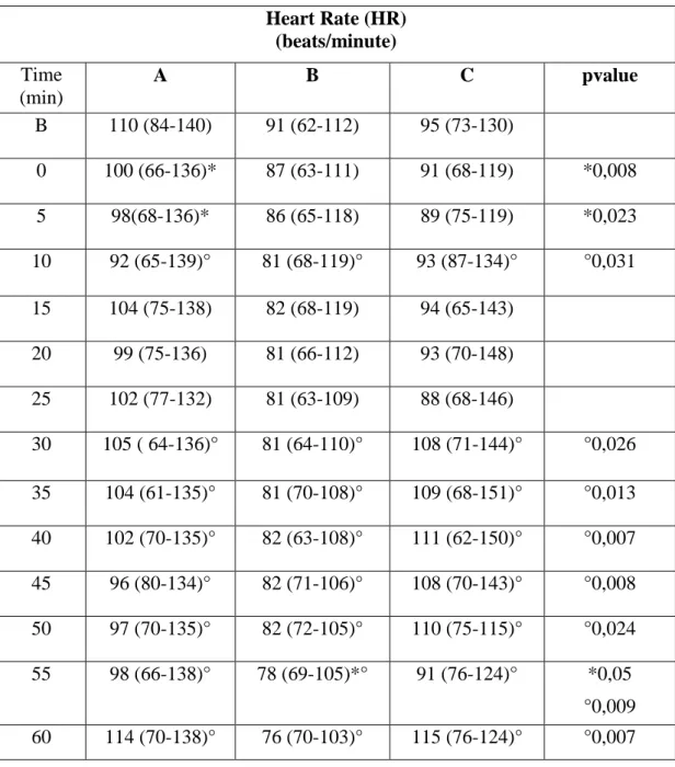
Heart and respiratory rates remain (HR, RR) within the normal physiological ranges in all measured time points and in all the three treatments; there are significant differences from baseline and between the three treatments in some time points. (Tables 8 and 9; Graphics 1 and 2).
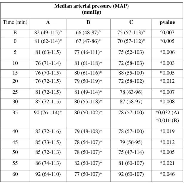
Median arterial pressure (MAP) is significantly higher than baseline point at almost all time points in Group B; there are significant differences in baseline and time 0 values between the three groups, and in Group B these values are lower than in both other two groups (Table 10; Graphic 3).
It was necessary to increase crystalloids perfusion rate from 5 to 10 mlkg-1h-1 in only one subject in Group A, in four subjects in Group B and C. In Group B three of the four subjects also received a 10 mlkg-1 crystalloid bolus and one of this a 2 mlkg-1 colloid bolus. In Group C three of the four subject also received a 10 mlkg-1 crystalloid bolus, one of these received a second crystalloid bolus, and another received a 4mlkg-1 colloid bolus; in two subjects 0,1 mgkg-1 of ephedrine was administered.
Expired isoflurane concentration (EtISO) in all time points and between three groups is not statistically significant (table 11; Graphic 4). The median scores in all time points and in the three groups were lower than conventional MACBAR. (59,60)
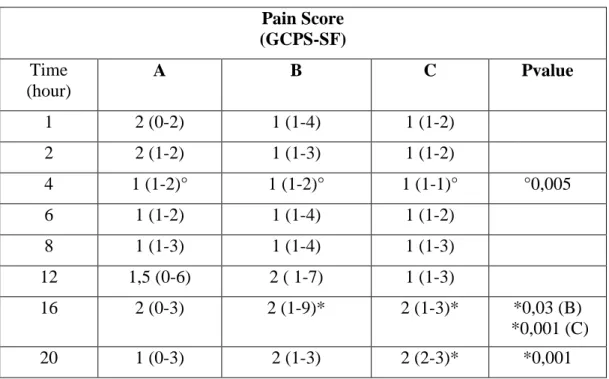
Statistical analysis of Pain Score shows that there is a significantly difference at 16th hour compared to the 1st hour in Group B, and at 16th and 20th hours compared to the 1st in Group C; no differences are found in Group A. Comparing the three groups there is a significant difference in the 4th hour (Table 12; Graphic 5). About rescue analgesia administration in intra- and post-operatively periods:
- In Group A, one subject received fentanyl two times and another three times in the intra-operatively period, but both do not received analgesics after; one dogs received methadone at 12th hour (GCPS=6);
- In Group B, two dogs received methadone at 12th hour and other two at 16th (GCPS=7, 6, 9, 5; this latter needed analgesia because it received a score of 3 in the section C);
- In Group C two dogs only received intra-operatively fentanyl.
No significant differences are found between the groups with concerning motor and sensory blocks; only in MT3 significant mildly difference is present for Kruskal Wallis test (p = 0,01), but not for Jonckheere-Terpstra tendency (Tables 13-16; Graphics 6-9).
Between motor and sensory block scores and dogs body weights there are some correlations at level:
- TP3, TP2 and TP1 and weights of subjects in Group B (p values = 0,005; 0,006 and 0,006 respectively);
- CM3, CM2 and CM1 and weights of subjects in Group B (p values = 0,004; 0,006 and 0,006 respectively).