Lactic Acid Fermentation of Cactus Cladodes
(Opuntia ficus-indica L.) Generates Flavonoid
Derivatives with Antioxidant and
Anti-Inflammatory Properties
Pasquale Filannino1, Ivana Cavoski2, Nadia Thlien2, Olimpia Vincentini3, Maria De Angelis1, Marco Silano3, Marco Gobbetti1, Raffaella Di Cagno1*
1 Department of Soil, Plant and Food Science, University of Bari Aldo Moro, Bari, Italy, 2 CIHEAM-MAIB, Mediterranean Agronomic Institute of Bari, Valenzano, Bari, Italy, 3 Unit of Human Nutrition and Health, Department of Veterinary Public Health and Food Safety, Istituto Superiore di Sanità, Roma, Italy *[email protected]
Abstract
Cactus pear (Opuntia ficus-indica L.) is widely distributed in the arid and semi-arid regions throughout the world. In the last decades, the interest towards vegetative crop increased, and cladodes are exploited for nutraceutical and health-promoting properties. This study aimed at investigating the capacity of selected lactic acid bacteria to increase the antioxi-dant and anti-inflammatory properties of cactus cladodes pulp, with the perspective of pro-ducing a functional ingredient, dietary supplement or pharmaceutical preparation.
Preliminarily, the antioxidant activity was determined through in vitro assays. Further, it was confirmed through ex vivo analysis on intestinal Caco-2/TC7 cells, and the profile of flavo-noids was characterized. Cactus cladode pulp was fermented with lactic acid bacteria, which were previously selected from plant materials. Chemically acidified suspension, with-out bacterial inoculum and incubated under the same conditions, was used as the control. Lactobacillus plantarumCIL6, POM1 and 1MR20, Lactobacillus brevis POM2 and POM4, Lactobacillus rossiae2LC8 and Pediococcus pentosaceus CILSWE5 were the best grow-ing strains. Fermentation of cladode pulp with L. brevis POM2 and POM4 allowed the high-est concentration ofγ-amino butyric acid. Lactic acid fermentation had preservative effects (P<0.05) on the levels of vitamin C and carotenoids. Two flavonoid derivatives (kaemferol and isorhamnetin) were identified in the ethyl acetate extracts, which were considered to be the major compounds responsible for the increased radical scavenging activity. After induc-ing oxidative stress by IL-1β, the increased antioxidant activity (P<0.05) of fermented clad-ode pulp was confirmed using Caco-2/TC7 cells. Fermented cladclad-ode pulp had also
immune-modulatory effects towards Caco-2 cells. Compared to the control, fermented clad-ode pulp exhibited a significantly (P<0.05) higher inhibition of IL-8, TNFα and prostaglan-dins PGE2 synthesis. The highest functional effect was found using ethyl acetate extracts. In conclusion, fermentation, especially with L. plantarum strains and L. brevis POM4, enhanced the antioxidant and immune-modulation features of cladode pulp.
OPEN ACCESS
Citation: Filannino P, Cavoski I, Thlien N, Vincentini O, De Angelis M, Silano M, et al. (2016) Lactic Acid Fermentation of Cactus Cladodes (Opuntia ficus-indica L.) Generates Flavonoid Derivatives with Antioxidant and Anti-Inflammatory Properties. PLoS ONE 11(3): e0152575. doi:10.1371/journal. pone.0152575
Editor: Muzamil Ahmad, Indian Institute of Integrative Medicine, INDIA
Received: September 2, 2015 Accepted: March 16, 2016 Published: March 29, 2016
Copyright: © 2016 Filannino et al. This is an open access article distributed under the terms of the
Creative Commons Attribution License, which permits unrestricted use, distribution, and reproduction in any medium, provided the original author and source are credited.
Data Availability Statement: All relevant data are within the paper and its Supporting Information files. Funding: The authors have no support or funding to report.
Competing Interests: The authors have declared that no competing interests exist.
Introduction
Cactus (Opuntia spp.), belonging to Cactaceae family, is cultivated in both hemispheres and all continents. Among all the species, Opuntia ficus-indica L. (known as cactus pear) is the most common. Native to Mexico, it is widely distributed and adapted to the arid and semi-arid regions of South and Central America, Africa and the Mediterranean area [1,2]. Because of the trend of Mediterranean area towards global desertification and decline of water resources, O. ficus-indica has an interesting potential as fruit and vegetative crop. The various vegetative parts of O. ficus-indica have marked economic importance, and are exploited as foods and pharmaceuticals [3]. Cactus fruits (prickly pear) are used for manufacturing juices, jams, jellies and alcoholic beverages. Young cladodes (stem segments) are commercialized mainly as mini-mally processed fresh foods [4]. Although less valuable than fruit crops, the interest towards vegetative crop of O. ficus-indica increased in the last decades. Cladodes were exploited for nutraceutical and health-promoting properties [5]. Since ancient times, O. ficus-indica was used in traditional folk medicine, and, recently, the popularity of cladodes increased also in developed countries, being recognized as source of phytochemicals and prebiotics [6]. Due to the high annual productivity of biomass per hectare (10–40 tones dry weight), cladodes repre-sent a cheap and suitable substrate for functional foods or dietary supplements [7]. Several studies showed the presence of natural compounds having anti-inflammatory, antioxidant, hypoglycemic, antimicrobial and neuro-protective activities [5,8]. The therapeutic potential of cladode extracts was shown in vitro or in vivo. A number of diseases were treated, including metabolic syndromes (diabetes type 2 and obesity), rheumatism, cerebral ischemia, renal dis-eases, cancers, and viral and bacterial infections [5,9–11]. Also the prebiotic activity of clad-odes-derived oligosaccharides was shown [6,11]. Cladodes are a source of phenolics, fibers, polyunsaturated fatty acids and vitamins [5,8]. Cladode extracts were used for technological applications as dietary supplements, ingredients for beverages, breakfast cereals and margarine or thickening agents in vegetable soups and dessert gels [11,12]. The cosmetic application is also wide. Cladode-gel exert a healing effect on dermal wounds [13].
The enhancement of the biogenic activity of cactus cladodes may rely into standardized and marketable products, having well-known or novel applications. For this purpose, bioprocessing of plant materials through microbial fermentation received considerable interest because of the better capacity to exploit inherent bioactivities with respect to industrial enzymatic processes. The only few studies investigated the effect of yeast fermentation on the health-promoting fea-tures of prickly pear cladodes [14]. To the best of our knowledge, no studies have considered the potential of the lactic acid fermentation. Besides, the identification and characterization of inherent components of O. ficus-indica were largely investigated [5,8] but very limited infor-mation are available regarding the metabolites that are generated during fermentation.
This study aimed at investigating the capacity of selected lactic acid bacteria to enhance the antioxidant and immune-modulatory features of cactus cladodes with the perspective of pro-ducing novel functional foods, dietary supplements or pharmaceutical preparations.
Material and Methods
Microorganisms and culture conditions
Thirteen strains lactic acid bacteria, belonging to the Culture Collection of the Department of Soil, Plant and Food Science, University of Bari Aldo Moro, Bari, Italy, were used as starters for fermentation (Table 1). All strains were previously isolated from fruits and vegetables. Strains were identified by partial sequencing of the 16S rRNA, recA, pheS, and rpoA genes. Cultures were maintained as stocks in 15% (vol/vol) glycerol at -80°C and routinely propagated at 30°C
for 24 h in MRS broth (Oxoid, Basingstoke, Hampshire, United Kingdom). Almost all the above strains were previously characterized for technology (e.g., acidifying capacity, growth and sensory profile) and functional features [15–23].
Cladode pulp processing and fermentation
Fresh cladodes of Opuntia ficus-indica (L.) Mill. (genotype Sanguigna) were collected in the autumn 2014 from an organically certified farm (Azienda agricola Ferarro Biofarm, Cod. Op. SUOLOESALUTE ASS34739AG7102), which is located in Santa Maria di Belice (Sicily, Italy). The owner of the farm gave permission to carry out the analyses on fresh cladodes. This study did not involve endangered or protected species. During the edible stage with tender and crispy structure, young cladodes were harvested three montgs after sprouting, before developing spines. After harvesting, cladodes were washed with water, cut into strips and blended by verti-cal food processor (mod R8 Robot Coupe, Bologna, Italy). Resulting cladode pulp (CP) was thoroughly mixed to ensure representative samples and stored at−20°C until processing and analyses. Lactic acid bacteria strains were singly used as starters. Cells were cultivated in MRS broth until the late exponential growth phase was reached (ca. 12 h), washed twice in 50 mM phosphate buffer, pH 7.0, and re-suspended in the CP at the initial cell density of ca. Log 8 CFU/g. CP was fermented at 30°C for 24 h. Samples were taken before and after fermentation. CP, without bacterial inoculum and chemically acidified with lactic acid (final pH of ca. 4.0), was incubated under the same conditions and used as the control (CP-CT).
Kinetics of growth and acidification
Growth was monitored by plating on MRS agar. The pH was measured by a Foodtrode elec-trode (Hamilton, Bonaduz, Switzerland). Kinetics of growth were determined and modeled according to the Gompertz equation as modified by Zwietering et al. [24]: y = k + A exp{-exp [(μmaxe/A)(λ - t) + 1]}, where k is the initial level of the dependent variable to be modeled (Log CFU/ml), A is the difference in cell density between inoculation and the stationary phase, μmax is the maximum growth rate (expressed as Log CFU/ml/h),λ is the length of the lag phase (expressed in hours), and t is the time. Experimental data were modelled by the non-linear regression procedure of the Statistica 8.0 software (Statsoft, Tulsa, USA).
Table 1. Lactic acid bacteria strains (n = 13) used in this study.
Strain Source Reference
Lactobacillus plantarumCIL6 Cherry [19]
L. plantarum POM1 Tomato [16]
L. plantarum 1MR20 Pineapple [18]
Lactobacillus brevisPOM2 Tomato [16]
L. brevis POM4 Tomato [16]
Lactobacillus fermentumF1 Freanch beans [15] Lactobacillus rossiae2LC8 Pineapple [18]
Lactobacillus curvatusPE5 Pepper [17]
Pediococcus pentosaceusF10 Freanch beans [15]
P. pentosaceus CILSWE5 Cherry [19]
Leuconostoc mesenteroidesKI6 Kiwi fruit Di Cagno (unpublished observations) Weissella cibaria confusaPOM12 Tomato [16]
W. cibaria/confusa P9 Papaya [20]
Determination of sugars, organic acids and free amino acids
Equal volumes of perchloric acid (5%, vol/vol) were added to fermented and unfermented CP aliquots as precipitating agent. The suspension was kept at 4°C overnight, centrifuged at 10,000 × g, 10 min, and filtered through a Millex-HA 0.22-mm pore size filter (Millipore Co., Bedford, MA). The concentration of glucose, fructose and sucrose was determined through HPLC analysis, using an ÄKTA Purifier system (GE Healthcare) equipped with a Spherisorb column (Waters, Millford, USA) and a Perkin Elmer 200a refractive index detector. Elution was at 32°C with a flow rate of 1 ml/min, using acetonitrile 80% as the mobile phase [25]. Organic acids were determined by HPLC, using an ÄKTA Purifier system (GE Healthcare) equipped with an Aminex HPX-87H column (ion exclusion, Biorad) and a UV detector operat-ing at 210 nm. Elution was at 60°C with a flow rate of 0.6 ml/min, usoperat-ing 10 mM H2SO4as the mobile phase [26]. Peaks were identified by comparing elution times and spiking samples with known quantities of standard solutions of acetic and lactic acid. Total and individual free amino acids were analyzed by a Biochrom 30 series Amino Acid Analyzer (Biochrom Ltd., Cambridge Science Park, England), with a Na-cation-exchange column (20 by 0.46 cm inner diameter) as described by Rizzello et al. [27].
DPPH radical scavenging activity
The antioxidant activity of fermented and unfermented CP was assayed as radical scavenging activity on 1,1-diphenyl-2- picrylhydrazyl radical (DPPH_). Analyses were carried out using water-soluble (WSE), ethyl acetate-soluble extracts (ESE) or hexane-soluble extracts (HSE) from raw CP, CP-CT and fermented CP. For WSE preparation, 50 g of sample were freeze-dried, re-suspended in 50 ml of distilled water, and incubated at room temperature for 30 min under stirring conditions. Suspensions were centrifuged at 14,000 × g for 20 min to recover the supernatants for scavenging activity assay. ESE were obtained as described by Filannino et al. [23], with some modifications. Fifty grams of sample were freeze-dried and mixed with 120 ml of aqueous methanol (70%, vol/vol). The mixture was shaken for 1 h and centrifuged at 4,225 × g for 10 min. The supernatant was removed, and the residue was further extracted as described above. Methanol was evaporated under vacuum at 30°C, and solids were re-dissolved with 30 ml of Milli-Q water and acidified to pH 1.5 with hydrochloric acid. The liquid-liquid extraction was carried out with 120 ml of ethyl acetate. The mixture was shaken every 10 min for 30 min. The liquid-liquid extraction was repeated, and the extract was evaporated under vacuum at 30°C. Solids were re-dissolved in 50 ml of methanol. HSE were prepared using the method described by Kubola et al. [28]. Fifty grams of sample were freeze-dried, placed in a vessel protected from light, and mixed with 100 ml of extraction solvent (hexane/acetone/etha-nol: 50:25:25 vol/vol/vol). The mixture was magnetically stirred for 30 min, and then 15 ml of water were added. The upper layer was placed in a round-bottomed flask, and evaporated to dryness. The residue was dissolved to a final volume of 50 ml. Free radical scavenging capacity was determined using the stable 2,2-diphenyl-1-picrylhydrazyl radical (DPPH_), as reported by Yu et al. [29]. The reaction mixture was prepared by diluting WSE and ESE in methanol, and HSE in acetone. The reaction was monitored by reading the absorbance at 517 nm every 2 min for 30 min through a spectrophotometer. HSE were analyzed in a quartz cuvette. A blank reagent was used to verify the stability of DPPH_over the test time. The absorbance value mea-sured after 10 min was used for the calculation of the scavenging activity by extracts. Scaveng-ing activity was expressed as follows: DPPH scavengScaveng-ing activity (%) = [(blank absorbance– sample absorbance) / blank absorbance] × 100. Butylated hydroxytoluene (BHT) 75 ppm was used as the antioxidant reference. Reaction mixture was prepared by diluting BHT in methanol or acetone. WSE were also subjected to protein hydrolysis with trypsin (EC 3.4.21.4;
Sigma-Aldrich Co.), as described by Atanassova et al. [30], and DPPH radical scavenging activity was also assayed on digested WSE as described above. Further analyses were carried out only for the best performing strains.
Purification of antioxidant compounds
Peptides from WSE were analyzed through Reversed-Phase Fast Performance Liquid Chroma-tography (RP-FPLC), using a Resource RPC column and an ÄKTA FPLC equipment, with the UV detector operating at 214 nm (GE Healthcare Bio-Sciences AB, Uppsala, Sweden). Aliquots of WSE, containing ca. 1 mg/ml of peptides, were added to 0.05% (vol/vol) trifluoroacetic acid (TFA) and centrifuged at 10,000 × g for 10 min. The supernatant was filtered with a 0.22μm pore size filter and loaded onto the column. Gradient elution was carried out at the flow rate of 1 ml/min, using a mobile phase composed of water and CH3CN, containing 0.05% TFA. The concentration of CH3CN was increased linearly from 5 to 46% between 16 and 62 min, and from 46 to 100% between 62 and 72 min. Fractions (2 ml) were recovered by using a FRAC 920 automatic fraction collector (GE Healthcare). Solvents were removed from collected fractions by freeze drying. The fractions were re-dissolved in distillated water and subjected to assays for antioxidant activity. The concentration of peptides in WSE and purified fractions was deter-mined by the o-phthaldialdehyde (OPA) method [31]. Free phenolic acids and flavonoids in ESE were analyzed through High Performance Liquid Chromatography (HPLC) using an Ulti-mate 3000 system equipped with a column Discovery C18 (250mm×4.6mm; 5μm). Solvent A (water/formic acid, 99.5/0.1, vol/vol) and B (methanol/formic acid, 99.5/0.1, vol/vol) were used for chromatographic separation. Samples were eluted with the following gradient: starting with A:B; 85:15 vol/vol, then linear gradient to 70% B in 25 min, then linear gradient till 95% B in 35 min maintained at 95% B for 5 min and equilibrate to initial mobile phase in 5 min. Twenty microliters of ESE were injected, and elution was carried out at 35°C with a flow rate of 1 ml/ min. A scan mode ranging from 245 to 550 nm wavelength was used. Phenolic acids and flavo-noids were detected at 280 and 360 nm, respectively. Flavonol aglycons (kaemferol and iso-rhamnetin), were identified and quantified using pure standards purchased from Sigma-Aldrich (Steinheim, Germany) by comparison of retention time and UV absorbance spectrum. Fractions were recovered every two minute. Solvents were removed from collected fractions and dried through a Speed-Vac centrifuge (Thermo Scientific, Waltham, MA) at 30°C. The fractions were re-dissolved in methanol and subjected to assays for antioxidant activity. The concentration of total phenols in ESE and purified fractions was determined as described by Slinkard and Singleton [32]. Data were expressed as equivalent of gallic acid.
Total carotenoids analysis
Total carotenoids content of CP was determined by measuring the absorption of HSE at 450 nm [28]. HSE were analyzed in a 1.5 mL quartz cuvette, through an UV–vis spectrophotome-ter. Total carotenoids concentration was calculated as equivalent of lutein.
Vitamin C analysis
Determination of vitamin C was done according to [33]. Briefly, extraction from sample was done using metaphosphoric acid solution. A reducing solution was used to transform dehydro L(+) ascorbic acid into L (+) ascorbic acid. Total L (+) ascorbic acid content was determined by the HPLC system Ultimate 3000 (Dionex, Germering, Germany) equipped with photodiode array detector (PAD 3000), low pressure pump Ultimate 3000, injector loop Rheodyne (Rheo-dyne, USA, volume 20μl), column Ascentis RP Amide (250 mm × 4.6 mm; 5 μm), and column oven. Chromeleon Software vs 6.8 (Dionex, Germering, Germany) was used to perform the
analysis and to elaborate the data. Solvent A (50 mM H3PO4, pH 3) and B (methanol) were iso-cratic eluted in 10 min for chromatographic separation. Twenty microliters of extract were injected and elution was carried out at 25°C with a flow rate of 1 ml/min. The analyses of L (+) ascorbic acid (Sigma-Aldrich, Steinheim, Germany) were performed at UV wavelengths of 245 nm. A scan mode ranging 230–450 nm was used. Quantity of ascorbic acid was expressed as mg/100 g of fresh weight.
Caco-2/Tc7 cell culture
Human intestinal Caco-2 cells (TC7 clone) [34] supplied by the Istituto Superiore di Sanità (Rome, Italy) were routinely cultured in Dulbecco Modified Eagle’s Medium supplemented with glucose (4.5 g/l), 2 mM L-glutamine, 1% (wt/vol) non-essential amino-acids, 50μg/ml penicillin/streptomycin and 10% (wt/vol) thermally inactivated fetal calf serum. Cells were maintained in 25 cm3culture flasks at 37°C under 95% of humidified atmosphere and 5% CO2. All the chemicals were supplied from Hyclone (Cramlington, UK). Cells were routinely passed every 7 days (Falcon, Free Lake, NJ) and the medium was changed at least twice a week. Pas-sage was performed at 80% of confluence. Experiments were carried out on pasPas-sages from 68 to 85. Caco-2/TC7 cells were seeded in 96-well plates at the density of 5 x 10³cells per well. Further, cells were allowed to reach 70% of confluence in about 3 days and then treated for 24 h. Cell viability was measured using the vital dye Neutral Red (NR) uptake assay [35].
Nitric oxide measurements
Nitric oxide (NO) was determined by measuring the stable oxidation products nitrite and nitrate [36]. Caco-2/TC7 cells were seeded in 24-well plates by re-suspending on DMEM medium (Gibco, BRL, Gaithersburg, MD, USA), without phenol red, at the density of 3.0 x 10⁴ cells per well and cultivated for 5 days at 37°C. After cultivation, different CP extracts (10 mg/ ml) alone or in combination with a cytomix solution (TNFα, 100 ng/ml; IL-1β 5 ng/ml; INF γ, 200 U/ml) were added to DMEM and cells were further treated for 48 h. Cytomix solution was added to induce oxidative stress. When added alone, citomix was used as positive control. Diluted CP extracts were sterilized through 0.22μm filter membrane (Millipore) to remove lac-tic acid bacteria cells. After 48 h of incubation, cell culture supernatants were first incubated for 1 h with nitrate reductase to convert nitrate to nitrite and then mixed with an equal volume of Griess reagent (Sigma) (1%, wt/vol, sulphanilic acid in 0.5 N HCl and 0.1%, wt/vol, N1 -1-napthylethylendiamine hydrochloride) and the absorbance at 540 nm was measured after 60 min [36]. The nitrite concentration was determined by reference to a standard curve of sodium nitrite. The percentage inhibition was calculated based on the ability of WSE, ESE, or HSE from CP unstarterd, CP-CT and CP fermented with lactic acid bacteria to inhibit NO forma-tion by cells compared with the control (cells in DMEM medium without CP extracts contain-ing respective solvents water, ethyl-acetate or hexane), which was considered as 0% inhibition. All these treatments did not alter cell viability as shown by NRU assay.
Measurement of transepithelial electrical resistance (TEER) of Caco-2/
TC7 cells
To allow differentiation, Caco-2/TC7 cells were seeded (1 x 10⁴cells/mL) onto 12-well insert plates with polyethylene terepthlate (PET) membrane (pore size of 0.4μm) (BD Falcon Frank-lin Lakes, NJ USA) and cultivated for 21 days at 37°C. After cultivation, cells were treated for 48 h with CP extracts alone or in combination with IL-1β (25 ng/ml). DMEM containing meth-anol (0.5%, vol/vol) and ethmeth-anol (0.5%, vol/vol) were used as the negative controls. DMEM containing IL-1β (25 ng/ml) was used as the positive control. The integrity of monolayer was
monitored by measuring the transepithelial electric resistance (TEER) through the Millicell-ERS device (Millipore, Bedford, MA, USA). Measurements were expressed in Ohms x cm2, after subtracting the mean values of the resistance from cell-free inserts. TEER data were recorded at room temperature.
Permeability measurement
Caco-2 cells were seeded at a density of 300 x 103cells/cm2on polycarbonate inserts (0.4 mm pore diameter, 0.9 cm2area). After 21 days, differentiated cells were treated from the apical compartment with WSE, ESE, or HSE from different CP samples. Fluorescein isothiocyanate-conjugated dextran (FITC-dextran; MW, 4.4 kd; Sigma, St. Luis, MO) was dissolved in culture medium and used at a final concentration of 2.2 mg/ml in the apical cell compartment. After 3 h of incubation the amount of fluorescence was measured in the basal compartment with a spectrofluorometer. The excitation and emission wavelengths were 490 and 520 nm, respectively.
Interleukin-8 (IL-8) and tumor necrosis factor alfa (TNF
α) detection
Caco-2/TC7 cells were incubated for 24 h at 37°C with INF-γ (2 ng/mL) (Peprotech) and then stimulated for other 24 h with WSE, ESE, or HSE from CP samples at the concentration of 10 mg/ml. DMEM containing methanol (0.5%, vol/vol) and ethanol (0.5%, vol/vol) were used as the negative controls. Synthesis of the pro-inflammatory IL-8 and TNFα was measured using the enzyme-linked immunosorbent assay (ELISA) (Bender MedSystems, Vienna, Austria). Quantification was carried out using a reference standard curve as provided by manufacturer.
Detection of intracellular reactive oxygen species (ROS)
The level of intracellular ROS was assessed by measuring the oxidation of the probe 20,70 -dichlorofluorescin diacetate (DCFH-DA) (Molecular Probes, Lifesciences), according to the method of Cathcart et al. [37]. Caco-2/TC7 cells were seeded in a 96-well microplate (5 × 10³ cell/well) in DMEM high glucose medium and allowed to reach 80% of confluence. Cells were treated with WSE, ESE or HSE from raw CP, CP-CT and CP fermented with lactic acid bacteria diluted in culture medium and co-incubated for additional 24 h with IL-1β (25 ng/ml). DMEM high glucose medium was used as the control. After incubation, CP extracts were removed and cells were washed twice with phosphate buffered saline (PBS) solution. Further, medium was replaced in the dark environment with DCFH-DA (25μM in PBS), then cells were incubated for 30 min. After incubation, the cells were washed and fluorescence was measured with a FL800 microplate fluorescent reader (Bioteck Instruments, USA) at excitation/emission wave-lengths of 485/528 nm. Values were expressed as fluorescence intensity (Fi) units.
Prostaglandin E2 (PGE2) quantification
PGE2 concentration was determined in the culture medium of differentiated Caco-2 cell monolayers after 24 h of incubation by using the PGE2-monoclonal enzyme immunoassay kit, as recommended by the manufacturer (Cayman Chemicals).
Statistical Analysis
Data were subjected to one-way ANOVA; pair-comparison of treatment means was achieved by Tukey’s procedure at P0.05, using the statistical software, Statistica for Windows (Statis-tica7.0 per Windows). Data for vitamin C, total carotenoids, GABA, kaemferol, and
isorhamnetin, antioxidant activity, nitric oxide release, TNFα, ROS, TEER, PGE2, IL-8 levels were subjected to permutation analysis using PermutMatrix.
Results
Cactus cladodes fermentation
Preliminarily, 13 strains of lactic acid bacteria, which were previously isolatedand identified from vegetables and fruits [15–20], and selected based on technology and functional features (Table 1), were assayed during single fermentation (30°C for 24 h) of cactus young cladode pulp (CP). After 24 h, Lactobacillus fermentum F1 and Leuconostoc mesenteroides K16 were the only strains that did not grow on CP. Lactobacillus curvatus PE5, Pediococcus pentosaceus F10, and Weissella cibaria POM12 and P9 showed an increase of the cell number by only ca. one half log cycle (from ca. 8.0 ± 0.3 to 8.3 ± 0.05–8.7 ± 0.03 Log CFU/ml). On the contrary, Lacto-bacillus plantarum CIL6, POM1 and 1MR20, LactoLacto-bacillus brevis POM2 and POM4, Lactoba-cillus rossiae 2LC8 and P. pentosaceus CILSWE5 grew from ca. 8.0 to 9.24 ± 0.2–9.58 ± 0.1 Log CFU/ml. Overall, the lag phase ranged from 5.96 ± 0.24 to 9.81 ± 0.28 h, andμmaxvaried from 0.18 ± 0.012 to 0.25 ± 0.019 Log CFU/ml/ h. CP had an initial value of pH of 4.3 ± 0.18. After fermentation, the value of pH remained almost constant or decreased to ca. 4.05. L. plantarum CIL6, POM1 and 1MR20 were the only strains, which decreased the value of pH to ca. 3.98. After fermentation, CP, without bacterial inoculum, contained ca. 2.5 and 3.0 Log CFU/ml of presumptive lactic acid bacteria and yeasts, respectively. Fermented CP contained ca. 1.0 Log CFU/ml of yeasts.
Based on the above data, further analyses on antioxidant activity, total phenols, vitamin C and total carotenoids were carried out only on CP fermented with the best growing strains. Therefore, L. fermentum F1, Leuc. mesenteroides K16, L. curvatus PE5, P. pentosaceus F10, and W. cibaria POM12 and P9 were excluded. Raw CP had concentrations of glucose, fructose and malic acid of 8 ± 1, 26 ± 2, and 28 ± 1 mmol/kg, respectively. During fermentation, such com-pounds were almost depleted. The synthesis of lactic acid ranged from 86 to 92 mmol/kg. The only exceptions were L. brevis POM2 and POM4, and L. rossiae 2LC8, which synthesized 57 ± 1–64 ± 2 mmol/kg of lactic acid. Ethanol was found mainly during CP fermentation with L. brevis POM2 and POM4 (ca. 20 mmol/kg) and L. rossiae 2LC8 (18 ± 2 mmol/kg). Only traces of ethanol were found in CP fermented with L. plantarum POM1 and CIL6, and P. pen-tosaceus CILSWE5. Acetic acid was only found in traces.
After fermentation, total concentration of free amino acids (FAA) was the highest for CP fermented with P. pentosaceus CILSWE5, L. rossiae 2LC8 and L. brevis POM2 (597 ± 29– 535 ± 29 mg/kg). CP fermented with L. plantarum CIL6, POM1and 1MR20 had the lowest concentration (302 ± 32–328 ± 29 mg/kg). Raw CP had a concentration of 467 ± 31 mg/kg and the control, without bacterial inoculum and chemically acidified (CP-CT), had a value of 453 ± 33 mg/kg. CP fermented with L. brevis POM2 and POM4 showed concentrations of glu-tamic acid (21.8 ± 5.8 and 21.0 ± 6.0 mg/kg, respectively) lower than those found in the other CP (51.5 ± 5.9–166.9 ± 7.2 mg/kg). γ-Amino butyric acid (GABA) was found in all fermented CP. The concentration ranged from 10.2 ± 0.8 to 11.9 ± 0.9 mg/kg. The only exceptions were found for L. brevis POM2 and POM4, which showed the highest level of GABA (45.7 ± 0.9 and 38.9 ± 0.9 mg/kg, respectively).
Antioxidant activity and total phenols, vitamin C and total carotenoids
Antioxidant activity was assayed as radical scavenging activity on DPPH radical. Apart from the sample (raw CP, CP-CT and fermented CP), the type of solvent markedly affected the extraction of bioactive compounds. Therefore, the analysis was carried out using water-soluble
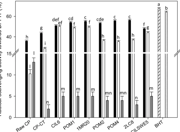
(WSE), ethyl acetate-soluble (ESE) and hexane-soluble (HSE) extracts. During radical scaveng-ing assay, the colored stable DPPH radical is reduced to non-radical DPPH-H, when in the presence of an antioxidant or a hydrogen donor. DPPH radical, without antioxidants, was sta-ble over the time. Under the assay conditions, the 100% of activity corresponded to the com-plete scavenging of DPPH radical (50μM final concentration) after 10 min of incubation with the antioxidant compounds. According to previous studies [38,39], the color intensity of DPPH•showed a logarithmic decline when it was in the presence of BHT. Apart from the extract used, the activity of raw CP and CP-CT was significantly (P<0.05) lower than that of BHT, which was used as the positive control. The radical scavenging activity of WSE, ESE and HSE from raw CP was 35.8 ± 0.56, 10.3 ± 0.41 and 13.5 ± 0.42%, respectively. WSE, ESE and HSE from CP-CT had a radical scavenging activity towards the stable radical DPPH of 43.6 ± 0.4, 28.7 ± 0.5 and 2.3 ± 0.1%, respectively (Fig 1).
Regardless of the strain used, the highest antioxidant activity was found for WSE, followed by ESE and HSE. Fermentation significantly (P<0.05) increased the radical scavenging activity
Fig 1. DPPH radical scavenging activity. DPPH radical scavenging activity of the water-soluble (black bars) (WSE), ethyl acetate-soluble (white bars) (ESE), and hexane-soluble (grey bars) (HSE) extracts from raw cladode pulp (raw CP), CP without bacterial inoculum and chemically acidified (CP-CT), and cladode pulp fermented with Lactobacillus plantarum POM1 (POM1), CIL6 (CIL6) and 1MR20 (1MR20), Lactobacillus brevis POM2 (POM2) and POM4 (POM4), Lactobacillus rossiae 2LC8 (2LC8) and Pediococcus pentosaceus CILSWE5 (CILSWE5). Butylatedhydroxytoluene (BHT) was used as positive control. The reaction mixture was prepared by diluting BHT in methanol (hatched bar) or acetone (dotted bar). Data are the means (± SD) of three independent experiments performed in triplicate. Data were subjected to one-way ANOVA; pair-comparison of treatment means was achieved by Tukey’s procedure at P0.05. Bars with different superscript letters differ significantly (P<0.05).
of all WSE. Except for CP fermented with P. pentosaceus CILSWE5 (48.1 ± 0.41%) and L. plan-tarum CIL6 (50.9 ± 0.38%), all the other strains caused a remarkable increase of the radical scavenging activity (53.4 ± 0.24–56.2 ± 0.51%). Compared to raw CP (10.3 ± 0.41%) and CP-CT (28.7 ± 0.53%), also the ESE from fermented CP showed a higher increase of the antiox-idant activity. The highest values were found for L. plantarum CIL6, POM1 and 1MR20 (48.7 ± 0.12, 49.5 ± 0. 22 and 50.3 ± 0.31%, respectively). Although the antioxidant activity from HSE was the lowest, the positive effect of the fermentation was confirmed also in this case (Fig 1).
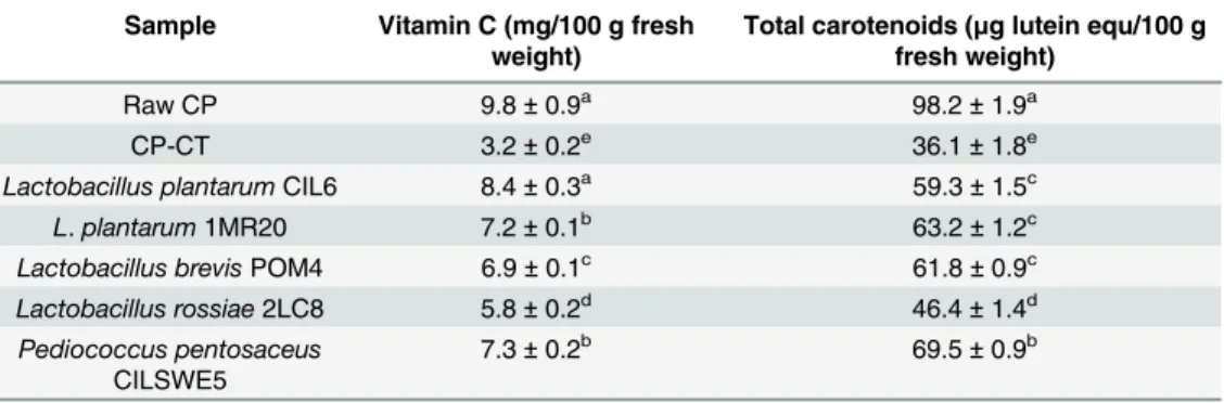
The analysis of total phenols, water soluble vitamin C and total carotenoids was carried out using ESE, WSE and HSE, respectively [28,32,33].
Total phenols of raw CP accounted for 37.4 ± 1.2 mg gallic acid equ/100 g of fresh weight. The concentration of total phenols from CP fermented with L. plantarum CIL6, POM1 and 1MR20 and P. pentosaceus CILSWE5 did not significantly (P>0.05) vary compared to raw CP. After fermentation with L. brevis POM2 and POM4, and L. rossiae 2LC8, a slight decrease was found. The concentrations of vitamin C and total carotenoids from fermented CP agreed with antioxidant activity (Table 2). The concentration of vitamin C of raw CP was 9.8 ± 0.8 mg/100 g fresh weight. Chemical acidification decreased the concentration of vitamin C of CP (3.1 ± 0.2 mg/100 g), whereas lactic acid fermentation had a preserving effect. The vitamin C concentration of fermented CP ranged from 5.75 ± 0.2 to 8.4 ± 0.3 mg/100 g. The trend was similar for total carotenoids.
Purification and identification of antioxidant compounds
To further investigate the radical scavenging activity of CP, WSE from fermented CP were digested with trypsin. After digestion, the antioxidant activity decreased between ca. 6 to 32% depending on the strain (S1 Fig). These findings suggested that protein compounds were somewhat related to activity. Aiming at purifying the antioxidant compounds, WSE was sub-jected to fractionation through RP-FPLC, and resulting fractions were assayed for antioxi-dant activity. Since the antioxiantioxi-dant activity of WSE was distributed within a large number of fractions (data not shown), no further peptide identification was carried out. Based on these
Table 2. Vitamin C and total carotenoids.
Sample Vitamin C (mg/100 g fresh weight)
Total carotenoids (μg lutein equ/100 g fresh weight)
Raw CP 9.8± 0.9a 98.2± 1.9a
CP-CT 3.2± 0.2e 36.1± 1.8e
Lactobacillus plantarumCIL6 8.4± 0.3a 59.3± 1.5c
L. plantarum 1MR20 7.2± 0.1b 63.2± 1.2c
Lactobacillus brevisPOM4 6.9± 0.1c 61.8± 0.9c
Lactobacillus rossiae2LC8 5.8± 0.2d 46.4± 1.4d
Pediococcus pentosaceus CILSWE5
7.3± 0.2b 69.5± 0.9b
Notes: Vitamin C (mg/100 g of fresh sample) and total carotenoids (μg lutein equ/100 g of fresh weight) in raw cladode pulp (raw CP), chemically acidified cladode pulp (CP-CT), and cladode pulp fermented with selected lactic acid bacteria strains. Data are the means (± SD) of three independent experiments performed in triplicate. Data were subjected to one-way ANOVA; pair-comparison of treatment means was achieved by Tukey’s procedure at P0.05. Means within the column with different superscript letters differ significantly (P<0.05).
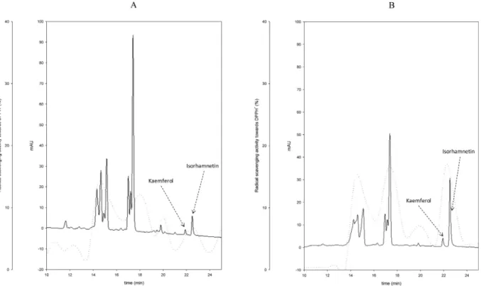
results, it was hypothesized a non-specific effect of CP protein derivatives, which were liber-ated through proteolysis during lactic acid bacteria fermentation. Aiming at identifying the antioxidant compounds from ESE, extracts were subjected to fractionation through HPLC analysis. The phenolic acid profiles of ESE from CP-CT and fermented CP were almost similar (data not shown). On the contrary, the flavonoid profiles of ESE from all fermented CP were diverse (Fig 2). Compared to CP-CT, the areas of two main peaks increased, which corresponded to kaemferol (retention time of 21.9 min) and isorhamnetin (retention time of 22.5). The concentration of kaemferol and isorhamnetin of CP-CT was 0.13 ± 0.04 and 0.55 ± 0.09μg/g, respectively. The increases were strain dependent. The highest concentra-tions were found for CP fermented with L. plantarum 1MR20 (0.23 ± 0.05 and 2.1 ± 0.15 μg/g, respectively) and L. brevis POM4 (0.22 ± 0.03 and 2.0 ± 0.18 μg/g, respectively). For all the other strains the concentration of isorhamnetin ranged from 1.19 ± 0.07 to 1.36 ± 0.05μg/g, whereas that of kaemferol did not significantly (P>0.05) vary compared to CP-Ct. Resulting fractions were also assayed for antioxidant activity. Compared to CP-CT, the highest increase of antioxidant activity was found in fractions matching with the retention times of kaemferol and isorhamnetin (Fig 2).
Viability of Caco-2/TC7 cells
Preliminarily, the cytotoxicity of CP samples was checked using the Neutral Red (NR) uptake assay. Apart from the lactic acid bacterium used for fermentation, CP behaved similarly to DMEM (negative controls) and did not significantly (P>0.05) affect the Caco-2/TC7 cell pro-liferation up to 10 mg/ml.
Fig 2. Flavonoid profiles of ESE. High Performance Liquid Chromatography (HPLC) profiles of ethyl acetate-soluble extracts (ESE) from cladode pulp (CP) without bacterial inoculum and chemically acidified control (CP-CT) (A) and CP fermented with Lactobacillus plantarum 1MR20 (B). The dashed line refers to the percentages of DPPH radical scavenging activity.
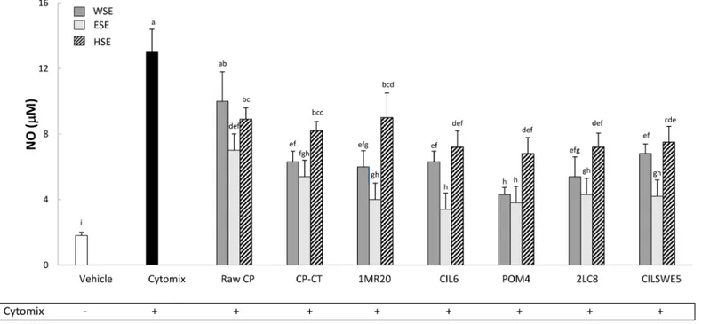
Nitric oxide (NO) release
Caco-2/TC7 cells were stimulated (48 h) with a cytomix solution containing TNFα, IL-1β and IFN-γ. Compared to the negative control (DMEM), the treatment significantly (P<0.05) increased the release of NO (Fig 3). On the contrary, treatments with WSE, HSE and, espe-cially, ESE from raw CP, CP-CT and fermented CP markedly (P<0.05) inhibited the release of NO. Apart from the extract, the highest anti-inflammatory activity was found for CP fer-mented with L. plantarum CIL6 and L. brevis POM4.
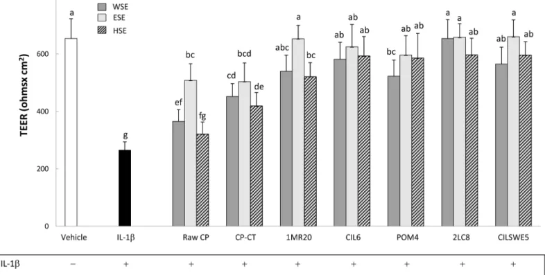
Transepithelial electric resistance (TEER)
Under culture conditions, Caco-2/TC7 cells develop morphological and functional characteris-tics of enterocytes, including intercellular tight junctions. Transepithelial electric resistance measured the integrity of the tight junctions. Preliminarily, TEER was measured in the pres-ence of WSE, HSE or ESE from raw CP, CP-CT and fermented CP. During 48 h of incubation, TEER was not affected (data not shown). Treatment of Caco-2/TC7 cells with IL-1β (25 ng/ mL) markedly (P<0.05) decreased the value of TEER (Fig 4). When Caco-2/TC7 cells were stimulated (basolateral compartment) with IL-1β and subsequently treated (apical compart-ment) with WSE, HSE or ESE from CP, the negative effect of IL-1β was markedly attenuated (P<0.05). Apart from the extract, all fermented CP showed the highest effect.
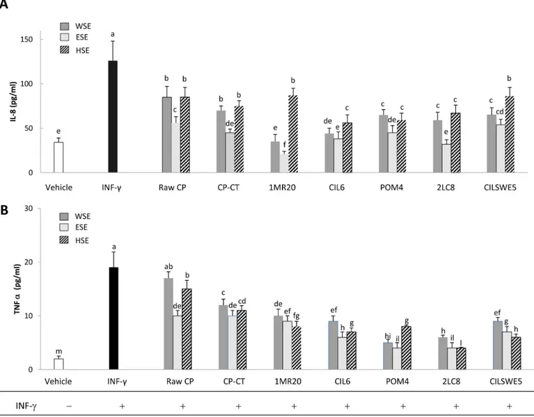
Interleukin-8 (IL-8) and tumour necrosis factor alfa (TNFα) release
IL-8 is a member of the C-X-C chemokine family and plays an essential role in the recruitment and activation of neutrophils, thereby initiating the inflammatory response. When Caco-2/
Fig 3. Nitric oxide (NO) (μM) release by Caco-2/TC7 cells. Caco-2/TC7 cells were treated for 48 h with water-soluble (WSE), ethyl-acetate (ESE), or hexane-soluble (HSE) extracts (10 mg/ml) from raw cladode pulp (raw CP), CP without bacterial inoculum and chemically acidified (CP-CT) and CP fermented with Lactobacillus plantarum 1MR20 (1MR20) and CIL6 (CIL6), Lactobacillus brevis POM4 (POM4), Lactobacillus rossiae 2LC8 (2LC8) and Pediococcus pentosaceusCILSWE5 (CILSWE5). Furthermore, cells were stimulated with a cytomix solution (TNFα, 100 ng/ml; IL-1β, 5 ng/ml; and INF γ, 200 U/ml). Data are the means (± SD) of three independent experiments performed in triplicate. Data were subjected to one-way ANOVA; pair-comparison of treatment means was achieved by Tukey’s procedure at P0.05. Bars with different superscript letters differ significantly (P<0.05).
TC7 cells were treated with INF-γ (2 ng/mL), a significant (P<0.05) increase of the synthesis of IL-8 and TNFα was found (Fig 5A and 5B). When Caco-2/TC7 cells were stimulated with INF-γ and also treated with WSE, HSE or ESE from raw CP, CP-CT and fermented CP, a significant (P<0.05) decrease of the synthesis of IL-8 and TNFα was found. Apart from the strain used, the synthesis of IL-8 and TNFα was more markedly inhibited (P<0.05) by treatment with ESE. The highest (P<0.05) inhibition of IL-8 was found with ESE from CP fermented with L. plan-tarum 1MR20 and L. rossiae 2LC8, whereas that of TNFα by ESE from CP fermented with L. rossiae 2LC8 and L. brevis POM4.
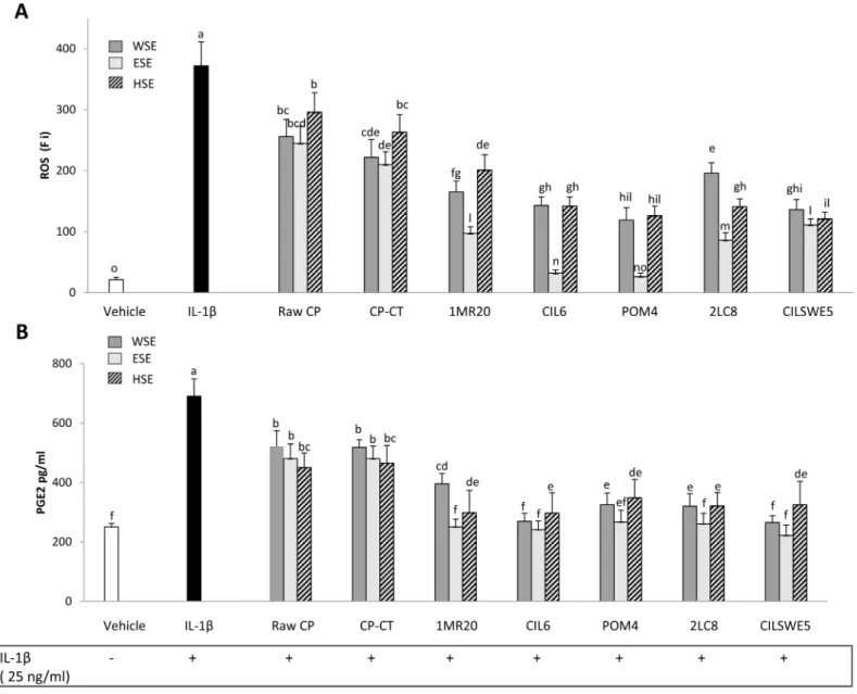
Reactive oxygen species (ROS) scavenging
The level of intracellular ROS was assessed by measuring the oxidation of the probe 20,70 -dichlorofluorescin diacetate (DCFH-DA). When Caco-2/TC7 cells were subjected to treatment with IL-1β (25 ng/mL), a significant (P<0.05) increase of the intracellular level of ROS was found (Fig 6A). When Caco-2/TC7 cells stimulated with IL-1β were also treated with WSE, HSE or ESE from raw CP, CP-CT and fermented CP, a significant (P<0.05) decrease of the intracellular level of ROS was found. Apart from the strain used, the highest decrease was found by treatment with ESE. CP fermented with L. plantarum CIL6 and L. brevis POM4 had the highest effect.
The formation of inflammatory eicosanoids such as prostaglandin E2 (PGE2), which is syn-thesized from arachidonic acid by COX- 2, was further determined. Treatment of cells with
IL-Fig 4. Transepithelial electric resistance (TEER) (Ohms x cm2) of Caco-2/TC7 cells. Caco-2/TC7 cells were incubated 24 h with IL-1β (25 ng/ml) and water-soluble (WSE), ethyl-acetate (ESE), and hexane-soluble (HSE) extracts (10 mg/ml) from raw cladode pulp (raw CP), CP without bacterial inoculum and chemically acidified (CP-CT) and CP fermented with Lactobacillus plantarum 1MR20 (1MR20) and CIL6 (CIL6), Lactobacillus brevis POM4 (POM4), Lactobacillus rossiae2LC8 (2LC8) and Pediococcus pentosaceus CILSWE5 (CILSWE5). Data are the means (± SD) of three independent experiments performed in triplicate. Data were subjected to one-way ANOVA; pair-comparison of treatment means was achieved by Tukey’s procedure at P0.05. Bars with different superscript letters differ significantly (P<0.05).
1β elicited markedly the synthesis of PGE2, whereas incubation with WSE, HSE or ESE from raw CP, CP-CT and, especially, fermented CP displayed high ability to decrease PGE2 accumu-lation in response to IL-1β (Fig 6B).
Discussion
The role of lactic acid fermentation for improving the nutritional and functional features of many vegetables is well-known [40]. The health benefits mostly result via ingestion of micro-bial metabolites that are synthesized during fermentation. Lactic acid bacteria, intended as microbial cell factories, increase the functionality of many fermented vegetables through their enzyme portfolio that promotes the synthesis of various metabolites and/or the release of
Fig 5. Interleukin-8 (IL-8) (A) and tumour necrosis alfa (TNFα) (B) release by Caco-2/TC7 cells. Caco-2/TC7 cells were stimulated for 24 h with interferon-gamma (INF-γ) (2 ng/mL) and subsequently treated with water-soluble (WSE), ethyl-acetate (ESE), or hexane-soluble (HSE) extracts (10 mg/ml) from raw cladode pulp (raw CP), CP without bacterial inoculum and chemically acidified (CP-CT) and CP fermented with Lactobacillus plantarum 1MR20 (1MR20) and CIL6 (CIL6), Lactobacillus brevis POM4 (POM4), Lactobacillus rossiae 2LC8 (2LC8) and Pediococcus pentosaceus CILSWE5 (CILSWE5). Data are the means (± SD) of three independent experiments performed in triplicate. Data were subjected to one-way ANOVA; pair-comparison of treatment means was achieved by Tukey’s procedure at P0.05. Bars with different superscript letters differ significantly (P<0.05).
biogenic compounds, which are mainly cryptic in the raw matrix [41]. The high concentration of functional compounds and the intrinsic features of Opuntia ficus-indica L. fruits and, espe-cially, cladodes may be suitable for preparations with health-promoting features [5,8,11]. First, this study investigated the potential of the lactic acid fermentation of cladodes. The func-tional features of fermented cladode pulp (CP) were compared to those of the raw CP (Fig 7). Thirteen strains of lactic acid bacteria, previously isolated from vegetable matrices that are par-ticularly rich of polyphenols [15–20], were used as starters. The concentration of fermentable carbohydrates of CP was enough to allow the bacterial growth of seven out the thirteen strains.
Fig 6. Intracellular reactive oxygen species (ROS) (fluorescence intensity units, Fi) (A) and Prostaglandin E2 (PGE2) (pg/ml) (B) on Caco-2/TC7 cells. Caco-2/TC7 cells were stimulated for 24 h at 37°C with IL-1β (25 ng/mL) and then treated for other 24 h with water-soluble (WSE), ethyl-acetate (ESE), or hexane-soluble (HSE) extracts (10 mg/ml) from raw cladode pulp (raw CP), CP without bacterial inoculum and chemically acidified (CP-CT) and CP fermented with Lactobacillus plantarum 1MR20 (1MR20) and CIL6 (CIL6), Lactobacillus brevis POM4 (POM4), Lactobacillus rossiae 2LC8 (2LC8) and Pediococcus pentosaceusCILSWE5 (CILSWE5). Only the best performing strains and representative species were considered. Data are the means (± SD) of three independent experiments performed in triplicate. Data were subjected to one-way ANOVA; pair-comparison of treatment means was achieved by Tukey’s procedure at P0.05. Bars with different superscript letters differ significantly (P<0.05).
Fig 7. Permutation analysis of compositional and functional profiles. Permutation analysis of compositional [vitamin C, total carotenoids,γ-amino butyric acid (GABA), kaemferol, and isorhamnetin], and functional [antioxidant activity, nitric oxide release, tumour necrosis alfa (TNFα), reactive oxygen species (ROS), transepithelial electric resistance (TEER), Prostaglandin E2 (PGE2), and interleukin-8 (IL-8)] profiles of raw cladode pulp (CP), CP without bacterial inoculum and chemically acidified (CP-CT), and fermented CP with Lactobacillus plantarum 1MR20 (1MR20) and CIL6 (CIL6), Lactobacillus brevis POM4 (POM4), Lactobacillus rossiae 2LC8 (2LC8) and Pediococcus pentosaceus CILSWE5 (CILSWE5). Differences are represented colorimetrically with red and green indicating the highest and lowest values of the standardized data, respectively, for each parameter. All data were shown as a percentage of dissimilarity using Euclidean distance.
Usually, plant autochthonous lactic acid bacteria better adapt to plant environments compared to allochthonous strains coming from other sources[40]. The functional composition O. ficus indica varies depending on the plant organ [8]. Cladodes are good sources of amino acids and proteins [8]. Fermentation of CP with L. brevis POM2 and POM4 allowed the highest concen-tration ofγ-amino butyric acid (GABA) (39–46 mg/kg), a non-protein amino acid with physio-logical functions (e.g., induction of hypotension, diuretic and tranquilizer effect) [42,43]. These concentrations are above the physiological threshold (ca. 10 mg/day), thus hypothesizing an in vivo health benefits [44]. Usually, the capacity to synthesize GABA is strain specific. It confers resistance to bacterial cells under acidic conditions like those of CP [45]. Lactic acid fer-mentation exerted also a preservative effect on the levels of vitamin C and carotenoids, which are inherent in CP. Ascorbic acid is one of the most sensitive vitamins in foods. The stability of vitamin C varies depending on environmental factors such as pH, concentration of metal ions and redox state [46]. The decrease of pH is one of the main mechanisms to prevent ascorbate autoxidation when the redox potential changes [47]. Nevertheless, the slight decreases of pH that were found after fermentation suggested the involvement of other mechanisms. Phyto-chemicals such as ascorbic acid, carotenoids and phenols determined the marked radical scav-enging activity of crude cladode cactus extracts [48]. The antioxidant activity of fermented cladodes was compared to that of a non-inoculated and chemically acidified control (CP-CT). Chemical acidification was done to exclude the effect of pH on the antioxidant activity and phenol extractability. As usual in herbal medicine, the solvent extraction of bioactive com-pounds from permeable solid plant materials is a key step to have phytochemical-rich products [8]. Water-soluble (WSE), ethyl acetate-soluble (ESE) and hexane-soluble (HSE) extracts were compared. The radical scavenging activity of CP was positively affected by lactic acid bacteria fermentation. The highest increase was found with ESE, suggesting a major role of ethyl acetate extractable compounds like phenols. When L. plantarum CIL6, POM1 and 1MR20 were used as starters, the DPPH radical scavenging activity was at least five and two times higher than that of raw CP and CP-CT, respectively.
Aiming at explaining the increase of antioxidant activity due to lactic acid fermentation, the contribution of various bioactive compounds was investigated. The preservative effect on vita-min C and carotenoids levels certainly gave a positive contribution to the activity from WSE and HSE. A consistent part of the antioxidant activity was lost after treatment with proteolytic enzyme, therefore, a further contribution by water-soluble peptides was not excluded. Never-theless, two flavonoid aglycone derivatives, kaemferol and isorhamnetin, which were identified from ESE were clearly responsible for the increased radical scavenging activity. Flavonols, the most ubiquitous flavonoids in vegetables, are usually present at relatively low concentrations and under glycosylated forms [49]. Esterase activities towards glycosylated forms increases the concentration of flavonols, which accumulate in the outer and aerial tissues of Cactus (skin and leaves) because the light stimulation [8,49]. Flavonol aglycones contain multiple hydroxyl free groups and possess higher antioxidant activity than their glycosides [49]. Furthermore, native glycosylated forms cannot be absorbed by the human organism, needing previous hydrolysis by intestinal enzymes or colonic microbiota. Aglycones are directly absorbed at the level of the small intestine [49]. Esterase enzymes are widespread in L. plantarum strains, allowing the metabolism of flavonol glycosides in plant matrices [50]. Several flavonoid tives such as kaempherol, quercetin and its corresponding glucuronides or methylated deriva-tive isorhamnetin are bioacderiva-tive [8]. The antioxidant capacity and modulatory effects on oxidative stress biomarkers and inflammatory mediators were studied using Caco-2/TC7 cells (human colon carcinoma), which are one of the in vitro systems most largely used to mimic the intestinal mucosa. Despite the neoplastic origin, these cells have the capacity to spontane-ously differentiate into mature enterocytes and to express brush border enzymes. Neutral Red
uptake assay on Caco-2 cells demonstrated the absence of cytotoxicity for a wide range of con-centrations of CP. The immune-modulatory and antioxidant effect of CP extracts was proven by in vitro and in vivo assays [51,52]. It might be associated with the presence of various com-pounds such as favonols and aglycone derivatives, phenolic acids and derivatives [8,48]. Extracts from fermented CP markedly inhibited the inflammatory status of Caco-2/TC7 cells, as induced by treatments with TNFα, IL-1β and IFN-γ. Fermented CP also contributed to maintain the integrity of the tight junctions, even if subjected to negative stimulation, and markedly inhibited the synthesis of IL-8 and TFNα, after treatment with IFN-γ. Natural poly-phenolic extracts, especially flavonoids, modulated the inflammation of Caco-2 cells, which were stimulated by cytokines and chemokines (e.g., IL-1, IL-6, IL-8 and TNF-α) [53]. The inhi-bition of these pathways at any point of the cascade repressed the synthesis of these proteins and/or of their reaction products [54]. TNFα is a pleiotropic inflammatory cytokine, which mediates inflammation, immune-response and apoptosis [55]. A large spectrum of diseases involved the over production or the persistent activation of TNFα [56]. Low levels of TNFα contribute to homeostasis by regulating the body circadian rhythm [57]. Fermentation of CP, especially with L. brevis POM4 and L. rossiae 2LC8, markedly affected the level of TNFα by Caco-2 cells. The antioxidant effect on cultured cells was higher than that found with extracts from CP and CP-CT. The protective effect was investigated through the determination of intra-cellular ROS and detoxification by DCFH-DA assay. Also in this case, ESE from fermented CP, mainly with L. plantarum CIL6 and L. brevis POM4, showed a markedly higher antioxidant activity on Caco-2 cells than ESE from CP-CT. Besides, the concentrations of prostaglandins PGE2, small-molecule derivatives of arachidonic acid produced by cyclooxygenases, and PG synthases were positively affected by fermented CP. PGE2 are mediators of active inflamma-tion by promoting local vasodilatainflamma-tion, attracinflamma-tion and regulainflamma-tion of multiple funcinflamma-tions of differ-ent immune cells. As expected, the incubation of Caco-2 cells with IL-1β resulted in an
increase in PGE2 synthesis [58], whereas the treatment with extracts from fermented CP inhib-ited the synthesis of PGE2 in vitro.
Conclusions
This study falls within the framework of the industrial exploitation of prickly pear by-prod-ucts. Lactic acid fermentation, one of the oldest and most low‐input biotechnologies used in food bio‐preservation, could be a valuable and innovative strategy to exploit the intrinsic fea-tures of Opuntia ficus-indica L. cladodes. The mechanisms by which selected lactic acid bac-teria fulfil the role of efficient cell factories to synthesize functional biomolecules from O. ficus-indica L. cladodes was hypothesized. Two flavonoid derivatives (kaemferol and iso-rhamnetin) were identified in the ethyl acetate extracts, which were considered to be the major compounds responsible for the increased radical scavenging activity and immune-modulation features. Flavonoid metabolites act at multiple levels, through the inhibition of IL-8 and TFNα pathways, decreasing the production of free radicals, suppressing the activity of COX- 2 and inhibiting the synthesis of PGE2. With the perspective of producing a func-tional ingredient, dietary supplement or pharmaceutical preparation, fermented CP extract may perform an anti-inflammatory adjuvant to restore homeostasis under conditions of cel-lular stress.
PermutMatrix analysis based on compositional (vitamin C, total carotenoids, GABA, kaem-ferol, and isorhamnetin), and functional (antioxidant activity, nitric oxide release, TNFα, ROS, TEER, PGE2, IL-8 levels) data showed that all fermented cladode pulp, especially those fer-mented with L. plantarum strains and L. brevis POM4, enhanced the antioxidant and immune-modulation features of cladode pulp (Fig 7).
Supporting Information
S1 Fig. DPPH radical scavenging activity of crude and enzimatically digested water-soluble extracts (WSE).DPPH radical scavenging activity of crude (black bars) and enzimatically digested (white bars) water-soluble extracts (WSE) from cladode pulp (CP) without bacterial inoculum and chemically acidified (CP-CT), and CP fermented with Lactobacillus plantarum CIL6 (CIL6) and 1MR20 (1MR20), Lactobacillus brevis POM4 (POM4), Lactobacillus rossiae 2LC8 (2LC8) and Pediococcus pentosaceus CILSWE5 (CILSWE5). Butylatedhydroxytoluene (BHT) was used as positive control. (± SD) of three independent experiments performed in triplicate. Bars with different superscript letters differ significantly (P<0.05).
(DOCX)
Acknowledgments
The authors thank Dott. Vincenzo Russo (Suolo & Salute S.R.L. Direzione Sicilia, Italy) for pro-viding fresh cladodes of Opuntia ficus-indica (L.) Mill. (genotype Sanguigna) from (Santa Maria di Belice (Sicily, Italy).
Author Contributions
Conceived and designed the experiments: RDC. Performed the experiments: PF IC NT OV MS. Analyzed the data: PF RDC. Wrote the paper: RDC MDA MG. Performed purification of active compounds and vitamin C determination: IC NT. Carried out ex-vivo assays: OV MS.
References
1. Mohamed-Yasseen Y, Barringer SA, Splittstoesser WE, Schnell RJ. Rapid propagation of tuna (Opun-tia ficus-indica) and plant establishment in soil. Plant Cell Tiss Org Cult. 1995; 42: 117–119.
2. Mohamed-Yasseen Y, Barringer SA, Splittstoesser WE. A note on the uses of Opuntia spp. in Central/ North America. J Arid Environ. 1996; 32: 347–353.
3. Feugang MJ, Konarski P, Zou D, Stintzing FC, Zou C. Nutritional and medicinal use of Cactus pear (Opuntia spp.) cladodes and fruits. Front Biosci. 2006; 11: 2574–2589. PMID:16720335
4. Guevara JC, Yahia EM, Brito de la Fuente E, Biserka SP. Effects of elevated concentrations of CO2in
modified atmosphere packaging on the quality of prickly pear cactus stems (Opuntia spp.). Postharvest Biol Technol. 2003; 29: 167–176.
5. Stintzing FC, Carle R. Cactus stems (Opuntia spp.): A review on their chemistry, technology, and uses. Mol Nutr Food Res. 2005; 49: 175–194. PMID:15729672
6. Cayupán YSC, Ochoa MJ, Nazareno MA. Health-promoting substances and antioxidant properties of Opuntiasp. fruits. Changes in bioactive-compound contents during ripening process. Food Chem. 2011; 126: 514–519.
7. Akanni G, Ntuli V, du Preez JC. Cactus pear biomass, a potential lignocellulose raw material for single cell protein production (SCP): a review. Int J Curr Microbiol Appl.Sci. 2014; 3: 171–197.
8. El-Mostafa K, El Kharrassi Y, Badreddine A, Andreoletti P, Vamecq J, El Kebbaj MH, et al. Nopal cactus (Opuntia ficus-indica) as a source of bioactive compounds for nutrition, health and disease. Molecules. 2014; 19: 14879–14901. doi:10.3390/molecules190914879PMID:25232708
9. Abd El-Razek FH, Hassan AA. Nutritional value and hypoglycemic effect of prickly cactus pear (Opun-tia ficus-indica) fruit juice in Alloxan-induced diabetic rats. 2011. Aust J Basic. Appl. Sci. 2011; 5: 356– 377.
10. Yeddes N, Chérif JK, Guyot S, Sotin H, Ayadi MT. Comparative study of antioxidant power, polyphe-nols, flavonoids and betacyanins of the peel and pulp of three Tunisian Opuntia forms. Antioxidants. 2013; 2: 37–51. doi:10.3390/antiox2020037PMID:26787622
11. Patel S. Opuntia cladodes (nopal): Emerging functional food and dietary supplement. Mediterranean J Nutr Metab. 2014; 7: 11–19.
12. Moreno–Álvarez MJ, Hernández R., Belén–Camacho DR, Medina–Martínez CA, Ojeda–Escalona CE, García–Pantaleón DM. Making of bakery products using composite flours: Wheat and cactus pear (Opuntia boldinghii Britton et Rose) stems (cladodes). J Prof Assoc Cactus. 2009; 11: 78–87.
13. Trombetta D, Puglia C, Perri D, Licata A, Pergolizzi S, Lauriano ER, et al. Effect of polysaccharides from Opuntia ficus-indica (L.) cladodes on the healing of dermal wounds in the rat. Phytomedicine. 2006; 13: 352–358. PMID:16635743
14. Akanni GB, du Preez JC, Steyn L, Kilian SG. Protein enrichment of an Opuntia ficus-indica cladode hydrolysate by cultivation of Candida utilis and Kluyveromyces marxianus. J Sci Food Agric. 2015; 95: 1094–1102. doi:10.1002/jsfa.6985PMID:25371280
15. Di Cagno R, Surico RF, Siragusa S, De Angelis M, Paradiso A, Minervini F, et al. Selection and use of autochthonous mixed starter for lactic acid fermentation of carrots, French beans or marrows. Int J Food Microbiol. 2008; 127: 220–228. doi:10.1016/j.ijfoodmicro.2008.07.010PMID:18710789
16. Di Cagno R, Surico RF, Paradiso A, De Angelis M, Salmon J- C, Buchin S, et al. Effect of autochtho-nous lactic acid bacteria starters on health-promoting and sensory properties of tomato juices. Int J Food Microbiol. 2009; 128: 473–483. doi:10.1016/j.ijfoodmicro.2008.10.017PMID:19028404
17. Di Cagno R, Surico RF, Minervini G, De Angelis M, Rizzello CG, Gobbetti M. Use of autochthonous starters to ferment red and yellow peppers (Capsicum annum L.) to be stored at room temperature. Int J Food Microbiol. 2009; 130: 108–116. doi:10.1016/j.ijfoodmicro.2009.01.019PMID:19217182
18. Di Cagno R, Cardinali G, Minervini G, Antonielli L, Rizzello CG, Ricciuti P, et al. Taxonomic structure of the yeasts and lactic acid bacteria microbiota of pineapple (Ananas comosus L. Merr.) and use of autochthonous starters for minimally processing. Food Microbiol. 2010; 27: 381–389. doi:10.1016/j. fm.2009.11.012PMID:20227603
19. Di Cagno R, Surico RF, Minervini G, Rizzello CG, Lovino R, Servili M, et al. Exploitation of sweet cherry (Prunus avium L.) puree added of stem infusion through fermentation by selected autochthonous lactic acid bacteria. Food Microbiol. 2011; 28: 900–909. doi:10.1016/j.fm.2010.12.008PMID:21569932
20. Di Cagno R, Minervini G, Rizzello CG, De Angelis M, Gobbetti M. Effect of lactic acid fermentation on antioxidant, texture, color and sensory properties of red and green smoothies. Food Microbiol. 2011; 28: 1062–1071. doi:10.1016/j.fm.2011.02.011PMID:21569953
21. Vitali B, Minervini G, Rizzello CG, Spisni E, Maccaferri S, Brigidi P, et al. Novel probiotic candidates for humans isolated from raw fruits and vegetables. Food Microbiol. 2012; 31: 116–125. doi:10.1016/j.fm. 2011.12.027PMID:22475949
22. Filannino P, Cardinali G, Rizzello CG, Buchin S, De Angelis M, Gobbetti M, et al. Metabolic responses of Lactobacillus plantarum strains during fermentation and storage of vegetable and fruit juices. Appl Environ Microbiol. 2014; 80: 2206–2215. doi:10.1128/AEM.03885-13PMID:24487533
23. Filannino P, Bai Y, Di Cagno R, Gobbetti M, Ganzle M. Metabolism of phenolic compounds by Lactoba-cillusspp. during fermentation of cherry juice and broccoli puree. Food Microbiol. 2015; 46: 272–279. doi:10.1016/j.fm.2014.08.018PMID:25475296
24. Zwietering MH, Jongeberger I, Roumbouts FM, Van’t Riet K. Modelling of bacterial growth curve. Appl Environ Microbiol. 1990; 56: 1875–1881. PMID:16348228
25. Rizzello CG, Nionelli L, Coda R, De Angelis M, Gobbetti M. Effect of sourdough fermentation on stabili-sation, and chemical and nutritional characteristics of wheat germ. Food Chem. 2010; 119: 1079– 1089.
26. Zeppa G, Conterno L, Gerbi V. Determination of organic acids, sugars, diacetyl, and acetoin in cheese by high-performance liquid chromatography. J Agric Food Chem. 2001; 49: 2722–2726. PMID:
11409957
27. Rizzello CG, Coda R, Mazzacane F, Minervini D, Gobbetti M. Micronized byproducts from debranned durum wheat and sourdough fermentation enhanced the nutritional, textural and sensory features of bread. Food Res Int. 2012; 46: 304–313.
28. Kubola J, Siriamornpun S. Phytochemicals and antioxidant activity of different fruit fractions (peel, pulp, aril and seed) of Thai gac (Momordica cochinchinensis Spreng). Food Chem. 2011; 127: 1138–1145. doi:10.1016/j.foodchem.2011.01.115PMID:25214106
29. Yu L, Perret J, Harris M, Wilson J, Haley S. Antioxidant properties of bran extracts from Akron wheat grown at different locations. J Agric Food Chem. 2003; 51: 1566–1570. PMID:12617585
30. Atanassova M, Choiset Y, Dalgalarrondo M, Chobert JM, Dousset X, Ivanova I, et al. Isolation and par-tial biochemical characterization of a proteinaceous anti-bacteria and anti-yeast compound produced by Lactobacillus paracasei subsp. paracasei strain M3. Int J Food Microbiol. 2003; 87: 63–73. PMID:
12927708
31. Church FC, Swaisgood HE, Porter DH, Catignani GL. Spectrophotometric assay using o-phthaldialde-hyde for determination of proteolysis in milk and isolated milk proteins. J Dairy Sci. 1983; 66: 1219– 1227.
32. Slinkard K, Singleton VL. Total phenol analysis: automation and comparison with manual methods. Am J Enol Vitic. 1997; 28: 49–55.
33. Foodstuffs-Determination of Vitamin C by HPLC, EN. 14130, European Committee for Standardization. 2003; Brussels, Belgium.
34. Chantret I, Rodolosse A, Barbat A, Dussaulx E, Brot-Laroche E, Zweibaum A, et al. Differential expres-sion of sucrose isomaltase in clones isolated from early and late passages of the cell line Caco-2: evi-dence for glucose-dependent negative regulation. J Cell Sci. 1994; 107: 213–225. PMID:8175910
35. Borenfreund E, Babich H, Martin-Alguacil N. Comparisons of two in vitro cytotoxicity assays-the neutral red (NR) and tetrazolium MTT tests. Toxicol in Vitro. 1988; 2: 1–6. PMID:20702351
36. Green LC, Wagner DA, Glogowski J, Skipper PL, Wishnok JS, Tannenbaum SR. Analysis of nitrate, nitrite and nitrate in biological fluids. Anal Biochem. 1982; 126: 131–138. PMID:7181105
37. Cathcart R, Schwiers E, Ames BN. Detection of picomole levels of hydroperoxides using a fluorescent dichlorofluorescein assay. Anal Biochem. 1983; 134: 111–116. PMID:6660480
38. Coda R, Rizzello CG, Pinto D, Gobbetti M. Selected lactic acid bacteria synthesize antioxidant peptides during sourdough fermentation of cereal flours. Appl Environ Microbiol. 2012; 78: 1087–1096. doi:10. 1128/AEM.06837-11PMID:22156436
39. Winata A, Lorenz K. Antioxidant potential of 5-N-pentadecylresorcinol. J Food Process Preserv. 1996; 20: 417–429.
40. Di Cagno R, Coda R, De Angelis M, Gobbetti M. Exploitation of vegetables and fruits through lactic acid fermentation. Food Microbiol. 2013; 33: 1–10. doi:10.1016/j.fm.2012.09.003PMID:23122495
41. Gobbetti M, Di Cagno R, De Angelis M. Functional microorganisms for functional food quality. Crit Rev Food Sci Nutr 2010; 508: 716–727.
42. Oh SH, Oh CH. Brown rice extract with enhanced levels of GABA stimulate immune cells. Food Sci Bio-technol. 2003; 12: 248–252.
43. Wong GT, Bottiglieri T, Snead OC. GABA,γ-hydroxybutyric acid, and neurological disease. Ann Neu-rol. 2003; 6: 3–12.
44. Inoue K, Shirai T, Ochiai H, Kassao M, Hayakawa K, Rimura M, et al. Blood-pressure-lowering effect of a novel fermented milk containing gamma aminobutyric acid (GABA) in mild hypertensive. Eur J Clin Nutr. 2003; 57: 490–495. PMID:12627188
45. Siragusa S, De Angelis M, Di Cagno R, Rizzello CG, Coda R, Gobbetti M. Synthesis ofγ-aminobutyric acid (GABA) by lactic acid bacteria isolated from Italian cheese varieties. Appl Environ Microbiol. 2007; 73: 7283–7290. PMID:17890341
46. Selman JD. Vitamin retention during blanching of vegetables. Food Chem. 1994; 49: 137–147. 47. Wang Y-C, Yu R-C, Chou C-C. Antioxidative activities of soymilk fermented with lactic acid bacteria
and bifidobacteria. Food Microbiol. 2006; 23: 128–135. PMID:16942996
48. Kim J, Soh SY, Shin J, Cho CW, Choi YH, Nama SY. Bioactives in cactus (Opuntia ficus-indica) stems possess potent antioxidants and pro-apoptotic activities through COX-2 involvement. J Sci Food Agric. 2014; doi:10.1002/jsfa.6968
49. Manach C, Scalbert A, Morand C, Rémésy C, Jiménez L. Polyphenols: food sources and bioavailability. Am J Clin Nutr. 2004; 79: 727–747. PMID:15113710
50. Esteban-Torres M, Reverón I, Mancheño JM, de las Rivas B, Muñoz R. Characterization of a feruloyl esterase from Lactobacillus plantarum. Appl Environ Microbiol. 2013; 79: 5130–5136. doi:10.1128/ AEM.01523-13PMID:23793626
51. Matiasa A, Nunes SL, Poejo J, Mecha E, Serra AT, Madeira P, et al. Antioxidant and anti-inflammatory activity of a flavonoid-rich concentrate recovered from Opuntia ficus-indica juice. Food Funct. 2014; 5: 3269–3280. doi:10.1039/c4fo00071dPMID:25347222
52. Antunes-Ricardo M, Gutiérrez-Uribe JA, Martínez-Vitela C, Serna-Saldívar SO. Topical anti-inflamma-tory effects of isorhamnetin glycosides isolated from Opuntia ficus-indica. Biomed Res Int. 2015; 2015:847320. doi:10.1155/2015/847320PMID:25821823
53. Chirumbolo S. The role of quercetin, flavonols and flavones in modulating inflammatory cell function. Inflamm Allergy Drug Targets. 2010; 9: 263–285. PMID:20887269
54. Romier-Crouzet B, Van De Walle J, During A, Joly A, Rousseau C, Henry O, et al. Inhibition of inflam-matory mediators by polyphenolic plant extracts in human intestinal Caco-2 cells. Food Chem Toxicol. 2009; 47: 1221–1230. doi:10.1016/j.fct.2009.02.015PMID:19233242
55. Locksley RM, Killeen N, Lenardo MJ. The TNF and TNF receptor superfamilies: integrating mammalian biology. Cell. 2001; 104: 487–501. PMID:11239407
56. McDermott MF. TNF and TNFR biology in health and disease. Cell Mol Biol. 2001; 47: 619–635. PMID:
11502070
57. Strieter RM, Kunkel SL, Bone RC. Role of tumor necrosis factor-alpha in disease states and inflamma-tion. Crit Care Med. 1993; 21: S447–S463. PMID:8403983
58. O’Leary KA, de Pascual-Tereasa S, Needs PW, Bao YP, O’Brien NM, Williamson G. Effect of flavo-noids and vitamin E on cyclooxygenase-2 (COX-2) transcription. Mutat Res. 2004; 551: 245–254. PMID:15225597
![Fig 7. Permutation analysis of compositional and functional profiles. Permutation analysis of compositional [vitamin C, total carotenoids, γ-amino butyric acid (GABA), kaemferol, and isorhamnetin], and functional [antioxidant activity, nitric oxide release](https://thumb-eu.123doks.com/thumbv2/123dokorg/5440413.60601/16.918.90.817.165.951/permutation-compositional-functional-permutation-compositional-carotenoids-isorhamnetin-antioxidant.webp)
