R E S E A R C H A R T I C L E
Open Access
Analysis of the association between CD40 and
CD40 ligand polymorphisms and systemic
sclerosis
María Teruel
1*, Carmen P Simeon
2, Jasper Broen
3, Madelon C Vonk
3, Patricia Carreira
4, Maria Teresa Camps
5,
Rosa García-Portales
6, Esmeralda Delgado-Frías
7, Maria Gallego
8, Gerard Espinosa
9,
the Spanish Scleroderma Group
10, Lorenzo Beretta
11, Paolo Airó
12, Claudio Lunardi
13, Gabriela Riemekasten
14,
Torsten Witte
15, Thomas Krieg
16, Alexander Kreuter
17, Jörg HW Distler
18, Nicolas Hunzelmann
16,
Bobby P Koeleman
19, Alexandre E Voskuyl
20, Annemie J Schuerwegh
21, Miguel Ángel González-Gay
22,
Timothy RDJ Radstake
23and Javier Martin
1Abstract
Introduction: The aim of the present study was to investigate the possible role of CD40 and CD40 ligand (CD40LG)
genes in the susceptibility and phenotype expression of systemic sclerosis (SSc).
Methods: In total, 2,670 SSc patients and 3,245 healthy individuals from four European populations (Spain,
Germany, The Netherlands, and Italy) were included in the study. Five single-nucleotide polymorphisms (SNPs) of
CD40 (rs1883832, rs4810485, rs1535045) and CD40LG (rs3092952, rs3092920) were genotyped by using a
predesigned TaqMan allele-discrimination assay technology. Meta-analysis was assessed to determine whether an
association exists between the genetic variants and SSc or its main clinical subtypes.
Results: No evidence of association between CD40 and CD40LG genes variants and susceptibility to SSc was
observed. Similarly, no significant statistical differences were observed when SSc patients were stratified by the
clinical subtypes, the serologic features, and pulmonary fibrosis.
Conclusions: Our results do not suggest an important role of CD40 and CD40LG gene polymorphisms in the
susceptibility to or clinical expression of SSc.
Introduction
Systemic sclerosis (SSc) is an autoimmune disease of the
connective tissue characterized by excessive fibrosis of
the dermis and vascular damage. It also affects internal
organs, such as the lung, gastrointestinal, and vascular
systems [1]. SSc is a complex polygenic disease in which
environmental and genetic factors are involved in the
susceptibility to this disease. Candidate gene and
gen-ome-wide association studies (GWASs) performed in
SSc have identified new loci implicated in the
suscept-ibility to SSc [2]. Nevertheless, the complete genetic
components of SSc remain unknown.
CD40 is a member of the tumor necrosis factor
recep-tor superfamily (TNFR), and it is expressed on the
sur-face of several immune and nonhematopoietic cells,
such as B cells, macrophages, dendritic cells, fibroblasts,
and endothelial cells in certain pathogenic conditions
[3]. Its ligand, CD40LG (CD154), is expressed mainly on
the surface of CD4
+T cells. CD40-CD40LG interactions
are necessary for the activation of both humoral and
cel-lular immune responses [3]. The CD40-CD40LG
path-way has been suggested to play an important role in the
pathogenesis of autoimmune diseases [4]. An increase of
soluble CD40LG (sCD40LG) has been observed in many
autoimmune diseases, such as systemic lupus
erythema-tosus (SLE) [5], rheumatoid arthritis (RA) [6], and
Graves disease (GD) [7]. Interestingly, patients with SSc
and limited cutaneous disease have higher levels of
* Correspondence: [email protected]
1
Instituto de Parasitología y Biomedicina López-Neyra, IPBLN-CSIC, Avda. del Conocimiento s/n. 18010, Granada, SpainArmilla (Granada), Spain Full list of author information is available at the end of the article
© 2012 Teruel et al.; licensee BioMed Central Ltd. This is an open access article distributed under the terms of the Creative Commons Attribution License (http://creativecommons.org/licenses/by/2.0), which permits unrestricted use, distribution, and reproduction in any medium, provided the original work is properly cited.
sCD40LG in the plasma than those of the diffuse
cuta-neous disease [8]. Similarly, high levels of CD40 protein
were observed in both the plasma [9] and the cell
sur-face of skin fibroblasts [10] from SSc patients. In
addi-tion, previous studies reported the association of
CD40
polymorphisms with susceptibility to a number of
auto-immune diseases, such as GD [11], multiple sclerosis
[12], RA [13,14], Crohn disease [15], and with visual
ischemic manifestations in individuals with
biopsy-pro-ven giant cell arteritis [16]. Nevertheless, in the case of
SLE, contradictory data exist [17-19]. In addition,
muta-tions of
CD40LG were observed in patients with the
hyper-immunoglobulin M (IgM) syndrome [20], and
genetic variations located at the 3UTR of the
CD40LG
gene were associated with two autoimmune diseases,
SLE [21] and RA [22].
Taking into account these considerations, we aimed to
investigate the potential association of
CD40 and
CD40LG genes polymorphism with SSc.
Materials and methods
Patients
In total, 2,670 SSc patients and 3,245 healthy individuals
from four European populations were included in this
study (Spain: cases, 1,103; controls, 1,610; Germany:
cases, 554; controls, 437; The Netherlands: cases, 380;
controls, 489; Italy: cases, 633; controls, 709). All the
patients fulfilled the 1980 American College of
Rheuma-tology (ACR) classification criteria for SSc [23]. Limited
cutaneous disease (lcSSc) was defined as definite skin
thickening confined to the distal extremities, whereas
cases of diffuse cutaneous disease (dcSSc) required also
the involvement of skin proximal to the knees and
elbow [24,25]. Measurement of main SSc-specific
auto-antibodies, centromere antibodies (ACAs), and
anti-topoisomerase I antibodies (ATAs), was performed by
using standard methods. Pulmonary fibrosis data were
investigated by using a computed tomography scan. The
main features of all populations included in this study
were previously reported [26-28].
Patients and controls were included in the study after
written informed consent, according to the declaration
of Helsinki. The study was approved by local ethical
committees from all the participating centers.
SNPs selection and genotyping
DNA from patients and controls was obtained by using
standard methods.
CD40 is located on chromosome
20q13.12. Three single-nucleotide polymorphisms
(SNPs) of
CD40 associated with other autoimmune
dis-eases were selected. rs1883832 had been associated with
GD [11]; whereas rs4810485, in linkage disequilibrium
with rs1883832 (
r
2= 0.95), has been identified as a new
risk factor for RA [13]. Furthermore, rs1535045 has
been associated with subclinical atherosclerosis in
dia-betes families [29].
CD40LG is found on chromosome
Xq26.3. Two genetic variants located in 5 UTR
(rs3092952) and 3 UTR (rs3092920) of
CD40LG (r
2=
0.38) were selected. These SNPs are located in different
haplotype blocks of
CD40LG [17]. The variant
rs3092920 is located near the 3 UTR microsatellite,
which was previously associated with RA and SLE
[21,22]; whereas rs3092952 is a functional variant related
to the levels of sCD40LG in plasma [30].
All SNPs were genotyped in the same center by using
a TaqMan SNP genotyping assay in a 7900HT
Real-Time polymerase chain reaction (PCR) system, by
fol-lowing the conditions recommended by the
manufac-turer (Applied Biosystems, Foster City, CA, USA).
About 10% of the patients were genotyped twice to
ver-ify the genotyping consistency, showing 99% identical
genotypes. The genotyping call-rate success was >95%
for both cases and controls in all populations.
Statistical analysis
The Hardy-Weinberg equilibrium (HWE) was tested by
means of the Fisher Exact test or
c
2when necessary.
The case-control association study was performed by
using a 2 × 2 contingency table with
c
2to obtain
P
values, odds ratios (ORs), and 95% confidence intervals
(CIs). Combined OR was calculated according to a
fixed-effects model (Mantel-Haenszel meta-analysis),
and the heterogeneity of OR among all populations was
calculated by using the Breslow-Day test.
P values <0.05
were considered statistically significant. Statistical
ana-lyses were carried out with PLINK [31].
The estimation of the power of the study to detect an
effect of a polymorphism in disease susceptibility was
performed by using The CaTS Power Calculator
soft-ware [32].
Results
CD40 gene variants in SSc
Three
CD40 polymorphisms were genotyped in four
European populations. First, we analyzed the cohorts
individually and then combined the samples in a pooled
analysis. Table 1 describes allelic distribution of the
three SNPs in the pooled analysis, and Additional file 1,
Table S1-3 contains detailed data for each population.
Both cases and controls were confirmed to be in HWE
in all populations. No evidence of association between
CD40 polymorphisms and susceptibility to SSc was
observed in the pooled analysis (allelic
P value
rs1883832:
P = 0.61; OR, 1.02; 95% CI, 0.94 to 1.11;
rs4810485:
P = 0.42; OR, 1.04; 95% CI, 0.95 to 1.13;
rs1535045:
P = 0.275; OR, 0.95; 95% CI, 0.88 to 1.04).
To investigate the possible influence of the
CD40
polymorphisms with clinical features, we stratified the
patients according to the main SSc manifestations.
How-ever, we did not observe evidence of association after
comparing lcSSc and dcSSc with healthy subjects (see
Table 1). Additionally, we compared the presence of
SSc-specific autoantibodies in the patients with the
healthy individuals, but no significant differences were
observed (Table 1). Likewise, no significant association
was observed when patients with pulmonary fibrosis
were compared with healthy controls (Table 1).
CD40LG gene variants in SSc
Because
CD40LG is located on the X-chromosome and
shows the sexual bias of this disease, we performed the
analysis separately for each gender. Table 2 shows the
allelic frequencies in SSc females of the two
CD40LG
polymorphisms in the pooled analysis, and Additional
file 1, Table S4-5 shows the frequencies for each
popula-tion. Deviations from HWE were not observed. With the
Mantel-Haenzel test, the genotype and allele frequencies
were similar between SSc patients and healthy
indivi-duals (allelic
P value rs3092952: P = 0.44; OR, 0.96; 95%
CI, 0.85 to 1.07; rs3092920:
P = 0.565; OR, 0.96; 95% CI,
0.83 to 1.11). No statistical differences were observed
when SSc patients were stratified by common subtype of
the disease, the presence of SSc-specific autoantibodies,
and pulmonary fibrosis (Table 2).
In addition, we analyzed these two polymorphisms in
SSc male patients, but no evidence of association was
found in the combined analyses (see Additional file 1,
Table S6-7). However, these results should be
inter-preted with caution because the statistical power is
insufficient to detect association because of low sample
size.
Discussion
The important role of the CD40-CD40LG pathway in
autoimmunity [4], together with the association of the
CD40 gene with a number of autoimmune diseases,
prompted us to investigate for the first time the
contri-bution of
CD40 and CD40LG genes in SSc.
Despite the previous findings [8-10], we observed no
evidence of association of the
CD40 or CD40LG gene
variants analyzed with SSc. It was also the case when
SSc patients were stratified by the SSc clinical subtypes,
specific autoantibodies, or pulmonary fibrosis. We
ana-lyzed a large European population from four different
countries to increase the robustness of the study. In this
regard, our combined study had an estimated power of
92% to detect the relative risk, with OR of 1.15 obtained
for RA susceptibility [13,14], at the 5% significance level.
Therefore, it seems unlikely that the absence of
associa-tion found in our study would be due to Type II error.
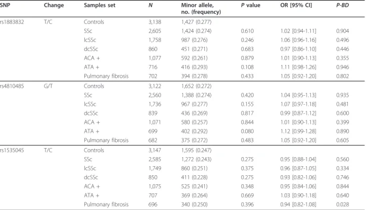
Table 1 Pooled analysis of CD40 polymorphisms
SNP Change Samples set N Minor allele,
no. (frequency) P value
OR [95% CI] P-BD rs1883832 T/C Controls 3,138 1,427 (0.277) SSc 2,605 1,424 (0.274) 0.610 1.02 [0.94-1.11] 0.904 lcSSc 1,758 987 (0.276) 0.246 1.06 [0.96-1.16] 0.496 dcSSc 860 451 (0.271) 0.683 0.97 [0.86-1.10] 0.446 ACA + 1,077 592 (0.261) 0.879 1.01 [0.90-1.13] 0.355 ATA + 716 416 (0.293) 0.108 1.11 [0.98-1.26] 0.946 Pulmonary fibrosis 702 394 (0.278) 0.433 1.05 [0.92-1.20] 0.802 rs4810485 G/T Controls 3,122 1,652 (0.272) SSc 2,560 1,388 (0.274) 0.420 1.04 [0.95-1.13] 0.935 lcSSc 1,736 967 (0.277) 0.155 1.07 [0.97-1.18] 0.481 dcSSc 839 436 (0.269) 0.817 0.99 [0.87-1.12] 0.600 ACA + 1,071 580 (0.257) 0.844 1.01 [0.90-1.13] 0.399 ATA + 699 402 (0.292) 0.080 1.12 [0.99-1.28] 0.890 Pulmonary fibrosis 682 375 (0.272) 0.483 1.05 [0.92-1.20] 0.605 rs1535045 T/C Controls 3,147 1,595 (0.247) SSc 2,585 1,272 (0.243) 0.275 0.95 [0.88-1.04] 0.560 lcSSc 1,749 860 (0.251) 0.375 0.96 [0.87-1.05] 0.334 dcSSc 850 411 (0.228) 0.275 0.93 [0.82-1.06] 0.746 ACA + 1,075 525 (0.241) 0.348 0.95 [0.84-1.06] 0.844 ATA + 707 369 (0.264) 0.669 1.03 [0.90-1.18] 0.640 Pulmonary fibrosis 696 340 (0.250) 0.396 0.94 [0.82-1.08] 0.028
Controls are used as reference for all comparisons. AllP values have been calculated for the allelic model by using the Mantel-Haenszel test under fixed effect. P_BD,P value by the Breslow-Day method.
In the present study, we analyzed the functional
CD40-1C/T polymorphism (rs1883832). This genetic variant is
located at -1 from the ATG, within a Kozak sequence, a
stretch of nucleotides essential for translation that flanks
the start codon in vertebrate genes [33]. The presence of a
major allele (C) in this SNP is associated with the increase
of the efficiency of CD40 translations [11]. Although
quan-titative differences between CD40 mRNA and proteins
have been observed in SSc skin fibroblasts [10], the
absence of association found for rs1883832 suggests that
this variant might not affect the translation of
CD40
mRNA. This process may be upregulated in these
abnor-mal skin fibroblasts, or other genes of the
CD40 signaling
pathway may influence the
CD40 expression. However,
functional studies in this way should be constructed before
excluding an association between this variant and the
CD40 expression in SSc.
Although
CD40 might be a common susceptibility
locus for some autoimmune diseases [11-16], our results
do not suggest an important role of
CD40 in the
sus-ceptibility to SSc. Several genes have been recently
dis-closed to play a function in the susceptibility to
autoimmune diseases, suggesting that these diseases
share a genetic background [34]. However, these loci
may not be universal genetic factors for autoimmune
disorders; therefore, the autoimmunity might result
from specific and multiple pleiotropic effects [19]. Also,
other genes are unique for each disease, reflecting a
spe-cific etiology [19,34]. Additional studies are required for
the identification of specific and shared genetic
path-ways that contribute to a better understanding of the
pathogenesis of the autoimmune diseases.
The role of the
CD40LG in the susceptibility to
auto-immune diseases has not been investigated as broadly as
that of
CD40, mainly because this gene is located on the
× chromosome. The different prevalences of these
dis-eases in both genders can suggest that genes located on
the × chromosome could be susceptibility factors in
autoimmune diseases; however, few studies analyzed
polymorphisms on this chromosome. Mutations on this
gene are associated with X-linked hyper-IgM syndrome,
a familial genetic disorder characterized by an increase
of IgM level and a decrease of IgG and IgA [20], but in
SLE, no evidence of association has been found [17].
Similarly, our results show that the
CD40LG gene may
not be SSc susceptibility loci.
Conclusions
Our results do not suggest an important role of
CD40
and
CD40LG genes in the susceptibility to SSc.
Addi-tional studies are required to draw firm conclusions
about the exact role of the
CD40 and CD40LG genes in
SSc susceptibility because other variants might be
involved in SSc. Future studies involving other genes of
the CD40-CD40LG pathway should be conducted to
elucidate fully the contribution of this pathway in the
pathogenesis of SSc.
Additional material
Additional file 1: Supplementary Tables 1 through 7. Genotype and allele distribution of the CD40 and CD40LG polymorphisms for each population included in the current study.
Abbreviations
ACA: anti-centromere antibody; ATA: anti-topoisomerase I antibody; CI: confidence interval; dcSSc: diffuse cutaneous subtype; GD: Graves disease;
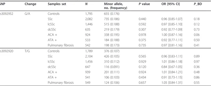
Table 2 Pooled analysis of CD40LG polymorphisms in female SSc patients and controls
SNP Change Samples set N Minor allele,
no. (frequency) P value
OR [95% CI] P_BD rs3092952 G/A Controls 1,795 655 (0.176) SSc 2,082 735 (0.186) 0.440 0.96 [0.85-1.07] 0.18 lcSSc 1,446 515 (0.188) 0.592 0.97 [0.85-1.10] 0.12 dcSSc 635 219 (0.179) 0.307 0.92 [0.77-1.09] 0.73 ACA + 924 338 (0.195) 0.978 1.00 [0.87-1.16] 0.06 ATA + 533 184 (0.188) 0.375 0.92 [0.77-1.11] 0.50 Pulmonary fibrosis 542 198 (0.173) 0.735 0.97 [0.81-1.16] 0.41 rs3092920 T/G Controls 1,789 376 (0.107) SSc 2,104 426 (0.105) 0.565 0.96 [0.83-1.11] 0.89 lcSSc 1,456 310 (0.112) 0.929 1.01 [0.86-1.18] 0.97 dcSSc 647 116 (0.091) 0.120 0.84 [0.67-1.05] 0.36 ACA + 939 201 (0.111) 0.924 1.01 [0.84-1.21] 0.48 ATA + 541 106 (0.103) 0.434 0.91 [0.73-1.15] 0.86 Pulmonary fibrosis 549 124 (0.106) 0.657 1.05 [0.84-1.31] 0.55
Controls are used as reference for all comparisons. AllP values have been calculated for the allelic model by using the Mantel-Haenszel test under fixed effect. P_BD, P value by the Breslow-Day method.
GWAS: genome-wide association study; HWE: Hardy-Weinberg equilibrium; lcSSc: limited cutaneous subtype; OR: odds ratio; P-BD: P value by Breslow-Day method; RA: rheumatoid arthritis; SLE: systemic lupus erythematosus; SNP: single-nucleotide polymorphism; SSc: systemic sclerosis.
Acknowledgements
We thank Sofia Vargas, Sonia Garcia, and Gema Robledo for their excellent technical assistance, and all the patients and healthy controls for kindly accepting their essential collaboration. We thank Banco Nacional de ADN (University of Salamanca, Spain) for supplying part of the control material. We also thank EUSTAR (The EULAR Scleroderma Trials and Research Group) and the German Network of Systemic Sclerosis for the facilitation of this project.
This work was supported by the following grants. JM was funded by SAF2009-11110 from the Spanish Ministry of Science, by CTS-4977 and PI-0590-2010 from Junta de Andalucía, and by RETICS Program, RD08/0075 (RIER) from Instituto de Salud Carlos III (ISCIII), within the VI PN de I+D+i 2008-2011 (FEDER). T.R.D.J.R. was funded by the VIDI laureate from the Dutch Association of Research (NWO) and Dutch Arthritis Foundation (National Reumafonds). JM and TRDJR were sponsored by the Orphan Disease Program grant from the European League Against Rheumatism (EULAR). TW was awarded grants by DFG WI 1031/6.1 and DFG KFO 250 TP03. MT was supported by Spanish Ministry of Science through the program Juan de la Cierva (JCI-2010-08227).
Spanish Scleroderma Group:
Norberto Ortego-Centeno, José Luis Callejas, and Raquel Ríos, Systemic Autoimmune Diseases Unit, Department of Internal Medicine, Hospital Clínico Universitario San Cecilio, Granada; Nuria Navarrete and Antonio Garcia, Department of Internal Medicine, Hospital Virgen de las Nieves, Granada; Antonio Fernández-Nebro, Department of Rheumatology, Hospital Carlos Haya, Málaga; María F. González-Escribano, Department of
Immunology, Hospital Virgen del Rocío, Sevilla; Julio Sánchez-Román and Mª Jesús Castillo, Department of Internal Medicine, Hospital Virgen del Rocío, Sevilla; Mª Ángeles Aguirre and Inmaculada Gómez-Gracia, Department of Rheumatology, Hospital Reina Sofía, Córdoba; Benjamín Fernández-Gutiérrez and Luis Rodríguez-Rodríguez, Department of Rheumatology, Hospital Clínico San Carlos, Madrid; Esther Vicente, Department of Rheumatology, Hospital La Princesa, Madrid; Mónica Fernández Castro and José Luis Andreu, Department of Rheumatology, Hospital Puerta del Hierro, Madrid; Paloma García de la Peña, Department of Rheumatology, Hospital Universitario Madrid Norte Sanchinarro, Madrid; Francisco Javier López-Longo and Lina Martínez-Estupiñán, Department of Rheumatology, Hospital General Universitario Gregorio Marañón, Madrid; Anna Pros, Department of Rheumatology, Hospital Del Mar, 08003 Barcelona; Vicente Fonollosa, Department of Internal Medicine, Hospital Valle de Hebrón, Barcelona; Carlos Tolosa, Department of Internal Medicine, Hospital Parc Tauli, Sabadell; Mónica Rodríguez Carballeira, Department of Internal Medicine, Hospital Universitari Mútua Terrasa, Barcelona; Ivan Castellví, Unidad de Reumatología, Department of Internal Medicine, Hospital de la Santa Creu i Sant Pau, Barcelona; Francisco Javier Narváez, Department of Rheumatology, Hospital Universitari de Bellvitge, Barcelona; Francisco Javier Blanco-García, Natividad Oreiro, and María Ángeles Robles, Department of Rheumatology, INIBIC-Hospital Universitario A Coruña, A Coruña; María Victoria Egurbide, Department of Internal Medicine, Hospital de Cruces, Vizcaya; Luis Sáez-Comet, Systemic Autoimmune Diseases Unit, Department of Internal Medicine, Hospital Universitario Miguel Servet, Zaragoza; Ricardo Blanco, Department of Rheumatology, Hospital Universitario Marqués de Valdecilla, Santander; Bernardino Díaz and Luis Trapiella, Department of Internal Medicine, Hospital Central de Asturias, Oviedo; Federico Díaz and Vanesa Hernández, Department of Rheumatology, Hospital Universitario de Canarias, Tenerife; Emma Beltrán, Department of Rheumatology, Hospital del Doctor Peset Aleixandre, Valencia; and José Andrés Román-Ivorra, Department of Rheumatology, Hospital Universitari i Politecnic La Fe, Valencia.
Author details
1Instituto de Parasitología y Biomedicina López-Neyra, IPBLN-CSIC, Avda. del
Conocimiento s/n. 18010, Granada, SpainArmilla (Granada), Spain.
2Department of Internal Medicine, Hospital Valle de Hebron, Passeig de la
Vall d’Hebron 119 08035 Barcelona, Spain.3Department of Rheumatology,
Radboud University Nijmegen Medical Centre, Comeniuslaan 4 6525 HP Nijmegen, The Netherlands.4Department of Rheumatology, Hospital 12 de
Octubre, Avda. de Córdoba s/n 28041, Madrid, Spain.5Department of
Internal Medicine, Hospital Carlos Haya, Avda Carlos Haya s/n 29010 Málaga, Spain.6Department of Rheumatology, Hospital Virgen de la Victoria, Campus de Teatinos s/n 29010 Málaga, Spain.7Department of Rheumatology,
Hospital Universitario de Canarias, Ctra. Cuesta-Taco, s/n 38320, La Cuesta, San Cristóbal de La Laguna, Tenerife, Canarias, Spain.8Department of
Internal Medicine, Hospital Central de Asturias, Celestino Villamil, s/n 33006 Oviedo, Spain.9Department of Autoimmune Diseases, Hospital Clinic, Carrer
de Villarroel, 170 08036 Barcelona, Spain.10See Acknowledgements.
11Referral Center for Systemic Autoimmune Diseases, Fondazione IRCCS Ca’
Granda Ospedale Maggiore Policlinico Via Francesco Sforza, 35 20122 and University of Milan, Via Festa del Perdono, 7 20122, Milan, Italy.
12Rheumatology Unit and Chair, Spedali Civili, Università degli Studi, Piazzale
Spedali Civili, 1 25123 Brescia, Italy.13Department of Medicine, Policlinico GB Rossi, Università degli studi di Verona, Via dell’Artigliere, 8 37129 Verona, Italy.14Department of Rheumatology and Clinical Immunology, Charité University Hospital and German Rheumatism Research Centre, a Leibniz Institute, Charitéplatz 1, 10117 Berlin, Germany.15Clinic for Immunology and Rheumatology Medical School, Carl-Neuberg-Str, 1 30625, Hannover, Germany.16Department of Dermatology, University of Cologne, Kerpener Str
62, 50924 Köln, Germany.17Department of Dermatology, Allergology, and
Venereology, Ruhr University of Bochum, Stiepeler Straße 129 44801 Bochum, Germany.18Department of Internal Medicine 3, Institute for Clinical Immunology, University of Erlangen-Nuremberg, Schillerstraße 1 91054, Erlangen, Germany.19Section Complex Genetics, Department of Medical Genetics, University Medical Center Utrecht, Universiteitsweg Stratenum 3508 AB, Utrecht, The Netherlands.20Department of Rheumatology, VU University Medical Center, De Boelelaan 1117 1081 HZ Amsterdam, The Netherlands.
21
Department of Rheumatology, Leiden University Medical Center, Albinusdreef 2 2300 RC, Leiden, The Netherlands.22Department of
Rheumatology, Hospital Universitario Marques de Valdecilla, IFIMAV, Avda. Valdecilla, 25, 39008 Santander, Spain.23Department of Rheumatology and
Clinical Immunology, University Utrecht Medical Center, Universiteitsweg 100 Stratenum 3508 AB, Utrecht, The Netherlands.
Authors’ contributions
MT and JM made substantial contributions to conception, design of study, and interpretation of data. MT carried out genotyping, analysis of data, and drafted the manuscript. CPS, JB, MCV, PC, MTC, RGP, EDF, MG, GE, LB, PA, CL, GR, TW, TK, AK, JHWD, NH, BPK, AEV, AJ, AJS, MAGG, TRDJR, and SSG had been involved in the acquisition of clinical data of the patients included in this study as well as the interpretation of the data. JM has been involved in revising of the final manuscript. All authors gave final approval of the version to be published.
Competing interests
The authors declare that they have no competing interests.
Received: 16 March 2012 Revised: 23 May 2012 Accepted: 25 June 2012 Published: 25 June 2012
References
1. Gabrielli A, Avvedimento EV, Krieg T: Scleroderma. N Engl J Med 2009, 360:1989-2003.
2. Martin JE, Bossini-Castillo L, Martin J: Unraveling the genetic component of systemic sclerosis. Hum Genet 2012, 131:1023-1037.
3. Elgueta R, Benson MJ, de Vries VC, Wasiuk A, Guo Y, Noelle RJ: Molecular mechanism and function of CD40/CD40L engagement in the immune system. Immunol Rev 2009, 229:152-172.
4. Peters AL, Stunz LL, Bishop GA: CD40 and autoimmunity: the dark side of a great activator. Semin Immunol 2009, 21:293-300.
5. Vakkalanka RK, Woo C, Kirou KA, Koshy M, Berger D, Crow MK: Elevated levels and functional capacity of soluble CD40 ligand in systemic lupus erythematosus sera. Arthritis Rheum 1999, 42:871-881.
6. Tamura N, Kobayashi S, Kato K, Bando H, Haruta K, Oyanagi M, Kuriyama M, Kipps TJ, Hashimoto H: Soluble CD154 in rheumatoid arthritis: elevated plasma levels in cases with vasculitis. J Rheumatol 2001, 28:2583-2590. 7. Faure GC, Bensoussan-Lejzerowicz D, Bene MC, Aubert V, Leclere J:
Coexpression of CD40 and class II antigen HLA-DR in Graves’ disease thyroid epithelial cells. Clin Immunol Immunopathol 1997, 84:212-215.
8. Allanore Y, Borderie D, Meune C, Lemarechal H, Weber S, Ekindjian OG, Kahan A: Increased plasma soluble CD40 ligand concentrations in systemic sclerosis and association with pulmonary arterial hypertension and digital ulcers. Ann Rheum Dis 2005, 64:481-483.
9. Komura K, Fujimoto M, Matsushita T, Yanaba K, Kodera M, Kawasuji A, Hasegawa M, Takehara K, Sato S: Increased serum soluble CD40 levels in patients with systemic sclerosis. J Rheumatol 2007, 34:353-358. 10. Fukasawa C, Kawaguchi Y, Harigai M, Sugiura T, Takagi K, Kawamoto M,
Hara M, Kamatani N: Increased CD40 expression in skin fibroblasts from patients with systemic sclerosis (SSc): role of CD40-CD154 in the phenotype of SSc fibroblasts. Eur J Immunol 2003, 33:2792-2800. 11. Jacobson EM, Concepcion E, Oashi T, Tomer Y: A Graves’
disease-associated Kozak sequence single-nucleotide polymorphism enhances the efficiency of CD40 gene translation: a case for translational pathophysiology. Endocrinology 2005, 146:2684-2691.
12. ANZgene: Genome-wide association study identifies new multiple sclerosis susceptibility loci on chromosomes 12 and 20. Nat Genet 2009, 41:824-828.
13. Raychaudhuri S, Remmers EF, Lee AT, Hackett R, Guiducci C, Burtt NP, Gianniny L, Korman BD, Padyukov L, Kurreeman FA, Chang M, Catanese JJ, Ding B, Wong S, van der Helm-van Mil AH, Neale BM, Coblyn J, Cui J, Tak PP, Wolbink GJ, Crusius JB, van der Horst-Bruinsma IE, Criswell LA, Amos CI, Seldin MF, Kastner DL, Ardlie KG, Alfredsson L, Costenbader KH, Altshuler D, et al: Common variants at CD40 and other loci confer risk of rheumatoid arthritis. Nat Genet 2008, 40:1216-1223.
14. Orozco G, Eyre S, Hinks A, Ke X, Wellcome Trust Case Control consortium YEAR Consortium, Wilson AG, Bax DE, Morgan AW, Emery P, Steer S, Hocking L, Reid DM, Wordsworth P, Harrison P, Thomson W, Barton A, Worthington J: Association of CD40 with rheumatoid arthritis confirmed in a large UK case-control study. Ann Rheum Dis 2009, 69:813-816. 15. Blanco-Kelly F, Matesanz F, Alcina A, Teruel M, Díaz-Gallo LM,
Gómez-García M, López-Nevot MA, Rodrigo L, Nieto A, Cardeña C, Alcain G, Díaz-Rubio M, de la Concha EG, Fernandez O, Arroyo R, Martín J, Urcelay E: CD40: novel association with crohn’s disease and replication in multiple sclerosis susceptibility. PLoS One 2010, 5:e11520.
16. Rodríguez-Rodríguez L, Castañeda S, Vázquez-Rodríguez TR, Morado IC, Marí-Alfonso B, Gómez-Vaquero C, Miranda-Filloy JA, Narvaez J, Ortego-Centeno N, Blanco R, Fernández-Gutiérrez B, Martín J, González-Gay MA: Influence of CD40 rs1883832 polymorphism in susceptibility to and clinical manifestations of biopsy-proven giant cell arteritis. J Rheumatol 2010, 37:2076-2080.
17. Chadha S, Miller K, Farwell L, Lightstone LB, Daly MJ, Rioux JD, Vyse TJ: Haplotype structure of TNFRSF5-TNFSF5 (CD40-CD40L) and association analysis in systemic lupus erythematosus. Eur J Hum Genet 2005, 13:669-676.
18. Vazgiourakis VM, Zervou MI, Choulaki C, Bertsias G, Melissourgaki M, Yilmaz N, Sidiropoulos P, Plant D, Trouw LA, Toes RE, Kardassis D, Yavuz S, Boumpas DT, Goulielmos GN: A common SNP in the CD40 region is associated with systemic lupus erythematosus and correlates with altered CD40 expression: implications for the pathogenesis. Ann Rheum Dis 2011, 70:2184-2190.
19. Ramos PS, Criswell LA, Moser KL, Comeau ME, Williams AH, Pajewski NM, Chung SA, Graham RR, Zidovetzki R, Kelly JA, Kaufman KM, Jacob CO, Vyse TJ, Tsao BP, Kimberly RP, Gaffney PM, Alarcón-Riquelme ME, Harley JB, Langefeld CD, International Consortium on the Genetics of Systemic Erythematosus: A comprehensive analysis of shared loci between systemic lupus erythematosus (SLE) and sixteen autoimmune diseases reveals limited genetic overlap. PLoS Genet 2011, 7:e1002406. 20. Lougaris V, Badolato R, Ferrari S, Plebani A: Hyper immunoglobulin M
syndrome due to CD40 deficiency: clinical, molecular, and immunological features. Immunol Rev 2005, 203:48-66.
21. Martin-Donaire T, Losada-Fernandez I, Perez-Chacon G, Rua-Figueroa I, Erausquin C, Naranjo-Hernandez A, Rosado S, Sanchez F, Garcia-Saavedra A, Citores MJ, Vargas JA, Perez-Aciego P: Association of the microsatellite in the 3’ untranslated region of the CD154 gene with rheumatoid arthritis in females from a Spanish cohort: a case-control study. Arthritis Res Ther 2007, 9:R89.
22. Citores MJ, Rua-Figueroa I, Rodriguez-Gallego C, Durantez A, Garcia-Laorden MI, Rodriguez-Lozano C, Rodriguez-Perez JC, Vargas JA, Perez-Aciego P: The dinucleotide repeat polymorphism in the 3’UTR of the CD154 gene has a functional role on protein expression and is
associated with systemic lupus erythematosus. Ann Rheum Dis 2004, 63:310-317.
23. Subcommittee for scleroderma criteria of the American Rheumatism Association Diagnostic and Therapeutic Criteria Committee: Preliminary criteria for the classification of systemic sclerosis (scleroderma). Arthritis Rheum 1980, 23:581-590.
24. LeRoy EC, Black C, Fleischmajer R, Jablonska S, Krieg T, Medsger TA Jr, Rowell N, Wollheim F: Scleroderma (systemic sclerosis): classification, subsets and pathogenesis. J Rheumatol 1988, 15:202-205.
25. LeRoy EC, Medsger TA Jr: Criteria for the classification of early systemic sclerosis. J Rheumatol 2001, 28:1573-1576.
26. Radstake TR, Gorlova O, Rueda B, Martin JE, Alizadeh BZ, Palomino-Morales R, Coenen MJ, Vonk MC, Voskuyl AE, Schuerwegh AJ, Broen JC, van Riel PL, van‘t Slot R, Italiaander A, Ophoff RA, Riemekasten G,
Hunzelmann N, Simeon CP, Ortego-Centeno N, González-Gay MA, González-Escribano MF, Spanish Scleroderma Group, Airo P, van Laar J, Herrick A, Worthington J, Hesselstrand R, Smith V, de Keyser F, Houssiau F, et al: Genome-wide association study of systemic sclerosis identifies CD247 as a new susceptibility locus. Nat Genet 2010, 42:426-429. 27. Gorlova O, Martin JE, Rueda B, Koeleman BP, Ying J, Teruel M,
Diaz-Gallo LM, Broen JC, Vonk MC, Simeon CP, Alizadeh BZ, Coenen MJ, Voskuyl AE, Schuerwegh AJ, van Riel PL, Vanthuyne M, van‘t Slot R, Italiaander A, Ophoff RA, Hunzelmann N, Fonollosa V, Ortego-Centeno N, González-Gay MA, García-Hernández FJ, González-Escribano MF, Airo P, van Laar J, Worthington J, Hesselstrand R, Smith V, et al: Identification of novel genetic markers associated with clinical phenotypes of systemic sclerosis through a genome-wide association strategy. PLoS Genet 2011, 7:e1002178.
28. Bossini-Castillo L, Martin JE, Broen J, Gorlova O, Simeón CP, Beretta L, Vonk MC, Callejas JL, Castellví I, Carreira P, García-Hernández FJ, Fernández Castro M, Spanish Scleroderma Group, Coenen MJ, Riemekasten G, Witte T, Hunzelmann N, Kreuter A, Distler JH, Koeleman BP, Voskuyl AE,
Schuerwegh AJ, Palm Ø, Hesselstrand R, Nordin A, Airó P, Lunardi C, Scorza R, Shiels P, van Laar JM, et al: A GWAS follow-up study reveals the association of the IL12RB2 gene with systemic sclerosis in Caucasian populations. Hum Mol Genet 2012, 21:926-933.
29. Burdon KP, Langefeld CD, Beck SR, Wagenknecht LE, Carr JJ, Rich SS, Freedman BI, Herrington D, Bowden DW: Variants of the CD40 gene but not of the CD40L gene are associated with coronary artery calcification in the Diabetes Heart Study (DHS). Am Heart J 2006, 151:706-711. 30. Malarstig A, Lindahl B, Wallentin L, Siegbahn A: Soluble CD40L levels are
regulated by the -3459 A>G polymorphism and predict myocardial infarction and the efficacy of antithrombotic treatment in non-ST elevation acute coronary syndrome. Arterioscler Thromb Vasc Biol 2006, 26:1667-1673.
31. Purcell S, Neale B, Todd-Brown K, Thomas L, Ferreira MA, Bender D, Maller J, Sklar P, de Bakker PI, Daly MJ, Sham PC: PLINK: a tool set for whole-genome association and population-based linkage analyses. Am J Hum Genet 2007, 81:559-575[http://pngu.mgh.harvard.edu/~purcell/plink/]. 32. Skol AD, Scott LJ, Abecasis GR, Boehnke M: Joint analysis is more efficient
than replication-based analysis for two-stage genome-wide association studies. Nat Genet 2006, 38:209-213[http://www.sph.umich.edu/csg/ abecasis/CaTS/].
33. Kozak M: An analysis of 5’-noncoding sequences from 699 vertebrate messenger RNAs. Nucleic Acids Res 1987, 15:8125-8148.
34. Cho JH, Gregersen PK: Genomics and the multifactorial nature of human autoimmune disease. N Engl J Med 2011, 365:1612-1623.
doi:10.1186/ar3890
Cite this article as: Teruel et al.: Analysis of the association between CD40 and CD40 ligand polymorphisms and systemic sclerosis. Arthritis Research & Therapy 2012 14:R154.