Amino Acids Activate Mammalian Target of Rapamycin
Complex 2 (mTORC2) via PI3K/Akt Signaling
*
□SReceived for publication, July 21, 2010, and in revised form, November 9, 2010Published, JBC Papers in Press, December 3, 2010, DOI 10.1074/jbc.M110.166991 Irantzu Tato1, Ramon Bartrons, Francesc Ventura, and Jose Luis Rosa2
From the Departament de Cie`ncies Fisiolo`giques II, Campus de Bellvitge, Institut d’Investigacio´ Biome`dica de Bellvitge (IDIBELL), Universitat de Barcelona, L’Hospitalet de Llobregat, Barcelona E-08907, Spain
The activity of mammalian target of rapamycin (mTOR) complexes regulates essential cellular processes, such as growth, proliferation, or survival. Nutrients such as amino ac-ids are important regulators of mTOR complex 1 (mTORC1) activation, thus affecting cell growth, protein synthesis, and autophagy. Here, we show that amino acids may also activate mTOR complex 2 (mTORC2). This activation is mediated by the activity of class I PI3K and of Akt. Amino acids induced a rapid phosphorylation of Akt at Thr-308 and Ser-473. Whereas both phosphorylations were dependent on the pres-ence of mTOR, only Akt phosphorylation at Ser-473 was de-pendent on the presence of rictor, a specific component of mTORC2. Kinase assays confirmed mTORC2 activation by amino acids. This signaling was functional, as demonstrated by the phosphorylation of Akt substrate FOXO3a. Interestingly, using different starvation conditions, amino acids can selec-tively activate mTORC1 or mTORC2. These findings identify a new signaling pathway used by amino acids underscoring the crucial importance of these nutrients in cell metabolism and offering new mechanistic insights.
Cell growth and proliferation are fundamental processes that are regulated by multiple signals, such as nutrients, hor-mones, and growth factors. One of the key components regu-lating these signal transduction pathways is mTOR,3which in higher eukaryotes forms two different complexes: mTORC1 and mTORC2 (1, 2). mTOR complexes display Ser/Thr kinase activity. mTORC1 is rapamycin-sensitive and includes the mTOR catalytic subunit, mLST8/GL, PRAS40, the regulatory-associated protein of mTOR (raptor), and DEPTOR (3–5). Instead, mTORC2 is rapamycin-insensitive, at least in short treatments (6), and includes mTOR, mLST8/GL, mSin1, rapamycin-insensitive companion of mTOR (rictor), the pro-tein observed with rictor (protor), and DEPTOR (4, 5, 7). mTORC1 regulates cell growth by controlling mRNA
transla-tion, ribosome biogenesis, autophagy, and metabolism (1, 8 –12). On the other hand, mTORC2 regulates cell survival and proliferation (13–18). Both hormones and growth factors have been described to regulate mTORC1 and mTORC2 (1, 19, 20). However, thus far only mTORC1 has been described to be regulated by nutrients such as amino acids (1, 2, 20 –22).
A number of studies point to the importance of nutrients in the etiology of some major diseases, such as insulin-resistant obesity (23) or cancer (24, 25) and in the aging process (11, 26). In humans, circulating amino acid levels are elevated in obese individuals, and this is related to an increase in insulin resistance (23, 27, 28). The morbidity of obesity not only ex-tends to diabetes and cardiovascular diseases but also has been linked to 20% of cancer deaths (29). During the last years, different studies have shown a relationship between nutrients, such as glucose or amino acids, and mTOR signal-ing. For example, it has been reported that amino acids acti-vate S6 kinase 1 protein (S6K1) and inhibit autophagy in an mTOR-dependent manner (30, 31). S6K1 activation involves its phosphorylation on multiple Ser/Thr amino acid residues, with the critical step being the phosphorylation at Thr-389 (3, 32–34). This phosphorylation is catalyzed by mTORC1, so there is a correlation between mTORC1 and S6K1 activation. Although the precise mechanism of mTORC1 activation by amino acids remains unclear, it appears to require the class III PI3K human vacuolar protein sorting-associated protein 34 (hVps34), as well as the serine/threonine kinase mitogen-acti-vated protein kinase kinase kinase kinase 3 (MAP4K3) and the Rag family of small GTPases (2, 28, 35–37). In turn, activation of hVps34 is mediated by Ca2⫹/calmodulin signaling (38). More recently, it has been demonstrated that amino acids may also activate S6K1 through MAPKs independently of mTOR (39).
Hormones and growth factors signal to both mTOR com-plexes. For instance, insulin binding to its receptor activates the canonical signaling cascade triggered by the activation of class I PI3K, leading to activation of Akt through its phosphor-ylation at Thr-308 and activation of mTORC1 and mTORC2 complexes (2, 35, 36, 40, 41). Thus, Akt-dependent mTORC1 activation leads to S6K1 and 4EBP1 phosphorylation regulat-ing cell growth and autophagy. On the other hand, mTORC2 activation regulates cell survival and proliferation by inducing further phosphorylation of its substrate Akt at Ser-473 (42), SGK1 phosphorylation (17, 18), and transcription regulation via Forkhead family transcription factor (FKHRL or FOXO) inhibition (14).
*This work was supported in part by Ministerio de Educacio´n y Ciencia (MEC) Grant BFU2008-02084/BMC, Instituto de Salud Carlos III Redes Tema´ticas de Investigacio´n Cooperativa en Salud Grant RD06/0020 and Generalitat de Catalunya Grant 2009SGR1059.
□S
The on-line version of this article (available at http://www.jbc.org) con-tainssupplemental Figs. S1 and S2.
1Supported by the Juan de la Cierva Program.
2To whom correspondence should be addressed. Tel.: 34-934021056; Fax: 34-934024268; E-mail: [email protected].
3The abbreviations used are: mTOR, mammalian target of rapamycin; DPBS, Dulbecco’s PBS; DMEM(⫺AA), DMEM without amino acids; RSK, 90 kDa ribosomal S6 kinase; MSK, mitogen- and stress-activated protein kinase.
THE JOURNAL OF BIOLOGICAL CHEMISTRY VOL. 286, NO. 8, pp. 6128 –6142, February 25, 2011 © 2011 by The American Society for Biochemistry and Molecular Biology, Inc. Printed in the U.S.A.
In the present study, we demonstrate that amino acids can also activate Akt signaling through class I PI3K, as in-sulin does. This new amino acid signaling pathway also leads to mTORC2 activation and phosphorylation of Fork-head transcription factor FOXO3a. Furthermore, we show the conditions under which amino acids may differentially activate mTOR complexes 1 and 2. Altogether, these data shed new light into the mechanisms underlying amino acid signaling and their role in cellular growth, proliferation, and survival.
EXPERIMENTAL PROCEDURES
Reagents—Insulin, PDGF, wortmannin, rapamycin, betaine, ␥-amino-n-butyric acid, protein A-peroxidase secondary anti-body, ATP, anti-FLAG, anti-HA, and anti-P-ERK1/2 antibod-ies, hVps34 class III PI3K inhibitor 3-methyladenine (Sigma-Aldrich); amino acid solutions (minimal essential medium and minimal essential medium nonessential) (Invitrogen); L-glutamine solution (Biological Industries); U0126 and SB 203580 (Calbiochem); mTOR, rictor (53A2), anti-raptor (24C12), anti-P-T308-Akt, anti-P-S473-Akt, anti-Akt, P-T37/46 – 4E-BP1, P-T24-Fox01/T32-Fox03a, anti-P-T389-S6K1 (1A5), anti-P-T421/S424-S6K1, anti-P-S380-RSK, anti-P-S376-MSK, anti-P-S235/236-S6, anti-S6 (54D2), and anti-PT180/Y182-p38 antibodies (Cell Signaling Technol-ogy); anti-TSC2 (C-20) and anti-S6K1 (C-18) antibodies (Santa Cruz Biotechnology, Inc.); anti-LC3 antibody (MBL); horseradish peroxidase-conjugated secondary antibodies (In-vitrogen); protein A-Sepharose (GE Healthcare); Immo-bilon-P PVDF transfer membrane (Millipore Corporation); class I PI3K isoform-specific inhibitors (PI3K-selective in-hibitor TGX-155 (43), PI3K␦-selective inhibitor IC87114 (44, 45), and PI3K␣-selective inhibitor AS702630 (46)) were kindly provided by M. Camps (Merck Serono); JNK inhibitor SP600125 (A. G. Scientific); and Akt Inhibitor VIII, Isozyme-Selective, Akti-1/2 (Calbiochem). pcDNA3-FLAG-HA-Akt1 was provided by Addgene (47). mTOR, rictor, and nontarget-ing control siRNAs were reported previously (16, 39).
Cell Culture and Transfection—HeLa, HEK293, and MCF7 cells were cultured at 37 °C in DMEM (Invitrogen) with 10% fetal bovine serum. Transfection of HeLa cells (plasmids and siRNAs) was carried out with calcium phosphate transfection system. Cell treatments such as serum starvation, amino acid deprivation, insulin, or amino acid stimulation were per-formed as reported previously (31, 36, 39, 48). For experi-ments with insulin, the cells were deprived of serum over-night and then incubated with 200 nMinsulin for 30 min. For experiments with PDGF, the cells were deprived of serum overnight and then incubated with 50 ng/ml of PDGF for 30 min. For experiments with amino acids, after overnight serum deprivation, the cells were incubated for an additional 2 h with DPBS (Dulbecco’s phosphate-buffered saline containing 1 g/literD-glucose and 36 mg/liter sodium pyruvate, calcium, and magnesium) (Invitrogen) (39), or, when noted, after over-night serum deprivation, the cells were incubated for an addi-tional 2 h with DMEM without amino acids, DMEM(⫺AA) (36). The cells were then stimulated with amino acids (a mix of essential amino acids, nonessential amino acids, andL
-glu-tamine solutions from Invitrogen and Biological Industries to 1⫻ stock final concentration) for 30 min. For experiments with amino acid supplementation, the cells were cultured for 48 h in complete DMEM, without starvation. After this, the cells were supplemented with amino acids once. The specific inhibitors were added 60 min before the stimulation with amino acids or insulin at a final concentration of 20 nM rapa-mycin, 100 nMwortmannin, 5MU0126, 10MSB 203580, 10MAkti-1/2, 10 mM3-methyladenine, 10MSP600125, and 1Mfor PI3K isoform-specific inhibitors.
Cell Lysate and Immunoblotting—Previously treated cells were lysed and processed as described previously (39). To si-multaneously analyze big (e.g. mTOR) and small (e.g. LC3) proteins in the same SDS/PAGE gel, we used a gel combina-tion named LAG gel (49). Band intensities were analyzed with a gel documentation system (LAS-3000 Fujifilm). Protein lev-els were normalized with respect to mTOR or TSC2 levlev-els and expressed as percentages of controls.
Immunoprecipitations and Kinase Assay—Lysates from HeLa cells were immunoprecipitated with mTOR, anti-rictor, or anti-raptor antibodies. Washed immunoprecipitates were used for in vitro kinase assays using previously purified FLAG-HA-Akt1 as substrate. All of the kinase assays were performed as described (17) with the following modifications: mTOR, rictor, and raptor immunoprecipitations were per-formed for 2 h, instead of 1 h, and the kinase assay for 40 min at 37 °C. FLAG-HA-Akt1 substrate purification was per-formed by immunoprecipitating FLAG-HA-Akt1 with anti-FLAG antibody for 1 h in HeLa cells previously starved of se-rum and amino acids. Band intensities were quantified with a gel documentation system (LAS-3000 Fujifilm).
Statistical Analysis—The results are expressed as the means⫾ S.E. Data for multiple variable comparisons were analyzed by one-way analysis of variance. For comparison of significance, Dunn’s test was used as a post hoc test according to the statistical program GraphPad Prism (*, p⬍ 0.05; **, p ⬍ 0.01; ***, p⬍ 0.001).
RESULTS
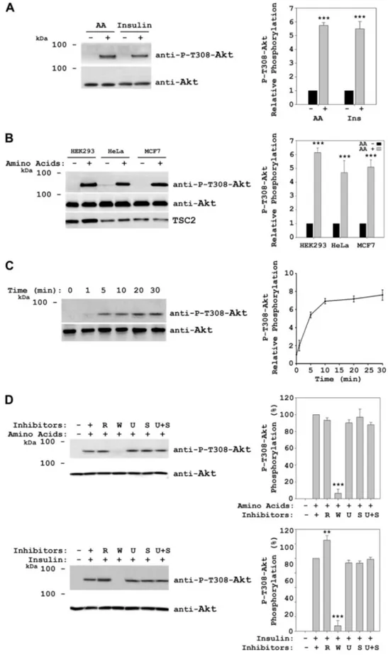
Amino Acids Induce Akt Phosphorylation at Thr-308—To analyze Akt activation by amino acids, HeLa cells were incu-bated overnight in DMEM without serum, followed by a 2-h incubation in Dulbecco’s PBS (DPBS) to deprive them of amino acids, and after this, the cells were stimulated with amino acids for 30 min. Lysates of these cells were analyzed by immunoblot, and Akt activation was measured by detec-tion of Thr-308 phosphoryladetec-tion. Under these condidetec-tions, we observed Akt Thr-308 phosphorylation in the presence of amino acids (Fig. 1A). As a positive control of Akt activation, serum-deprived cells were stimulated with insulin. In fact, Akt Thr-308 phosphorylation levels obtained with amino ac-ids were similar to the levels observed in the presence of insu-lin (Fig. 1A). We obtained the same result when we used dif-ferent cell lines such as HEK293, MCF7, Huh7, C2C12, and mouse embryonic fibroblasts (Fig. 1B and data not shown), indicating that Akt phosphorylation did not depend on the cell line studied. Time course experiments showed that Akt phosphorylation was rapid (1–5 min after the addition of
FIGURE 1. Akt is phosphorylated at Thr-308 in response to amino acids. A–D, HeLa cells were deprived of serum overnight and stimulated with 200 nM insulin for 30 min, or after serum withdrawal, they were deprived for 2 h of amino acids (AA) with DPBS medium, followed by stimulation with 1⫻ amino acids for 30 min or the indicated times (C). In B, MCF7 or HEK293 cells were treated with amino acids as in A. Where indicated, the cells were pretreated with 20 nMrapamycin (R), 100 nMwortmannin (W), 5MU0126 (U), or 10MSB 203580 (S) for 60 min before insulin or amino acid stimulation. Cellular lysates were analyzed by Western blot with the indicated antibodies. Molecular mass markers are indicated on the left. Quantification and statistical analyses (right
panels) were performed as described under “Experimental Procedures.” These data are representative of at least three independent experiments.
Amino Acids Activate mTOR Complexes
amino acids) and reached maximum levels at 10 –30 min (Fig. 1C). Similar to insulin, Akt phosphorylation by amino acids was dependent on wortmannin (inhibitor of PI3K activity) and independent of rapamycin (inhibitor of mTORC1 activ-ity), U0126 (inhibitor of MEK activactiv-ity), and SB 203580 (inhib-itor of p38 activity) (Fig. 1D). Rapamycin and wortmannin were tested to different concentrations (10 –100 and 10 –200 nM, respectively) and times (30 –120 min). Except for the lower wortmannin concentration, similar results were ob-tained (supplemental Fig. S1). These results indicate sufficient dosage and time duration of the treatment used with these inhibitors.
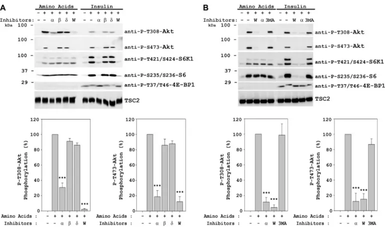
Amino Acids Induce Akt Phosphorylation at Ser-473—To analyze the Akt signaling induced by amino acids, we then checked substrates of mTORC complexes: phosphorylations of 4E-BP1 (Thr-37/Thr-46) and p70 S6K1 (Thr-389) for mTORC1 complex and phosphorylation of Akt (Ser-473) for mTORC2 complex. Stimulation by insulin was used as posi-tive control of the activation of these complexes. Under these conditions, we observed phosphorylation of Akt at Ser-473 by amino acids to a similar extent to that obtained with insulin (Fig. 2A). We also observed phosphorylation of p70 S6K1 (Thr-421/Ser-424) and S6 ribosomal protein (Ser-235/Ser-236) by amino acids, as we described previously (39). As ex-pected, insulin activated 4E-BP1 (Thr-37/Thr-46), p70 S6K1 (Thr-389), p70 S6K1 (Thr-421/Ser-424), and S6 ribosomal protein (Ser-235/Ser-236), in agreement with previous reports (1, 19, 28). However, we observed that amino acid addition did not result in 4E-BP1 37/Thr-46) or p70 S6K1 (Thr-389) phosphorylation. Note that the band detected in the presence of amino acids with the anti-P-T389-S6K1 antibody (asterisk) is phosphorylated RSK/MSK (39). Akt phosphoryla-tion at Ser-473 was also observed in other cell lines such as HEK293, MCF7, Huh7, C2C12, and mouse embryonic fibro-blasts, indicating that Akt activation was not restricted to HeLa cells (Fig. 2B and data not shown). Time course experi-ments showed rapid phosphorylation of Akt at Ser-473 by amino acids (1–5 min), with maximum levels between 10 and 30 min. When we compared the activation course for Thr-308 and Ser-473 phosphorylation sites (Fig. 2C, right panel), we did not detect significant differences in their phosphorylation kinetics. To ascertain that the observed Akt activation on both Thr-308 and Ser-473 was not due to an osmotic effect and was instead a specific response to amino acids, we tested
the effects of non-nutrient osmolytes betaine and
␥-amino-n-butyric acid. Thr-308 and Ser-473 phosphorylations were only observed in the presence of amino acids, thus ruling out an osmotic effect (Fig. 2D). We had previously shown similar results for p70 S6K1 (Thr-421/Ser-424) and S6 (Ser-235/Ser-236) phosphorylations (39). In agreement with the results in Fig. 1D, Akt phosphorylation at Ser-473 by amino acids was only blocked in the presence of wortmannin (Fig. 2E). In insu-lin-induced cells, we observed a slight increase of Akt phos-phorylation at Ser-473 in the presence of rapamycin (Fig. 2E), similar to what was observed for Akt phosphorylation at Thr-308 (Fig. 1D). This increase in Akt phosphorylations with in-sulin in the presence of rapamycin has previously been de-scribed as a result of mTORC1 inhibition (10, 50 –53). We
could not observe a similar increase in Akt phosphorylations with amino acids in the presence of rapamycin, suggesting different downstream substrates and pathway regulation for amino acid signaling. In Fig. 2E, we could also observe the different activation mechanisms, as reflected by different in-hibitor sensitivities, for S6K1 (Thr-389 and Thr-421/Ser-424) and S6 (Ser-235/Ser-236) proteins in response to insulin or amino acids, as published previously (39). Rapamycin and wortmannin were also tested to different concentrations (10 – 100 and 10 –200 nM, respectively) and times (30 –120 min) (supplemental Fig. S1). Therefore, these results suggest a common activation mechanism for Akt in response to both insulin and amino acids (probably via PI3K pathway) and dif-ferent activation mechanisms for S6K1 and S6 ribosomal pro-tein: via Akt for the insulin-induced signal and via MAPK for the amino acid-induced signal.
Amino Acids Activate Akt via the␣ Isoform of Class I PI3K—
To assess whether Akt phosphorylations occur downstream of PI3K, specific inhibitors for class I PI3K isoforms were tested (Fig. 3A). The inhibitors were PI3K␣-selective inhibitor AS702630 (46), PI3K-selective inhibitor TGX-155 (43), and PI3K␦-selective inhibitor IC87114 (44, 45). We observed a great inhibition, for both Thr-308 and Ser-473 phosphoryla-tions, in the presence of the PI3K␣-selective inhibitor. We obtained the same results in response to both amino acids and insulin, reinforcing the notion that both of them signal through the same class I PI3K isoform. That p110␣ is essen-tial for insulin signal transduction has been described previ-ously (54). These experiments confirm the involvement of the ␣ isoform of class I PI3K in the amino acids signal as well. We also analyzed the involvement of class III PI3K (hVps34) in amino acid signaling (Fig. 3B) using the hVps34 inhibitor 3-methyladenine. We did not observe inhibition of Akt phos-phorylation with amino acids nor with insulin.
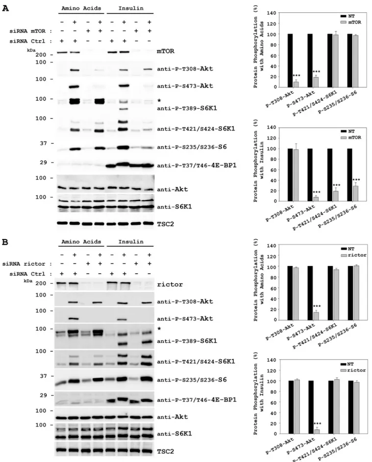
Amino Acids Signal to Akt Ser-473 through mTORC2—It has been described that mTORC2 phosphorylates Akt at Ser-473 downstream of PI3K, (16, 40, 42, 55, 56). We also showed that amino acids may activate Akt in the same way insulin does (Figs. 2E and 3). To test whether mTOR complexes me-diate Akt Ser-473 phosphorylation induced by amino acids, we used an mTOR siRNA to knock down mTOR protein lev-els. HeLa cells were transfected with mTOR-specific siRNA and stimulated with amino acids. Stimulation with insulin was used as a positive control. Knockdown of mTOR clearly in-hibited the amino acid-induced phosphorylations of Akt in Ser-473 (downstream of mTORC2) and in Thr-308 (upstream of mTORC2) (Fig. 4A). In insulin-induced cells, knockdown of mTOR only inhibited Ser-473 phosphorylation and not Thr-308. Moreover, mTOR knockdown did not affect phos-phorylations of S6K1 (Thr-421/Ser-424) or S6 ribosomal pro-tein (Ser-235/Ser-236) in response to amino acids, but S6K1 and S6 ribosomal protein activation was clearly inhibited in response to the insulin signal, as described previously (10, 42, 57). In agreement with the decrease of mTORC1 activity, acti-vation of 4E-BP1 (Thr-37/Thr-46) was also abolished in insu-lin signainsu-ling.
To check whether the Akt activation was specific of mTORC2, we used a rictor siRNA (Fig. 4B). Knockdown of
rictor completely abolished the amino acid-induced phosphor-ylation of Akt at Ser-473. Akt phosphorphosphor-ylation at Thr-308 was not inhibited. Rictor knockdown did not affect phosphor-ylations of S6K1 or S6 ribosomal protein in response to amino acids. Similar results were obtained by stimulating cells with insulin (Fig. 4B). In these conditions, knockdown of rictor did not affect the mTORC1 activation (phosphorylations of 4E-BP1 at Thr-37/Thr-46 and of p70 S6K1 at Thr-389) in re-sponse to insulin in agreement with the above data and previ-ous reports (10, 42, 57).
To confirm the mTORC2 role in amino acid signal trans-duction to Akt, we performed in vitro kinase assays using FLAG-HA-Akt fusion protein as substrate. Lysates from HeLa cells stimulated or not with amino acids were cipitated with anti-mTOR antibodies. Purified immunopre-cipitates were incubated with previously purified FLAG-HA-Akt in the presence or absence of Mg-ATP for 40 min. Under these conditions, phosphorylation of Akt at Ser-473 was sig-nificantly increased in mTOR immunoprecipitates stimulated by amino acids (Fig. 5A). The mTOR immunoprecipitates contained rictor and raptor (Fig. 5A). To assess which of the
two mTOR complexes was involved in this phosphorylation, we performed immunoprecipitations from HeLa cell lysates using specific antibodies against mTORC1 (using anti-raptor antibodies) and mTORC2 (using anti-rictor antibodies). In the presence of Mg-ATP and FLAG-HA-Akt substrate, rictor immunoprecipitates stimulated by amino acids increased sig-nificantly the phosphorylation of Akt substrate at Ser-473 (Fig. 5C). In parallel experiments, raptor immunoprecipitates stimulated by amino acids failed to increase Akt tion (Fig. 5B). We could not detect Akt Thr-308 phosphoryla-tion in kinase assays (Fig. 5), raptor in rictor immunoprecipi-tates (Fig. 5C), or rictor in raptor immunoprecipiimmunoprecipi-tates (Fig. 5B). Taken together, these data support the specific activation of Akt at Ser-473 by amino acids via mTORC2.
Amino Acids Inactivate FOXO3a Downstream of Akt and Independently of the JNK Activity—Akt has been described as a mediator of cell survival and proliferation in response to extracellular stimuli such as insulin or growth factors (14, 58 – 61). These signals suppress the death machinery and prevent apoptosis. Akt plays a central role in promoting cell survival by phosphorylating proapoptotic substrates and suppressing
FIGURE 2. Amino acids induce Akt phosphorylation at Ser-473. HeLa (A–E), MCF7 (B), or HEK293 (B) cells were treated with insulin or amino acids (AA) as in Fig. 1. D, cells deprived of amino acids as in A were stimulated with amino acids (1⫻), betaine (10 mM), or␥-amino-n-butyric acid (AIB, 10 mM) for 30 min. Where indicated, the cells were pretreated with 20 nMrapamycin (R), 100 nMwortmannin (W), 5MU0126 (U), or 10MSB 203580 (S) for 60 min before in-sulin or amino acid stimulation. Cellular lysates were analyzed by Western blot with the indicated antibodies. *, anti-P-T389-S6K1 antibody recognized phosphorylated RSK or MSK in amino acid-stimulated cells (39). Molecular mass markers are indicated on the left. These data are representative of three independent experiments. Quantification and statistical analyses (right panels) were performed as described under “Experimental Procedures.”
FIGURE 3. Amino acids induce Akt phosphorylations via class I PI3K. HeLa cells were treated with insulin or amino acids as in Fig. 1. Cells were pretreated with 100 nMwortmannin (W), 1Mclass I PI3K isoform-specific inhibitors (␣, , and ␦) (panel A), or with 100 mMwortmanin (W), 1Mclass I PI3K isoform-specific inhibitor (␣), or 10 mMhVps34 class III PI3K-specific inhibitor 3-methyladenine (3MA) for 60 min before insulin or amino acid stimulation. Molecular mass markers are indicated on the left. These data are representative of three independent experiments. Quantification and statistical analyses (lower
FIGURE 4. mTOR and rictor are required for the phosphorylation of Akt Ser-473 induced by amino acids. HeLa cells were transfected with either mTOR siRNA (A), rictor siRNA (B), or nontargeting (NT) control siRNA (A and B) 48 h before serum deprivation. Deprivation and stimulation with insulin or amino acids were performed as in Fig. 1. Cellular lysates were analyzed by Western blot with the indicated antibodies. *, phospho-RSK/MSK band (see Fig. 2). Mo-lecular mass markers are indicated on the left. Quantification and statistical analyses (right panels) were performed as described under “Experimental Proce-dures.” These data are representative of three independent experiments.
their function. One of these downstream substrates belongs to the Forkhead family of transcription factors: FKHRL or FOXO. The phosphorylations in Thr-24 of FOXO1 and Thr-32 of FOXO3a (and two other phosphorylation sites) result in exclusion of FOXO from the nucleus, which prevents transcription of its proapoptotic targets and thus promotes cell survival and proliferation (14, 62– 64). These effects have been described with insulin and other growth factors. Because we have shown here that amino acids induce Akt phosphory-lation and activation, it seems plausible that amino acids might also inactivate FOXO by inducing its phosphorylation in an Akt-dependent manner. To confirm this hypothesis, we checked FOXO phosphorylation. As shown in Fig. 6A, in HeLa cells stimulated with amino acids, we could observe Akt (Thr-308 and Ser-473) and FOXO3a (Thr-32) phosphoryla-tions. To check the specificity of this phosphorylation, we
used the PI3K inhibitor wortmannin and the Akt-specific in-hibitor Akti-1/2 (65). Both of them blocked the Thr-32 phos-phorylation of FOXO3a, confirming the downstream effect of Akt in the amino acid signal. The Akti-1/2 inhibitor con-firmed that FOXO3a phosphorylation was due to Akt. We could also detect FOXO1 phosphorylation and the same inhi-bition profile with a longer exposure (data not shown). When using Akti-1/2 we observed, as expected, an inhibition of Akt phosphorylation at Ser-473 but also a marked inhibition of Akt phosphorylation at Thr-308. This may be due to the same feedback regulatory loop observed in Fig. 4A for the mTOR siRNA assay.
Some previous studies suggest that Akt activation is achieved through a series of phosphorylation steps: first, Akt is phosphorylated at Thr-450 by JNK to prime its activation; then PDK1 phosphorylates Akt at Thr-308 to expose the Ser-FIGURE 5. Amino acids induce phosphorylation of Akt Ser-473 through mTORC2. HeLa cells were treated as in Fig. 1. Postnuclear supernatants (PNS) were immunoprecipitated (IP) with anti-mTOR (A), anti-raptor (B), or anti-rictor (C) antibodies. Immunocomplexes were resuspended in kinase buffer and incubated with purified FLAG-HA-Akt1 (substrate) in the presence or absence of ATP for 40 min at 37 °C. The reactions were stopped in ice with SDS/PAGE buffer and analyzed by Western blot with the indicated antibodies. Band intensities from three independent experiments were quantified and normalized relative to mTOR (A), raptor (B), or rictor (C). The data represent the ratio of P-S473-FLAG-HA-Akt1 phosphorylation and are expressed as the means⫾ S.E. of percentage of respective control.
473 residue; and finally, Akt is phosphorylated at Ser-473 by mTORC2 to achieve its full activation (66). To test whether Akt phosphorylation at Thr-450 is involved in the Akt activa-tion by amino acids, we used the JNK inhibitor SP600125 (66). The Akt activation by amino acids (phosphorylations of Akt at Thr-308 and Ser-473, and phosphorylation of its substrate FOXO3a) was not modified in the presence of the JNK inhibi-tor (Fig. 6B). As positive control, HeLa cells were stimulated with PDGF in the presence or absence of the inhibitor. Under these conditions, Akt activation was JNK-dependent (Fig. 6B). These data suggest that the Akt activation by amino acids is JNK-independent.
Amino Acid Signaling Mechanisms Depend on Starvation Conditions—Several groups have reported different results about the activation of Akt, mTORC1, mTORC2, or S6K1 proteins in the presence of amino acids (Refs. 3, 22, 31, 36, 38, 39, 48, and 67– 69 and data shown here). If we analyzed the experimental conditions used in these reports, we observed some differences: for example, the starvation media, the star-vation time, the concentration of amino acids, the addition of insulin, or the previous preload with some amino acid. Al-though it is difficult to test and compare all of these condi-tions, we have tried to analyze some of them. Thus, to test whether some differences were due to different experimental FIGURE 6. Amino acids inactivate FOXO3a independently of JNK activity. HeLa cells were deprived of serum overnight and stimulated with 50 ng/ml PDGF for 30 min, or after serum withdrawal, they were deprived for 2 h of amino acids (AA) with DPBS medium, followed by stimulation with 1⫻ amino ac-ids for 30 min. Where indicated, the cells were pretreated with 100 nMwortmannin (W) and 10MAkti-1/2 (panel A), or with 100 nMwortmanin (W) and 10 MSP600125 (SP) for 60 min before amino acid stimulation. Cellular lysates were analyzed by Western blot with the indicated antibodies. Molecular mass markers are indicated on the left. Quantification and statistical analyses (right panels) were performed as described under “Experimental Procedures.” In this figure, the significance (***, p⬍ 0.001) in the statistical analyses is indicated by †. These data are representative of three independent experiments.
conditions used to deprive cells from nutrients, we decided to compare the effects of two different starvation media upon subsequent amino acid-induced responses in HeLa and HEK293 cells. The two starvation media used after overnight serum deprivation were DPBS and DMEM(⫺AA). Under these conditions (Fig. 7A), in DPBS-starved HeLa cells amino acids induced the phosphorylation of Akt (Thr-308 and Ser-473), S6K1 (Thr-421/Ser-424), S6 (Ser-235/Ser-236), p38 (Thr-180/Tyr-182), RSK (Ser-380), and MSK (Ser-376), but not S6K1 (Thr-389) and 4E-BP1 (Thr-37/Thr-46). By con-trast, when DMEM(⫺AA) was used, amino acids induced the phosphorylation of S6K1 (Thr-389 and Thr-421/Ser-424), S6 (Ser-235/Ser-236), and 4E-BP1 (Thr-37/Thr-46), but not the phosphorylation of Akt (Thr-308 and Ser-473), p38 (Thr-180/ Tyr-182), RSK (Ser-380), or MSK (Ser-376). Amino acids in-hibited ERK1/2 phosphorylation in both starvation condi-tions. Interestingly, we also observed a similar autophagy regulation (measured as LC3-II levels (70)) with both media (Fig. 7B), in agreement with one of the earliest physiological observations in the field of amino acid signaling (71, 72).
One could argue that at least some of these effects could be mediated by a differential activation of AMP kinase in the two starvation conditions. To check this point, we analyzed the phosphorylation of AMP kinase (Thr-172) with both media. We did not find differences in AMP kinase activation between both conditions (data not shown).
To further confirm the differential activation observed with amino acids, we performed inhibitor assays with both starva-tion media (Fig. 7, C and D). Rapamycin, wortmannin, and the hVps34 inhibitor 3-methyladenine inhibited the stimulation by amino acids of S6K1, S6 ribosomal protein, and 4E-BP1 phosphorylations only when cells were previously starved in DMEM(⫺AA), in agreement with previous reports (36, 38). In these conditions, this pathway is regulated by mTORC1 and the class III PI3K hVps34 (2). Instead, when cells were starved in DPBS, the stimulation by amino acids of S6K1 and S6 ribo-somal protein was regulated by the MAPK pathway, inde-pendently of mTORC1 (39). In these conditions, amino acids induced Akt phosphorylations through Class I PI3K and mTORC2, as we have shown above. siRNA assays confirmed FIGURE 7. Amino acid signaling mechanisms depend on starvation conditions. After serum deprivation, HeLa (A, C, and D) or HEK293 (B) cells were deprived for 2 h of amino acids using either DPBS or DMEM(⫺AA) medium (DMEM without serum and without amino acids). After this, the cells were stimulated with 1⫻ amino acids for 30 min. Where indicated, the cells were pretreated with 20 nMrapamycin (R), 100 nMwortmannin (W), or 10 mMhVps34 class III PI3K-specific inhibitor 3-methyladenine (3MA) for 60 min before amino acid stimulation. Cellular lysates were analyzed by West-ern blot with the indicated antibodies. *, phospho-RSK/MSK band (see Fig. 2). Molecular mass markers are indicated on the left. Quantification and statistical analyses (supplemental Fig. S2) were performed as described under “Experimental Procedures.” These data are representative of three in-dependent experiments.
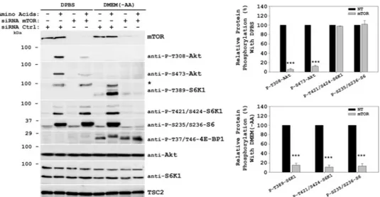
this differential pathway activation (Fig. 8). In DMEM( ⫺AA)-starved cells, knockdown of mTOR greatly inhibited S6K1 (Thr-389 and Thr-421/Ser-424), S6 ribosomal protein (Ser-235/Ser-236), and 4E-BP1 (Thr-37/Thr-46) phosphorylations, confirming the mTOR dependence of this pathway, as de-scribed previously (3). On the other hand, in DPBS-starved cells, knockdown of mTOR only inhibited Akt phosphoryla-tion (Fig. 8).
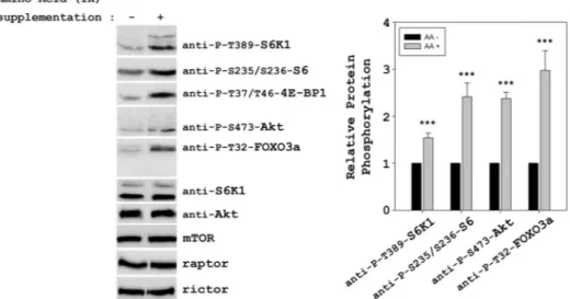
To test whether the starvation time used could affect the Akt/mTORC2 activation, we analyzed amino acid-induced responses at different times of starvation with DPBS. Fig. 9 shows how amino acids may activate Akt/mTORC2 when cells were starved for 2–3 h with DPBS, pointing out that the starvation time is a important factor to activate this pathway. The relevance of mTORC activation was also analyzed with a more physiological assay: supplementation of cells with amino acids. HeLa cells were incubated in DMEM complete medium for 48 h. After this time, amino acids were added to 1⫻ final concentration. Under these conditions, we could ob-serve a significant activation of mTORC1 (phosphorylations of 4E-BP1 at Thr-37/Thr-46, S6K1 at Thr-389, and S6 at
235/236) and mTORC2 (phosphorylations of Akt at Ser-473 and FOXO3a at Thr-32) complexes (Fig. 10).
Altogether, these data demonstrate that both mTOR sig-naling pathways (mTORC1 and mTORC2) may be used by cells to respond to amino acids. Activation of these pathways is highly dependent upon the precise starvation conditions to which cells have been subjected prior to amino acid
stimulation. DISCUSSION
The activity of mTOR complexes regulates essential cellu-lar processes, such as growth, proliferation, or survival. At the organismal level, deregulation of mTOR complex activity has been linked to aging and to diseases like diabetes or cancer (11, 23–26). Furthermore, mTOR inhibitors have been used to extend lifespan in mice and in clinical treatments of several human cancers, including mantle cell lymphoma, endometrial cancer, and renal cell carcinoma (26, 73, 74). Nutrients, and in particular amino acids, have arisen as important regulators of mTORC1 activation, thus affecting cell growth, protein syn-FIGURE 8. mTOR dependence in the signaling by amino acids. HeLa cells were transfected with either mTOR siRNA or nontargeting (NT) control siRNA 48 h before serum deprivation. Starvation and stimulation by amino acids were as in Fig. 7. Cellular lysates were analyzed by Western blot with the indi-cated antibodies. *, phospho-RSK/MSK band (see Fig. 2). Molecular mass markers are indiindi-cated on the left. Quantification and statistical analyses (right
pan-els) were performed as described under “Experimental Procedures.” These data are representative of three independent experiments.
FIGURE 9. Amino acid signaling depends on DPBS starvation time. HeLa cells were deprived of serum overnight. After serum withdrawal, they were de-prived with DPBS medium for the indicated times. Finally the cells were stimulated with 1⫻ amino acids for 30 min. Cellular lysates were analyzed by West-ern blot with the indicated antibodies. Molecular mass markers are indicated on the left. Quantification and statistical analyses (right panels) were per-formed as described under “Experimental Procedures.” These data are representative of at least three independent experiments.
thesis, and autophagy via phosphorylations of S6K1 and 4EBP1 (2, 8, 10 –12, 75).
Here, we demonstrate that amino acids can also induce Akt phosphorylation at both Thr-308 and Ser-473 and that both phosphorylations are mediated by PI3K␣, an isoform that is crucial for embryogenesis (76). Our finding that amino acids can signal via class I PI3K was unexpected, because previous results had described amino acids to signal via the class III PI3K hVps34 (35, 36, 38). This newly identified class I PI3K/ Akt signaling pathway for amino acids may explain some ef-fects reported for nutrients in different physiological/patho-logical situations. For example, activating mutations of PI3K are often found in cancer (77) and so is overexpression of Akt (78). Interestingly, it has recently been shown that tumors harboring a constitutively active PI3K/Akt pathway become resistant to dietary restriction (77), as opposed to other tu-mors, whose growth is severely curtailed by dietary restriction (79, 80). In view of our results, these previously unex-plained data may reflect the activation of PI3K/Akt signal-ing by amino acids in dietary restriction-sensitive tumors, but this still needs to be addressed experimentally. In an-other example, high blood levels of branched chain amino acids have been shown to correlate with insulin-resistant obesity (23). Given our data, it is conceivable that such an increase in amino acids could raise PI3K/Akt pathway ac-tivity in obese individuals, thus contributing to the insulin-resistant obese phenotype. Outside the realm of disease, dietary restriction has also been associated with an in-crease in lifespan (81– 83), and more experiments will be needed to elucidate what role amino acid-induced PI3K/ Akt signaling plays in that context.
We also checked whether amino acid-induced Akt Ser-473 phosphorylation was due to mTORC2, as reported for insulin signaling (42). Using mTOR and rictor siRNAs and in vitro kinase assays, we demonstrate that Ser-473 is indeed phos-phorylated by mTORC2 in response to amino acids. Interest-ingly, we also observed that Akt Thr-308 phosphorylation
depended on the presence of mTOR but not of rictor (Figs. 4 and 8). Kinase assays show that Akt Thr-308 is not a substrate of mTOR complexes activated by amino acids (Fig. 5). Al-though more work is needed to comprehend this exclusive mechanism for the amino acid signaling (insulin did not show this mechanism), these data seem to suggest that Akt Ser-473 phosphorylation stabilizes Akt Thr-308 phosphorylation in an mTOR-dependent manner but independent of kinase activi-ties of mTORC1 and mTORC2. We also observed that Akt phosphorylations in response to amino acids lead to the full Akt activation. Thus, we observed the phosphorylation of Akt substrate FOXO3a (Fig. 6). This is an important fact, because in this way amino acids may regulate cell survival and apopto-sis. In agreement with these data, dietary restriction has been described to inhibit the PI3K/Akt pathway and induce a prominent pro-apoptotic effect through FOXO (84). On the other hand, the mTORC2 complex has been described to be sensitive to rapamycin in prolonged treatments (6), and such rapamycin treatments extend lifespan in mice (26). The pro-longed lifespan seems to be due to a combination of anti-neo-plastic effects and effects on cellular stress resistance in re-sponse to nutrients. These effects were achieved in
rapamycin-fed mice by inhibiting mTORC1, but it cannot be ruled out that such prolonged treatments also affect
mTORC2 (6), and thus the transcriptional activity of FOXO. However, with shorter treatments, any rapamycin inhibitory effect could not be detected upon mTORC2 signal ( supple-mental Fig. S1). Therefore, this new FOXO regulation path-way induced by amino acids may provide new and valuable insights into the mechanisms of aging, apoptosis, and tumor growth regulation.
It was previously shown that amino acids may regulate the activity of S6K1, S6 ribosomal protein, and 4EBP1 pro-teins through activation of mTORC1 and not through acti-vation of Akt or mTORC2 (3, 22, 36 –38). Recently, it has been reported that some basal activity of p38 is required for mTORC1 activity (85). Another signaling pathway has
FIGURE 10. Amino acid supplementation leads to activation of mTORC1 and mTORC2. HeLa cells were supplemented with 1⫻ amino acids for 30 min after 48 h in DMEM complete medium. Cellular lysates were analyzed by Western blot with the indicated antibodies. Molecular mass markers are indicated on the left. Quantification and statistical analyses (right panels) were performed as described under “Experimental Procedures.” These data are representa-tive of at least three independent experiments.
also been described to S6K1 stimulated by amino acids and independent of mTOR (39). This last activation pathway leads to S6K1 and S6 phosphorylations through MAPKs instead of via mTORC1 or hVps34. Now, we demonstrate that amino acids can also signal to Akt and FOXO via class I PI3K and mTORC2 (Fig. 11). To understand and inte-grate all of these results, we analyzed the specific condi-tions leading to activation of each of these pathways. We show here some critical differences between these studies such as the amino acid deprivation medium (DPBS or DMEM(⫺AA)) and the starvation time. Thus, we demon-strate that it is possible to activate, with the same nutri-tional stimuli, different signaling pathways depending on the deprivation medium used and the starvation time (Figs. 7–9 and 11). When DPBS is used for 2–3 h, amino acids activate S6K1 and S6 ribosomal protein independently of mTORC1. In these conditions, amino acids also activate mTORC2, thus leading to Akt and FOXO phosphoryla-tions, affecting cell survival and proliferation. On the other hand, when DMEM (⫺AA) is used, amino acids regulate
S6K1, S6 ribosomal protein, and 4EBP1 in an mTORC1-de-pendent manner but indemTORC1-de-pendently of the PI3K/Akt path-way. Interestingly, autophagy was activated in a similar manner with both deprivation media, decreasing after stimulation with amino acids, thus suggesting that autoph-agy can be regulated by mechanisms dependent and inde-pendent of mTORC1 (39) (Figs. 7C and 11). We could also demonstrate that when DMEM (⫺AA) is used, amino acids signal through class III PI3K pathway (2, 28, 35–37), whereas when DPBS is used, amino acids signal through class I PI3K and MAPK. A systematic analysis of the com-ponents of amino acid deprivation media will probably al-low us to identify the factors responsible for these differen-tial effects.
Recently, DEPTOR has been described as an mTOR-bind-ing protein that negatively regulates mTORC1 and mTORC2 activities (5). In serum-stimulated cells, the loss of DEPTOR activates S6K1, Akt, and SGK1, promoting cell growth and survival. Conversely, DEPTOR overexpression in the presence of nutrients inhibits mTORC1 but unexpectedly activates FIGURE 11. A putative model of how amino acids may stimulate cell proliferation and survival pathways. When cells are supplemented with amino acids, kinase activities of mTORC1 (measured as the phosphorylation of their substrates: 4E-BP1 at Thr-37/Thr-46 and S6K1 at Thr-389) and mTORC2 (mea-sured as the phosphorylation of its substrate: Akt at Ser-473) are stimulated. Previous amino acid starvation with DMEM(⫺AA) or DPBS selects the further activation of mTORC1 or mTORC2 when amino acids are added. Under these conditions, mTORC1 activation is dependent on the class III PI3K (hVps34), and mTORC2 activation is dependent on the␣ isoform of the class I PI3K. Additionally, Akt Thr-308 phosphorylation is also dependent of the ␣ isoform of the class I PI3K and of the presence of mTOR but independent of mTORC1 and mTORC2 complexes (not shown). Both starvation conditions stimulate autoph-agy, which can be inhibited either by mTORC1-dependent (71, 72) and mTORC1-independent mechanisms (39). The dashed lines indicate that several steps may be involved. Activation or inhibition is indicated with arrows or blunt-ended arrows, respectively.
mTORC2 via PI3K/Akt signaling (5). This mTORC2 activa-tion could be due to a block in the inhibitory feedback signal transmitted from mTORC1 to PI3K (5). An attractive hypoth-esis is that a similar mechanism could account for mTORC2 activation by amino acids. Interestingly, these authors show a relationship between mTORC2 activation and cancer. They find that deregulated overexpression of DEPTOR in multiple myeloma cells is a mechanism for activating PI3K/Akt signal-ing and promotsignal-ing cell survival (5).
Finally, this report shows the crucial importance of dietary restriction/starvation conditions for understanding the amino acid signaling. Several studies show the effects of amino acid intake in obesity (23, 27, 28) and of dietary restriction in hu-man cancers (79, 80). Although more physiological studies are needed to link these effects to mTOR complex regulation, it is noteworthy that a study in human muscle shows activation of both mTORC1 and mTORC2 by ingestion of a leucine-en-riched amino acid-carbohydrate mixture (86). It has been re-cently described that branched chain amino acid dietary sup-plementation increased the average life span in mice and cardiac and skeletal muscle improvement (87), validating the physiological relevance of amino acid supplementation. In this context, we now report that cell supplementation with amino acids can activate both mTOR complexes (Figs. 10 and 11). In summary, this manuscript shows for the first time that amino acids can activate mTORC1 and mTORC2 complexes, thus underscoring the crucial importance of these nutrients in cell metabolism and offering new mechanistic insights with potential therapeutic applications in cancer, obesity, and aging.
Acknowledgments—We thank F. R. Garcia-Gonzalo, F. Amair-Pinedo, and M. Cubillos-Rojas for critical reading of the manu-script; E. Adanero for technical assistance; and M. Camps for gener-ously providing PI3K inhibitors.
REFERENCES
1. Wullschleger, S., Loewith, R., and Hall, M. N. (2006) Cell. 124, 471– 484 2. Laplante, M., and Sabatini, D. M. (2009) J. Cell Sci. 122, 3589 –3594 3. Kim, D. H., Sarbassov, D. D., Ali, S. M., King, J. E., Latek, R. R.,
Erdju-ment-Bromage, H., Tempst, P., and Sabatini, D. M. (2002) Cell. 110, 163–175
4. Dann, S. G., Selvaraj, A., and Thomas, G. (2007) Trends Mol Med. 13, 252–259
5. Peterson, T. R., Laplante, M., Thoreen, C. C., Sancak, Y., Kang, S. A., Kuehl, W. M., Gray, N. S., and Sabatini, D. M. (2009) Cell. 137, 873– 886 6. Sarbassov, D. D., Ali, S. M., Sengupta, S., Sheen, J. H., Hsu, P. P., Bagley,
A. F., Markhard, A. L., and Sabatini, D. M. (2006) Mol Cell. 22, 159 –168 7. Pearce, L. R., Huang, X., Boudeau, J., Pawlowski, R., Wullschleger, S.,
Deak, M., Ibrahim, A. F., Gourlay, R., Magnuson, M. A., and Alessi, D. R. (2007) Biochem. J. 405, 513–522
8. Kanazawa, T., Taneike, I., Akaishi, R., Yoshizawa, F., Furuya, N., Fu-jimura, S., and Kadowaki, M. (2004) J. Biol. Chem. 279, 8452– 8459 9. Meijer, A. J., and Codogno, P. (2004) Int. J. Biochem. Cell Biol. 36,
2445–2462
10. Sarbassov, D. D., Ali, S. M., and Sabatini, D. M. (2005) Curr. Opin. Cell
Biol. 17,596 – 603
11. Drummond, M. J., Dreyer, H. C., Fry, C. S., Glynn, E. L., and Rasmussen, B. B. (2009) J. Appl. Physiol. 106, 1374 –1384
12. Yamaoka, I., Doi, M., Kawano, Y., Nakayama, M., Watanabe, Y., Oba, K., Sugahara, K., and Yoshizawa, F. (2009) Biochem. Biophys. Res. Commun.
386,252–256
13. Franke, T. F., Kaplan, D. R., and Cantley, L. C. (1997) Cell 88, 435– 437 14. Brunet, A., Bonni, A., Zigmond, M. J., Lin, M. Z., Juo, P., Hu, L. S.,
Anderson, M. J., Arden, K. C., Blenis, J., and Greenberg, M. E. (1999)
Cell 96,857– 868
15. Sabatini, D. M., Barrow, R. K., Blackshaw, S., Burnett, P. E., Lai, M. M., Field, M. E., Bahr, B. A., Kirsch, J., Betz, H., and Snyder, S. H. (1999)
Sci-ence 284,1161–1164
16. Sarbassov, D. D., Ali, S. M., Kim, D. H., Guertin, D. A., Latek, R. R., Erd-jument-Bromage, H., Tempst, P., and Sabatini, D. M. (2004) Curr. Biol.
14,1296 –1302
17. García-Martínez, J. M., and Alessi, D. R. (2008) Biochem. J. 416, 375–385
18. Alessi, D. R., Pearce, L. R., and García-Martínez, J. M. (2009) Sci. Signal.
2,pe27
19. Avruch, J., Hara, K., Lin, Y., Liu, M., Long, X., Ortiz-Vega, S., and Yon-ezawa, K. (2006) Oncogene 25, 6361– 6372
20. Bhaskar, P. T., and Hay, N. (2007) Dev. Cell. 12, 487–502 21. Beugnet, A., Wang, X., and Proud, C. G. (2003) J. Biol. Chem. 278,
40717– 40722
22. Nicklin, P., Bergman, P., Zhang, B., Triantafellow, E., Wang, H., Nyfeler, B., Yang, H., Hild, M., Kung, C., Wilson, C., Myer, V. E., MacKeigan, J. P., Porter, J. A., Wang, Y. K., Cantley, L. C., Finan, P. M., and Murphy, L. O. (2009) Cell 136, 521–534
23. Newgard, C. B., An, J., Bain, J. R., Muehlbauer, M. J., Stevens, R. D., Lien, L. F., Haqq, A. M., Shah, S. H., Arlotto, M., Slentz, C. A., Rochon, J., Gal-lup, D., Ilkayeva, O., Wenner, B. R., Yancy, W. S., Jr., Eisenson, H., Mus-ante, G., Surwit, R. S., Millington, D. S., Butler, M. D., and Svetkey, L. P. (2009) Cell Metab. 9, 311–326
24. Toden, S., Bird, A. R., Topping, D. L., and Conlon, M. A. (2007)
Carcino-genesis 28,2355–2362
25. Mrkonjic, M., Chappell, E., Pethe, V. V., Manno, M., Daftary, D., Green-wood, C. M., Gallinger, S., Zanke, B. W., Knight, J. A., and Bapat, B. (2009) Br. J. Cancer 100, 1966 –1974
26. Harrison, D. E., Strong, R., Sharp, Z. D., Nelson, J. F., Astle, C. M., Flur-key, K., Nadon, N. L., Wilkinson, J. E., Frenkel, K., Carter, C. S., Pahor, M., Javors, M. A., Fernandez, E., and Miller, R. A. (2009) Nature 460, 392–395
27. Krebs, M. (2005) Eur. J. Clin. Invest. 35, 351–354
28. Um, S. H., D’Alessio, D., and Thomas, G. (2006) Cell Metab. 3, 393– 402 29. Calle, E. E., and Kaaks, R. (2004) Nat. Rev. Cancer 4, 579 –591
30. Blommaart, E. F., Luiken, J. J., Blommaart, P. J., van Woerkom, G. M., and Meijer, A. J. (1995) J. Biol. Chem. 270, 2320 –2326
31. Hara, K., Yonezawa, K., Weng, Q. P., Kozlowski, M. T., Belham, C., and Avruch, J. (1998) J. Biol. Chem. 273, 14484 –14494
32. Dennis, P. B., Pullen, N., Kozma, S. C., and Thomas, G. (1996) Mol. Cell.
Biol. 16,6242– 6251
33. Weng, Q. P., Kozlowski, M., Belham, C., Zhang, A., Comb, M. J., and Avruch, J. (1998) J. Biol. Chem. 273, 16621–16629
34. Nojima, H., Tokunaga, C., Eguchi, S., Oshiro, N., Hidayat, S., Yoshino, K., Hara, K., Tanaka, N., Avruch, J., and Yonezawa, K. (2003) J. Biol.
Chem. 278,15461–15464
35. Byfield, M. P., Murray, J. T., and Backer, J. M. (2005) J. Biol. Chem. 280, 33076 –33082
36. Nobukuni, T., Joaquin, M., Roccio, M., Dann, S. G., Kim, S. Y., Gulati, P., Byfield, M. P., Backer, J. M., Natt, F., Bos, J. L., Zwartkruis, F. J., and Thomas, G. (2005) Proc. Natl. Acad. Sci. U.S.A. 102, 14238 –14243 37. Findlay, G. M., Yan, L., Procter, J., Mieulet, V., and Lamb, R. F. (2007)
Biochem. J. 403,13–20
38. Gulati, P., Gaspers, L. D., Dann, S. G., Joaquin, M., Nobukuni, T., Natt, F., Kozma, S. C., Thomas, A. P., and Thomas, G. (2008) Cell Metab. 7, 456 – 465
39. Casas-Terradellas, E., Tato, I., Bartrons, R., Ventura, F., and Rosa, J. L. (2008) Biochim. Biophys. Acta 1783, 2241–2254
40. Jacinto, E., Facchinetti, V., Liu, D., Soto, N., Wei, S., Jung, S. Y., Huang, Q., Qin, J., and Su, B. (2006) Cell 127, 125–137
41. Engelman, J. A. (2009) Nat. Rev. Cancer 9, 550 –562
Science 307,1098 –1101
43. Jackson, S. P., Schoenwaelder, S. M., Goncalves, I., Nesbitt, W. S., Yap, C. L., Wright, C. E., Kenche, V., Anderson, K. E., Dopheide, S. M., Yuan, Y., Sturgeon, S. A., Prabaharan, H., Thompson, P. E., Smith, G. D., Shep-herd, P. R., Daniele, N., Kulkarni, S., Abbott, B., Saylik, D., Jones, C., Lu, L., Giuliano, S., Hughan, S. C., Angus, J. A., Robertson, A. D., and Salem, H. H. (2005) Nat. Med. 11, 507–514
44. Sadhu, C., Dick, K., Tino, W. T., and Staunton, D. E. (2003) Biochem.
Biophys. Res. Commun. 308,764 –769
45. Billottet, C., Grandage, V. L., Gale, R. E., Quattropani, A., Rommel, C., Vanhaesebroeck, B., and Khwaja, A. (2006) Oncogene 25, 6648 – 6659 46. Ru¨ckle, T., Biamonte, M., Grippi-Vallotton, T., Arkinstall, S., Cambet,
Y., Camps, M., Chabert, C., Church, D. J., Halazy, S., Jiang, X., Martinou, I., Nichols, A., Sauer, W., and Gotteland, J. P. (2004) J. Med. Chem. 47, 6921– 6934
47. Hsieh, A. C., Bo, R., Manola, J., Vazquez, F., Bare, O., Khvorova, A., Scaringe, S., and Sellers, W. R. (2004) Nucleic Acids Res. 32, 893–901 48. Wang, X., Beugnet, A., Murakami, M., Yamanaka, S., and Proud, C. G.
(2005) Mol. Cell. Biol. 25, 2558 –2572
49. Casas-Terradellas, E., Garcia-Gonzalo, F. R., Hadjebi, O., Bartrons, R., Ventura, F., and Rosa, J. L. (2006) Electrophoresis 27, 3935–3938 50. Haruta, T., Uno, T., Kawahara, J., Takano, A., Egawa, K., Sharma, P. M.,
Olefsky, J. M., and Kobayashi, M. (2000) Mol. Endocrinol. 14, 783–794 51. Takano, A., Usui, I., Haruta, T., Kawahara, J., Uno, T., Iwata, M., and
Kobayashi, M. (2001) Mol. Cell. Biol. 21, 5050 –5062
52. Carlson, C. J., White, M. F., and Rondinone, C. M. (2004) Biochem.
Bio-phys. Res. Commun. 316,533–539
53. Harrington, L. S., Findlay, G. M., Gray, A., Tolkacheva, T., Wigfield, S., Rebholz, H., Barnett, J., Leslie, N. R., Cheng, S., Shepherd, P. R., Gout, I., Downes, C. P., and Lamb, R. F. (2004) J. Cell Biol. 166, 213–223 54. Chaussade, C., Rewcastle, G. W., Kendall, J. D., Denny, W. A., Cho, K.,
Grønning, L. M., Chong, M. L., Anagnostou, S. H., Jackson, S. P., Daniele, N., and Shepherd, P. R. (2007) Biochem. J. 404, 449 – 458 55. Guertin, D. A., Stevens, D. M., Thoreen, C. C., Burds, A. A., Kalaany,
N. Y., Moffat, J., Brown, M., Fitzgerald, K. J., and Sabatini, D. M. (2006)
Dev. Cell 11,859 – 871
56. Huang, J., Dibble, C. C., Matsuzaki, M., and Manning, B. D. (2008) Mol.
Cell. Biol. 28,4104 – 4115
57. Toschi, A., Lee, E., Xu, L., Garcia, A., Gadir, N., and Foster, D. A. (2009)
Mol. Cell. Biol. 29,1411–1420
58. Dudek, H., Datta, S. R., Franke, T. F., Birnbaum, M. J., Yao, R., Cooper, G. M., Segal, R. A., Kaplan, D. R., and Greenberg, M. E. (1997) Science.
275,661– 665
59. Kauffmann-Zeh, A., Rodriguez-Viciana, P., Ulrich, E., Gilbert, C., Coffer, P., Downward, J., and Evan, G. (1997) Nature. 385, 544 –548
60. Songyang, Z., Baltimore, D., Cantley, L. C., Kaplan, D. R., and Franke, T. F. (1997) Proc. Natl. Acad. Sci. U.S.A. 94, 11345–11350
61. Birkenkamp, K. U., and Coffer, P. J. (2003) Biochem. Soc. Trans. 31, 292–297
62. Huang, H., and Tindall, D. J. (2007) J. Cell Sci. 120, 2479 –2487 63. Obsil, T., and Obsilova, V. (2008) Oncogene 27, 2263–2275
64. Essaghir, A., Dif, N., Marbehant, C. Y., Coffer, P. J., and Demoulin, J. B. (2009) J. Biol. Chem. 284, 10334 –10342
65. DeFeo-Jones, D., Barnett, S. F., Fu, S., Hancock, P. J., Haskell, K. M.,
Le-ander, K. R., McAvoy, E., Robinson, R. G., Duggan, M. E., Lindsley, C. W., Zhao, Z., Huber, H. E., and Jones, R. E. (2005) Mol. Cancer Ther.
4,271–279
66. Xiao, L., Gong, L. L., Yuan, D., Deng, M., Zeng, X. M., Chen, L. L., Zhang, L., Yan, Q., Liu, J. P., Hu, X. H., Sun, S. M., Liu, J., Ma, H. L., Zheng, C. B., Fu, H., Chen, P. C., Zhao, J. Q., Xie, S. S., Zou, L. J., Xiao, Y. M., Liu, W. B., Zhang, J., Liu, Y., and Li, D. W. (2010) Cell Death
Dif-fer. 17,1448 –1462
67. Campbell, L. E., Wang, X., and Proud, C. G. (1999) Biochem. J. 344, 433– 441
68. Wang, X., Fonseca, B. D., Tang, H., Liu, R., Elia, A., Clemens, M. J., Bommer, U. A., and Proud, C. G. (2008) J. Biol. Chem. 283, 30482–30492
69. Foster, K. G., Acosta-Jaquez, H. A., Romeo, Y., Ekim, B., Soliman, G. A., Carriere, A., Roux, P. P., Ballif, B. A., and Fingar, D. C. (2010) J. Biol.
Chem. 285,80 –94
70. Kabeya, Y., Mizushima, N., Ueno, T., Yamamoto, A., Kirisako, T., Noda, T., Kominami, E., Ohsumi, Y., and Yoshimori, T. (2000) EMBO J. 19, 5720 –5728
71. Mortimore, G. E., and Schworer, C. M. (1977) Nature. 270, 174 –176 72. Klionsky, D. J. (2007) Nat. Rev. Mol. Cell Biol. 8, 931–937
73. Guertin, D. A., and Sabatini, D. M. (2007) Cancer Cell 12, 9 –22 74. Bjedov, I., Toivonen, J. M., Kerr, F., Slack, C., Jacobson, J., Foley, A., and
Partridge, L. (2010) Cell Metab. 11, 35– 46
75. Yoshizawa, F. (2004) Biochem. Biophys. Res. Commun. 313, 417– 422 76. Bi, L., Okabe, I., Bernard, D. J., Wynshaw-Boris, A., and Nussbaum, R. L.
(1999) J. Biol. Chem. 274, 10963–10968
77. Kalaany, N. Y., and Sabatini, D. M. (2009) Nature 458, 725–731 78. Rychahou, P. G., Kang, J., Gulhati, P., Doan, H. Q., Chen, L. A., Xiao,
S. Y., Chung, D. H., and Evers, B. M. (2008) Proc. Natl. Acad. Sci. U.S.A.
105,20315–20320
79. Kritchevsky, D. (2001) J. Nutr. Sci. Vitaminol. 47, 13–19
80. Thompson, H. J., Zhu, Z., and Jiang, W. (2003) J. Mammary Gland. Biol.
Neoplasia 8,133–142
81. Panowski, S. H., Wolff, S., Aguilaniu, H., Durieux, J., and Dillin, A. (2007) Nature 447, 550 –555
82. Grandison, R. C., Piper, M. D., and Partridge, L. (2009) Nature 462, 1061–1064
83. Selman, C., Tullet, J. M., Wieser, D., Irvine, E., Lingard, S. J., Choudhury, A. I., Claret, M., Al-Qassab, H., Carmignac, D., Ramadani, F., Woods, A., Robinson, I. C., Schuster, E., Batterham, R. L., Kozma, S. C., Thomas, G., Carling, D., Okkenhaug, K., Thornton, J. M., Partridge, L., Gems, D., and Withers, D. J. (2009) Science. 326, 140 –144
84. Fu, Z., and Tindall, D. J. (2008) Oncogene 27, 2312–2319
85. Cully, M., Genevet, A., Warne, P., Treins, C., Liu, T., Bastien, J., Baum, B., Tapon, N., Leevers, S. J., and Downward, J. (2010) Mol. Cell. Biol. 30, 481– 495
86. Fujita, S., Dreyer, H. C., Drummond, M. J., Glynn, E. L., Cadenas, J. G., Yoshizawa, F., Volpi, E., and Rasmussen, B. B. (2007) J Physiol. 582, 813– 823
87. D’Antona, G., Ragni, M., Cardile, A., Tudesco, L., Dossena, M., Bruttini, F., Caliaro, F., Corsetti, G., Bottinelli, R., Carruba, M. O., Valerio, A., and Nisoli, E. (2010) Cell Metab. 12, 362–372