UNIVERSITY OF NAPLES FEDERICO II DOCTORATE
MOLECULAR MEDICINE AND MEDICAL BIOTECHNOLOGY XXX CICLO
Selection of aptamers targeting a hypoxia marker:
Carbonic Anhydrase-IX (CA-IX)
Tutor Candidate
Dott. Vittorio de Franciscis Silvia Nuzzo
COORDINATOR
Prof. Vittorio Enrico Avvedimento
“Selection of aptamers targeting a hypoxia marker:
4
TABLE OF CONTENTS
LIST OF PUBLICATIONS ... 5 ABBREVIATIONS: ... 6 ABSTRACT ... 7 1. BACKGROUND ... 8 1.1 Hallmarks of cancer ... 8 1.5 Aptamers ... 101.5.1 Composition and advantages of aptamers ... 10
1.5.2 Aptamer production: SELEX technology ... 12
1.5.3 Cell-based SELEX ... 14
1.6.1 Modifications of aptamers for clinical applications ... 17
1.6.2 Aptamers as therapeutics ... 19
1.6.3 Aptamers as delivery agents ... 21
1.6.4 Aptamers in cancer imaging, diagnosis and biomarker discovery. ... 24
3. MATERIALS AND METHODS ... 26
3.1 Cell culture ... 26
3.2 Hypoxia induction ... 26
3.3 CA-IX cDNA transfection and acidic condition ... 26
3.4 Immunoblotting ... 27
3.5 Immunofluorescence ... 27
3.7 Specific cell-SELEX ... 30
3.8 RT-PCR, mutagenic and non-mutagenic PCR for cell-SELEX method ... 30
3.9 Cell binding assay by RT-qPCR ... 31
3.10 In vitro human serum stability ... 31
3.11 ELONA (Enzyme-linked oligonucleotide assay) assay for human serum albumin ... 32
3.12 Flow cytometry analysis ... 32
4. RESULTS ... 33
5. DISCUSSION ... 33
6. CONCLUSIONS ... 33
5
LIST OF PUBLICATIONS
1. Esposito CL, Nuzzo S., CatuognoS., Romano S., de NigrisF., de FranciscisV. STAT3 gene silencing by Aptamer-siRNA chimera as selective therapeutic for Glioblastoma. Under revision to MolTherNucleic Acids.
2. Iaboni M, Fontanella R, Rienzo A, Capuozzo M, Nuzzo S, Santamaria G, Catuogno S, Condorelli G, de Franciscis V, Esposito CL. Targeting Insulin Receptor with a Novel Internalizing Aptamer. Mol Ther Nucleic Acids. 2016 Sep 20;5(9):e365. doi: 10.1038/mtna.2016.73.
3. Esposito CL, Nuzzo S, Kumar SA, Rienzo A, Lawrence CL, Pallini R, Shaw L, Alder JE, Ricci-Vitiani L, Catuogno S, de Franciscis V. A combined microRNA-based targeted therapeutic approach to eradicate glioblastoma stem-like cells. J Control Release. 2016 Sep 28;238:43-57. doi: 10.1016/j.jconrel.2016.07.032. Epub 2016 Jul 21.
6
ABBREVIATIONS:
2’-F-Py, 2′-Fluoro pyrimidine 2’NH2-Py, 2’-amino pyrimidine
ELISA, enzyme-linked immunosorbent assay FDA, Food and Drug Administration
99mTc, Technetium-99m
SELEX, Systematic Evolution of Ligands by Exponential enrichment mTOR, mammalian target of rapamycin
Akt, Protein Kinase B
MAPK, Mitogen-activated protein kinases
PI3K, Phosphatidylinositol-4,5-bisphosphate 3-kinase VEGF, vascular endothelial growth factor
GLUT1/3, Glucose transporter 1/3 MMPs, Matrix metalloproteinases IGF-2, Insulin-like growth factor 2
TGF-α, Transforming growth factor alpha TM, transmembrane region
IC, cytoplasmic tail
LDH5, Lactate dehydrogenase 5
7
ABSTRACT
Carbonic anhydrases (CAs) family are metalloenzymes, involved in pH control, catalysing the reversible conversion of carbon dioxide to protons and bicarbonate. Until now, 16 isoenzymes have been identified with different tissue distribution, subcellular localization and molecular biophysical properties.
Among them, carbonic anhydrase IX (CA-IX) is a hypoxic marker because the master regulator of hypoxia, named Hypoxia Inducible Factor 1 (HIF-1), promotes its expression.
Thanks to its catalytic activity, CA-IX reduces extracellular pH. The acid microenvironment increases cancer cell proliferation and invasion, giving to CA-IX an important role during the cancer progression.
Unlike other CAs, many studies have demonstrated that CA-IX is expressed only in few normal tissues, whereas it becomes overexpressed in many types of cancer. Therefore, the targeting of CA-IX, using specific tools, opens new important fields to improve the conventional therapies and the early diagnosis in cancer.
Based on these evidences, we would generate new strategies able to target CA-IX. To this end, nucleic acid-based aptamers are emerging as new tools perfectly suitable to different fields.
Aptamers are selected by an in vitro combinatorial chemistry approach, named Systematic Evolution of Ligands by Exponential enrichment (SELEX), and they are able to bind with high affinity and specificity virtually any given molecule. Compared to monoclonal antibodies, they have a small size that results in their rapid tumour penetration; they are not immunogenic and could be easily modified to increase their in vivo stability.
Thus, in this study we describe the selection of RNA-based aptamers directed against the extracellular domain of CA-IX.
Firstly, we performed two different cell-based SELEX protocols that allowed the enrichment for CA-IX specific aptamers.
Furthermore, we characterized and improved the two best sequences selected, named S-47s1 and S-51s1, that we are still studying to handle a final product usable for diagnostic and therapeutic purposes.
8
1. BACKGROUND
1.1 Hallmarks of cancerCancer is a class of diseases characterized by out-of-control cell growth [1].
It is the second leading cause of death in the world after cardiovascular diseases.
By the mid-1980s, since when researchers defined genomic alterations (nucleotide substitutions, chromosomal copy number alterations, and DNA rearrangements) [2] of two principal cancer-causing genes, named oncogenes and tumour suppressor genes , lot of new progresses have been made to better understand the molecular basis of the cancer.
In 2000, Hanahan and Weinberg proposed six hallmarks of cancer to clarify how the normal cells can progressively evolve to a neoplastic phenotype.
They postulated that cells become able to acquire a succession of hallmark capabilities (sustaining proliferative signalling, evading growth suppressors, activating invasion and metastasis, enabling replicative immortality, inducting angiogenesis, resisting to cell death), that improve their tumorigenesis and malignancy [3]
In 2010, always Hanahan and Weinberg added other two emerging hallmarks [4]:
the capability to modify the cellular metabolism to support neoplastic proliferation.
the capability of cancer cells to evade immunological response, in particular by T and B lymphocytes, macrophages, and natural killer cells.
In the last few years, as Caroline Wigerup shows (Fig.1), the eight hallmarks seems to be regulated by tumour hypoxia including cell proliferation, apoptosis, altered metabolism, immune responses, genomic instability, vascularization, invasion and metastasis [5].
Thus, the linkage emerged between the hypoxic condition and cancer could represent a new prognostic/predictive markers to improve the conventional therapies and to improve the early diagnosis of the cancer.
9 Fig.1: Hypoxia and HIFs regulate hallmarks of cancer and multiple cancer
phenotypes. The hypoxia regulates treatment resistance, vascularization, dedifferentiation, genomic instability, survival, immune response, invasion and metastasis. (From Wigerup C. et al. 2016)
10
1.5 Aptamers
1.5.1 Composition and advantages of aptamers
Aptamers are short synthetic single stranded nucleic acids able to bind tightly to a target molecule [55].
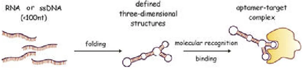
The “aptamer” means “to fit” (aptus) in latin, that reflects two their important characteristics (Fig. 8):
ability to fold into complex tertiary structures;
ability to bind with high affinity and specificity to their targets.
Fig. 8 Aptamers: Schematic representation of the aptamer functionality. (From Stoltenburg R. et al. 2007)
Aptamers act by directly binding to the protein target without interfering with its expression, unlike other small noncoding RNAs either natural or artificial, such as antisense, ribozymes, siRNAs and microRNAs (miRNA) that inhibit gene expression [56].
Aptamers are small nucleic acids of DNA or RNA, showing different characteristics.
The DNA aptamers are more stable due to the lack of 2′-OH groups. However, there are numerous advantages of RNA aptamers.
Firstly, RNA aptamers make more structure thanks to strong intra strand RNA–RNA interactions [57].
Moreover, their stability can be enhanced using different types of modifications, such as the substitution of the 2’-ribose [58] with 2′-fluoro, 2’-amino pyrimidine (2’-F-Py, 2’-NH2-Py) or 2′-O-alkyl.
Modified RNA aptamers may survive, to degradation by nucleases, in
vivo for several hours.
Thus, the RNA aptamers represent an attractive alternative as diagnostics and therapeutic tools compared to their peptide and monoclonal antibody counterparts [59-62] such as (Fig. 9):
11 1. easy chemical synthesis that results in a little or no batch-to-batch
variation;
2. small size around 15-40kDa that allows a good penetration in tumour tissue;
3. low or no immunogenicity;
4. easy editing to improve their stability and half-life.
Furthermore, the oligonucleotides composition of the aptamers gives them a great stability at high temperatures compared to monoclonal antibodies and peptides that have limited shelf life, undergoing to denaturation at high temperature.
12
1.5.2 Aptamer production: SELEX technology
The SELEX technology was developed in 1990. It is an evolutionary in
vitro combinatorial chemistry process used to identify aptamers as
specific ligands of a given target from large pools of different oligonucleotides [63].
The starting point for the generation of aptamers is the chemical synthesis of a single-stranded nucleic acid (RNA, DNA) library of large sequence complexity. A typical oligonucleotide library contains random sequences of 20–50 bases flanked by two constant regions that include primer sites for PCR amplification (Fig.10). Randomization is used to create possible sequences of enormous diversity (i.e. with n nucleotides in randomized region, 4n different molecules), which generates a vast array of different conformations with different binding properties. As schematized in figure 10, the SELEX method includes several steps: (i) incubation of the library with the target molecule under favourable binding conditions; (ii) partitioning of molecules that, under the employed conditions, adopt conformations that permit binding to a specific target from other sequences; (iii) dissociation of the nucleic acid-protein complexes and (iv) amplification of the nucleic acids pool. The pool obtained from the first cycle will be then the starting pool for the next rounds of selection, thus reiterating these steps the library enriched in sequences that bind to the target is generated. After the final round, the resulting pool is subjected to DNA sequencing. Sequences corresponding to the initially variable region of the library are screened for conserved regions and structural elements indicative of potential binding sites and, subsequently, tested for their ability to bind specifically to the target molecule.
SELEX technology usually requires different cycles of selection, around eight or more, in order to isolate aptamers with nanomolar affinity.
Particularly, if the SELEX technology is performed on a protein, the rounds could be lesser than the SELEX technology performed on cells or tissues.
Even if many aptamers are still selected by the traditional in vitro methodology, over the last few years considerable efforts have been focused on automating in vitro selection procedures [64], thereby accelerating aptamers discovery.
13 Fig. 10. Schematic representation of the SELEX technology. The RNA/DNA aptamers library contains a random sequence of 20–100 bases flanked by two constant regions. These constant regions include primer sites for PCR/RT-PCR amplification and transcription. The library is incubated with the target, not binding sequences are discarded whereas bound aptamers are recovered and amplified. (Adapted from Esposito C.L. et al 2011)
14
1.5.3 Cell-based SELEX
A great promise in developing specific molecular probes for disease biomarkers is recently represented by the intact cell-based SELEX strategy, that allows to select nucleic acid aptamers against living cells [65].
Aptamer selection approach that targets the cell surface open a new path which presents two major advantages: i) direct selection without prior purification of membrane-bound targets, ii) access to membrane proteins in their native conformation similar to the in vivo conditions. By using living cells as targets, aptamers able to discriminate cells from distant tumour types like small lung cancer cells versus large cell lung cancer [66], T-cell acute lymphocytic leukemia (ALL) versus B-cell lymphoma [67] and colon cancer cells versus other cancer cells [68] have been generated. Furthermore, by the SELEX technology against whole-living cells in culture [69] it was demonstrated that even by using complex targets as intact cells, it is possible to obtain aptamers against rare antigens if specifically expressed on the target cell.
In this regard, a panel of aptamers that bind a type of human malignant glioblastoma cells, discriminating them from non-tumorigenic glioblastoma cells, was isolated by a differential cell-SELEX approach [70]. In addition, the great advances in cell-SELEX offer also the opportunity to develop innovative approaches to identify and isolate cancer stem cells that are emerging as important target to develop more effective cancer therapy [71].
During the cell-SELEX strategy, to avoid the selection of ligands that recognize multiple surface proteins along with the target of interest, the counter- or negative-selection is critical.
A negative selection step using negative cells was included to enhance the specificity of the aptamers and prevent the enrichment of aptamers for abundant non-specific proteins.
Thus, for each cycle of the cell-SELEX strategy, the library is first incubated with non-target cells as counter-selection step (negative selection). Unbound aptamers are, then, recovered and incubated with the target cells. Bound aptamers are recovered and amplified (see Fig 11). Moreover, to improve the affinity and specificity of the aptamers the selective pressure during the cell-SELEX protocol is increased changing several conditions:
15 1. The increase of the washes number after the incubation is important to eliminate weakly bound or unbound aptamers.
2. the reduction of the cells number is important to reduce the quantity of the target molecule in order to recover only the aptamers with the best affinity and discard the aptamers with the limited binding capability.
3. the increase of the counter-selection numbers instead is important to discard the most of aptamers recognizing non-specific target molecules.
4. the addition of polyanionic nucleic acid, as yeast tRNA, salmon spermidine or polyinosine, avoids binding depending only on the opposite charge between basic proteins and the negatively charged nucleic acids. The cell-SELEX approach has been further developed to discriminate even different properties in the same cancer cell type (such as malignancy, therapeutic response, metastatic potential).
Moreover, to cell-SELEX, even a tumour implanted in mice (in vivo-SELEX) have been used to select aptamers [72].
Recently, more sophisticated approaches combine fluorescence activated cell sorting (FACS) technology with in vitro selection (FACS–SELEX) has been performed [73], thus enabling a live-cell/dead-cell separation within a cultured cell mixture.
16 Fig. 11. Cell-based SELEX. a) A pool of RNAs is incubated with non-target cells (counter-selection). Unbound sequences in the supernatant were recovered, and incubated with cells overexpressing the target for the selection step (positive selection). Unbound sequences were discarded by washings and bound sequences were recovered by total RNA extraction. b) Cell-based SELEX protocol to identify multiple ligands specifically. recognizing a cell phenotype, without prior knowledge of the target protein. (Catuogno S. et al. 2017)
17
1.6 Aptamers in therapy and diagnosis
1.6.1 Modifications of aptamers for clinical applications
Aptamers usable for clinical applications are usually modified in order to optimize their pharmacokinetic (PK) and pharmacodynamic (PD) profiles (see Fig. 12).
Since aptamers, especially RNA-based aptamers, are rapidly degraded by nucleases in whole organisms, major efforts have been addressed to improve their stability by a variety of approaches [74].
The most typical modifications to enhance the stability of aptamers are the substitution of the 2’-ribose [17]. RNA aptamers with 2′-fluoro, 2’-amino pyrimidine (2’-F-Py, 2’-NH2-Py) or 2′-O-alkyl nucleotides modifications may survive for several hours in vivo against degradation by nucleases.
Another example of modified nucleic acids is represented by the Spiegelmers. They do not contain additional groups added to the sugar moieties, but are enantiomers of natural nucleic acids [75]. In particular, the natural D-nucleic acids are substituted with enantiomeric L-nucleic acids. This property prevents recognition by nucleases, increasing the stability.
A hurdle in administering of aptamers to patients for many therapeutic applications is a short circulating half-life due to their small size. While a low molecular weight can be an advantage because it allows economical chemical synthesis and better target accessibility, it promotes rapid clearance by the renal system. By simply increasing the molecular weight of the aptamers, the circulating half-life can be significantly extended. The most common method to increase the aptamers size is to add a polyethylene glycol (PEG) moiety or cholesterol tail [76].
18 Fig. 12. Aptamer modifications. Scheme of the most typical modifications used to improve aptamer nuclease resistance (red) or its pharmacokinetic profile (green).
19
1.6.2 Aptamers as therapeutics
In the last years, the development of aptamers as therapeutics has primarily involved aptamers that bind and inhibit the activity of their protein targets.
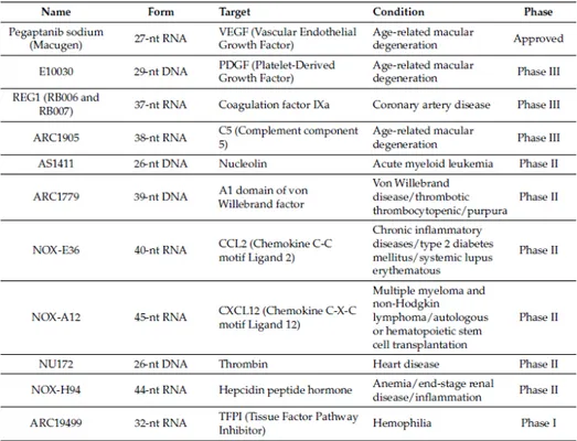
The list of aptamers against important therapeutic targets is growing rapidly and a handful of aptamers is now in clinical trials as therapeutic agents (see Table 1).
To date, the most successful therapeutic application is represented by an RNA aptamer, named Macugen, binding and antagonizing the action of Vascular Endothelial Growth Factor (VEGF), a growth factor that promotes the blood vessel formation (vascularization) [77].
Macugen (or Pegaptanib, marketed by Pfizer) has been demonstrated in phase III clinical trials to be effective for diabetic retinopathy treatment. The aptamer has been fully approved by the Food and Drug Administration (FDA) in December 2004 for the treatment of age-related macular degeneration (AMD). It is characterized by the formation of a neovascular membrane leaking blood and fluid under the retina with consequent destruction of the macula and loss of vision [78]. The aptamer binds and antagonizes the action of VEGF-165, the VEGF isoform preferentially involved in pathological ocular neovascularization. With the intent to improve the pharmacodynamic and pharmacokinetic properties of this 28-mer aptamer, it was chemically modified with 2′-F-Py and 2′-OMe-Pu, capping, and linkage to a 40kDa branched PEG molecule, which increases the intravitreal residence time of the molecule. Different studies have been carried out to assess the clinical cost-effectiveness of Macugen comparing to Ranibizumab (Lucentis, Genentech), a monoclonal antibody targeting all isoforms of human VEGF-A, approved in 2006 by the FDA for the treatment of exudative AMD.
Both drugs show comparable therapeutic efficacy and mild adverse events, while the economic evaluation varies considerably depending on the methodology for cost-effectiveness used in different studies.
Many other aptamers, not yet approved by the FDA, are currently in clinical trials.
Among them, it is very interesting for cancer therapy the AS1411 DNA aptamer (AGRO100) directed against nucleolin [79], a protein often overexpressed on the surface of cancer cells. This DNA aptamer is part of the guanine-rich oligonucleotide class of aptamers that form G-quartets, a structural element that exhibits a proliferative activity.
20 Once bound to nucleolin, the AS1411 aptamer is taken into the cancer cell, where it causes cellular death by apoptosis through inhibition nuclear factor-κB (NF-κB) [80] and Bcl-2 [81] pathways.
It showed its effectiveness as an anticancer therapy for different solid human malignancies as well as for acute myeloid leukemia (AML) and it is currently in phase IIb clinical trial to evaluate its effectiveness in combination with high-dose of cytarabine in patients with relapsed and refractory AML.
Table 1. Aptamers in on-going or completed clinical trials. (From Zhenjian Z. et al. 2017)
21
1.6.3 Aptamers as delivery agents
The aptamers can be also internalized together with their target receptor. For this reason, they can be used to deliver any kind of secondary reagents to a given cancer cell or tissue (Fig. 13).
Fig. 13. Aptamers as delivery agents. Aptamers that bind to cell surface receptors can be used to deliver small interfering RNAs (siRNAs and miRNAs), toxins, radioisotopes, and chemotherapeutic agents to target cells. (From Catuogno S. 2013)
In this way, only targeted cells will be exposed to the secondary reagent, thus increasing the efficacy of a given therapy as well as attenuating the overall toxicity of the drug [82]. In this regard, currently an increasing number of aptamers targeting cancer cell surface epitopes have been successfully used for the specific delivery of active drug substances both
in vitro and in vivo, including nanoparticles, anti-cancer therapeutics,
small interfering RNAs (siRNAs), microRNAs, toxins [83], enzymes [84], radionuclides [85], viruses [86].
Several cell-internalizing aptamers against surface epitopes of cancer cells have been successfully used as targeting vehicles. These include aptamers against the protein tyrosine kinase 7 (PTK7), nucleolin, prostate specific membrane antigen (PSMA), mucin 1 (MUC1) and EGFRwhich have been selected through either protein- or cell-SELEX strategies [87].
22 To date, the 2’F-Py-RNA aptamers A9 and A10 have been characterized for targeted delivery.. These aptamers, binding to extracellular domain of PSMA, have been used to deliver secondary molecule such as nanoparticles, quantum dots, toxin or siRNA to prostate cancer cells [88]. As shown in figure 14, A9 and A10 have been linked to siRNAs by covalent or non-covalent conjugation. Another promising delivery molecule is the phototoxic aptamer against MUC1, a membrane specific marker expressed on a broad range of epithelial cancer, that carries a cytotoxic cargo such as the light-activated PDT drug, chlorin e6, that produces cytotoxic singlet oxygen species. After light activation, the complex kills selectively MUC1 expressing cells [89]. In addition, John Rossi’s group used a RNA aptamer against gp120 for targeted delivery of siRNA against Human Immunodeficiency Virus (HIV) infections [90]. Aptamer-siRNA provides new promising therapeutic options [91].
Furthermore, recent papers also explored the use of aptamers to deliver microRNAs as cancer therapeutics.
Recently, Esposito et al. demonstrated the in vivo effectiveness of an aptamer-miRNA conjugate for lung cancer targeting. The authors designed a dual-function molecules containing an RNA aptamer (GL21.T), directed against the RTK Axl, covalently linked to miRNA let-7g [92]. The same aptamer was also used for the delivery of miR-212 to restore TRAIL-mediated cytotoxicity in NSCLC cells [93]. On the other end, Catuogno et al. described for the first time an aptamer-based system for the delivery of therapeutic single strand antimiRs [94]. Furthermore, very recently miRNA and antimiR delivery has been integrated developing a novel combined therapeutic approach for glioblastoma stem-like cells (GSCs) therapeutic targeting [95].
Moreover, the non-covalently link of the drugs to the aptamers was studied in order to understand if there is an abolishment of the aptamer binding and if the drug is released from the aptamer.
This model was studied with doxorubicin (DOX) intercalated with the aptamers against PSMA for prostate cancer [96], MUC1 [97].
This doxorubicin approach has shown toxicity levels compared to the free doxorubicin, but only in the cancer cells, reducing general toxicity or cardiotoxicity thanks to the specific aptamer delivery.
23 Fig. 14. A9-A10 conjugated with siRNA . (a) Using biotin, two aptamers and biotinylated siRNAs are non-covalently assembled via streptavidin; (b) Added at the end of aptamer complementary sequence to the antisense strand of the siRNA sequence, the chimera is formed by annealing of the aptamer to the siRNA antisense strand; (c) the aptamer portion of the chimera is truncated, and the sense and antisense strands of the siRNA portion are swapped. A two-nucleotide 3′-overhang and a PEG tail are added to the chimera; (d) the 3′-terminus of the aptamer is conjugated to the sense strand of the siRNA, followed by a 10-mer loop sequence and then by the antisense strand of the siRNA. (From Cerchia L. et al. 2011)
24
1.6.4 Aptamers in cancer imaging, diagnosis and biomarker discovery.
In the early stage of tumorigenesis, the cancer cells number is very low and for this reason, the detection is an important challenge.
Thus, to find new methods high sensitive to detect cancer cells are very important.
Thanks to their characteristics, aptamers have also started to play increasingly important roles in diagnosis of human disease [98].
Indeed, aptamers can be functionalized using fluorophores, superparamagnetic iron oxide nanoparticles, Mn3O4- or gold
nanoparticles, radioisotopes and biotin. These characteristics make the aptamers suitable as ligands for protein detection in a great number of different methodologies.
Due to their small size (8-15kDa) in comparison to antibodies (150kDa), aptamers havea rapid tumour penetration and blood clearance.
Hicke et al. were the first that published the use of a radiolabelled aptamer, named TTA1, for in vivo tumour target imaging [99].
TTA1 is a RNA modified aptamer against tenascin-C, an extracellular matrix protein upregulated in a number of tumours such as breast, lung, colon, prostate, glioblastoma, and lymphoma [100]. The aptamer was conjugated to the Technetium-99m (99mTc) and it was intravenously injected in xerographs mice bearing glioblastoma (U251) and breast (MDA-MB-435) [99].The authors, using single photon emission-computed tomography (SPECT), obtained a good tissue penetration and an important ratio tumour-to-blood of TTA1 aptamer.
This study suggests a quickly blood clearance and long tumour retention. As shown in table 2, many other DNA or RNA aptamers have been developed for diagnosis of human diseases.
Moreover, aptamers are useful in many imaging techniques such as fluorescence and bioluminescence imaging, magnetic resonance imaging (MRI), positron emission tomography (PET), single photon emission tomography (SPECT), computed tomography (CT) and ultrasound (US) [101,102].
25 Table 2. Recently developed aptamers for the diagnosis of human diseases. (From Zhuo Z. et al. 2017)
Until now, the aptamers are used for in vitro purpose such as dot-blot [103], western blot applications [104, 105], in the sandwich assay called ELONA (Enzyme Linked Oligonucleotide Assay).
In addition to their role as imaging tools, aptamers can also aid in clinical
in vitro diagnosis of diseases and discovery of new biomarkers.
Recently, several groups developed aptamer-based biomarker discovery platforms.
For example, Gold co-workers described an aptamer chip for the biomarker discovery. The system is able to measure thousands of proteins together from serum or plasma samples [106].
Novel aptamer-based technologies will continue to evolve and they could provide enormous opportunities in different diagnostic area.
26
3. MATERIALS AND METHODS
3.1 Cell cultureCell lines were purchased from the ATCC (LG Standards, Milan, Italy).
COS7, MDA-MB-231 and U87MG cells were grown in Dulbecco’s
modified Eagle’s medium (DMEM) supplemented with 10% heat-inactivated fetal bovine serum (FBS) and 100 U/ml penicillin/streptomycin. All cell culture reagents were purchased from Sigma (St Louis, MO).
3.2 Hypoxia induction
Physical method: 2x10⁵ U87MG cells were seeded in p35 cell plate in serum-free medium, and put in the hypoxic incubation chamber maintained at 5% O2. The cells were recovered at different times (6, 24,
48h).
Chemical methods: 2x10⁵ U87MG or MDA-MB-231 cells were seeded in
p35 cell plate. U87MG or MDA-MB-231 cells were incubated with
100-150-200μM of Cobalt (II) Chloride hexahydrate (Sigma, St Louis, MO) dissolved in the serum-free medium and put in a conventional incubator (37°C; 5% C02). At different times (1, 6, 24h) the cells were recovered.
3.3 CA-IX cDNA transfection and acidic condition
Human CA-IX ORF clone (NM_001216) (Origene, Rockville, USA) was transfected with Lipofectamine 2000 (Invitrogen, Waltham, MA, USA),
according to the manufacturer’s protocol.
For the acid condition, the MES buffer (morpholinoethanesulfonic acid) (Sigma, St Louis, MO) was added to the serum-free DMEM from 45 to 90mM for different incubation times (30, 90, 180 minutes). The pH medium was measured by a pHmeter (Mettler Toledo, Columbus, Ohio,USA).
27
3.4 Immunoblotting
Cells were washed twice in ice-cold phosphate-buffered saline (PBS) (Sigma St Louis, MO), and lysed in the JS buffer containing:
• 50 mM Tris-HCl pH 7.5 • 150 mM NaCl • 1% Nonidet P-40 • 2 mg/ml aprotinin • 1 mg/ml pepstatin • 2 mg/ml leupeptin • 10 mM Na3VO4
Protein concentration was determined by Bradford assay (Biorad, Hercules, CA, USA).
The cell lysates were denatured in Laemmli buffer (2% SDS , 5% β-mercaptoethanol, 0,001% Bromophenol Blue, 10 % glycerol) for 5 minutes at 100°C, and then subjected to SDS-PAGE.
12% Acrylamide/bis-acrylamide gels were electroblotted into polyvinylidene difluoride (PVDF) membranes (Millipore Co., Bedford, MA, USA)
Filters were probed with primary antibodies as indicated.
The primary antibodies used are: anti-CA-IX (R&D Systems,
Minneapolis, MN. USA); anti-HIF-1α (BD Biosciences, Qume Drive San
Jose, USA); anti-α-tubulin (Santa Cruz Biotechnology, CA, USA); anti-vinculin (Cell Signaling Technology, Danvers, MA); anti-β-actin (Santa Cruz Biotechnology, CA, USA). Donkey anti-goat, goat anti-mouse and goat anti-rabbit (Santa Cruz Biotechnology, CA, USA) were used as secondary antibodies.
3.5 Immunofluorescence
To assess CA-IX expression on the cell surface, MDA-MB-231 cells in
normoxic condition or stimulated with CoCl2 (150μM) for 24h to mimic
hypoxic condition, were seeded on poly-l-Lysine coated glass coverslips
and incubated with anti-CA-IX antibody for 30 min at 37 °C prior to
fixation. Cells were, then, fixed with paraformaldehyde 4% in PBS for 10 minutes and incubated at 37°C for 30 minutes with Alexa568 secondary antibody (Invitrogen, Waltham, MA, USA). Coverslips were then mounted on microscope slides with Prolong Gold Antifade Reagent with DAPI (Invitrogen, Waltham, MA, USA) and visualized by confocal
28 microscopy. Images were obtained using a Zeiss 510 LSM confocal microscope with a 40 × oil objective.
29
3.6 Physiological cell-SELEX
The SELEX cycle was performed essentially as described (Fitzwater and
Polisky 1996). Transcription was performed in the presence of 1mM 2’-F
pyrimidines and mutant T7 RNA polymerase (2.5u/ml T7 R&DNA polymerase, Epicentre Biotechnologies, Madison, Wisconsin) to improve yields.
2’F-Py RNAs were used to increase the resistance of the aptamers to
degradation by sieric nucleases. 2’F-Py RNAs were heated at 85°C for 5
min, snap-cooled on ice for 2 min, and allowed to warm up to 37°C. The Hypoxic cell-SELEX protocol is composed by twelve cycles. Counter-selection step against normoxic MDA-MB-231 cells.
To avoid the selection of aptamers recognizing normoxic targets on MDA-MB-231 surface, the pool was firstly incubated on normoxic MDA-MB-231 for 30 min (up to round 6) or for 15 min (for the following rounds) at 37°C.
In each cycle the SELEX conditions were changed such as the dimension of the cell culture dishes (150mm, 100mm, 50mm, 35mm). Unbound sequences were recovered for the selection step.
Selection step against hypoxic MDA-MB-231cells.
The recovered sequences were incubated on CoCl2-stimulated
MDA-MB-231 cells (to mimic the hypoxic condition).
After several washings, sequences were recovered by total RNA extraction.
During the selection process, the selective pressure was changed increasing washings number (from one for the first cycle up to five for the last cycles), decreasing the incubation time (from 30 to 15 min from round 7) and the dimension of the cell culture dishes (150mm, 100mm, 50mm, 35mm). It was also increased the number of counter-selection steps from one to two from round 8.
Moreover, the use of a non-specific competitor, named yeast tRNA (Sigma, St Louis, MO), was introduced at different concentrations: 100μg/ml for the round 10 and 200μg/ml for the round 11; in the last two
cycles (11 and 12) the pre-treatment with yeast tRNA before of the 2
30
3.7 Specific cell-SELEX
The starting pool is represented by the 9th cycle of Physiological
cell-SELEX.
The counter-selection step was performed against COS7 wild type (COS7-WT) cells to avoid the selection of aptamers that recognize proteins normally expressed on COS7 cells.
The selection step was performed against transient transfected COS7 with the CA-IX cDNA (called COS7-CAIX) in order to select specific aptamers for the target.
For each cycle, the pool of 2’F-Py RNAs was firstly incubated on the
COS7 wild type cells at 37°C, the 2’F-Py RNAs that not bind these cells
are recovered and incubated on CA-IX-COS7 for the selection step. After several washes, the sequences were recovered by total RNA extraction.
During the SELEX process, the selective pressure was progressively enhanced increasing washings number (from five for the round 10a up to six for the rounds 11a and 12a), increasing the number of counter-selection steps from two in 10a and 11a cycles to three for the last cycle (12a). The yeast tRNA 100μg/ml as a nonspecific competitor (Sigma, St Louis, MO) was used in 10a and 11a cycles; in the last two cycles (11aand 12a) it was also added the pre-treatment with yeast tRNA.
Each cycle was checked for CA-IX expression.
3.8 RT-PCR, mutagenic and non-mutagenic PCR for cell-SELEX method
The RNA extracted from each SELEX cycle was retro-transcribed using Tetro Reverse Transcriptase Enzyme (Bioline, London, UK) according to the manufacturer’s protocol. The retro-transcription reaction was as follow: 90°C for 3min, 42°C for 15min and 50°C for 30min.
The product was used for mutagenic PCR, characterized by higher concentration of MgCl2 (6mM) and dNTP 1mM, using 0.5U/μl of
FIREpol DNA Polymerase (Microtech, Milan, Italy) and 0,3μM primers: N40 (Forward):5’-TTCAGGTAATACGACTCACTATAGGGAAGAGA AGGACATATGAT-3’
N40 (Reverse): 5’-TCAAGTGGTCATGTACTAGTCAA -3’
The reaction was as follow: 95°C for 5min, 10 cycles of 95°C for 1 min, 65°C for 1min and 72°C for 1 min, and a final extension of 72°C for 1min.
31 The last SELEX cycle is amplified by non-mutagenic PCR using 0.1U/μl of FIREpol DNA Polymerase and 200μM dNTP, not adding MgCl2 to that contained in the Taq Buffer.
The reaction was as follow: 95°C for 5min, 8 cycles of 95°C for 30 sec, 65°C for 1min and 72°C for 1 min, and a final extension of 72°C for 5min.
Amplified DNA was purified using Amicon Ultra Centrifugal Filters (Millipore, Massachusetts, USA).
At the end of SELEX method, sequences of the pools were subjected to cloning with TOPO-TA cloning kit (Invitrogen, Waltham, MA, USA). The sequences have been analysed for multiple alignment (ClustalW by EMBL-EBI) and structural elements (MFOLD by unafold) indicative of potential binding sites.
3.9 Cell binding assay by RT-qPCR
The binding assay of individual aptamers was performed in 6-well plates in triplicate on COS7-CA-IX cells as positive cells and COS7-WT as
negative cells. 105 cells per well were seeded and, 24 hours after
transfection, were incubated for 30 minutes with serum-free medium in presence of yeast tRNA 200μg/μl and MES buffer (60mM). Then, 100nM
of aptamers were added for 15 min at 37°C. Following three washes with
PBS to remove unbound aptamers, the bound sequences were recovered by TRIzol (Life Technologies, Carlsbad, CA, USA) containing 0.5pmol/ml of reference control. The amount of bound RNAs was determined by RT-qPCR.
At each experiment, cells cultured were counted. The obtained data were normalized to the reference control and to cell number.
3.10 In vitro human serum stability
2’-F-RNA S-47s1 and S-51s1 aptamers were incubated at 4μmol/l concentration in 87% human serum (Type AB Human Serum provided by Euroclone, Milan, Italy) from 1 to 72 hours at 37°C.
At each time point 2μl (8 pmoles RNA) were recovered and treated with 2,5μl of proteinase K solution (600 mAU/ml) for 1 hour at 37°C in order to remove serum proteins that interfere with electrophoretic migration. Following the addition of 9 μl of denaturing gel loading buffer (1× TBE, 95% formamide, EDTA 10mM and bromophenol blue), samples were stored at −80°C. All serum-RNA samples were analyzed on 15%
32 polyacrylamide/urea 7M denaturing gel. The gel was stained with ethidium bromide and visualized by UV exposure.
3.11 ELONA (Enzyme-linked oligonucleotide assay) assay for human serum albumin
Human serum albumin: C96 maxisorp nunc-immunoplate (Thermo Fisher Scientific, Waltham, MA, USA) were left untreated or coated with 25nM of human serum albumin (HAS) (Sigma, St Louis, MO) in Coating/Washing buffer (20 mM Tris HCl pH 7.4; 150 mM NaCl; 1 mM MnCl2; 0.5 mM MgCl2; 2 mM CaCl2) overnight at 4°C.
Wells were washed once with Coating/Washing buffer and blocked for 2 hours at room temperature (RT), with Coating/Washing buffer containing 5% non-fat dried milk. Following two washes with Coating/Washing buffer, the biotinylated 2’-F-RNA S-47s1 and S51s1 aptamers were heated at 95 °C for 5’, snap-cooled on ice for 3min and allowed to warm up to 37°C for 10’ and, then, incubated for 2 hours at room temperature at different concentrations (10-100-1000nM) in Coating/Washing buffer containing 1,6% non-fat milk. After three washes with Coating/Washing buffer, samples were incubated for 1 hour at room temperature with horseradish peroxidase (HRP)-conjugated Streptavidin (Thermo Fisher Scientific, Waltham, MA, USA) diluted 1:1000 in PBS.
Following four washes with PBS, signals were reveled with 3,3',5,5'-tetrametilbenzidina (TMB) substrate solution (Thermo Fisher Scientific, Waltham, MA, USA) and stopped with stop solution (H2SO4 0.16M) for
TMB substrate (Thermo Fisher Scientific, Waltham, MA, USA). Absorbance at 450nm was measured with Multiskan FC Microplate Photometer (Thermo Fischer Scientific, Waltham, MA, USA).
As positive control, wells were incubated with an anti-HSA biotinylated polyclonal antibody (concentration: 1:1000, Abcam; Cambridge, MA). The assay was performed in duplicate.
33
3.12 Flow cytometry analysis
CA-IX expression in COS7 transfected with human CA-IX plasmid was
detected by anti-human CA-IX primary antibody (R&D Systems, Milan,
Italy). Alexa Fluor 488 donkey anti-goat IgG (Invitrogen, Milan, Italy)
was used as secondary antibody. Cells were analyzed with FACS BD Accuri C6 (BD Biosciences, Franklin Lakes, New Jersey, USA). IgG antibody (Millipore, Burlington, MA, USA) was used as negative control.
34
4. RESULTS
5. DISCUSSION
6. CONCLUSIONS
In conclusion, this study has identified new possible tools that are able to discriminate between CA-IX positive cells and CA-IX negative cells.
Considering that the hypoxia phenotype and CA-IX expressing cells are involved in chemotherapy resistance and poor prognosis, the aptamers identified in this study could have an important role in detecting only the hypoxic cells and acid microenvironment.
The study provide a new Selection strategy to successfully isolate aptamers with a very high specificity under physiological condition for in
vivo applicability.
Although more studies are needed to better understand the usefulness of the selected molecules, taken together, our findings represent an initial development of novel aptamer-based tools fo
35
7. REFERENCES
[1] Boveri T. Concerning the origin of malignant tumours by Theodor Boveri. Translated and annotated by Henry Harris. J Cell Sci. 2008 Jan;121 Suppl 1:1-84.
[2] Macconaill LE, Garraway LA. Clinical implications of the cancer genome. J Clin Oncol. 2010 Dec 10;28(35):5219-28.
[3] Hanahan D, Weinberg RA. The hallmarks of cancer. Cell. 2000 Jan 7;100(1):57-70.
[4] Hanahan D, Weinberg RA. Hallmarks of cancer: the next generation. Cell. 2011 Mar 4;144(5):646-74.
[5] Wigerup C, Påhlman S, Bexell D. Therapeutic targeting of hypoxia and hypoxia-inducible factors in cancer. Pharmacol Ther. 2016 Aug;164:152-69.
[55] Hermann T, Patel DJ. Adaptive recognition by nucleic acid aptamers. Science.2000 Feb 4;287(5454):820-5.
[56] Tucker WO, Shum KT, Tanner JA. G-quadruplex DNA aptamers and their ligands: structure, function and application. Curr Pharm Des. 2012;18(14):2014-26.
[57]. Dua P, Kim S, Lee DK. Nucleic acid aptamers targeting cell-surface proteins. Methods. 2011 Jun;54(2):215-25.
[58] Keefe AD, Cload ST. SELEX with modified nucleotides. Curr Opin Chem Biol. 2008 Aug;12(4):448-56.
[59] Foy JW, Rittenhouse K, Modi M, Patel M. Local tolerance and systemic safety of pegaptanib sodium in the dog and rabbit. J Ocul Pharmacol Ther. 2007 Oct;23(5):452-66.
[60] Higashimoto Y, Matsui T, Nishino Y, Taira J, Inoue H, Takeuchi M, Yamagishi S. Blockade by phosphorothioate aptamers of advanced glycation end products-induced damage in cultured pericytes and endothelial cells. Microvasc Res. 2013 Nov;90:64-70.
[61] Bates PJ, Laber DA, Miller DM, Thomas SD, Trent JO. Discovery and development of the G-rich oligonucleotide AS1411 as a novel treatment for cancer. Exp Mol Pathol. 2009 Jun;86(3):151-64.
[62] Lauridsen LH, Rothnagel JA, Veedu RN. Enzymatic recognition of 2'-modified ribonucleoside 5'-triphosphates: towards the evolution of versatile aptamers. Chembiochem. 2012 Jan 2;13(1):19-25.
[63] Ellington AD, Szostak JW. In vitro selection of RNA molecules that bind specific ligands. Nature. 1990 Aug 30;346(6287):818-22.
36 [64] Eulberg D, Buchner K, Maasch C, Klussmann S. Development of an automated in vitro selection protocol to obtain RNA-based aptamers: identification of a biostable substance P antagonist. Nucleic Acids Res. 2005 Mar 3;33(4):e45.
[65] Zhu G, Ye M, Donovan MJ, Song E, Zhao Z, Tan W. Nucleic acid aptamers: an emerging frontier in cancer therapy. Chem Commun (Camb). 2012 Nov 4;48(85):10472-80.
[66] Chen HW, Medley CD, Sefah K, Shangguan D, Tang Z, Meng L, Smith JE, Tan W. Molecular recognition of small-cell lung cancer cells using aptamers. ChemMedChem. 2008 Jun;3(6):991-1001.
[67] Shangguan D, Li Y, Tang Z, Cao ZC, Chen HW, Mallikaratchy P, Sefah K, Yang CJ, Tan W. Aptamers evolved from live cells as effective molecular probes for cancer study. Proc Natl Acad Sci U S A. 2006 Aug 8;103(32):11838-43.
[68] Sefah K, Meng L, Lopez-Colon D, Jimenez E, Liu C, Tan W. DNA aptamers as molecular probes for colorectal cancer study. PLoS One. 2010 Dec 10;5(12):e14269.
[69] Ye M, Hu J, Peng M, Liu J, Liu J, Liu H, Zhao X, Tan W. Generating aptamers by cell-SELEX for applications in molecular medicine. Int J Mol Sci. 2012;13(3):3341-53.
[70] Cerchia L, Esposito CL, Jacobs AH, Tavitian B, de Franciscis V. Differential SELEX in human glioma cell lines. PLoS One. 2009 Nov 24;4(11):e7971.
[71] Guo KT, Schäfer R, Paul A, Ziemer G, Wendel HP. Aptamer-based strategies for stem cell research. Mini Rev Med Chem. 2007 Jul;7(7):701-5.
[72] Mi, J. et al. In vivo selection of tumor targeting RNA motifs. Nat Chem Biol 2010, 6(1):22-24.
[73] Raddatz MS, Dolf A, Endl E, Knolle P, Famulok M, Mayer G. Enrichment of cell-targeting and population-specific aptamers by fluorescence-activated cell sorting. Angew Chem Int Ed Engl. 2008;47(28):5190-3.
[74] Klein R, Conway D, Parada LF, Barbacid M. The trkB tyrosine protein kinase gene codes for a second neurogenic receptor that lacks the catalytic kinase domain. Cell. 1990 May 18;61(4):647-56.
[75] Ni X, Castanares M, Mukherjee A, Lupold SE. Nucleic acid aptamers: clinical applications and promising new horizons. Curr Med Chem. 2011;18(27):4206-14.
[76] Amoresano A, Incoronato M, Monti G, Pucci P, de Franciscis V, Cerchia L. Direct interactions among Ret, GDNF and GFRalpha1
37 molecules reveal new insights into the assembly of a functional three-protein complex. Cell Signal. 2005 Jun;17(6):717-27.
[77] Ng EW, Shima DT, Calias P, Cunningham ET Jr, Guyer DR, Adamis AP. Pegaptanib, a targeted anti-VEGF aptamer for ocular vascular disease. Nat Rev Drug Discov. 2006 Feb;5(2):123-32.
[78] Esposito CL, Catuogno S, de Franciscis V, Cerchia L. New insight into clinical development of nucleic acid aptamers. Discov Med. 2011 Jun;11(61):487-96.
[79] Bates PJ, Laber DA, Miller DM, Thomas SD, Trent JO. Discovery and development of the G-rich oligonucleotide AS1411 as a novel treatment for cancer. Exp Mol Pathol. 2009 Jun;86(3):151-64.
[80] Girvan AC, Teng Y, Casson LK, Thomas SD, Jüliger S, Ball MW, Klein JB, Pierce WM Jr, Barve SS, Bates PJ. AGRO100 inhibits activation of nuclear factor-kappaB (NF-kappaB) by forming a complex with NF-kappaB essential modulator (NEMO) and nucleolin. Mol Cancer Ther. 2006 Jul;5(7):1790-9.
[81] Soundararajan S, Chen W, Spicer EK, Courtenay-Luck N, Fernandes DJ. The nucleolin targeting aptamer AS1411 destabilizes Bcl-2 messenger RNA in human breast cancer cells. Cancer Res. 2008 Apr 1;68(7):2358-65.
[82] Cerchia L, de Franciscis V. Targeting cancer cells with nucleic acid aptamers. Trends Biotechnol. 2010 Oct;28(10):517-25.
[83] Catuogno S, Esposito CL, de Franciscis V. Aptamer-Mediated Targeted Delivery of Therapeutics: An Update. Pharmaceuticals (Basel). 2016 Nov 3;9(4).
[84] Chen CH, Dellamaggiore KR, Ouellette CP, Sedano CD, Lizadjohry M, Chernis GA, Gonzales M, Baltasar FE, Fan AL, Myerowitz R, Neufeld EF. Aptamer-based endocytosis of a lysosomal enzyme. Proc Natl Acad Sci U S A. 2008 Oct14;105(41):15908-13.
[85] Hicke BJ, Stephens AW, Gould T, Chang YF, Lynott CK, Heil J, Borkowski S, Hilger CS, Cook G, Warren S, Schmidt PG. Tumor targeting by an aptamer. J Nucl Med. 2006 Apr;47(4):668-78.
[86] Tong GJ, Hsiao SC, Carrico ZM, Francis MB. Viral capsid DNA aptamer conjugates as multivalent cell-targeting vehicles. J Am Chem Soc. 2009 Aug12;131(31):11174-8.
[87] Cerchia L, Esposito CL, Camorani S, Catuogno S, Franciscis V. Coupling Aptamers to Short Interfering RNAs as Therapeutics. Pharmaceuticals (Basel). 2011Oct 27;4(11):1434-1449.
[88] Dhar S, Gu FX, Langer R, Farokhzad OC, Lippard SJ. Targeted delivery of cisplatin to prostate cancer cells by aptamer functionalized
38 Pt(IV) prodrug-PLGA-PEG nanoparticles. Proc Natl Acad Sci U S A. 2008 Nov 11;105(45):17356-61.
[89] Ferreira CS, Cheung MC, Missailidis S, Bisland S, Gariépy J. Phototoxic aptamers selectively enter and kill epithelial cancer cells. Nucleic Acids Res. 2009 Feb;37(3):866-76.
[90] Neff CP, Zhou J, Remling L, Kuruvilla J, Zhang J, Li H, Smith DD, Swiderski P, Rossi JJ, Akkina R. An aptamer-siRNA chimera suppresses HIV-1 viral loads and protects from helper CD4(+) T cell decline in humanized mice. Sci Transl Med. 2011 Jan 19;3(66):66ra6.
[91] Kruspe S, Giangrande PH. Aptamer-siRNA Chimeras: Discovery, Progress, and Future Prospects. Biomedicines. 2017 Aug 9;5(3). pii: E45. [92] Esposito CL, Cerchia L, Catuogno S, De Vita G, Dassie JP, Santamaria G, Swiderski P, Condorelli G, Giangrande PH, de Franciscis V. Multifunctional aptamer-miRNA conjugates for targeted cancer therapy. Mol Ther. 2014 Jun;22(6):1151-1163.
[93] Iaboni M, Russo V, Fontanella R, Roscigno G, Fiore D, Donnarumma E, Esposito CL, Quintavalle C, Giangrande PH, de Franciscis V, Condorelli G. Aptamer-miRNA-212 Conjugate Sensitizes NSCLC Cells to TRAIL. Mol Ther Nucleic Acids. 2016 Mar 8;5:e289. [94] Catuogno S, Rienzo A, Di Vito A, Esposito CL, de Franciscis V. Selective delivery of therapeutic single strand antimiRs by aptamer-based conjugates. J Control Release. 2015 Jul 28;210:147-59.
[95] Esposito CL, Nuzzo S, Kumar SA, Rienzo A, Lawrence CL, Pallini R, Shaw L, Alder JE, Ricci-Vitiani L, Catuogno S, de Franciscis V. A combined microRNA-based targeted therapeutic approach to eradicate glioblastoma stem-like cells. J Control Release. 2016 Sep 28;238:43-57. [96] Bagalkot V, Farokhzad OC, Langer R, Jon S. An aptamer-doxorubicin physical conjugate as a novel targeted drug-delivery platform. Angew Chem Int Ed Engl. 2006 Dec 11;45(48):8149-52. [97] Hu Y, Duan J, Zhan Q, Wang F, Lu X, Yang XD. Novel MUC1 aptamer selectively delivers cytotoxic agent to cancer cells in vitro. PLoS One. 2012;7(2):e31970.
[98] Soontornworajit B, Wang Y. Nucleic acid aptamers for clinical diagnosis: cell detection and molecular imaging. Anal Bioanal Chem. 2011 Feb;399(4):1591-9.
[99] Hicke BJ, Stephens AW, Gould T, Chang YF, Lynott CK, Heil J, Borkowski S,Hilger CS, Cook G, Warren S, Schmidt PG. Tumor targeting by an aptamer. J Nucl Med. 2006 Apr;47(4):668-78.
[100] Levy-Nissenbaum E, Radovic-Moreno AF, Wang AZ, Langer R, Farokhzad OC. Nanotechnology and aptamers: applications in drug delivery. Trends Biotechnol. 2008 Aug;26(8):442-9.
39 [101] Dougherty CA, Cai W, Hong H. Applications of aptamers in targeted imaging: state of the art. Curr Top Med Chem. 2015;15(12):1138-52.
[102] Sun H, Tan W, Zu Y. Aptamers: versatile molecular recognition probes for cancer detection. Analyst. 2016 Jan 21;141(2):403-15.
[103] Zhu J, Li T, Hu J, Wang E. A novel dot-blot DNAzyme-linked aptamer assay for protein detection. Anal Bioanal Chem. 2010 Aug;397(7):2923-7.
[104] Tombelli S, Mascini M. Aptamers as molecular tools for bioanalytical methods. Curr Opin Mol Ther. 2009 Apr;11(2):179-88. [105] Shin S, Kim IH, Kang W, Yang JK, Hah SS. An alternative to Western blot analysis using RNA aptamer-functionalized quantum dots. Bioorg Med Chem Lett. 2010 Jun 1;20(11):3322-5.
[106] Gold L, Ayers D, Bertino J, Bock C, Bock A, Brody EN, Carter J, Dalby AB, Eaton BE, Fitzwater T, Flather D, Forbes A, Foreman T, Fowler C, Gawande B, Goss M, Gunn M, Gupta S, Halladay D, Heil J, Heilig J, Hicke B, Husar G, Janjic N, Jarvis T, Jennings S, Katilius E, Keeney TR, Kim N, Koch TH, Kraemer S, Kroiss L, Le N, Levine D, Lindsey W, Lollo B, Mayfield W, Mehan M, Mehler R, Nelson SK, Nelson M, Nieuwlandt D, Nikrad M, Ochsner U, Ostroff RM, Otis M, Parker T, Pietrasiewicz S, Resnicow DI, Rohloff J, Sanders G, Sattin S, Schneider D, Singer B, Stanton M, Sterkel A, Stewart A, Stratford S, Vaught JD, Vrkljan M, Walker JJ, Watrobka M, Waugh S, Weiss A, Wilcox SK, Wolfson A, Wolk SK, Zhang C, Zichi D.Aptamer-based multiplexed proteomic technology for biomarker discovery. PLoS One. 2010 Dec 7;5(12):e15004.