biological activity of
alkyl-guanidine oligomers as potent
antibacterial agents
C. Zamperini
1,2, G. Maccari
1, D. Deodato
1, C. Pasero
1, I. D’Agostino
1, F. Orofino
1, F. De
Luca
3, E. Dreassi
1, J. D. Docquier
2,3& M. Botta
1,2,4In the last two decades, the repertoire of clinically effective antibacterials is shrinking due to the rapidly increasing of multi-drug-resistant pathogenic bacteria. New chemical classes with innovative mode of action are required to prevent a return to the pre-antibiotic era. We have recently reported the identification of a series of linear guanidine derivatives and their antibacterial properties. A batch of a promising candidate for optimization studies (compound 1) turned out to be a mixture containing two unknown species with a better biological activity than the pure compound. This serendipitous discovery led us to investigate the chemical nature of the unknown components of the mixture. Through MS analysis coupled with design and synthesis we found that the components were spontaneously generated oligomers of the original compound. Preliminary biological evaluations eventually confirmed the broad-spectrum antibacterial activity of this new family of molecules. Interestingly the symmetric dimeric derivative (2) exhibited the best profile and it was selected as lead compound for further studies.
Bacterial resistance to antibiotics currently represents one of the most relevant threat to global public health1 and is responsible for 700,000 deaths every year worldwide2. Moreover, the development of new antibiotics, espe-cially active against Gram-negative pathogens, significantly decreased in the last two decades3–8. Several factors contributing to the present paucity of new antibacterial were evoked, including poor economic returns9, lim-ited throughput of increasingly challenging drug discovery programmes10 and the complexity of the regulatory process to obtain drug approval6, 11–13. Consequently, few new antibacterials with novel mechanisms of action have been approved by FDA during the last 20 years, for example linezolid in 200014, daptomycin in 200315 and ceftaroline fosamil in 201016, all being active only against Gram-positive pathogens17–20. The rapid evolution of relevant Gram-negative pathogens (Klebsiella pneumoniae, Pseudomonas aeruginosa and Acinetobacter bauman-nii) exhibiting extensively drug- and pandrug-resistance phenotypes ultimately limits the efficacy of most avail-able antibiotics, including the life-saving carbapenems and colistin4, 21–27. In the frame of this unprecedented “antibiotic crisis”28, and to avoid a return to the pre-antibiotic era, the discovery and development of new active antibacterials, belonging to novel chemical classes, is urgently needed1, 29.
It is known that many compounds which show a significant antibacterial activity are characterized by the presence of one or more guanidine functions in their structure30–32. At physiological pH, the guanidine moiety carries a positive charge33 and the supposed mechanism of action involves an electrostatic interaction between the negatively charged bacterial cell surface and the positively charged compounds, potentially resulting in disruption of the bacterial membrane integrity. The increased membrane permeability thus leads to lysis and cell death30. Amphiphilic molecules with a hydrophobic surface and a net positive charge are called CADs or CAPs (Cationic
1Department of Biotechnology, Chemistry and Pharmacy, University of Siena, I-53100, Siena, Italy. 2Lead Discovery
Siena s.r.l., Via Vittorio Alfieri 31, I-53019, Castelnuovo, Berardenga, Italy. 3Department of Medical Biotechnology,
University of Siena, I-53100, Siena, Italy. 4Sbarro Institute for Cancer Research and Molecular Medicine, Temple
University, BioLife Science Building, Suite 333, 1900 North 12th Street, Philadelphia, Pennsylvania, 19122, United States of America. Correspondence and requests for materials should be addressed to M.B. (email: botta.maurizio@ gmail.com)
Received: 3 May 2017 Accepted: 17 July 2017 Published: xx xx xxxx
Amphiphilic Drugs or Peptides) and act through the above described interaction, inducing an alteration of phos-pholipids storage34–36.
In our previous work, we reported the synthesis and the biological evaluation of a series of linear alkyl-biguanylated compounds showing a subnanomolar affinity (Ki ranging from 0.08 to 3.00 nM) as competitive
inhibitors of Maize polyamine oxidase (PAO). The selective binding with this enzyme plays a crucial role in the inhibition of cell proliferation, in particular in tumor cell lines37.
Considering this important correlation between the guanidine moiety and the antimicrobial properties, we decided to evaluate the antibacterial profile of some of the above mentioned anti-PAO derivatives. The activ-ity of some selected molecules was tested on a panel of different bacteria, including representatives of both Gram-positive and Gram-negative organisms and clinical isolates, allowing the identification of compound 1 (Fig. 1) as promising candidate for further development. Interestingly, it exhibited a potent antibacterial activity on Gram-positive strains with MIC values ranging from 0.12 to 4 µg/mL and a remarkable activity on multi-drug resistant clinical isolates of E. cloacae and A. baumannii (MIC values of 2 and 4 µg/mL respectively)38.
In order to perform further analysis on compound 1, a new synthetic strategy (“Chemistry”) has been set up to overcome the withdrawal from the market of some commercial starting materials. The biological assays con-ducted on all the newly synthesized batches of compound 1 surprisingly showed a significantly lower antibacterial activity, when compared to that of the original batch of compound 138. In the light of these results, we turned our attention to this first batch to understand the reason of its higher activity. By means of analytical procedures (HPLC-MS) carried out on this sample, it emerged that it actually consisted of a multicomponent mixture includ-ing three different chemical identities. Initial attempts to separate the main components of the mixture with pre-viously optimized HPLC-MS protocols did not allow a complete separation of all species but successfully separate only compound 1 from the other analytes that were collected together and tested, revealing to have the better activity profile. Hence, we investigated about their chemical nature and through accurate mass measurements and MSn experiments, we hypothesized that they could be dimer and trimer of compound 1. We designed two
possible isomers for each oligomer, as reported in Fig. 139. At least, dimers (2 and 3) and trimers (4 and 5) have been synthesized and tested separately to identify the real responsible for the high antibacterial activity.
Results and Discussion
HPLC and MS analysis.
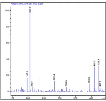
Fig. 2 shows the mass spectrum obtained by a direct injection of a sample of the original batch of compound 1. Signals detected were attributable to molecular structures heavier than compound 1. Further in-depth studies allowed the assignment of each MS signal to multiple charged ions of oligomeric derivatives, characterized as shown in Table 1.Attempts of identification and separation of the main components of the mixture were performed through LC–MS method, using a C18 column with a linear gradient elution, which gave us the best results. The chroma-tographic profile obtained is reported in Fig. 3.
The HPLC trace shows three main components, as the UV signals A, B and C, corresponding respectively to compound 1 and two other larger species, with m/z values approximatively twice and three times higher than 1 (see Supplementary Fig. S1). The eluate A was isolated and identified as compound 1, while the analytes B and C were collected together in a single fraction because of their close retention times, not giving us the information Figure 1. Structures of compound 1 and of the dimers (symmetric and asymmetric: 2 and 3 respectively) and trimers (symmetric and asymmetric: 4 and 5 respectively) studied in this work.
necessary to identify them. The eluted fractions were tested separately, revealing in the case of compound 1 (elu-ate A) the same moder(elu-ate antibacterial activity of its freshly synthesized batches. On the other side, the fraction containing the eluates B and C showed MIC values comparable to that of the original batch, confirming that its good antibacterial profile was due to the components contained in this latter fraction. Hence, we decided to investigate about their chemical nature. Preliminary fragmentation studies obtained by changing the fragmentor voltage were performed on a sample of the original batch (see Supplementary Figs S2 and S3) and showed, at higher fragmentation energy, the presence of fragments of compound 1 in all the three chromatographic peaks; this observation demonstrates that the unknown components could be derivatives of compound 1. At lower fragmentation energy, instead, the double, triple and quadruple charged cations prevailed. Chromatographic and Figure 2. Mass spectrum obtained by direct injection of a sample of the mixture. Conditions described in “Methods - General chemistry”.
Eluate Compound Mean tR ± SD a (min) Principal ions (m/z)b [M + H]+ [M + 2 H]2+ [M+3 H]3+ [M+4 H]4+ A Monomer (1) 12.44 ± 0.027 410.1 205.5 137.4 — B Dimer 13.48 ± 0.032 845.8 423.1 282.5 212.1 C Trimer 13.88 ± 0.025 1281.1 641.1 427.5 320.9
Table 1. Chromatographic and mass spectral data of compounds present in the analyzed mixture.
aChromatographic conditions reported in “Experimental section - Chromatographic separation”. bIons detected
with Agilent 1100 series MSD single-quadrupole instrument with fragmentation voltage 30 mV, reported in “Experimental section - Preliminary characterization studies”.
Figure 3. Chromatographic profile obtained after blank substraction of a sample of the first batch (10 mg/ mL) by LC–MS:A (tR = 12.40 min) monomer (1); B (tR = 13.34 min) = dimer; C (tR = 13.74 min) = trimer.
mass data, resumed in Table 1, led us to hypothesize that the unknown mixture components could be a dimer and a trimer, characterized by a carbonyl group as the linker between the monomers (compound 1).
Although the factors favoring the formation of these derivatives were unclear, we assumed that the generation of this mixture occurred during the storage of the sample in DMSO solution before the biological evaluation, especially considering that the characterization analysis of compound 1, performed immediately after its synthe-sis, confirmed its purity and authenticity.
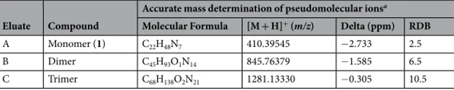
Moreover, in-depth studies were performed to elucidate the chemical formula and the structure of the main components. Through MS (ES+) analysis nowadays it is possible to obtain structurally significant fragment pat-terns40. LTQ-Orbitrap is a LC/MS technique usually used in analysis of unknown or peptidic mixtures because of its very high-resolution and high mass accuracy measurements on molecular ions41. One of its recent approaches is reported as the structural identification of drug metabolites42, 43. In this study accurate mass measurements and empirical formula calculations for the molecular ions were conducted using LTQ-Orbitrap XL mass spectrom-eter (Table 2). The ring and double bond (RDB) values and the difference between the theoretical (dimeric and trimeric) and experimental m/z for product ions (Delta) supported our hypothesis.
On the basis of the detected properties, we designed two possible structural isomers for each oligomer: a sym-metric and an asymsym-metric one, as reported in Fig. 1. We refer to asymmetric structure (compounds 3 and 5) when the connection between the monomers involves the central amine of one monomer and the guanidine group of the other, generating an amidinourea moiety. On the other side, the symmetric structure (compounds 2 and 4) is characterized by a urea function, involving both the central amines of the two monomers.
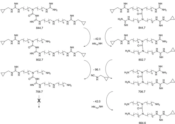
To establish which was the actual structure of dimer and trimer between the two hypothesized isomers, the mixture was analyzed by per infusion MSn technique, using an ion trap coupled with the Orbitrap mass
ana-lyzer, that allows fast, sensitive and reliable detection and identification of small molecules regardless of rela-tive ion abundance analytes44, 45. The MS2 and MS3 spectra obtained from the precursor ion 845.7 m/z showed
the formation of several product ions, in particular we detected 803.9 and 707.8 m/z, derived from the loss of methanediimine and N-(cyclopropylmethyl)-cyanamide fragments respectively, which are characteristic of both symmetric and asymmetric isomers (see Supplementary Figs S4 and S5). The detection in MS4 experiments of the
signal at 665.8 m/z, due to the loss of another methanediimine fragment (see Supplementary Fig. S6), confirmed the symmetric structure of the dimer (2) and/or the trimer (4), since this fragmentation is not possible for the asymmetric isomers (3 and 5), as reported in Fig. 4.
From the isolation of the trimer signal at 1281.1 m/z, MS2 spectrum showed 845.8 and 410.5 m/z as the main
signals, corresponding to dimer and monomer respectively (see Supplementary Fig. S7).
This per infusion MSn technology led us to observe the presence of a symmetric moiety that could belong to
the dimer or the trimer. Unfortunately, this moiety was not assignable to one of the two compounds since the retention times were too close to perform this kind of experiment during the chromatographic run. For this reason, to confirm the structure of the dimer and the trimer present in the original mixture we turned to the synthesis of all the possible isomers shown in Fig. 1.
Compounds 2–5 obtained by the synthesis39 were analyzed through HPLC-MS to compare their retention time with the ones of the initial mixture. The chromatograms showed a perfect correspondence between the two symmetric isomers (2 and 4) and the eluates B and C of the mixture. Accurate mass experiments of compounds 2 and 4 have been conducted, revealing that these two compounds were the mixture predominant components.
Moreover, a quantitative analysis of the fraction containing eluates B and C was performed with the same separation method above mentioned and it revealed that the B/C ratio, corresponding to the molar ratio of com-pound 2/comcom-pound 4, was 7/3. This molar ratio was extrapolated from appropriate standard calibration curves of compounds 2 and 4 obtained through HPLC-UV/MS signals.
Chemistry.
The synthetic procedure for the preparation of compound 1 reported in the literature37 is not accessible since the starting material 1,17-diamino-9-azaheptadecane is no longer commercially available. Thus, we reported in Fig. 5 a new strategy which begins with the guanylation of 1,8-diaminooctane, using 1,3-Bis(tert-butoxycarbonyl)-2-methyl-2-thiopseudourea, furnishing 7. The monoguanylated derivative was reacted with 6, previously obtained from 1,8-dibromooctane via nucleophilic substitution. The azido group of the resulting 8 was reduced and guanylated with N,N′-Di-Boc-N-methylcyclopropyl-pyrazole-1-carboxamidine, giving 10. Final compound 1 was obtained as trifluoroacetate salt after deprotection of all the Boc protecting groups with TFA in DCM.The guanylating agent, N,N′-Di-Boc-N-methylcyclopropyl-pyrazole-1-carboxamidine, have been obtained through Mitsunobu reaction between cyclopropanemethanol and di-Boc-pyrazole-1-carboximidamide, as reported in our previous work38, 39.
Eluate Compound
Accurate mass determination of pseudomolecular ionsa Molecular Formula [M + H]+ (m/z) Delta (ppm) RDB
A Monomer (1) C22H48N7 410.39545 −2.733 2.5
B Dimer C45H93O1N14 845.76379 −1.585 6.5
C Trimer C68H138O2N21 1281.13330 −0.305 10.5
Table 2. Accurate mass data. aAccurate mass measurements and chemical formulas calculation were found
through LTQ-Orbitrap XL and proposed chemical formulas, m/z values, RDB and Delta values were obtained from the software Xcalibur (Thermo Scientific, Bremen, Germany), as reported in “Experimental section - Accurate mass and fragmentation studies”.
As reported in Fig. 6, to synthesize the symmetric dimer 2, compound 10 was reacted with its carbamoyl derivative (11) affording the urea function. At the end the dimer 12 was deprotected under acidic condition, furnishing the trifluoroacetate salt of the final product (2).
The preparation of the asymmetric dimer 3 (Fig. 7) was more challenging, because required an orthogonal reaction between the central amine of a first monomer and the carbonyl group of a Boc of a second one46, 47. In order to promote the cross-reaction over oligomerization and self-cyclization, we designed and syn-thesized two different monomers (14 and 15) in such a way that they would react in an orthogonal fashion. Thus, the secondary amine of 14 was protected with a p-methoxybenzyl (PMB) group and, since the coupling step requires a di-Boc-guanidine moiety to be successful46, 47 the guanidine function of 15 was inactivated as mono-Boc-protected. The two building blocks were synthesized from compound 7 that was first protected with the PMB group and then reacted with 1-azido-8-bromooctane 6, furnishing 14. The other monomer (15) was synthesized from compound 14 through an oxidative deprotection via cerium ammonium nitrate. This reaction Figure 4. Proposed fragmentation pattern for the structures of dimeric moiety and their calculated exact mass. Spectral details are reported in “Supplementary Information”.
Figure 5. Synthesis of monomer (1). Reagents and conditions: (i) NaN3, DMF, 50 °C, 16 h; (ii)
1,3-Bis(tert-butoxycarbonyl)-2-methyl-2-thiopseudourea, DIPEA, CH3CN/CH3OH, 40 °C, 16 h; (iii) CsOH·H2O, molecular
sieves, 6, dry DMF, r.t., 24 h; (iv) H2, Pd/C, i-PrOH, r.t., 4 h; (v)
led to the simultaneous removal of the PMB and one Boc on the guanidine moiety. 15 was then reacted with compound 14 to give the dimeric compound 16. Reduction of the azido groups and their following guanylation afforded 17, that was eventually deprotected to give the asymmetric dimer 3 as trifluoroacetate salt.
In the synthesis of trimer 4, to selectively remove the PMB group from 17, without removing the Boc protect-ing groups, the oxidative deprotection via cerium ammonium nitrate was set up with different conditions
and reaction time, allowing the obtainment of 18. Its free central amine was reacted with carbamoyl derivative 11 to obtain, after acidic deprotection, the trifluoroacetate salt of the symmetrical trimer 4. (Fig. 8)
Central amine of monomer 10 was protected with FMOC, affording 22, and reacted with 21, which was obtained with the same synthetic procedure described for 8. The resulting 23 was first deprotected from FMOC and then reacted with 22, affording 24. 25 was easily prepared with subsequent reduction, guanylation and FMOC deprotection. Final removal of Boc protecting groups, under acidic condition, gave the asymmetric trimer 5 as trifluoroacetate salt. (Fig. 9)
Antibacterial activity.
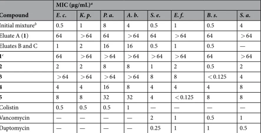
The antibacterial activity of eluate A (compound 1 isolated from the mixture), elu-ates B and C together and compounds 1–5 was investigated and their MIC values determined using a panel of eight organisms representative of both Gram-positive and Gram-negative bacteria. Compound 1, both the newly synthesized and the one isolated as eluate A, surprisingly showed a lower activity, when compared to that of the original batch published in our previous work38(reported as “Initial mixture” in Table 3), supporting the fact that the observed antibacterial activity was actually due to the presence of the other chemical species. In fact, the frac-tion containing both eluates B and C exhibited a significant antibacterial activity, demonstrating to be composed by the active molecule(s) of the original batch. All the synthesized oligomers also showed a notable antibacte-rial activity, especially on Gram-positive organisms but showed a different biological profile according to their isomerism: the symmetric isomers (2 and 4) were more active than their asymmetric counterparts (3 and 5). The symmetric dimer (2) exhibited the most potent activity on all the tested organisms, instead the asymmetric dimer Figure 6. Synthesis of symmetric dimer (2). Reagents and conditions: (i) Triphosgene, DIPEA, dry THF, 0 °C to r.t., 10 min; (ii) 10, DIPEA, NaI, dry DCM, 40 °C, 48 h; (iii) TFA 10%, dry DCM, r.t., 7 h.Figure 7. Synthesis of asymmetric dimer (3). Reagents and conditions: (i) 1) p-Anisaldehyde, CH3OH, r.t., 3 h;
2) NaBH4, 0 °C, 1 h; (ii) 6, NaI, DMF, r.t., 48 h; (iii) CAN, t-BuOH/CH3OH, 55 °C, 5 h; (iv) 14, TEA, dry THF,
ref., 2 h; (v) H2, Pd/C, i-PrOH, r.t., 16 h; (vi) N,N′-Di-Boc-N-methylcyclopropyl-pyrazole-1-carboxamidine,
(3) apparently lost most of its activity on Gram-negative ones. The symmetric trimer (4) was moderately active against all the tested species, while the asymmetric one (5) had a good activity on Gram-positive pathogens, in particular E. faecalis.
These antibacterial activity data overall support that the two symmetric oligomers were most likely the active components of the original mixture, thus confirming our hypothesis.
The biological profile of compound 2 which was the most active of the series was further investigated through Minimal Bactericidal Concentration (MBC) assay to distinguish whether it is bactericidal or bacteriostatic. Upon analysis, the MBC values of compound 2 on some bacterial strains were determined and subsequently compared to the corresponding MIC values: compound 2 was found to be bactericidal at the same concentrations to its MICs. As per CLSI standards48, a MBC/MIC ratio of 1 to 2 is considered indicative of bactericidal behaviour; Figure 8. Synthesis of symmetric trimer (4). Reagents and conditions: (i) CAN,CH3CN/H2O, r.t., 16 h; (ii) 11,
DIPEA, NaI, dry DCM, ref., 8 h; (iii) TFA 10%, dry DCM, r.t., 7 h.
Figure 9. Synthesis of the asymmetric trimer (5). Reagents and condition: (i)
N,N′-Di-Boc-N-methylcyclopropyl-pyrazole-1-carboxamidine, DIPEA, CH3CN/CH3OH, 50 °C, 16 h; (ii) CsOH·H2O, molecular
sieves, 6, dry DMF, r.t., 24 h; (iii) FmocCl, DIPEA, dry DCM, 0 °C to r.t., 2 h; (iv) 21, TEA, THF, ref., 10 h; (v) Piperidine 20%, DMF, r.t., 1 h; (vi) 22, TEA, dry THF, ref., 16 h; (vii) H2, Pd/C, i-PrOH, r.t., 1.5 h; (viii)
N,N′-Di-Boc-1H-pyrazole-1-carboxamidine, DIPEA, THF, r.t.,16 h; (ix) Piperidine 20%, DMF, r.t., 5 h; (x) TFA 10%, dry DCM, r.t., 10 h.
while a ratio ≥ 8 is indicative of bacteriostatic activity. Compound 2 showed a strong bactericidal activity with MBC and MIC values found to be identical for all the tested organisms.
Furthermore, the determination of killing curves showed that compound 2 was a relatively fast bactericidal agent, with a reduction of viable microbial load of > 3 log10 after only 1 h of exposure to the compound (final
con-centration, 10 × MIC) (see Supplementary Fig. S10). A further reduction of the viable count was progressively observed (>5 log10 after 4 h) and no viable cells could be detected after 24 h. Interestingly, compound 2 did not
show any detectable haemolytic activity (up to 50 µg of compound tested in the assay).
Conclusion
In summary, in-depth MS studies allowed the identification of the composition of a spontaneously gener-ated mixture derived from a batch of compound 1. We assume it origingener-ated after the re-suspension and the storage of the pure compound in DMSO prior to the evaluation of its antibacterial properties, as explained in Supplementary Information. We designed four possible isomers of the main components and synthesized them separately. Biological data have highlighted compound 2 as the actual responsible for the antibacterial activity with MIC values ranging from 1 to 8 µg/mL. It showed a strong bactericidal activity on both Gram-positive and Gram-negative clinically-relevant pathogens, while no haemolytic activity could be detected. Remarkably, this work originated from a serendipitous discovery and contributed to the identification of a new chemical scaffold showing a broad-spectrum antibacterial activity. Compound 2 was chosen as lead compound for further inves-tigations to generate a library of derivatives. These findings represent a significant achievement considering the current need of novel classes of antibacterials to fight resistant bacteria.
Methods
General Chemistry.
All commercially available chemicals and solvents were used as purchased. DCM was dried over calcium hydride and THF was dried over sodium and benzophenone prior to use. Anhydrous reac-tions were run under a positive pressure of dry nitrogen. Chromatographic separareac-tions were performed on col-umns packed with silica gel (230–400 mesh, for flash technique). 1H NMR and 13C NMR were recorded at 400and 100 MHz respectively on a Bruker AC200F spectrometer and are reported in parts per million (δ scale) and internally referenced to the CDCl3 or CD3OD signal, respectively at δ 7.24 ppm and 3.31 ppm. Chemical
shifts for carbon are reported in parts per million (δ scale) and referenced to the carbon resonances of the sol-vent (CDCl3 at δ 77.00 and CD3OD at δ 49.00 ppm). Data are shown as following: chemical shift, multiplicity
(s = singlet, d = doublet, t = triplet, m = multiplet and/or multiplet resonances, br = broad), coupling constant (J) in Hertz (Hz) and integration. Mass spectra (LC-MS) were acquired using an Agilent 1100 LC-MSD VL system (G1946C) by direct injection with a 0.4 mL/min flow rate using a binary solvent system of 95/5 CH3OH/H2O. UV
detection was monitored at 254 nm. Mass spectra were acquired in positive mode scanning over the mass range 105–1500 m/z, using a variable fragmentor voltage of 10–70 mV.
Determination of the purity.
The purity of final products (1–5) was 95% or higher and it was assessed by HPLC-MS, using an Equivalence 3 C18 column (ACE EQV-8977: 150 × 4.6 mm, 5 μm particle size) at a flow rate of 0.6 mL/min with a linear gradient elution from 100/0 to 50/50 v/v CH3CN (formic acid 0.1% v/v)/H2O (formicacid 0.1% v/v) for 23 min. UV detection was monitored at 210 nm. Mass spectra were acquired in positive mode scanning over the mass range 105–1500 m/z, using a fragmentor voltage of 70 mV.
Interference compound prediction.
The behaviour of all the final compounds (1–5) as PAINS was pre-dicted using the web-server FAFDrugs3. Through its tool Bank-Formater the compound library was prepared andCompound MIC (µg/mL)a E. c. K. p. P. a. A. b. S. e. E. f. B. s. S. a. Initial mixtureb 0.5 1 8 4 0.5 1 0.5 4 Eluate A (1) 64 > 64 64 > 64 64 > 64 64 > 64 Eluates B and C 1 2 16 16 0.5 1 0.5 — 1c 64 > 64 > 64 > 64 > 64 > 64 64 > 64 2 2 2 8 8 1 2 0.5 2 3 > 64 > 64 > 64 > 64 8 8 < 0.125 4 4 4 4 16 8 4 4 4 8 5 8 8 32 32 4 < 0.125 8 8 Colistin 0.5 0.5 0.5 1 — — — — Vancomycin — — — — 2 1 0.5 1 Daptomycin — — — — 0.25 1 1 0.5
Table 3. Minimum Inhibitory Concentration (MIC) of compounds 1–5, eluates A, B and C and control antibiotics (colistin, vancomycin and daptomycin) against representative strains of positive and Gram-negative bacteria. -.: not determined. aMICs are expressed as median values calculated from experiments
performed at least in triplicate. Tested bacterial strains: E.c.: E. coli CCUGT; K. p.: K. pneumoniae ATCC 13833;
P.a.: P. aeruginosa ATCC 27853; A.b.: A. baumannii ATCC 17978; S.e.: S. epidermidis ATCC 14990; E.f.: E. faecalis ATCC 29212; B.s.: B. subtilis ATCC 6633; S.a.: S. aureus ATCC 25923. bFirst batch of compound 1 as
umn (ACE EQV-8977: 150 × 4.6 mm; 5 µm) using linear gradient elution for 23 min with a mobile phase of 0.1% v/v formic acid in H2O and 0.1% v/v formic acid in CH3CN (from 0/100 to 50/50 v/v in 23 min) at the flow rate
of 0.6 mL/min. Analytes B and C were eluted after compound 1 (eluate A, tR = 12.40 min), with retention times of
13.34 and 13.74 min respectively. Eluates from 13.10 to 13.90 min were collected and tested. Identification of the two main components of the fraction was performed through in-depth MS analysis. With the same method, the retention time of each pure synthesized compound (2–5) was determined and it is reported in Table 4.
Preliminary characterization studies.
Chromatography-mass spectrometry (LC-MS) system consisted of an Agilent 1100 series liquid chromatograph system (Agilent Technologies, Palo Alto, CA) including a vac-uum solvent degassing unit, a binary high-pressure gradient pump, an UV detector, and an 1100 MSD model VL benchtop mass spectrometer with API-ES interface. The UV detector was set at 210 nm. The Agilent 1100 series MSD single-quadrupole instrument was equipped with the orthogonal spray API (Agilent Technologies, Palo Alto, CA). Nitrogen was used as nebulizer gas and drying gas (350 °C). The LC-API-MS determination was performed by operating the MSD in the positive ion mode. Mass spectra were acquired over the scan range 105–1500 m/z using a step size of 0.1 u. The nebulizer gas, the drying gas, the capillary voltage, and the vaporizer temperature were set at 40 psi, 9 L/min, 3000 V and 350 °C, respectively. For the fragmentation study the fragmen-tor voltage was set in the range 30–200 mV.Accurate mass and fragmentation studies.
The accurate masses were measured by the LTQ-Orbitrap XL (Thermo Scientific, Bremen, Germany) mass spectrometer interfaced with an electrospray ionization (ESI) source characterized by a spray voltage of 4.5 kV and nitrogen as sheat gas (10 a.u.). The resolution of accurate masses is 30000. MS/MS spectra were recorded with an isolation window of 2 mass units, collision energy of 15, 16 or 17 V and helium as collision gas.All data were processed using Xcalibur (Thermo Scientific, Bremen, Germany). The elementar composition tool was used to calculate the proposed chemical formulas, (ring and double bond) RDB values and the difference between the theoretical and experimental m/z for product ions.
Synthetic procedures and characterizations of compounds 1–5. For the synthetic procedures and characteriza-tions of compounds 6–25 see Supplementary Information.
N‐{8‐[(8‐carbamimidamidooctyl)amino]octyl}‐N′‐(cyclopropylmethyl)guanidine (1). Compound 10 (10.0 mg, 0.12 mmol) was dissolved in dry DCM (1.8 mL) and TFA (10%, 0.2 mL) was added. The reaction mixture was stirred at room temperature for 7.5 h. Then the solvent was evaporated and the crude product was dissolved and evaporated several times first with CH3OH to remove TFA residue and then with Et2O to precipitate the desired
compound. No further purification followed; the product was obtained as a colourless oil. 1H NMR (CD 3OD, 400 MHz): δ 0.26 (d, J = 4.8 Hz, 2 H); 0.58 (d, J = 7.2 Hz, 2 H); 1.05 (s, 1 H); 1.28 (s, 24 H); 2.96–3.00 (m, 2 H); 3.05 (d, J = 7.2 Hz, 2 H); 3.12–3.20 (m, 4 H). 13C NMR (CD 3OD, 100 MHz): δ 2.4; 9.5; 25.8; 26.0; 26.1; 28.3; 28.4; 28.6; 29.2; 29.4; 40.9; 41.1; 45.8; 46.9; 48.1; 157.0; 159.7. LC-MS m/z (ES+) = 410.1 [M + H]+; 205.5 [M + 2 H]2+ YIELD: quantitative. 1,3‐bis(8‐carbamimidamidooctyl)‐1,3‐bis({8‐[N′‐(cyclopropylmethyl)carbamimidamido]octyl})urea (2). The same procedure for the synthesis of compound 1 was followed. 1H NMR (CD
3OD, 400 MHz): δ 0.23–0.30 (m,
4 H); 0.53–0.61 (m, 4 H); 1.01–1.10 (m, 2 H); 1.31–1.45 (m, 32 H); 1.48–1.55 (m, 8 H); 1.55–1.63 (m, 8 H); 3.05 (d, J = 6.8 Hz, 4 H); 3.08–3.21 (m, 12 H); 3.28–3.32 (m, 4 H). 13C NMR (CD
3OD, 100 MHz): δ 2.4; 9.5; 26.2; 26.5;
27.5; 28.4; 28.4; 28.8; 28.9; 29.2; 41.0; 41.1; 45.8; 46.9; 47.1; 47.3; 47.5; 47.7; 47.9; 48.1; 155.6; 165.4. LC-MS m/z (ES+) = 845.8 [M + H]+; 423.1 [M + 2 H]2+; 282.5 [M + 3 H]3+; 212.1 [M + 4 H]4+ YIELD: quantitative.
1-(8-carbamimidamidooctyl)-1-[8-[[N-(cyclopropylmethyl)carbamimidoyl]amino]octyl]-3-[N-[8-[8-[[N-(cyclo-propylmethyl)carbamimidoyl]amino]octylamino]octyl]carbamimidoyl]urea (3). The same procedure for the synthesis of compound 1 was followed. 1H NMR (CD
3OD, 400 MHz): δ 0.23–0.27 (m, 4 H); 0.54–0.58 (m, 4 H); 1.00-1-10 (m, 2 H); 1.26–1.40 (m, 32 H); 1.44–1.62 (m, 16 H); 2.52 (t, J = 8.0 Hz, 4 H); 3.03 (d, J = 8.0 Hz, 4 H); 3.11–3.20 (m, 12 H). 13C NMR (CD 3OD, 100 MHz): δ 3.4; 11.2; 26.7; 27.1; 28.8; 29.3; 30.4; 30.6; 41.5; 42.0; 42.3; 44.7; 49.9; 155.4; 155.8; 157.8; 158.0. LC-MS m/z (ES+) = 845.8 [M + H]+; 423.1 [M + 2 H]2+; 282.5 [M + 3 H]3+; 212.1 [M + 4 H]4+ YIELD: quantitative.
3‐(8‐carbamimidamidooctyl)‐1‐{8‐[({[(8‐carbamimidamidooctyl)({8‐[N′‐(cyclopropylmethyl)carbamimid- amido]octyl})carbamoyl]amino}methanimidoyl)amino]octyl}‐1,3‐bis({8‐[N′‐(cyclopropylmethyl)carbamimid-amido]octyl})urea (4). The same procedure for the synthesis of compound 1 was followed. 1H NMR (CD
3OD, 400 MHz): δ 0.24–0.28 (m, 6 H); 0.56–0.59 (m, 6 H); 1.02–1.11 (m, 3 H); 1.22–1.46 (m, 48 H); 1.49–1.70 (m, 24 H); 3.00 (d, J = 7.2 Hz, 6 H); 3.12–3.27 (m, 24 H). 13C NMR (CD 3OD, 100 MHz): δ 3.4; 11.1; 26.6; 26.7; 28.8; 29.3; 30.4; 41.5; 41.9; 42.3; 44.7; 49.8; 50.5; 155.4; 155.8; 157.9; 158.0; 164.5. LC-MS m/z (ES+) = 641.1 [M + 2 H]2+; 427.5 [M + 3 H]3+; 320.9 [M + 4 H]4+ YIELD: quantitative. 1-(8-carbamimidamidooctyl)-1-[8-[[N-(cyclopropylmethyl)carbamimidoyl]amino]octyl]-3-[N-[8-[8-[[N-(cyclo-propylmethyl)carbamimidoyl]amino]octyl-[[N-[8-[8-[[N-(cyclopropylmethyl)carbamimidoyl]amino]octylamino] octyl]carbamimidoyl]carbamoyl]amino]octyl]carbamimidoyl]urea (5). The same procedure for the synthesis of compound 1 was followed. 1H NMR (CD
3OD, 400 MHz): δ 0.23–0.27 (m, 6 H); 0.54–0.59 (m, 6 H); 1.08–1.15
(m, 3 H); 1.26–1.37 (m, 48 H); 1.50–1.70 (m, 24 H); 2.95 (t, J = 8.0 Hz, 4 H) 3.03 (d, J = 4.0 Hz, 6 H); 3.13–3.33 (m, 20 H). 13C NMR (CD
3OD, 100 MHz): δ 3.5; 11.2; 26.7; 27.1; 28.8; 29.3; 30.4; 30.6; 41.6; 41.9; 42.3; 44.7; 50.0; 155.5;
157.8; 158.0. LC-MS m/z (ES+) = 641.1 [M + 2 H]2+; 427.5 [M + 3 H]3+; 320.9 [M + 4 H]4+ YIELD: quantitative.
Antibacterial susceptibility testing. Bacterial strains, including representatives of both Gram-positive and Gram-negative bacteria, were obtained from the ATCC or CCUG culture collections. Compounds were re-suspended in DMSO at a final concentration of 50 or 100 mg/mL and subsequently diluted in the culture medium. The minimum inhibitory concentration (MIC) and the minimum bactericidal concentration (MBC) of the compounds were determined using the micro-dilution broth method using Mueller-Hinton broth as recom-mended by the Clinical Laboratory Standards Institute (CLSI)48. Bacterial inoculum was 5 × 10⁴ CFU/well. MICs
were recorded after 18 hours of incubation at 35 °C after visual observation of solution transparency. MIC and MBC values are reported as median values and were obtained from experiments performed at least in triplicate.
The haemolytic activity of selected compounds was estimated using the method described by Bechlars et al49. 10 µL of compound (concentrations, 0.25 to 25 mg/mL) were spotted on the surface of a blood agar plate. Triton X-100 was used as a positive haemolysis control.
References
1. WHO. Antimicrobial resistance. Bull. World Health Organ. 61, 383–94 (2014).
2. O’neill, J. Tackling Drug-Resistant Infections Globally: Final Report and Recommendations the Review on Antimicrobial Resistance. (2016).
3. Rice, L. B. Federal Funding for the Study of Antimicrobial Resistance in Nosocomial Pathogens: No ESKAPE. J. Infect. Dis. 197, 1079–1081 (2008).
4. Spellberg, B. & Gilbert, D. N. The future of antibiotics and resistance: A tribute to a career of leadership by John Bartlett. Clin. Infect.
Dis. 59, S71–S75 (2014).
5. Spellberg, B. et al. The epidemic of antibiotic-resistant infections: a call to action for the medical community from the Infectious Diseases Society of America. Clin.Infect.Dis. 46, 155–164 (2008).
6. Silver, L. L. Challenges of antibacterial discovery. Clin. Microbiol. Rev. 24, 71–109 (2011).
7. Projan, S. J. Why is big Pharma getting out of antibacterial drug discovery? Curr. Opin. Microbiol. 6, 427–430 (2003).
8. Fair, R. J. & Tor, Y. Perspectives in Medicinal Chemistry Antibiotics and Bacterial Resistance in the 21st Century. Perspect. Medicin.
Chem. 25–64 (2014). doi:10.4137/PMC.S14459.
9. Shlaes, D.M. Antibiotics - The perfect Storm. 29–50 (Springer-Verlag, 2010).
10. Bush, K. Introduction to antimicrobial therapeutics reviews: Infectious diseases of current and emerging concern. Ann. N. Y. Acad.
Sci. 1323, v–vi (2014).
11. Bartlett, J. G., Gilbert, D. N. & Spellberg, B. Seven ways to preserve the Miracle of antibiotics. Clin. Infect. Dis. 56, 1445–1450 (2013). 12. Piddock, L. J. V. The crisis of no new antibiotics—what is the way forward? Lancet Infect. Dis. 12, 249–253 (2017). 13. Gould, I. M. & Bal, A. M. New antibiotic agents in the pipeline and how they can help overcome microbial resistance. Virulence 4,
185–91 (2013).
14. U.S. Food and Drug Administration, Center for Drug Evaluation and Research. Zyvox NDA 21-130, 21-131 & 21-132 approval letter, April 18, 2000 from https://www.accessdata.fda.gov/drugsatfda_docs/nda/2000/21130_Zyvox_approv.PDF Accessed 29/06/2017.
15. U.S. Food and Drug Administration, Center for Drug Evaluation and Research. Cubicin NDA 21-572 approval letter, October 29, 2010, from https://www.accessdata.fda.gov/drugsatfda_docs/nda/2003/21-572_Cubicin_Approv.pdf Accessed 29/06/2017. 16. U.S. Food and Drug Administration, Center for Drug Evaluation and Research. Teflaro NDA 200327 approval letter, December 9,
2003, from https://www.accessdata.fda.gov/drugsatfda_docs/nda/2010/200327Orig1s000Approv.pdf Accessed 29/06/2017. 17. Zurenko, G. E., Ford, C. W., Hutchinson, D. K., Brickner, S. J. & Barbachyn, M. R. Oxazolidinone antibacterial agents: development
of the clinical candidates eperezolid and linezolid. Expert Opin. Investig. Drugs 6, 151–8 (1997).
18. Tally, F. P. & DeBruin, M. F. Development of daptomycin for gram-positive infections. J. Antimicrob. Chemother. 46, 523–526 (2000). 19. Zhanel, G. G. et al. Ceftaroline. Drugs 69, 809–831 (2009).
20. Ventola, C. L. The antibiotic resistance crisis: part 1: causes and threats. P T A peer-reviewed J. Formul. Manag. 40, 277–83 (2015). 21. Tommasi, R., Brown, D. G., Walkup, G. K., Manchester, J. I. & Miller, A. A. ESKAPEing the labyrinth of antibacterial discovery. Nat.
Rev. Drug Discov. 14, 529–542 (2015).
22. Rossolini, G. M., Arena, F., Pecile, P. & Pollini, S. Update on the antibiotic resistance crisis. Curr. Opin. Pharmacol. 18, 56–60 (2014). 23. Poirel, L., Jayol, A. & Nordmann, P. Polymyxins: Antibacterial Activity, Susceptibility Testing, and Resistance Mechanisms Encoded
by Plasmids or Chromosomes. Clin. Microbiol. Rev. 30, 557–596 (2017). 24. McKenna, M. Antibiotic resistance: the last resort. Nature 499, 394–396 (2013).
25. Boucher, H. W. et al. Bad bugs, no drugs: no ESKAPE! An update from the Infectious Diseases Society of America. Clin. Infect. Dis. 48, 1–12 (2009).
26. CDC. Antibiotic resistance threats in the United States, 2013. Current 114 doi:CS239559-B (2013).
27. Boucher, H. W. et al. 10 x ’20 Progress–development of new drugs active against gram-negative bacilli: an update from the Infectious Diseases Society of America. Clin. Infect. Dis. 56, 1685–94 (2013).
28. Viswanathan, V. K. Off-label abuse of antibiotics by bacteria. Gut Microbes 5, 3–4 (2014).
29. World Health Organization 2017 Global priority list of antiobiotic-resistant bacteria to guide research, discovery, and development of new antibiotics. Available at www.who.int/medicines/publications/WHO-PPL-Short_Summary_25Feb-ET_NM_WHO.pdf. Accessed 29/06/2017.
40. Fatta-Kassinos, D., Vasquez, M. I. & Kümmerer, K. Transformation products of pharmaceuticals in surface waters and wastewater formed during photolysis and advanced oxidation processes – Degradation, elucidation of byproducts and assessment of their biological potency. Chemosphere 85, 693–709 (2011).
41. Makarov, A., Denisov, E., Lange, O. & Horning, S. Dynamic range of mass accuracy in LTQ orbitrap hybrid mass spectrometer. J.
Am. Soc. Mass Spectrom. 17, 977–982 (2006).
42. Peterman, S. M., Duczak, N., Kalgutkar, A. S., Lame, M. E. & Soglia, J. R. Application of a linear ion trap/orbitrap mass spectrometer in metabolite characterization studies: Examination of the human liver microsomal metabolism of the non-tricyclic anti-depressant nefazodone using data-dependent accurate mass measurements. J. Am. Soc. Mass Spectrom. 17, 363–375 (2006).
43. Haddad, T. & Kümmerer, K. Characterization of photo-transformation products of the antibiotic drug Ciprofloxacin with liquid chromatography-tandem mass spectrometry in combination with accurate mass determination using an LTQ-Orbitrap.
Chemosphere 115, 40–46 (2014).
44. Dunn, W. B. et al. Metabolic profiling of serum using Ultra Performance Liquid Chromatography and the LTQ-Orbitrap mass spectrometry system. J. Chromatogr. B 871, 288–298 (2008).
45. Zhang, J. et al. Neutral Fragment Filtering for Rapid Identification of New Diester-Diterpenoid Alkaloids in Roots of Aconitum carmichaeli by Ultra-High-Pressure Liquid Chromatography Coupled with Linear Ion Trap-Orbitrap Mass Spectrometry. PLoS One 7, (2012).
46. Miel, H. & Rault, S. Conversion of N,N′-bis(tert-butoxycarbonyl)guanidines to N-(N′-tert-butoxycarbonylamidino)ureas.
Tetrahedron Lett. 39, 1565–1568 (1998).
47. Castagnolo, D., Raffi, F., Giorgi, G. & Botta, M. Macrocyclization of di-Boc-guanidino-alkylamines related to guazatine components: Discovery and synthesis of innovative macrocyclic amidinoureas. European J. Org. Chem. 334–337, doi:10.1002/ejoc.200801109
(2009).
48. M. P. Weinstein, Methods for Dilution Antimicrobial Susceptibility Tests for Bacteria That Grow Aerobically; Approved Standard- Ninth Edition 32 (2012).
49. Bechlars, S. et al. Toxicon Cell-free synthesis of functional thermostable direct hemolysins of Vibrio parahaemolyticus q. Toxicon 76, 132–142 (2013).
Author Contributions
C.Z. and E.D. designed and performed analytical chemistry experiments and analysed the data. G.M. designed the compounds. D.D., C.P., I.D., F.O. synthesized and characterized the compounds. F.D. and J.D.D. performed and analysed microbiological assays. M.B., J.D.D., C.P. and I.D. wrote the manuscript. All authors have read and approved the manuscript.
Additional Information
Supplementary information accompanies this paper at doi:10.1038/s41598-017-08749-6
Competing Interests: The authors declare that they have no competing interests.
Publisher's note: Springer Nature remains neutral with regard to jurisdictional claims in published maps and institutional affiliations.
Open Access This article is licensed under a Creative Commons Attribution 4.0 International License, which permits use, sharing, adaptation, distribution and reproduction in any medium or format, as long as you give appropriate credit to the original author(s) and the source, provide a link to the Cre-ative Commons license, and indicate if changes were made. The images or other third party material in this article are included in the article’s Creative Commons license, unless indicated otherwise in a credit line to the material. If material is not included in the article’s Creative Commons license and your intended use is not per-mitted by statutory regulation or exceeds the perper-mitted use, you will need to obtain permission directly from the copyright holder. To view a copy of this license, visit http://creativecommons.org/licenses/by/4.0/.