Il presente lavoro di ricerca, svolto presso il Laboratorio di Chimica Inorganica e di Coordinazione (LaCIC) dell'Università della Calabria, sotto la supervisione del Dott. Massimo La Deda, e in parte nel Laboratoire de Physico-Chimie des Matériaux Luminescents (Université Claude Bernard, Lyon, France), si colloca all'interfaccia tra la Biomedicina, la Chimica di Coordinazione e la Fotochimica, alla ricerca di un comune denominatore.
L'obiettivo del nostro lavoro è stato quello di sviluppare una metodologia ed un set-up sperimentale per collegare l'esperienza del LaCIC nella sintesi organometallica, con le applicazioni di composti di coordinazione in campo biomedico.
Abbiamo scelto tre aree di ricerca in grado di mettere in evidenza la relazione tra "composti di coordinazione", "luce" e "biomedicina": l'applicazione di complessi metallici incapsulati in polimeri o in nanoparticelle di oro e silice per la generazione di ossigeno di singoletto nella Terapia Fotodinamica (Capitoli 3 e 4), l'utilizzo dei processi a trasferimento di energia che coinvolgono i composti di coordinazione per lo studio delle interazioni farmaco-proteina (applicazioni di “sensing”, capitolo 2), l'utilizzo della luminescenza di nanoparticelle contenenti complessi di metalli di transizione nell’imaging cellulare.
Le proprietà uniche dei composti metallici, soprattutto la rilevante fotochimica e fotofisica dei composti di metalli di transizione, li rendono idonei per applicazioni in fotomedicina.
Capitolo 2 - Applicazione di “sensing” dei composti di coordinazione: interazione farmaco-proteina. Un nuovo complesso di zinco, recentemente sintetizzato presso
il LaCIC, ha evidenziato un’interessante attività antiproliferativa in vitro nei confronti di alcune linee cellulari tumorali. Tuttavia, i test in vitro rappresentano solo il primo step per l’applicazione di questo complesso come farmaco antineoplastico; una fase successiva richiede uno studio della sua biodistribuzione, dunque la sua interazione con biomolecole quali l’ Albumina sierica umana, la proteina più abbondante presente nel torrente circolatorio, la quale aumenta la solubilità di farmaci idrofobici nel plasma e ne modula il rilascio a livello cellulare.
complesso metallico. Inoltre, la "struttura speciale" del composto di coordinazione, la sua luminescenza intrinseca, ha reso possibile lo studio dell’interazione di legame da un’altra prospettiva, giungendo ad una interessante conclusione, che evidenzia l'aspetto multifattoriale del complesso: terapeutico e sensoristico.
Capitolo 3 - Processi attivati dalla luce in composti di coordinazione: fotogenerazione di ossigeno di singoletto. La Terapia Fotodinamica (PDT) fa
riferimento all’applicazione di luce al fine di ottenere un effetto terapeutico, in particolare fa riferimento alla capacità di fotogenerare 1O2, una specie altamente
reattiva (il “vero” agente terapeutico) da una molecola cosiddetta “fotosensibilizzante”. Tra gli effetti terapeutici dell’ 1O2 si pongono in evidenza la
terapia antimicrobica e, soprattutto, la terapia antitumorale: in entrambe è preferibilmente richiesto l’utilizzo di fotosensibilizzanti solubili in acqua.
I Complessi di Metalli di Transizione (TMC), grazie alle loro “speciali” proprietà fotofisiche, sono fotosensibilizzanti eccellenti, ma per la maggior parte scarsamente idrofilici. Per rendere TMC solubili in acqua si può procedere per esempio inserendoli in un polimero biocompatibile, senza che gli stessi perdino la loro capacità di generare ossigeno di singoletto. Seguendo questo criterio, è stato sintetizzato e caratterizzato il primo esempio di un polimero solubile in acqua legante un complesso di Pt(II) in grado di generare ossigeno di singoletto.
Capitolo 4 - Il paradigma “theranostic”: complessi di metalli di transizione e nanoparticelle. Un’altra alternativa per ottenere un fotosensibilizzante solubile in
acqua con le “speciali” proprietà dei TMC è di incapsularlo all’interno di nanoparticelle (NPs), le quali stanno sempre più acquisendo una crescente importanza in ambito medico, grazie alla capacità di agire da sistema di rilascio e alla loro bassa tossicità.
Su questa base, sono state sintetizzate e caratterizzate un certo numero di NPs aventi un “core” d’oro e una “shell” di silice con intrappolati nella matrice complessi di Ir (III) e Ru (II), aventi la capacità di generare ossigeno di singoletto. Come prova preliminare, un campione di NPs contenenti un complesso di Ru (II), è stato
Inoltre, le "speciali" proprietà fotofisiche dei TMC consentono una disattivazione non radiativa degli stati eccitati (fenomeno necessario per la generazione di 1O2
mediante un processo a trasferimento di energia) senza perdere la luminescenza. In virtù di questo, è stato possibile localizzare le NPs fotosensibilizzanti all'interno della cellula mediante microscopia a fluorescenza, rendendo le NPs sintetizzate un nuovo materiale per “theranostic purposes”.
COMPOUNDS: A PHOTOPHYSICAL POINT OF VIEW
Chapter 1 INTRODUCTION 1
Chapter 2 SENSORISTIC APPLICATIONS OF COORDINATION
COMPOUNDS: DRUG-PROTEIN INTERACTION
10
2.1 Coordination Compounds in Cancer Therapy 10
2.2 The serum albumin proteins: a capital role in drug bioavailability 14
2.2.1 Protein fluorescence 15
2.3 Drug-albumin interaction: the protein’s fluorescence quenching as method of investigation
17
2.4 Photophysical studies of a cytotoxic Zn(II) complex and Human Serum Albumin
21
2.4.1 Photophysical characterization of Human Serum Albumin 21
2.4.2 Photophysical characterization of (Bpy9)Zn(Cur)(Cl) 22 2.4.3 Study on the interaction between Zn(II) complex and Human
Serum Albumin by fluorescence spectroscopy
23
2.5 Conclusion 30
Chapter 3 LIGHT-ACTIVATED PROCESSES IN COORDINATION COMPOUNDS: PHOTOGENERATION OF SINGLET OXYGEN
32
3.1 Singlet oxygen: a guest ghost in everyday life 32
3.1.1 Electronic structure and the lifetime of singlet oxygen 32
3.2 Photosensitizers 40
3.3 Coordination compounds as photosensitizers: a viable route in singlet oxygen generation
43
3.4 Photophysical characterization of Pt (II), Ir(III) and Ru(II) complexes as photosensitizers
48
3.4.1 Platinum compound 52
3.4.2 Iridium and Ruthenium compounds 56
3.4.2.1 Synthesis 56
3.4.2.2 Photophysical characterization in solution 58
3.5 Transition Metal Complexes in PDT 68
3.6 Strategy to obtain water solubility 71
3.7 A new water-soluble polymer binding a platinum complex showing oxygen photosensitizing properties
74
3.8 Conclusion 76
Chapter 4 THE THERANOSTIC PARADIGM: TRANSITION METAL COMPLEXES AND NANOPARTICLES
78
4.1 Nanoparticles: a therapeutical aspect 79
4.1.1 Nanoparticles developed for Photodynamic Therapy 79
4.1.2 Gold nanoparticles: a multifunctional material 84
4.2 Nanoparticles: a sensoristic aspect 89
4.2.1 Transition metal complexes in fluorescence imaging 89
4.3.1.2 Synthesis of Au core - Ru and Ir complexes doped silica shell nanoparticles
102
4.3.1.3 Incorporation of dyes and gold nanoparticles in silica beads and surface modification
102
4.3.2 Photophysical characterization 107
4.3.2.1 Absorption spectra 107
4.3.2.2 Luminescence and photosensitizing properties 111
4.3.3 GSNPs entrapping other TMCs 118
4.3.4 In-vitro test of nanoparticles cytotoxicity 119
4.3.5 Gold-Dye Silica Nanoparticles and imaging 120
4.4 Conclusion 124
Final conclusions and perspectives 127
Appendix MATERIALS, METHODS, EXPERIMENTAL APPARATUS AND SUPPLEMENTARY PHOTOPHYSICAL DATA
130
A1 Photophysical Measurements 130
A2 Synthetic Procedures 134
A3 Supplementary Photophysical Data 135
References 148
1
A glance at the Thesis: scope and limitations
The present book collects the principal results obtained during the three-year PhD course in Inorganic Chemistry Methods at the “Bernardino Telesio” Doctorate School (Cosenza, Italy). The research work, performed in the Laboratory of Inorganic and Coordination Chemistry (LaCIC) of University of Calabria under the supervision of Dr. Massimo La Deda, and partially in the Laboratoire de Physico-Chimie des Matériaux Luminescents (Université Claude Bernard, Lyon, France), lies at the interface between biomedicine, coordination chemistry and photochemistry, looking for a common denominator of these three fields. This has been identified in the light, as a carrier of energy and information. We have tried to highlight the role of coordination compounds in their dual aspect of sensors and therapeutic agents in photomedicine, where you try to use the light for healing. More modestly, the goal of our work was to develop a methodology and an experimental set-up in order to connect up the experience of LaCIC in the organometallic synthesis with the applications of coordination compounds in biomedical field.
We have choose three areas of research able to demonstrate the link between "coordination compounds", "light" and "biomedicine": the application of metal complexes entrapped in polymers or gold-silica nanoparticles in the generation of singlet oxygen in photodynamic therapy (Cancer PDT and Antimicrobial PDT, Chapters 3 and 4); the use of coordination compounds energy-transfer aptitude for the study of drug-protein interaction (sensing applications, Chapter 2); the use of the luminescence of nanoparticles encapsulating transition metal complexes in cell imaging.
2
Metallopharmaceuticals: the special properties of Coordination
Compounds
Metals are essential cellular components selected by nature to function in several indispensable biochemical processes for living organisms. Metals are endowed with unique characteristics that include redox activity, variable coordination modes, and reactivity towards organic substrates. Due to their reactivity, metals are tightly regulated under normal conditions and aberrant metal ion concentrations are associated with various pathological disorders, including cancer. The use of metals and their salts for medicinal purposes, from iatrochemistry to modern day, has been present throughout human history. In particular, the discovery of platinum anticancer drugs and the appearance of organ specific diagnostic-imaging agents containing technetium were prominent developments that have stimulated further interest in so-called “metallopharmaceuticals”. Today, the metallopharmaceuticals industry has a global market measured in billions of euros and has well-established applications in both diagnostic and therapeutic medicine.
Metal ions or metal compounds important for our health are of both endogenous and exogenous origin. Endogenous metal compounds are required for many critical
3
processes such as respiration, much of metabolism, development, neural transmission, muscle contraction, signal transduction, gene expression, protection against toxic and mutagenic agents. Exogenous compound can be introduced into the organism through the diet, via interaction with the environment, or in order to induce a predetermined alteration of the system. It is notable that some organic pharmaceuticals or pollutants may be directed toward metal targets in the body or require metal binding to function.
Coordination compounds are the most suitable chemicals to use the metal special properties through chemical design, so that coordination chemistry plays a significant role in the development of metallopharmaceuticals involving metals, especially transition metals. Coordination compounds allow for the synthesis of structures with unique stereochemistry and orientation of organic ligands and structures which are not accessible through purely organic, carbon-based compounds. The kinetic inertness of the coordination/organometallic bonds make these compounds in principle similar to organic compounds. This approach immensely expands the ability to chart biologically-relevant chemical space. A coordination compound is a sort of multifunctional agent in which, by a proper
choice of metal and ligands, it is possible to conjugate a variety of properties inaccessible to single molecule. The importance of ligands in modifying the biological effects of metal-based drugs cannot be overestimated. Ligands can modify the oral/systemic bioavailability of metal ions, can assist in targeting specific tissues or enzymes; can deliver, protect, or sequester a particular metal ion, depending on the requirements, for therapy or diagnosis. Ligands can also ensure protection of tissues from toxic metal ions or, in a contrasting strategy, enhance uptake of pharmacologically beneficial metal ions. In this way, the properties of a coordination compound are something more than the simple sum of the properties of the ligand and metal. The concept of the metal as scaffold for the construction of unique, yet well-defined three-dimensional structures, rather than reactive centre, holds much promise. This highly modular approach, combined with currently available combinatorial techniques and knowledge of supramolecular chemistry, yields a very powerful method for optimizing drug interactions with carefully selected targets.
4
Supramolecular systems are constituted of a number of discrete molecular components with definite individual properties held together by various interactions. In natural systems the molecular components are very often assembled by intermolecular forces (hydrogen bonds, donor-acceptor interactions, van der Waals forces, etc.), whereas in artificial systems covalent or coordination bonds are used to achieve a better control of the supramolecular structure. The development of supramolecular chemistry has allowed construction of structurally organized and functionally integrated chemical systems capable of elaborating the energy and information input photons to perform complex functions. During the past decade research on transition metal supramolecular systems has experienced extraordinary progress. In terms of bonding strength, the moderate coordination bonds between transition metals and ligands are between strong covalent bonding in carbon-based systems and weak interactions in biological systems. Some advantages of employing transition metals to build supramolecular systems include the following: (i) involvement of d orbitals which offer more bonding modes and geometric symmetries than simple organic molecules; (ii) a range of electronic and steric properties which can be fine-tuned by employing various ancillary ligands; (iii) easily modified size of the desired supramolecules by utilizing various lengths of bridging ligands; and (iv) incorporation of their distinct spectral, magnetic, redox, photophysical, and photochemical properties.
The (special) photochemical properties of Coordination Compounds
offered to biomed
Light can be used to alter the electronic structure of molecules, inducing changes in both physical and chemical properties. The excited state which is generated is typically short-lived; however, as the molecule returns to the ground state, the energy can be dissipated in a wide variety of ways, in the form of light or heat, a chemical modification of the structure or transfer of energy to another species. Luminescent transition metal complexes possess many useful photophysical and
photochemical properties that enable them to serve as unique biological labels and probes. First, many transition metal complexes, especially those with a charge-transfer excited state, show intense and long-lived emission in the visible region,
5
which is an advantage for detection and imaging studies. Although numerous organic compounds also exhibit intense emission in the visible region, their lifetimes are very short due to their singlet excited state. Thus, background interference in the samples cannot be easily removed by time-resolved detection. Förster-type resonance energy transfer (RET) quenching is commonly employed to study interactions between biological molecules because the range of usual Förster distances is similar to the dimensions of common biomolecules. Fluorescent organic labels in these studies rely essentially on steady-state emission measurements. The longer excited-state lifetimes of transition metal complexes mean that RET quenching can be readily examined by lifetime measurements as an additional monitoring method. Furthermore, metal complexes of long excited-state lifetimes are attractive candidates in the development of new anisotropic probes to study the hydrodynamics of proteins and new assays involving high-molecular antigens and antibodies. A number of transition metal complexes exhibit rich photoredox properties, which allow studies of photocleavage of DNA and photoinduced electron-transfer reactions in metalloproteins. Most importantly, the choice of various metal centres and a wide range of ligands renders it possible to fine-tune the ground-state and excited-state redox potentials of transition metal complexes, which facilitates the development of new photoredox-active biological labelling reagents and probes in specific investigations. Additionally, many transition metal complexes respond sensitively to their local environment, and can thus serve as luminescent reporters of their surroundings. In particular, complexes with a charge-transfer excited state usually show significant changes in their emission energy, intensities and lifetimes in different media. These favorable properties enable the complexes to act as luminescent molecular probes to report biomolecular recognitions that are associated with a change of hydrophobicity. Furthermore, owing to the phosphorescent nature of most luminescent transition metal complexes, their Stokes shifts are much larger than those of the organic fluorophores. Thus, biological molecules can be multiply labelled with metal complexes without reduced fluorescence intensities due to self-quenching, which often occurs in organic fluorophores.
6
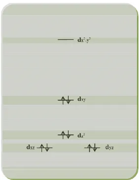
In contrast to organic species, metals have excited states that are often easily accessible by irradiation with visible and UVA light. Transition metal complexes with d6 and d8 electronic configurations are particularly promising, due to the favorable photophysical properties and the relative non-lability of complexes with these configurations. In particular, d6 transition metal complexes can be used to exemplify the diversity of excited states that can be generated by light excitation, and the chemistry that is associated with their generation. Excitation leads to electronically- and vibrationally-excited states with the same multiplicity as the ground state. The transitions to the excited electronic states are formally classified according to the character of the orbitals involved in the electronic transition:
• Metal-centred (MC) transitions, i.e. d-d or ligand-field (LF) transitions. These are orbitally (Laporte)-forbidden, and can also be
spin-forbidden if the spin state changes. Consequently, they give rise to weak absorptions (ε ~ 1-20 x 103
M-1cm-1) which can be masked by stronger, formally allowed charge-transfer transitions. Since MC transitions typically populate antibonding orbitals, the excited states generated often lead to bond lengthening and favor ligand substitution. Photochemical lability is commonly a feature of complexes in which a MC excited state is lowest in energy, such as those metal complexes which photorelease a bioactive molecule (e.g. CO, NO).
• Charge-transfer (CT) transitions, metal-to-ligand (MLCT), ligand-to-metal (LMCT) or to-solvent (TS). These give rise to more intense
transitions (typically ε ~ 1-100 x 103 M-1cm-1) and can lead to redox reactions (of both the complex and molecules in the local environment
e.g. solvent) and also result in homolytic bond cleavage, reducing the
metal centre and generating radicals. Production of radicals under biological conditions is a well established mechanism for causing damage to cellular components (e.g. DNA).
7
• Ligand-centred (LC) transitions, or interligand (IL) transitions. These
generally involve only ligand-centered orbitals and are often seen in large delocalized systems.
Once these excited states are generated they can undergo a series of physical radiationless processes which ultimately lead to the ground-state electronic structure: intersystem crossing (ISC), internal conversion (IC) vibrational relaxation, intramolecular vibrational redistribution and solvation dynamics (reorganization of solvent shells). Radiative processes such as fluorescence (singlet–singlet) and phosphorescence (triplet–singlet) result in a return to the ground state, with emission of light of longer wavelength than was used for the excitation. Beside these monomolecular radiative or non-radiative processes, an excited state can undergo a bimolecular process involving a ground-state molecule that receives the energy content of the excited molecule. Fluorescence RET quenching is an example.
The unique properties of metal compounds, especially very rich photochemistry and photophysics of transition-metal compounds, make them suitable candidates for selected applications in photomedicine.
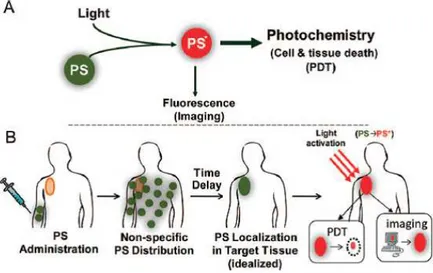
The Thesis in a nutshell
The use of metal compounds in bio-medicine explored in the present work concerns the properties of their excited states; as above mentioned an excited state deactives along two paths: a non radiative decay and/or a radiative decay.
8
Chapter 2 - Sensoristic applications of coordination compounds: drug-protein interaction. A new zinc complex, recently synthesized in the LaCIC, showed
interestingly in vitro cytotoxic properties versus some types of tumor cells. Nevertheless, the in vitro test is only the first step to propose this complex as antineoplastic drug; a successive step is to study its biodistribution. Unfortunately, this Zn complex is scarcely water soluble, so a way to reach a good bioavailability is through its interaction with Human Serum Albumin, the most abundant protein in the bloodstream that increases the solubility of hydrophobic drugs in plasma and modulates their delivery to cell in vivo.
Thanks to the protein fluorescence, it was possible to study its luminescence quenching correlating it to the binding interaction with the Zn complex, and to determine the bimolecular rate constant value, which account for a static quenching. But, the “special property” of the zinc coordination compound, its intrinsic luminescence, has made possible to study the binding interaction from another point of view, with interesting conclusion, that highlight the multifactorial aspect of the complex: therapeutical and sensoristic.
Chapter 3 - Light-activated processes in coordination compounds: photogeneration of singlet oxygen. Photodynamic Therapy (PDT) refers to the application of light
9
to obtain a therapeutical effect; more precisely it refers to the ability of photogenerate the highly reactive species1O2 (which is the “true” therapeutical
agent) by a molecule called photosensitizer. Among the therapeutical effect exerted by 1O2 there are an antimicrobial therapy and, above all, an anticancer
therapy: both prefer water soluble photosensitizer.
Transition metal complexes (TMC), due to their “special” photophysical properties, are excellent photosensitizers, but many of them are scarcely hydrophilic. A way of making TMC water soluble can be to entrap them into a biocompatible polymer, without losing their capacity to generate singlet oxygen. Following this criterion, it was synthesized and characterized the first example of a water soluble polymer grafting a Pt(II) complex able to generate singlet oxygen.
Chapter 4 - The theranostic paradigm: transition metal complexes and nanoparticles.
Another way to obtain a water soluble photosensitizer with the “special” properties of TMCs, is to entrap them into nanoparticles (NP). Nanoparticles are having an increasing importance in the medical field, deserving a new branch of medicine, the so-called “nanomedicine”. This is due to the ability of the NPs to have a little toxicity and to act as drug-delivery system. Moreover, gold NP carried out therapeutical effect thanks to the intriguing properties of the gold plasmon (photothermal therapy).
On this basis, it was synthesised and characterized a number of silica-shell/gold-core NPs entrapping Ir(III) and Ru(II) complexes, which show qualitative ability to generate singlet oxygen. As preliminary test, a sample of gold-silica NP entrapping a Ru(II) complex was characterized in vitro to measure the cytotoxicity against tumor cells, giving promising results.
Moreover, the “special” photophysical properties of TMCs allow having non-radiative deactivation of the excited states (necessary to generate 1O2 by energy
transfer process) without losing luminescence. By virtue of this, it was possible to localize the photosensitizing NPs inside the cell by fluorescence microscope, making the synthesized NPs a new material for theranostic purposes.
10
COORDINATION COMPOUNDS:
DRUG-PROTEIN INTERACTION
2.1 Coordination Compounds in Cancer Therapy
Medicinal inorganic chemistry (Hambley 2007; Orvig 1999; Guo 1999) is a field of increasing prominence as metal-based compounds offer possibilities for the design of therapeutic agents not readily available to organic compounds. The wide range of coordination numbers and geometries, accessible redox states, thermodynamic and kinetic characteristics, and the intrinsic properties of the cationic metal ion and ligand itself offer the medicinal chemist a wide spectrum of reactivities that can be exploited. Although metals have long been used for medicinal purposes in a more or less empirical fashion (Thompson 2006), the potential of metal-based anticancer agents has only been fully realized and explored since the landmark discovery of the biological activity of cisplatin, cis-(NH3)2Pt(Cl)2. To date, this prototypical anticancer
drug remains one of the most effective chemotherapeutic agents in clinical use.
Early investigations on the action mechanism of cisplatin suggested a formation of an adduct with nuclear DNA by a covalent bond of the metal complex with two adjacent guanines on the same DNA strand (Fig. 1) that, causing a distortions in the DNA structure, interfere with replication, triggering cellular events that lead to the death of cancer cell (Wang 2005).
11
Figure 1. DNA and cisplatin form an adduct leaving two amino groups coordinated to the
platinum atom.
Cisplatin is particularly active against testicular cancer and, if tumours are discovered early, an impressive cure rate of nearly 100% is achieved. Besides cisplatin, several other platinum complexes (carboplatin, oxaliplatin and more recently, picoplatin and satraplatin) have been screened as potential antitumor agents (Kelland 2007); however, it must be noted that only a limited number of tumours can be treated with platinum-based anti-cancer drugs. Moreover they produce several side effects including bone marrow suppression, neurotoxicity and above all nephrotoxicity (Jung 2007). In fact, platinum complexes are known to react not only with DNA but also with many other cell components such as glutathione and other sulphur-containing biomolecules, present in relatively high doses inside the cell, and recent studies have shown that the inactivation of the thiol-containing enzymes causes serious side effects in the kidney. In addition to the high systemic toxicity, inherent or acquired resistance is a second problem often associated with platinum-based drugs, with further limits their clinical use (Galanski 2005). Much effort has been devoted to the development of new platinum drugs and the elucidation of cellular responses to them to alleviate these limitations (van Zutphen 2005; Bruijnincx 2008).
These unresolved disadvantages stimulate research on the development of novel non- platinum metal complexes as anticancer agents, with mechanisms of action different
12
from cisplatin, i.e. based on non-covalent interactions with DNA, such as groove binding, insertion or intercalation. Ruthenium compounds, for example, are considered to be promising candidates for anticancer drug design, with two Ru(III) complexes already entered in clinical trials, NAMI-A and KP1019 (Fig. 2); these new octahedral complexes differ structurally from the square planar platinum (II) drugs, offering a more elaborate interaction with double-helical DNA, not only forming coordination bonds, but also H-bonds and intercalation between DNA base pairs (Peacock 2008). Despite modest cytotoxic activity, these complexes have attracted significant interest because of their ability to prevent the formation of metastases and inhibit their growth.
Figure 2. Structure formula of NAMI-A (A) and KP1019 (B).
Homoleptic complexes of Cu(II) and Zn(II) (Wang 2009) have proved to be great examples of intercalating metal compounds and, in addition, the presence of intrinsic fluorescence allows in a single molecule combination of anticancer properties with an excellent tool for investigating their mechanism of action through optical methods. Moreover, in the chemically diverse environment within an organism, issues of metal release can be very important as a complex will subject to a wide variety of enzymatic degradation processes. In this light, the use of zinc, for example, compared to other metals such as platinum, leads to a reduction of the correlated side-effects. In fact, as well known, zinc is an essential nutrient for all organisms, because it serves as a catalytic or structural cofactor for several proteins and is involved in the regulation of mitochondrial apoptosis of many mammalian cells (Franklin 2007; Franklin 2009).
13
Recently, it has been reported the synthesis, characterization, biological evaluation and mode of interaction with DNA of a heteroleptic pentacoordinated Zn(II) complex, (Bpy9)Zn(Cur)(Cl), which bears a bipyridine functionalized in para position with an
aliphatic 9-members chain as N,N’-donor ligand, and a curcumin (1,7-bis-(4-hydroxy-3-methoxyphenyl)-1,6-heptadienes-3,5-dione) as O,O chelating ligand (Fig. 3) showing promising anticancer properties (Pucci 2012). This complex exhibits an antiproliferative activity, observed in vitro against prostatic tumor cells, with an IC50
value of ~12 µM, very interesting compared to that of cisplatin (IC50= 33). Moreover,
the complex shows a significant green luminescence, and this property was useful to demonstrate some form of intercalation with the DNA helices. Unfortunately, (Bpy9)Zn(Cur)(Cl) is insoluble in water, so the in vitro test was carried out in
ethanol/water mixture; this can be a serious problem for any in vivo tests.
Figure 3. Structure formula of (Bpy9)Zn(Cur)(Cl) complex.
The IC50 determination is only the first step to propose a drug for a clinical trial; very
interesting is the study of the biochemical behaviour of a drug, including its interaction with blood plasma proteins, which would be the primary targetmolecules, when it is administered intravenously. In fact the determination of drug binding to plasma proteins (in particular serum albumins) is considered another important factor in pre-clinical drug studies (Zhou 2007; Sulkowska 2002; Yang 2007): this type of interaction can influence the drug stability and toxicity during the chemotherapeutic process; moreover, the adduct drug-plasma protein can overcome problem linked to a scarce water solubility of the drug (Kratz 2008).
14
2.2 The serum albumin proteins: a capital role in drug
bioavailability
Human serum albumin (HSA) is the most abundant protein in the bloodstream, which constitutes up to 60% of the total protein and contributes for 80% to the colloid osmotic blood pressure maintenance (Carter 1994). One of its most extraordinary properties is the ability to bind reversibly a large variety of endogenous and exogenous ligands, such as nutrients, hormones, fatty acids, a great number of therapeutic drugs such as penicillins, sulfonamides, indole compounds, and benzodiazepines to name just a few (Peters 1996). In particular, it increases the solubility of hydrophobic drugs in plasma and modulates their delivery to cell in vivo and in vitro (Yue 2009). Consequently, binding to this protein controls the free, active concentration of a drug, provides a reservoir for a long duration of action, and strongly affects its absorption, metabolism, distribution and excretion.
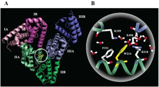
HSA is a globular protein consisting of a single peptide chain of 585 amino acids (He 1992; Dugaiczyk 1982), with three structurally similar α-helical domains I-III, each containing two subdomains A and B. The crystal structure analyses indicate that the main regions of ligand binding sites in albumin are located in hydrophobic pockets in IIA and IIIA subdomains (Fig. 4A). These binding sites are known as Sudlow I and Sudlow II, respectively (Sudlow 1975; Sudlow 1976). HSA contains a single tryptophan residue (W214) within the hydrophobic binding pocket of subdomain IIA (Fig. 4B), which significantly contributes to the intrinsic fluorescence of the protein.
15
Figure 4. X-ray crystallographic structure of human serum albumin (A), and local
configuration around the only one tryptophan residue (W214) of the protein (B) (Qiu 2006).
2.2.1 Protein fluorescence
Among biopolymers, proteins are unique in displaying useful intrinsic fluorescence. They contain three aromatic amino acids residues that contribute to their ultraviolet luminescence, phenylalanine, tyrosine and tryptophan, used as fluorescent probes for the investigation of protein conformation, structure and function (Lakowicz 2003). These chromophores have different absorption and emission spectra because of their structure (Fig. 5).
16
Figure 5. Absorption (A) and emission spectra (F) of the aromatic amino acids in pH 7
aqueous solution (Lakowicz 2003).
Due to tryptophan’s largest extinction coefficient, highest quantum yield, and absorption at the longest wavelength, the fluorescence spectrum of a protein, containing the three amino acids, strongly resembles that of tryptophan. Indeed, because of their spectral properties, resonance energy transfer can occur from phenylalanine to tyrosine to tryptophan resulting in a reduced contribution of phenylalanine and tyrosine to the emission of proteins, dominated by the tryptophan residues.
The emission maximum of tryptophan in aqueous solution occurs around 350 nm (Fig. 5). This fluorescence signal, mainly due to the excitation and transition processes of π electrons in the benzene ring, is highly sensitive to the microenvironment with changes in response to protein conformational transitions, subunit association, substrate binding or denaturation.
17
In particular, as previously mentioned, the Human Serum Albumin has the advantage to have only one tryptophan residue, located in position 214 along the chain in the binding pocket of the subdomain IIA, which significantly contributes to the strong fluorescence of the protein (§ 2.4.1). Therefore, the fluorescence spectroscopy can easily reveal changes in the surrounding environment of tryptophan and be used for determining the binding affinities (Lakowicz 2003).
2.3 Drug-albumin interaction: the protein’s fluorescence quenching
as method of investigation
Many types of analytical methods can be applied to determine the binding constant of the drug-HSA interaction, but fluorescence spectroscopy offers many advantages (high sensitivity, rapidity and ease of implementation) over conventional techniques such as affinity and size exclusion chromatography, dialysis and ultrafiltration (Zhang 2008; Mandeville 2009; Yue 2009; Nanda 2007; Faridbod 2011; Qi 2008). It offers a simple method without needing to separate the bound and unbound molecule and this reduces the time required for the experiment and eliminates the need for a size selective membrane. In addition, dialysis and ultrafiltration cannot be used when the drugs bind extensively to the membrane, a frequent and serious problem with highly hydrophobic drugs. By measuring the intrinsic fluorescence quenching of HSA, the accessibility of quenchers to the fluorophore groups of HSA can be estimated. This information can help to predict the binding mechanisms of drug to human serum albumin.
Fluorescence quenching is a decrease in the quantum yield of fluorescence from a fluorophore induced by a variety of molecular interactions with quencher molecules. The different mechanisms of fluorescence quenching are usually classified as either
dynamic or static quenching, the first resulting from collisional encounters between
the fluorophore in the excited state and the quencher, the second resulting from the formation of a ground-state complex between the fluorophore and the quencher (Lakowicz 2003). In both cases, molecular contact is required between the fluorophore and the quencher for fluorescence quenching to occur. The application of fluorescence quenching as a technique can also reveal the accessibility of the
18
quenchers to the fluorophores.
Generally, the mechanism accepted to be responsible in fluorescence quenching of protein is the fluorescence resonance energy transfer (FRET). This process occurs whenever the emission spectrum of a fluorophore, called the donor (D), overlaps with the absorption spectrum of another molecule, called the acceptor (A). The extent of energy transfer is determined by the distance between the donor and the acceptor, and the extent of spectral overlap.
FRET usually occurs over distances comparable to the dimensions of most biological macromolecules, that is, about 10 to 100 Å. The following equations consider energy transfer between a single linked D/A pair separated by a fixed distance r and originate from the theoretical treatment of Forster (Lakowicz 2003). The energy transfer rate kT(r) between a single D/A pair is dependent on the distance r between D and A and
can be expressed in terms of the Forster distance R0.
kT(r) = (1/τD) (R0/r)6
τD is the lifetime of the donor in absence of energy transfer; R0 is the distance
between D and A at which 50% of the excited D molecules decay by energy transfer, while the other half decay through other radiative or non-radiative channels. R0 can
be calculated from the spectral properties of the D and A species:
R0 = 9.78 x 10³ [κ2 n-4 ΦD J(λ)]1/6 (in Å)
The factor κ2 describes the D/A transition dipole orientation and can range in value from 0 (perpendicular) to 4 (collinear/parallel). There has been much debate about which dipole orientation value to assign for particular FRET formats. Only in few cases the crystal structure of the D/A molecules can be determined; there is no other reliable experimental method to measure absolute or fixed κ2 values, which leads to potential uncertainties in subsequent calculations (Lakowicz 2003). Fortunately, the accumulated evidence has shown that the mobility and statistical dynamics of the dye linker lead to a κ2
value of approximately 2/3 in almost all biological formats. The refractive index n of the medium is ascribed a value of 1.4 for biomolecules in aqueous solution. ΦD is the quantum yield of the donor in the absence of the acceptor
19
and J(λ) is the overlap integral, which represents the degree of spectral overlap between the donor emission and the acceptor absorption. The values for J(λ) and R0
increase with higher acceptor extinction coefficients and greater overlap between the donor emission spectrum and the acceptor absorption spectrum.
There have already been several studies on the fluorescence quenching of HSA induced by the interaction with drugs (Faridbod 2011; Liu, X., 2009; Qi 2008; Trynda-Lemiesz 2010; Katrahalli 2010), small molecules (Zhang 2008; Mandeville 2009; Wu 2009; Nanda 2007; Trnková 2011) or metal complexes (Yue 2009; Wu 2010; Tarushi 2010; Divsalar 2009; Wu 2008). All experiments are performed by studying the decrease of the HSA fluorescence intensity by adding increasing amount of drugs that act as quencher. The fluorescence quenching data are analyzed using the Stern-Volmer equation:
FD /FDA = 1 + kq τD[Q]
where FD and FDA are the fluorescence intensities before and after the addition of the
quencher Q, kq is the bimolecular quenching rate constant and τD is the lifetime of the
fluorophore in the absence of the quencher.
Just to remind some example, Faridbod et al. (2011) reported an interaction study of
pioglitazone (a drug with hypoglycemic action) with albumin using fluorescence
emission and UV-Vis absorption spectra. Experimental results revealed that this drug have an ability to quench the intrinsic fluorescence of HSA tryptophan residue through a static quenching mechanism. Fluorescence quenching data, analyzed by Stern-Volmer equation, give a binding constant value of 4.45 x 10⁵ M-1 (at 300 K). Most non steroidal anti-inflammatory drugs (NSAID) show a high degree of binding to albumin, which is a primary determinant of their pharmacokinetic properties (Trynda-Lemiesz 2010). The interaction of meloxicam, an NSAID, with HSA at physiological conditions (pH 7.4) was evaluated by measuring the intrinsic fluorescence intensity of protein before and after its addition. The strong quenching of the fluorescence clearly indicated that the binding of the drug to HSA changed the microenvironment of tryptophan residue and the tertiary structure of the protein. The interaction between human serum albumin and fluoxetine hydrochloride (a psychotropic drug) have been studied by using different spectroscopic technique
20
(fluorescence, UV-vis absorption, circular dichroism) under simulated physiological conditions, and the fluorescence quenching analyses support a static mechanism (Katrahalli 2010). The study showed that the drug binds the protein most likely at the hydrophobic pocket located in subdomain IIA of site I. Fluorescence data give a binding constant value of 1.64 x 10³ M-1 (at 300 K).
The HSA fluorescence quenching study (Mandeville 2009) was determinant to determine the binding constants K of curcumin and genistein, two molecules present in turmeric and in soybean, respectively, having a wide spectrum of physiological and pharmacological functions. Based on the spectroscopic results curcumin and genistein bind human serum albumin via both hydrophilic and hydrophobic interactions with stronger affinity for curcumin (Kcurcumin = 5.5 x 10⁴ M-1) than genistein (Kgenistein = 2.4
x 104 M-1).
An accurate HSA-binding study of eight catechins, including the epigallocathechin gallate, contained in green tea, has been performed (Trnková 2011) by fluorescence quenching; a static mechanism was clarified, and the Stern-Volmer analyses give a series of binding constant values ranging from 0.34 x 10⁴ to 10.57 x 10⁴ M-1, that were correlated to the chemical structures of the catachins: a balance between their hydrophilic hydroxyl groups and the aromatic moieties resulted determinant to give a good arrangement within the hydrophobic pocked of the albumin.
Instead, examples of HSA binding investigations with metal complexes are rare. Platinum complex oxaliplatin, an antineoplastic drug, show a binding constant of 3.3 x 10³ M-1, determined by Ster-Volmer analysis of the HSA fluorescence quenching , and a static mechanism was reported (Yue 2009). A study involving an antibacterial drug based on a zinc complex (Tarushi 2010) accounted for a static quenching mechanism with a binding constant of 6.9 x 10⁴ M-1.
21
2.4 Photophysical studies of a cytotoxic Zn(II) complex and Human
Serum Albumin
The interaction of the (Bpy9)Zn(Cur)(Cl) complex (Fig. 3) with Human Serum
Albumin has been investigated by fluorescence spectroscopy. The emissive properties of the zinc complex allow possible to study the binding from a dual perspective: (i) the HSA fluorescence quenching, and (ii) the change of (Bpy9)Zn(Cur)(Cl) intrinsic
luminescence.
2.4.1 Photophysical characterization of Human Serum Albumin
The basic photophysics of HSA was studied by preparing a 4.4 x 10⁻⁶ M buffer solution (pH = 7.4) of the protein, purchased from Sigma-Aldrich (purity 96-99%). The buffer solution was prepared by dissolving phosphate buffer saline tablet (Sigma-Aldrich) in 200 mL of water. The applied general methods are reported on § Appendix.
The absorption spectrum of the buffer solution showed a maximum at 278 nm, and a strong fluorescence emission band at 345 nm was recorded by fixing the excitation wavelength at 280 nm, due to the HSA single tryptophan residue de-excitation (Fig. 6). 250 275 300 325 350 375 400 425 0,00 0,05 0,10 0,15 0,20 0,25 E m is s io n I n te n s it y ( a . u .) A b s o rb a n c e Wavelength (nm) A B 0 100 200 300 400 500
Figure 6. Absorption (A) and emission (B) spectra of HSA in buffer solution (pH = 7.4) at
22
The emission quantum yield measurement, performed by optically-diluted method by using 2-aminopyridine in ethanol as standard (§ Appendix), gave Φ = 0.20, while the excited-state decay profile (Fig. 7) was fitted by a biexponential function, giving an average lifetime of 5.71 ns (reduced χ2 = 0.98; § Appendix). A quick rise is noted in the first part of the time profile, which is due to rapid energy transfer occurring from phenylalanine to tyrosine to tryptophan (§ 2.2.1).
Figure 7. HSA in buffer solution (pH = 7.4): time-resolved fluorescence decay recorded at
345 nm upon irradiation by a 265 nm Nanoled (§ Appendix).
2.4.2 Photophysical characterization of (Bpy
9)Zn(Cur)(Cl)
The Zn(II) complex (Bpy9)Zn(Cur)(Cl), synthesized in the Laboratory of Inorganic
and Coordination Chemistry (University of Calabria) by using the method reported in Scheme 1, was available from previous study (Pucci 2012).
N N C9H19 C9H19 N N C9H19 C9H19 Zn O O OCH3 OCH3 HO OH Cl ZnCl2 CH2Cl2, r.t., 5 days N N C9H19 C9H19 Zn Cl Cl curcumin Triethylamine CH2Cl2, N2, r.t., 6 days
Scheme 1. Synthesis of (Bpy9)Zn(Cur)(Cl) complex.
0 5 10 15 20 25 30 35 40 45 50 55 100 1000 10000 C o u n ts ( lo g ) Time (ns)
23
The photophysical characterization of the complex shows a green luminescence with a fluorescence emission band centered 540 nm in EtOH solution, confirmed by the excitation (Fig. 8); the emission quantum yield measurement, performed by optically-diluted method by using Ru(bipy)3Cl2 (bipy = 2,2’-bipyridine) in water as standard (§
Appendix), gives a value Φ = 0.20; the lifetime value (τ = 1.2 ns) is typical of a fluorescence decay. 250 300 350 400 450 500 550 600 650 700 0 5 10 15 20 25 30 E m is s io n I n te n s it y ( a . u .) Wavelength (nm)
Figure 8. Excitation (λem = 540 nm) and emission (λex = 430 nm) spectra of (Bpy9)Zn(Cur)(Cl) in EtOH solution.
2.4.3 Study on the interaction between Zn(II) complex and Human Serum
Albumin by fluorescence spectroscopy
The interaction between the Zn(II) complex and the HSA was achieved by adding, to a buffer solution 4.4 x 10⁻⁶ M of albumin (§ 2.4.1), an increasing amount of Zn(II) complex dissolved in ethanol to a final concentration not exceeding 10%. Higher concentrations of organic solvent, as reported in literature (Lin 2004), may denature the protein by a great exposition of its hydrophobic areas, and reducing its solubility. Five solutions (Table 1) having an increasing [(Bpy9)Zn(Cur)(Cl)]/[HSA] ratio
ranging from 0 to 5 were prepared, and fluorescence emission of HSA at 345 nm were observed by exciting at 295 nm.
24
Sample Added volume of (Bpy9)Zn(Cur)(Cl) ethanol
solution to HSA buffer solution
Mol of (Bpy9)Zn(Cur)(Cl)
contained in the HSA solution
1 0 0
2 50 µL 1.14 x 10⁻⁸
3 150 µL 3.42 x 10⁻⁸
4 200 µL 4.56 x 10⁻⁸
5 250 µL 5.70 x 10⁻⁸
Table 1. Composition of five examined samples of HSA in buffer solution (2.5 mL, 1.12 E-8
mol) obtained by adding different aliquots of a (Bpy9)Zn(Cur)(Cl) 1.14 x 10 -4
M ethanol solution.
The gradual addition of Zn(II) complex to the protein solution caused a gradual decrease in the HSA fluorescence intensity (Fig. 9); due to the comparative low volume of the added Zn complex solutions, the HSA absorption intensity of the five samples remained unvaried, so excluding any dilution effect on the fluorescence intensity. The HSA fluorescence intensity decreases until the ratio [(Bpy9)Zn(Cur)(Cl)]/[HSA] = 5; successive additions of the zinc complex solution do
not cause further reduction of the fluorescence intensity. Furthermore, there was a slight blue-shift at the maximum wavelength of HSA fluorescence emission when the solution of (Bpy9)Zn(Cur)(Cl) was added. This suggests that the chromophore of
protein was placed in a more hydrophobic environment after addition of the zinc complex (Zhang 2008).
25
Figure 9. Fluorescence emission spectra of HSA (λex = 295 nm) in buffer solution (pH 7.4) in the presence of increasing concentrations of the Zn(II) complex. [(Bpy9)Zn(Cur)(Cl)]/[HSA] in samples 1-5: 0, 1, 3, 4, 5.
Quenching of the HSA intrinsic fluorescence is attributed to an interaction with the Zn(II) complex. In fact, the spectral overlap of the absorption spectrum of the complex with the emission spectrum of the protein (Fig. 10) suggests that a FRET from the protein donor (Trp residue, specifically) to the complex acceptor occurs, that is responsible of the albumin fluorescence quenching.
325 350 375 400 425 450 475 500 0,00 0,03 0,06 0,09 0,12 0,15 0,18 0,21 0,00 0,03 0,06 0,09 0,12 0,15 0,18 0,21 N o rm a li z e d E m is s io n I n te n s it y ( a . u .) Wavelength (nm) A b s o rb a n c e HSA (Bpy9)Zn(Cur)(Cl)
Figure 10. Spectral overlap of the emission spectrum of HSA with the absorption spectrum
of Zn(II) complex in a buffer solution (pH = 7.4) : EtOH (10:1) mix. 320 340 360 380 400 420 440 0 20 40 60 80 100 120 140 E m is s io n I n te n s it y ( a . u .) Wavelength (nm) 1 5
26
As already mentioned (§ 2.3), the FRET Forster’s model (Lakowicz 2003) predicts that the energy can be reliably transferred whenever the fluorescence emission spectrum of the donor (in our case, the HSA) and the absorption spectrum of the acceptor (the Zn(II) complex) have enough overlap, and the distance between donor and acceptor is not longer than 10 nm. These distances are comparable to the diameter of many proteins, e.g. HSA 5.9-6.2 nm at pH 7.4 (Lakowicz 2003).
Energy Transfer efficiency (η) is typically measured using the relative fluorescence intensity of the donor, in the absence and presence of acceptor, according to the formula
η= 1- FDA /FD.
Using fluorescence intensities (that, taking into account the practically unvaried volume of the HSA samples, correspond to the areas under the emission spectral curves) of the HSA in the absence (FD) and in presence (FDA) of Zn(II) complex, the η
was calculated, obtaining a value of 48%. Moreover, when increasing concentration of the Zn(II) complex was added to the protein, the lifetime of HSA varies within experimental error remaining almost identical to the value of the unquenched one (τD
= 5.71 ns). The decrease of the emission intensity by adding the quencher with a constancy of the lifetimes values, account for a static quenching.
The Stern-Volmer equation (§ 2.3) was applied to determine the bimolecular quenching rate constant by plotting FD /FDA versus quencher concentration (Fig. 11);
the linearity of this plot, proposes a single type of quenching process (Lakowicz 2003). The obtained value of kq is 8.5 x 10¹² L mol-1s-1 is typical of a binding
interaction, and it indicated that the quenching was not initiated from dynamic collision but from an adduct formation, i.e. it is a static quenching (Lakowicz 2003). The binding constant,obtained from kq taking into account τD = 5.71 ns, results 4.8 x
104 M-1, in agreement with values reported for several other ligand-protein complexes.
27
Figure 11. Stern-Volmer plot for the (Bpy9)Zn(Cur)(Cl) quenching of the HSA fluorescence.
As above mentioned (§ 2.2.1), the intrinsic fluorescence of HSA is very sensitive to its microenvironment: it would be quenched obviously even if there is a slight change of the local surroundings of the protein, such as molecular binding, protein conformation variation and denaturation. To better confirm that the fluorescence quenching is due to a binding interaction of the protein with the complex, which penetrates into the hydrophobic binding site, and not to a simple interaction which modifies the protein conformation, a new set of measures was conducted that take advantage from the Zn complex fluorescence.
Emission spectra from solutions 2-5 (Table 1) containing HSA and (Bpy9)Zn(Cur)(Cl) were recorded exciting at 430 nm (i.e. on the Zn complex
absorption band, although it should be considered a solvatochromic shift, see below), and they report a 627 and a 510 nm bands (Fig. 12A). At the same time, four solutions of (Bpy9)Zn(Cur)(Cl) in buffer were prepared, by dissolving in 2.5 mL of
buffer solution, 50, 150, 200, 250 µL of (Bpy9)Zn(Cur)(Cl) 1.14 x 10⁻⁴ M ethanol
solution; in this way a new set of solutions (2’-5’) with the same concentration of the Zn complex of sample 2-5, but without HSA, was prepared: these solutions showed, upon excitation at 430 nm, a unique band at 627 nm (Fig. 12B).
0,0 5,0x10⁻⁶ 1,0x10⁻⁵ 1,5x10⁻⁵ 2,0x10⁻⁵ 1,0 1,2 1,4 1,6 1,8 2,0 F D / FD A
28
A B
Figure 12. Emission spectra obtained by 430 nm excitation light of samples 2-5 (HSA +
(Bpy9)Zn(Cur)(Cl)), A, and of samples 2’-5’ ((Bpy9)Zn(Cur)(Cl)), B.
The obtained results,summarized in a simplified manner in Figure 13, account for the formation of an adduct between HSA and the Zn(II) complex: in fact, the emission spectrum of the (Bpy9)Zn(Cur)(Cl)+HSA solution has been interpreted by
attributing the band at 627 nm to the free complex in the hydrophilic buffer solution, and the band at 510 nm to the complex bound to the hydrophobic pockets of HSA.
450 500 550 600 650 700 750 0 2 4 6 8 10 12 14 16 18 bound quencher (Bpy9)Zn(Cur)(Cl)
(Bpy9)Zn(Cur)(Cl) + HSA
E m is s io n I n te n s it y ( a . u .) Wavelength (nm) free quencher
Figure 13. Comparison between the emission spectra of Zn(II) complex (λex = 430 nm) with or without HSA in the mix buffer solution: EtOH (10:1). The two spectra are not in scale.
450 500 550 600 650 700 750 0,0 2,5 5,0 7,5 10,0 12,5 15,0 17,5 50 µµµµL 150 µµµµL 200 µ µ µ µL 250 µµµµL E m is s io n I n te n s it y ( a . u .) Wavelength (nm) 500 550 600 650 700 750 0,0 2,5 5,0 7,5 10,0 12,5 50 µµµµL 150 µµµµL 200 µµµµL 250 µµµµL E m is s io n I n te n s it y ( a . u .) Wavelength (nm)
29
The blue-shift of the emission is due to a solvatochromic behavior of the Zn(II) complex, already evidenced by comparing the maximum band in ethanol (λem = 540
nm, Fig. 8) with that in buffer (λem = 627 nm, Fig. 12B). To confirm the
solvatochromic properties of (Bpy9)Zn(Cur)(Cl), emission spectra of the complex
were recorded dissolving it in different solvents; actually, solvatochromic band shifts are observed: in particular a red shift of the maximum emission by increasing the solvent polarity was observed (Fig. 14).
0,0 0,1 0,2 0,3 0,4 0,5 0,6 0,7 470 480 490 500 510 520 530 540 550 E m is s io n p e a k ( n m ) ENT Cyclohexane Chloroform Dimethyl sulfoxide Ethanol
Figure 14. Linear plot of wavelength maximum of (Bpy9)Zn(Cur)(Cl) emission versus the Reichard’s parameter ET
N
values of some organic solvents where the complex was dissolved.
Finally, to explore a further effect of the FRET from HSA to Zn complex, it was recorded the emission of the acceptor Zn complex by exciting the donor HSA, i.e. by recording the emission of 2-5 samples reported in Table 1 by exciting at 280 nm. The obtained results showed the emission maximum of the free complex at 627, instead of the maximum of the bound complex at 510 nm. Taking into account that the free complex has an absorption band at about 280 nm (due to the bipyridine-localized levels), it can be concluded that the amount of the free complex in solution show an emission more intense that those deriving from the energy transfer from HSA to the bound complex.
30
2.5 Conclusion
Since the discovery of cisplatin, many metal complexes are used as chemotherapic agents against cancer diseases. The different metal coordination geometry exploitable by organometallic compounds is the key factor that allows these drugs to be effective: in fact, traditional anticancer drugs target DNA preventing cell reproduction. Besides direct coordinative binding of metallo-agents to DNA bases, other potential DNA binding modes, non-covalent in nature, include intercalation and groove binding, in which metal role is prevalently structural, like a scaffold for a variety of ligands. But the coordination compounds versatility can be exploited by inserting another function, i.e. to add a fluorescent ligand to the drug, so coupling the therapeutical to the sensoristic action in a unique compound. In fact, to have a simple and economical method for monitoring the biodistribution of a drug is a great advantage. In this light, fluorescence spectroscopy offers a direct connection between the spectral evidences and the molecular features of a sample.
The cytotoxic properties of many coordination complexes are test in vitro; nevertheless, this test is only the first step to propose a complex as antineoplastic drug; a successive step is to study its biodistribution. Moreover, a few of coordination compound is scarcely water soluble, so a way to reach a good bioavailability is through its interaction with Human Serum Albumin (HSA), the most abundant protein in the bloodstream that increases the solubility of hydrophobic drugs in plasma and modulates their delivery to cell in vivo. The protein fluorescence has opened the way to numerous studies that correlate the luminescence quenching to the binding interaction with a complex.
Unfortunately, the intrinsic fluorescence of HSA is very sensitive to its microenvironment: it would be quenched even if there is a slight change of the local surroundings of the protein, such as molecular binding, protein conformation variation and denaturation. To overcome this drawback, we have studied the interaction between HSA and a new Zn(II) complex, with twofold function: an excellent antitumor activity and an intense fluorescence. So, we have been able to look at the binding interaction from a dual perspective: the luminescence quenching of the protein and the fluorescence variation of the complex.
31
The fluorescence-resonance energy-transfer from protein to complex was observed by monitoring the decreases of luminescence by increasing the complex concentration; the Stern-Volmer equation was applied to determine the bimolecular quenching rate constant, obtaining a value of kq = 8.5 x 10¹² L mol-1s-1 is typical of a binding
interaction, and it indicated that the quenching was not initiated from dynamic collision but from an adduct formation, i.e. it is a static quenching. While it was impossible to evidence an increase of the complex fluorescence intensity ̶ as result of the energy-transfer ̶ because the acceptor absorbs at the same exciting wavelength of the donor, it has been evidenced a solvatochromic shift of the emission complex as result of the hydrophobic environment experienced by the Zn compound inside the protein binding site.
The investigated Zinc compound represents an example of the multitude of applications that metal complexes can successfully offer, and the obtained results concerning the Zn(II) complex–HSA interaction can contribute to the current knowledge in the area of protein-ligand binding and confirm the capital role of the fluorescence spectroscopy in biomedical research.
32
COORDINATION COMPOUNDS:
PHOTOGENERATION OF SINGLET
OXYGEN
3.1 Singlet oxygen: a guest ghost in everyday life
3.1.1 Electronic structure and the lifetime of singlet oxygen
Despite its apparent simplicity, molecular oxygen (O2) exhibits a number of rather
unusual properties due to its unique electronic structure. Unlike many molecules, oxygen has an open-shell triplet ground state (3Ʃg-) with two unpaired p-electrons
distributed in the highest occupied π-antibonding orbitals (Fig. 1A). Rearrangement of the electron spins within these two degenerate orbitals results in two possible singlet excited states, the 1∆g and the1Ʃg+ states, which lie 22.5 Kcal and 31.5 Kcal
mol-1 respectively, above the ground state (Fig. 1B). In the first excited state (1∆g),
both electrons are paired in a single orbital, leaving the other empty while the higher singlet state (1Ʃg+) comes from the spin pairing electrons in different orbitals (Ogilby
2010). In both forms of 1O2, the spin restriction is removed so that the oxidizing
ability is greatly increased (Halliwell 2007).
Figure 1. (A) Molecular orbital diagram showing the electron distribution in 3Ʃg, 1
∆g and 1
Ʃg (Josefsen 2008); (B) potential energy curves of molecular oxygen (De Rosa 2002).
33
The higher-energy singlet oxygen state (1Ʃg+) is very short lived and due to a
spin-allowed transition rapidly relaxes to the lower-energy 1∆g state with unit efficiency,
before chemical reactions can occur (Wilkinson 1993). Conversely, the transition from the 1∆g state to the ground state is spin forbidden, thus the 1∆g oxygen is a
relatively long-lived species with a lifetime strongly dependent on the nature of the solvent (Table 1). Solvent τ/µs Solvent τ/µs H2O 4 D2O 68 CH3OH 10 CH3CN 75 C4H8O 23 CHCl3 244 C6H6 31 C6F6 3900 C6H5Cl 45 CS2 34000 CH3COCH3 54 CCl4 87000
Table 1. Lifetime of singlet oxygen in several solvents (Gorman 1989; Schmidt 1989;
Wilkinson 1995; Montalti 2006).
In fact, in most solvents, singlet oxygen’s lifetime is reduced because the electronic excitation energy of 1O2 is dissipated as heat by coupling with the vibrational
frequencies of the solvent molecules. The most probable energy-accepting oscillator of a solvent molecule is its terminal atom pairs with the highest vibrational energy (e.g., O-H, C-H); molecules with low energy oscillators, as C-F and C-Cl, act as poor quenchers (Fujii 2004; Schmidt 1989).
The 1∆g state decays to the triplet ground state through a radiative deactivation and
the spectral profile of its phosphorescence is unique, with a distinct and narrow band centered at ~1275 nm (Fig. 2).