International PhD Program in Neuropharmacology
XXIV Cycle
INTERACTION BETWEEN ESTROGEN RECEPTOR AND METABOTROBIC GLUTAMATE RECEPTOR 1 MEDIATES THE
DUAL EFFECT OF ESTROGEN IN NEUROPROTECTION AND NEURODEGENERATION
Doctorate thesis Simona Federica Spampinato
Coordinator: Prof. Filippo Drago Tutor: Prof.ssa Maria Angela Sortino
2
Table of contents
TABLE OF CONTENTS 2
ABSTRACT 4
ESTROGEN: PHYSIOLOGICAL ROLE AND SIGNALING 6
ESTROGEN RECEPTORS 7
Nuclear receptors 7
Membrane receptors 8
ESTROGEN ACTIVITY IN THE CNS 11
Neuroprotection 12
Parkinson’s disease 14
Alzheimer’s Disease 15
Preclinical evidence of estrogen effects in AD 18
Clinical evidence on estrogen effects in AD 20
Mechanisms of action of estrogen 23
METABOTROPIC GLUTAMATE RECEPTOR 24
ESTROGEN RECEPTOR AND MGLUR 29
CHAPTER I 32
CHAPTER II 70
GENERAL DISCUSSION AND CONCLUSIONS 95
3
Acknowledgment
I want to thank my tutor, Professor Maria Angela Sortino, for all that she does for me: she believes in me, encourages and gives me advice useful for the work, but especially for life.
Professor Ferdinando Nicoletti that welcomed me in his lab in Pozzilli and is always available for clarification and suggestion.
Professor Agata Copani and all her group for the precious advices and their availability for comparison ideas.
Professor Filippo Drago for the possibility to attend this PhD program that has been an important moment for my scientific and personal growth. Thank to Doctor Sara Merlo e Gemma Molinaro, that shared with me their knowledge and that taught how to work and behave in a lab.
Finally thank to all my friends and colleagues at Dep. Farmacologia University of Catania, INMR Neuromed Pozzilli and BRC University of Cambridge, with whom I shared very important moments, not only related to work.
4
Abstract
Estrogen receptors (ER) are known to exert important action in the central nervous system, including neuroprotection. These effects are due to interaction of estrogen with membrane localized receptors that signal through rapid transduction pathways. How membrane ERs initiate rapid signal transduction has not been clarified, but they have been reported to interact with other membrane receptors, including metabotropic glutamate receptors (mGluR).
The role of estrogen in neuroprotection is not always defined and above all, neuroprotective data provided by preclinical studies have not been confirmed by the use of estrogen in humans: estrogen treatment can in fact be neuroprotective but also responsible for increased neurodegeneration. The dual role ascribed to estrogen, can be observed also with mGluR1 agonists. 3,5-Dihydroxyphenylglycine (DHPG) behaves both as a neuroprotective and neurodegenerative factor. The aim of this study has been to point out whether the similar behaviour of the estrogen and mGlu1R agonists depends on the interaction between their receptors.
Neuroprotective activity of both drugs was demonstrated in an in vitro model of cortical cultures exposed to beta amyloid (Aβ) toxicity. Pre-treatment with either 17β-estradiol (E2) and DHPG reduced Aβ-induced neuronal death. The neuroprotective effect was due to strict interaction between mGluR1 and ERα, as treatment with the mGluR1 antagonist, JNJ 16259685, or ER antagonist, ICI 182,780, prevented the effect induced by the respective as well as by the reciprocal agonist. Moreover, E2 and DHPG shared a common signalling pathway, as they stimulated to a similar extent phosphoinositide hydrolysis and induced enhanced phosphorylation of AKT. Both these effects were not additive, when the two agonists were
5 added together and they were prevented by the reciprocal antagonists. In addition, a similar effect was reproduced when each receptor was expressed in recombinant cells.
The interaction between the two receptors seems to be involved also in the neurotoxic effect of E2 and DHPG. Both compounds exacerbated NMDA induced toxicity when added after the neurotoxic challenge, while being still neuroprotective when administered before. The effect of E2 was not modified by co-treatment with DHPG and was sensitive to ICI 182,780, but also to JNJ16259685. Moreover, this potentiating effect is abolished by pre-treatment with a calpain III inhibitor. The ability of E2 to phosphorylate AKT and ERK is reduced after NMDA pulse, but this effect is partially reverted by pre-treatment with the calpain inhibitor.
Altogether the present data demonstrate that membrane ERs and mGluR1 interact to induce both neuroprotection and neurodegeneration. The different outcome may depend on the cellular conditions, thus pointing out the importance of the choice of the timing of intervention.
6
General introduction
Estrogen: physiological role and signaling
Estrogens belong to the class of steroid hormones representing the primary female’s sex hormones. 17-beta Estradiol (E2) is the most potent compound, although there are two different metabolites, estrone (E1) and estriol (E3), whose activity has tissue-specific roles (Gruber DM et al., 1999). They are produced primarily in the ovaries and placenta, through the synthesis of androstenedione from cholesterol that is then converted to estrone or estradiol, either immediately or through testosterone. The conversion of testosterone to estradiol and of androstenedione to estrone, is catalyzed by the enzyme aromatase. Estrogen can also be produced in small amounts in the liver, adrenal glands and breast, and astrocytes have been found to express aromatase, justifying brain derived-estrogen (Gulinello M et al., 2006, Saldanha CJ et al.2005).
The role of estrogen is not only related to the development of sexual and reproductive function; its activity is essential, in both males and females, in cardiovascular, skeletal, immune functions as well as in the central nervous systems (CNS) (Gustafsson JA. et al., 2003).
In the late 50s (Jensen EV et al., 1962) the nature of an estrogen binding protein, estrogen receptor α, (ERα), was characterised, and after several years the nature of a second estrogen receptor, ERβ, was pointed out (Kuiper GG et al., 1996).
Using the models of recombinant mice, either ERα-/- , ERβ-/- or the double knockout mice, it has been demonstrated that life is possible without either or both receptors, while the reproductive system is severely impaired (Couse JF et al., 1999).
7 Studies on the role of the two receptors in the CNS, in the skeletal, cardiovascular and immune systems pointed out that the two receptors have distinct and not overlapping roles (Gustafsson JA. Et al., 2003, Harris H et al., 2007). It has been suggested that the two receptors are involved in opposite actions, so the effect of E2 could be the result of the balance between ERα and ERβ signaling (Liu MM et al., 2002; Heldring N et al., 2007).
Estrogen Receptors Nuclear receptors
ERα and ERβ belong to the class of nuclear receptors. They are the products of two different genes located on different chromosomes (Enmark E et al.,1997; Menasce LP et al.,1993) and several splice variants have been described for both receptors. Similarly to other nuclear receptors, they contain evolutionary conserved domains. The NH2 terminus is not conserved and it represents the area with most variables both in length and sequence. Except for the N-terminal area, ERα and ERβ share high sequence homology, have the same affinity for E2 and bind the same DNA Estrogen Response Elements (ERE) (Nilsson S et al., 2001). When the receptor is not coupled to estrogen it binds to heat shock proteins, in a transcriptionally inactive state (Hall JM et al., 2001). Upon binding to estrogen the receptor undergoes conformational changes, homodimeriz-zation and nuclear translocation, where the complex E2/ER directly binds the ERE sequence, or interacts with other transcriptor factors, such as Fos/Jun or SP-1(Kushner PJ et al., 2000; Saville B et al., 2000). The complex ligand/receptor alters chromatin structure and allows the activity of RNA polymerase II transcriptional machinery.
8
Membrane receptors
In addition to the classical genomic pathway, evidence exists to suggest that estrogen is responsible for rapid responses occurring within few seconds after addition of E2. These effects include activation of kinases, phosphatases and ion fluxes across membranes (Simoncini T et al., 2006; Song RX et al., 2005; Warner M et al., 2006; Wong CW et al., 2002). The first line of evidence of membrane-initiated signaling by estrogen were reported back in 1967, when Szego and Davis described increased cyclic andenosine mono-phosphate (cAMP) formation in uterus following exposure to E2 for 15 seconds (Szego CM et al., 1967). These rapid effects are initiated at the membrane surface, as demonstrated by the use of membrane impermeable estrogen analogs, estrogen conjugated either to serum bovine albumine (E-BSA) or to horse radish peroxidase (E-HRP) (Zheng J et al., 1996).
In the CNS, membrane-initiated effects of estrogen and activation of classical intracellular signaling cascades are involved in the control of reproductive functions, neuronal excitability, neuroprotection, neurotrophism (reviewed in Vasudevan D et al., 2008); furthermore E2 is involved in the modulation of putative nociceptive signaling in dorsal root ganglion (DRG) neurons (review in Mermelstein P et al., 2008).
The effects of estrogen stimulation are not only related to the activation of ERα or ERβ; removal of classical receptors, using double knockout mice, does not prevent all estradiol binding (Shughrue P.J et al., 2002), and estrogen maintains its activity (Dominguez-Salazar E. et a., 2006; Fugger H.N. et al., 2001). Accordingly, it has been suggested that other receptors are able to bind E2 and mediate its activity. (Kelly M.J. et al.,2008; Li L et al., 2003; Qiu J et al., 2003; Razandi M et al.,1999; Thomas P et al., 2005;
9 Toran-Allerand C.D et al.,2002). A new receptor, ER-X, has been described in uterus, lung and neocortex, localised at the plasma membrane and associated with caveolin proteins (Toran-Allerand C.D et al.,2005). ER-X binds 17α estradiol, and its activity is not modified by the non selective ER antagonist ICI 182,780 (both stereospecific activation by 17β estradiol and antagonism by ICI 182,780 are classical hallmarks of ERα and ERβ); the receptor is functionally coupled to Mitogen Activated Protein Kinase (MAPK) signaling (Toran-Allerand et al., 2002). In the cortex, its expression peaks in the post natal period (days 7-10), but drops off within a month; in contrast it appears to be upregulated after ischemic injury or in animal models of Alzheimer disease (reviewed in Micevych P et al., 2009). More recently, the activity of G coupled membrane receptors, likely activated by estradiol has been described (Filardo E.J. et al., 2002; Evinger A.J. III et al., 2005). Among others, GPR30, an integral membrane protein, is the best characterised. Although initially thought to be localised at plasmatic membrane level, recently its localization has been restricted to Golgi apparatus and endoplasmatic reticulum (Matsuda K et al., 2008; Otto C. et al., 2008).
Estrogen activation of GPR30 increases activity of the cAMP pathway (Revankar C.M. et al., 2005; Thomas P. et al., 2005). The ability of ICI 182,780 to modify this effect has not been clarified as contrasting data are reported (Thomas P. et al., 2005). However there are still controversies on the possible role of GPR30 as an estrogen receptor (reviewed in Langer G et al., 2010).
Kelly and co-authors (2003) analyzed the activity of another membrane protein that is activated by a diphenylacrylamide compound, STX (Kelly MJ et al., 2003; Qiu J et al., 2006). This STX binding protein is stereospecifically activated by E2 and blocked by ICI 182,780. When
10 activated by STX, the receptor affects calcium oscillations and modulates gonadotropin releasing hormone (GnRH) release from hypothalamic neurons in mice and primates, with effects that are similar to those caused by estrogen treatment. To date, the molecular structure of the STX-sensitive receptor remains to be identified(Kenealy BP et al., 2011), but it has been hypothesized that the STX-activated protein is a GPCR, since downstream effects are sensitive to G protein modulation.
Finally, as mentioned, despite their classical nuclear localization, ERα and ERβ can also be associated with the plasma membrane and can be responsible for the rapid non genomic effects of estrogen (Razandi M et al.,1999). It is not fully understood how the nuclear receptors are trafficked to the membrane, and if they undergo post-transcriptional modification that allow their insertion into the membrane (Acconcia F et al., 2005; Milner T.A et al., 2001).
Membrane ERα and ERβ can transactivate different classes of tyrosine kinase receptors, including epidermal growth factor receptors (EGF) (Song et al., 2010) and type-I insulin-like growth factor receptors (IGF-I) (Marin et al., 2009; Varea et al., 2010).
E2 is able to modulate ionic movements through the membrane, particularly potassium and calcium, due to activation of cAMP and Protein Kinase A (PKA), suggesting the occurrence of mechanisms mediated by a Gs coupled protein (Aronica S.M. et al., 1994, Gu Q., et al.., 1996, Nabekura J. et al., 1986). It can also activate phospholipase C (PLC)/PKC thus modulating a Gq protein (Vasudevan, D.W et al., 2008; Boulware M.I et al.,2005; Dewing P et al., 2007; Dewing P. et al., 2008; Kelly MJ et al., 2003), and signaling related to Gi/o protein has been also demonstrated (Navarro C.E et al.,2003; Wyckoff M.H et al., 2001).
11 How estrogen receptors activate G protein-coupled proteins is not fully understood; recently, it has been suggested that ERs interact with membrane receptors coupled to G proteins; the ensuing transactivation of a G protein-coupled receptor involve also metabotropic glutamate receptors (see paragraph Estrogen receptor and mGluR)
Fig 1.
Blas et al., 2009
Estrogen activity in the CNS
The role of estrogen in the CNS has been largely studied both in physiological and pathological conditions. Estrogen activities in the brain are related not only to the regulation of hormonal feedback in the hypothalamic pitituary system (Kalra SP et al., 1983; MacLusky NJ et al.,
12 1978). Estrogen in fact can modulate motor behaviour, mood and mental state, pain perception, etc (Beatty WW et al., 1978; Fink G et al., 1996; Harlan RE et al., 1988; Harlan RE et al., 2001).
In rats, estrogen treatment increases synapse formation in the arcuate nucleus during postnatal development (Arai Y et al., 1978), while in adults it is responsible for synapse remodelling. This occurs not only in response to axonal injury (Matsumoto A et al., 1978), but also in a continuously ongoing process involved in memory and learning, as demonstrated by synapse remodelling in the arcuate nucleus and CA1 area in hippocampus, depending on hormonal fluctuation during the estrous cycle (Olmos G et al., 1989).
The effects of estrogen on synaptic morphology and number of dendritic spines, occurring both in postnatal period and adulthood, have been described in midbrain (Reisert I et al., 1987), cortex (Garcia-Segura LM et al., 1989), hippocampus (Gould E et al., 1990), spinal cord (VanderHorst VG et al., 1997), pituitary (Chun TY et al., 1998) and are related to changes in brain morphology, sexual behaviour, learning and memory. The increased transcription of neurotrophic factors is one of the mechanisms through which E2 induces these effects: in physiological conditions, in fact, E2 increases the transcription of Nerve Growth Factor (NGF) and its receptors in cholinergic neurons (Toran-Allerand CD et al., 1996) and sensory neurons (Sohrabji F et al., 1994), while it modulates the release of Transforming Growth Factor Beta 1 (TGF-β1; Ma YJ et al., 1992) and IGF-1 (Pons S et al., IGF-1993) in hypothalamic neurons and Brain Derived Neurotrophic Factor (BDNF) in cortex (Sohrabji F et al., 1995).
Neuroprotection
In 1991 Hall and coworkers pointed out that female gerbils had less severe brain damage after occlusion of carotid artery versus male animals. These
13 data were confirmed using different models of brain ischemia (middle cerebral artery occlusion, MCAO ) in mice and rats (Alkayed N et al., 1998; Park EM et al., 2006); all females had greater survival rates compared to males after brain injuries.
In human beings, women, at least before menopause onset, are less exposed to stroke risks, (Murphy SJ et al., 2004; Niewada M et al., 2005) while a worse outcome in women parallels the reduced estrogen levels that occurs after menopause (Hochner-Celnikier D et al., 2005).
Evidence that E2 is the neuroprotective factor responsible for the gender-related difference in the outcome of brain ischemia is provided by studies in which estrogen administration reduced infarct volume after global or focal ischemia in both ovariectomized female or male mice, as well as in rats and gerbils (Jover T et al., 2002; Miller NR et al., 2005; Alkayed NJ et al., 1999).
The effects of estrogen consist of a significant improvement in recognition, working and spatial memory, and a reduction in the sensorimotor dysfunctions (Gulinello M et al., 2006; Plamondon H et al., 2006; Li X et al., 2004).
Several studies (Jover T et al., 2002; Plamondon H et al., 2006; Dubal DB et al., 1998) demonstrated that at least a 24 hours estrogen pretreatment is needed to achieve the reduction in the volume of ischemic area. ERα seems to be more implicated in estrogen neuroprotective activity, due to the lack of protective effects in ERα-/- mice (Dubal DB et al., 2001; Sampei K et al., 2000), and the upregulation of ERα in the penumbra area following MCAO in rats (Dubal DB et al., 1999). However, the involvement of ERβ cannot be completely excluded (Miller NR et al., 2005; Sampei K et al., 2000; Carswell HV et al., 2004).
14 The effects of estrogen are related to its ability to increase the expression of anti-apoptotic genes, in particular bcl-2 in the penumbra following global ischemia or MCAO, in in vivo models (Dubal DB et al., 1998). Accordingly, in vitro, E2 increases the expression of bcl-2 in neuronal hippocampal cultures (Zhao L et al., 2004; Wu TW et al., 2005) and neuronal continuous cell lines (human NT2) (Singer CA et al., 1998); furthermore, E2 reduces the expression of the pro-apoptotic protein BAD (Dubal DB et al., 1999; Zhao L et al., 2004), cytochrome c translocation (Bagetta G et al., 2004) and DNA fragmentation (Rau SW et al., 2003).
The neuroprotective activity of estrogen could be extended to chronic or acute brain injuries. It has in fact protective effects in seizure models where it reduces NMDA- and kainate-induced seizure numbers and duration (Hoffman GE et al., 2003; Velisek L et al., 2002; Kalkbrenner KA et al., 2003). Estrogen prevents cerebellar damage and behavioural decline after ethanol withdrawal (Jung ME et al., 2002) and ameliorates the outcome in models of amyotrophic lateral sclerosis (Kruman II et al., 1999).
There is also evidence suggesting a role of estrogen in different models of brain injury. Through reduction of inflammation, estrogen protects against spinal cord injury (Yune TY et al., 2008; Sribnick EA et al., 2003) and reduces edema and blood brain barrier permeability after traumatic brain injury (Sribnick EA et al., 2005).
Parkinson’s disease
Differences in symptoms severity and treatment outcome between males and females have been reported in Parkinson’s disease. More specifically males have higher risk in developing the pathology and generally they are affected by more severe symptoms, whereas women have a better outcome,
15 although they exhibit more frequently levodopa-induced dyskinesia (Rajput MI et al., 2004; Baba Y et al., 2005; Scott B et al., 2000).
Studies in animal models of Parkinson’s Disease demonstrate that E2, acting on ERα, reduces toxicity induced in the striatum by 1-Methyl-4-phenyl-1,2,3,6-tetrahydropyridine (MPTP) (D’Astous M et al., 2004), and maintains motor function after 6-hydroxydopamine lesions. In nigrostriatal dopaminergic neurons the latter effect has been related to the interaction with the IGF-1 system (Quesada A, et al., 2004).
In humans, there is evidence that correlates reduced estrogen levels during life to higher risk in developing Parkinson’s Disease (Currie LJ et al., 2004). In line with this, estrogen replacement therapy seems to protect against this condition (Westberg L et al., 2004), also through an increased availability of the dopamine transporter in putamen (Gardiner SA et al., 2004).
Finally estrogen treatment induces human neuronal stem cell proliferation and enhances their differentiation in dopaminergic neurons (tyrosine hydroxylase positive cells) (Kishi Y, et al., 2005).
Alzheimer’s Disease
Alzheimer’s disease (AD) is a progressive neurodegenerative disorder, characterised by severe cognitive decline. AD is a multi-factorial disease influenced by a combination of genetic and environmental factors. According to the age of onset, AD has been classified into two forms. A rare familial form of AD (FAD), accounting for about 5% of cases, characterized by early onset (45–60 years of age) and linked to causative genetic mutations (Bertram L et al., 2001; Williamson J.,2009). Sporadic AD accounts for the remaining 95% of AD cases and is characterized by late onset (>65 years). This form has not been associated with specific gene
16 mutations, but with genetic risk factors that seem to underlie an increased chance to develop the disease (Williamson J.,2009).
Pathological hallmarks of AD are extracellular senile plaques and intracellular neurofibrillary tangles (NFTs), results of the aberrant aggregation of misfolded proteins, respectively amyloid beta protein (Aβ) and hyper-phosphorylated tau protein ( Selkoe D et al., 2001).
Tau is a microtubule-associated protein that is involved in microtubule assembly and stabilization. Hyperphosphorylation and abnormal phosphorylation are major biochemical abnormalities of the protein. They are early events in the development of the neurofibrillary lesions (Braak E et al., 1994) and, as a result, tau is unable to bind to microtubules (Bramblett GT et al., 1993; Yoshida H et al., 1993). The hyperphosphorylated, insoluble, filamentous tau is the main component of NFTs. Although associations per se cannot prove cause-effect relationships, tau inclusions are widely thought to contribute to the pathogenesis of the disease because they occur in specific brain regions whose functions are altered and NFT formation correlates with the duration and progression of AD (Giannakopoulos et al., 2003; Ihara,2001). Tau inclusions also appear to modulate the clinical features of other neurodegenerative diseases, known as tauopathies (Iqbal K et al., 2005).
The classical and widely accepted mechanism proposed for AD pathogenesis is the amyloid cascade theory (De Strooper d et al., 2010; Pimplikar SW et al., 2009).
Aβ is a short peptide (39-42 amino acids) derived from the proteolytic cleavage of the transmembrane amyloid precursor protein (APP). APP is cleaved at different sites by transmembrane proteolytic complexes known as secretases. Cleavage of the N-terminal ectodomain of APP could be alternatively generated by either α- or β-secretases (β-site APP-cleaving
17 enzyme, BACE). The activity of α-secretase produces a non-amyloidogenic soluble fragment (sAPPα), and prevents the formation of amyloidogenic fragments due to the activity of BACE. γ- Secretase, a complex of at least 5 different proteins (presenilin 1 and 2, nicastrin, APH1 and PEN2), operates a transmembrane C-terminal cut at alternative sites to generate, when coupled to BACE, Aβ species of 39–42 amino acids. The 40 and 42 aminoacids isoforms have an increased propensity to self aggregate. According to the amyloid theory, an imbalance leading to an over-production of the highly aggregation-prone Aβ 42 species triggers its accumulation and aggregation first into low-molecular-weight oligomers, then into fibrils and finally into plaques, in specific brain regions.
Plaques contain Aβ and degenerating neurites, and evoke strong inflammatory response; activated astrocytes and microglia are attracted to the area of the plaques and release cytokines, chemokines and complement components. The significance of the resulting inflammatory state is not fully understood, since it could represent a protective mechanism against neurodegeneration or rather it is responsible for increased neuronal damage in the areas surrounding the plaques (Schlachetzki JC et al., 2009; Zilka N et al.,2006). Recently it has been shown that oligomers may be highly synapto-toxic species able to cause neuronal synaptic dysfunction and degeneration (De Strooper D et al., 2010, Walsh DM et al., 2009, Sakono m et al., 2010). The use of estrogen in AD is justified by the effects that E2 exerts in memory functions, through the modulation of synaptic morphology and density (Gould E et al., 1990; Woolley, C et al., 1990), and the regulation of neurotransmission, including catecholaminergic, GABAergic, cholinergic and serotoninergic systems (McEwen, B et al., 2002). Furthermore E2 modulates the expression of apoptotic proteins (Pike, C. J et al., 1999) and exerts effects as antioxidant agent (Greene R et al., 2000).
18 The effects of estrogen therapy on AD have been widely studied in different preclinical and clinical settings.
Preclinical evidence of estrogen effects in AD
Several in vitro studies demonstrate that E2 treatment is able to reduce Aβ toxicity in neuronal cultures (Chen S et al., 2006; Goodman Y et al., 1996; Green SG et al., 1996; Cordey M et al., 2005; Sortino MA et al., 2004), and to reduce the toxic effects caused by oxidative stress (Sawada H et al., 1998; Behl C et al., 1998) and excitotoxicity (Goodman Y et al., 1996; Singer CA et al., 1999; Singer CA et al., 1991; Weaver CE et al., 1997), both events related to neuronal damage in AD.
Furthermore it has been demonstrated that estrogens are involved in the modulation of APP metabolism: E2 increases the activity of the non amyloidogenic α-secretase (Jaffe AB et al., 1994; Manthey D et al., 2001) and stimulates the activity of enzymes responsible of Aβ clearance. Enzymes involved in the degradation of Aβ are matrix metalloproteases (MMPs) and the synaptic zinc metallo-endopeptidase neprilysin (NEP). NEP is considered the dominant Aβ-degrading enzyme in the brain and it recognizes 5 different Aβ cleavage sites (Howell S et al., 1995). NEP has been shown to fully degrade Aβ(1-42) peptide injected into rat hippocampus (Iwata N et al., 2000). Accordingly, infusion with NEP inhibitors produces a significant increase of Aβ levels, leading to formation of extracellular deposits similar to plaques (Takaki Y et al., 2000; Iwata N et al., 2000; Marr RA et al., 2004). In different AD mouse models transgenic NEP was overexpressed using viral vectors: increased NEP levels inhibit Aβ deposition by significantly increasing its degradation, and such event totally prevents neurodegeneration and plaque deposition in these animals (Leissring MA et al., 2003, Marr RA et al., 2004, El Amouri SS et al.,
19 2008). Convincing evidence finally comes from several studies on brains from AD patient, all showing a significant decrease of NEP mRNA and protein levels in high plaque load areas (Yasojima K et al., 2001; Reilly CE 2001). There are several reports indicating that estrogen regulates the expression and activity of NEP; the NEP gene promoter contains more than one steroid response element, and estrogen is able to up-regulate the transcription of NEP gene through both ERα and ERβ (Xiao ZM et al., 2009). In particular, ERα has proven to be more efficient than ERβ in activating gene transcription in this system. Finally estrogen has also been shown to directly affect enzymatic activity of NEP in different brain areas (hippocampus, cerebellum and caudatum; Huang J et al., 2004).
The matrix metalloproteinases (MMPs) belong to a large family of zinc-dependent endopeptidases and are also involved in the metabolism of Aβ. MMP2 and -9 are the major type found in the CNS. MMP2 is prevalently expressed by astrocytes whereas MMP9 is the prevalent type expressed by neurons (Roher AE et al., 1994; Backstrom JR et al., 1996). Both MMPs have been shown, in vitro, to hydrolyze Aβ(1-42) peptides, purified from AD patients, at several specific sites (Roher AE et al, 1994; Backstrom JR , et al., 1996) and MMP9 has been shown to degrade both soluble and fibrillar Aβ in APP/PS1 transgenic mice (Yan P et al., 2006).
Although estrogen action has been related with modulation of MMPs in areas other than the CNS, only recent data have highlighted the involvement of MMPs in the Aβ-degrading activity of estrogen (Merlo and Sortino, unpublished). Furthermore, a study on healthy post-menopausal women subjected to estrogen replacement therapy with conjugated equine estrogens showed increased plasma levels of both MMP2 and MMP9 compared to untreated women (Lewandowski KC et al., 2006).
20 E2 induces dephosphorylation of tau protein and prevents its phosphorylation in neurons (Alvarez de la Rosa M et al 2005); it also increases phosphorylation of AKT (Znamensky V et al., 2003; Zhang L et al., 2001), while reducing the activity of BAD and GSK3β (Singer CA et al., 1991; Goodenough S et al., 2005). In vivo studies on different AD transgenic mice showed the protective effect of estrogen in reducing Aβ levels and its aggregation in plaques (Amtul Z et al., 2007; Yue X et al., 2005; Carroll JC et al., 2007), as well as tau hyperphoshorylation (Carroll JC et al., 2007).
Clinical evidence on estrogen effects in AD
The effects of estrogen treatments in cross-sectional, longitudinal studies and randomised, control trials have been reviewed by Sherwin and collaborators (Sherwin B et al., 2009).
Evidence has emerged from cross sectional studies showing that elderly women (average age 75) treated with an estrogen substitutive therapy started just after surgically induced menopause (mean 45 years) (Verghese, J et al., 2000) or exposed to an early estrogen initiation treatment (before they were 56 years old in the case of natural menopause, or within 5 years after oophorectomy) (MacLennan, A. H et al., 2006), had a better outcome in cognitive functions, compared to women that did not undergo any estrogen treatment or were subjected to a late initiation therapy.
Improved performances in short and long term verbal memory, verbal fluency and abstract formation were also confirmed by longitudinal studies comparing elderly women that had initiated estrogen therapy at the time of
21 menopause (ever users) to women who had not received estrogen treatment (never users) (Jacobs, D. M et al., 1998).
All these studies suggest that estrogen treatment can reduce the cognitive decline occurring 20-25 years later, especially when an early initiation therapy is established. This has been also demonstrated by another longitudinal study, the study of osteoporotic fractures (Matthews K et al., 1999), that pointed out that past users (women who started therapy at 46) performed better on Mini Mental state examination and mental flexibility tests compared to current users (that started the therapy at 52) and never
users.
However, in contrast with these encouraging results, the WHIMS (Women Health’s Initiative Memory Study), the largest randomised controlled trial, failed to demonstrate the beneficial effects of estrogen treatment and pointed out that the effects of the therapy could also cause cognitive decline and dementia in women who at the time of therapy initiation were older than 65 (Espeland, M. A et al., 2004; Shumaker, S. A et al., 2004).
The WHI involved the use of estrogen plus a progestin (conjugated equine estrogen [CEE] 0.625 mg daily plus medroxyprogesterone acetate [MPA] 2.5 mg daily) or placebo in women in menopause, or CEE or placebo in hysterectomized women. The combined estrogen and progestin treatment group exhibited a higher risk in developing all-cause dementia compared with the placebo group (Espeland, M. A et al., 2004). In the estrogen-only group, in contrast, no significant differences were found in the incidence of probable dementia or mild cognitive impairment compared with the placebo group, although a non significant increase was observed in the risk of probable all-cause dementia (Shumaker, S. A et al., 2004).
Several criticisms have been moved on the design of the study, in particular it is now known that progestin, and MPA in particular, might counteract the
22 protective effect of estrogen treatment (Gould, e et al., 1990). Moreover CEE is predominantly comprised of estrone and at least 10 other hormones (Rocca, W et al., 2007). Estrone is biologically less active than estradiol and its affinity for ER is less than 50%, so its activity is not comparable to estradiol (Kuiper, G et al., 1997). Finally the use of Mini Mental State examination, used to test the cognitive function in women in treatment, could not be the most appropriate exam, due to the fact that it fails to examine some specific neuropsychological tasks that are improved by estrogen treatment (verbal and working memory) (LeBlanc et al., 2001). However the major criticism that can be moved on the study is the involvement of women older than 65. This study in association with others that examined the different impact of estrogen therapy depending on treatment timing (MacLennan, A. H et al., 2006; Matthews, K et al., 1999) suggested the ‘critical period hypothesis’: according to this theory, estrogen therapy effectively decreases the cognitive decline associated with normal aging if the treatment is initiated at the time of menopause or very early in the postmenopausal period and is continued for several years. In contrast, estrogen therapy has no effect, or might even be harmful, when treatment is initiated decades after the menopause begins (Sherwin, B et al., 2009). This hypothesis has found support in in vitro models, that have generated the so called “healthy cell bias of estrogen benefit” ( Zhao L & Brinton RD 2004): if estrogen treatment is started while neurons are healthy, their response to estrogen is beneficial for their function and survival, but if neurons are exposed to estrogen when their health is already compromised, the hormone exacerbates neuronal death. To support this hypothesis, hippocampal neuronal cultures were exposed to different treatment paradigms (acute, continuous or intermittent E2 exposure before Aβ), or to acute E2 treatment either before or after Aβ exposure. When cultures were
23 pretreated with E2 in an acute, continuous or intermittent protocol, the neuronal death induced by Aβ exposure was reduced, while E2 treatment after Aβ insult was ineffective to reverse Aβ-induced neurodegeneration, and even exacerbated Aβ-induced cell damage (Chen S et al., 2006).
Mechanisms of action of estrogen
As already described the neuroprotective activity of estrogen could be due to induction of gene transcription that can, for example, increase the expression of neurotrophic factors (NGF, TGF-β, BDNF, IGF-1), and modulate the expression of genes related to apoptotic process (increases of bcl-2 and reduction of BAD expression).
In addition to these genomic effects, estrogen is also able to exert non genomic effects: activation of extracellular signal-regulated kinases (ERK) and phosphoinositol-3-kinase (PI3K)-Akt pathways by E2 have been reported (Singer CA et al., 1999, Singh M et al., 2001). The Akt pathway could in turn phosphorylate and inactivate BAD and GSK3β ( Goodenough S et al., 2006; Datta SR et al., 1997; Cross, D et al., 1995).
The effect of estrogen is not confined only to neurons, but it can involve also astrocytes and microglia.
Astrocytes in vitro express both ERα and ERβ and E2 induces increased release of TGF-β, from 6 to 36 hours after estrogen treatment (Zhu Y et al., 2002). The increased production of TGF-β at astrocytic level protects cortical and hippocampal neuronal cultures exposed to Aβ toxicity or serum deprivation (Sortino MA et al., 2004; Dhandapani KM et al., 2003a). Astrocyte-derived TGF-β induces also protection after brain ischemia (Ruocco A et al., 1999; Dhandapani KM et al 2003b). Furthermore, estrogen increases the activity of glutamine synthetase, an astrocyte specific enzyme necessary for producing glutamine, that neurons reuptake to increase the
24 formation of glutamate (Mong JA et al., 2006). Estrogen enhances also the expression of GLAST (glutamate aspartate transporter) and GLT1 (glutamate transporter 1), reducing the levels of extracellular glutamate, potentially harmful (Pawlak J et al., 2005). As astrocytes express aromatase they likely increase the brain derived-estrogen (Gulinello M et al., 2006, Saldanha CJ et al., 2005).
Estrogen has important activity also on microglia, cells that are activated after neuronal insults in different neurodegenerative diseases. Estrogen treatment reduces microglial release of IK-β, NFK-β, PGE2, COX2 and iNOS after brain ischemia (Wen Y et al., 2004; Bruce-Keller AJ et al., 2000; Morale MC et al., 2006; Lewis DK et al., 2008); furthermore estrogen pretreatment reduces the release of superoxide anion, phagocytic activity and expression of inflammatory markers in microglia primary cultures (Bruce-Keller AJ et al., 2000).
E2 seems to be able to reduce also oxidative stress, a condition that affects specifically neurons, and that can lead to both necrotic and apoptotic death. Estrogen reduces brain mitochondrial ROS production, inhibiting mitochondrial superoxide production (Razmara A et al., 2007). It stabilises ATP production increasing mitochondrial efficiency (Simpkins JW et al., 2004), and at physiological concentration modulates antioxidant enzyme activity, including superoxide dismutase, catalase, and glutathione sintetase (Azevedo RB et al., 2001).
Metabotropic glutamate receptor
Metabotropic glutamate receptors (mGluRs) are a class of membrane receptors belonging to class C G Protein Coupled Receptor (GPCRs).
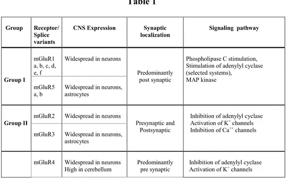
25 Genes encoding for 8 different mGluR subtypes have been identified and they are widely expressed in different cell types in the CNS. The receptors are subdivided into three different groups, based on sequence and pharmacological homologies (Table 1).
Class I receptors, coupled to a Gq protein, include mGluR1 and mGluR5; class II, coupled to a Gi/o protein, includes mGluR2 and mGluR3, whereas class III, coupled to a Gi/o protein, includes mGluR4, mGluR6, mGluR7 and mGluR8 (reviewed by Niswender and Conn 2010).
mGluRs modulate also other signaling pathways: class I receptors activate phospholipase D, Jun kinase, components of the MAPK/ERK pathway, and the mammalian target of rapamycin (mTOR)/p70 S6 kinase pathway (Li XM et al., 2007; Saugstad, JA et al., 2008, Page G et al., 2006; Hou Let al., 2004). Group II and III receptors also activate MAPK pathway and PI3 kinase pathways (Iacovelli L et al., 2002).
Table 1 Group Receptor/ Splice variants CNS Expression Synaptic localization Signaling pathway Group I mGluR1 a, b, c, d, e, f Widespread in neurons Predominantly post synaptic Phospholipase C stimulation, Stimulation of adenylyl cyclase (selected systems), MAP kinase mGluR5 a, b Widespread in neurons, astrocytes Group II
mGluR2 Widespread in neurons
Presynaptic and Postsynaptic
Inhibition of adenylyl cyclase Activation of K+ channels
Inhibition of Ca++ channels
mGluR3 Widespread in neurons, astrocytes
mGluR4 Widespread in neurons High in cerebellum
Predominantly pre synaptic
Inhibition of adenylyl cyclase Activation of K+ channels
26
GroupIII
mGluR6
a, b, c Retina ON-bipolar retinal Postsynaptic in cell Inhibition of Ca++ channels Stimulation of cGMP phosphodiesterase (mGluR6) mGluR7 a, b, c, d, e
Widespread in neurons Active zone of presynaptic
terminals mGluR8
a, b, c Lower and more restricted expression than mGluR4/7
Predominantly pre synaptic
Mod from Nicoletti et al., 2010
Several proteins can interact with the C-terminal tails of mGluR, regulating their signaling. In particular Homer proteins interact with the last several aminoacids of mGluR1-5 (Tu JC et al., 2008); through the interaction with Homer, class I mGluRs are associated with the long isoform of protein PI3 kinase enhancer (PIKE-L), preventing neuronal apoptosis (Rong R et al., 2003).
mGluRs are diffused in the CNS, where they can be localised at synaptic and extrasynaptic levels in both neurons and glia. They mainly modulate neuronal excitability and synaptic transmission, through their control on ion fluxes and regulation of signalling proteins.
Group I mGluRs are generally localised postsynaptically and their activation leads to cell depolarization and synaptic excitability. Group II and III are localised at presynpatic levels where they reduce the release of neurotrasmitters in both excitatory (glutamate), inhibitory (GABA) and neuromodulatory (Ach) terminals (reviewed inNiswender and Conn 2010). Preclinical studies suggested a potential role for drugs targeting mGluRs in multiple CNS disorders including depression (Pilc A et al., 2008), anxiety (Swanson CJ, et al., 2005), schizophrenia (Conn PJ et al., 2008; Moghaddam B et al., 2004), pain syndromes (Bleakman D et al., 2006), epilepsy (Alexander GM et al., 2006), Alzheimer’s disease (Lee HG et al.,
27 2004), and Parkinson’s disease (Conn PJ et al., 2005). These data are now beginning to be validated also in clinical studies.
mGluR II agonists have been proved, both in preclinical and clinical trials, to have good effects in the treatment of schizophrenia and anxiety disorders, (Conn PJ et al., 2008; Swanson CJ, et al., 2005,174, 175 ,176) as well as in pain, additive disorders, depression and epilepsy (reviewed in Brady, AE et al., 2008). In contrast, mGluR3 seems to be selectively implicated in neuroprotection, through the release of neurotrophic factors by astrocytes (Corti C et al., 2007).
mGluR4 agonists are promising drugs for the treatment of PD (Conn PJ et al., 2005), and evidence exists to suggest a role for class III mGluRs in depression, anxiety (Pilc A et al., 2008), neuroblastoma treatment (Iacovelli L et al., 2006) and neuronal differentiation (Saxe JP et al., 2007).
mGluR5 antagonists are useful tools in anxiety disorders (Swanson CJ, et al., 2005), chronic pain, addiction, depression, some neurodegenerative disorders (Slassi A, et al., 2005), migraine (Keywood C et al., 2008a) and gastroesofageal reflux (Keywood C et al., 2008b). mGluR5 is also a target in the treatment of X fragile syndrome (Bear MF et al., 2004; Yan QJ et al., 2005).
The role of mGluR1 in neurodegeneration is ambiguous. Due to the fact that mGluR1 activity produces excitatory effects in neurons, its activation has been related to induction and progression of excitotoxic neuronal death. Accordingly, mGluR1 agonists have been shown to have neurotoxic effects both in in vivo and in vitro models (Camon L et al., 1998; Bruno V et al., 1995). However, evidence also suggests that mGluR1 agonists induce neuroprotection (Pizzi M et al., 2000; Battaglia et al., 2001; Emery et al., 2010; Pshenichkin et al., 2008; Scartabelli et al., 2008; Zhou et al., 2009 ).
28 The role of mGluR1 in neurodegeneration or neuroprotection has been largely examined (Nicoletti F et al., 1999, Bruno V et al., 2001b) and it can be concluded that the dual activity observed depends on the cellular context and the experimental paradigm applied.
In an attempt to explain the dual role of mGluR1, Baudry and coworkers have proposed a model in which mGluR1 conformation can be modified as a consequence of high intracellular calcium concentration (Xu et al.,2007). As already described, mGluR1a signaling involves activation of a Gq protein, responsible for the stimulation of phospholipase C (PLC), which hydrolyzes membrane phosphoinositides and leads to inositol trisphosphate (IP3)-mediated Ca2+ release from intracellular stores (Masu et al., 1991). The neuroprotective effect of mGluR1 seems instead to be mediated by activation of PI3 K-Akt pathway through the formation of a mGluRI-Homer-PIKE-L signaling complex (Rong et al., 2003).
According to Baudry’ s group (2007) the increased calcium influx mediated by NMDA receptor activation, leads to calpain-mediated cleavage of the C-terminal tail of mGluR1, where the Homer binding motif is located, preventing the activation of the neuroprotective signaling. When activated after the cleavage, mGluR1 can only activate the PLC pathway, leading neurons to death (Fig 2).
29
Xu W et al., 2007 Neuron
Estrogen receptor and mGluR
As previously reported membrane ERs can interact with other receptors. In 2005 for the first time evidence emerged that ERs could transactivate mGluRs (Boulware MI et al., 2005).
In cultured hippocampal neurons, membrane ERα interacts with mGluR1 to increase, within few seconds, the phosphorylation of CREB. E2 also decreases L-type calcium channel-dependent CREB phosphorylation, through the interaction between either membrane ERα or ERβ and mGluR2/3 (Boulware MI et al., 2005). These effects were the result of treatment at physiological estrogen concentrations and were mimicked using E2 conjugated to BSA, highlighting the involvement of a membrane receptor, and inhibited by the ER antagonist, ICI 182,780. Moreover agonists/antagonists of mGluRs mimicked or prevented the effect of estrogen, respectively ( Kuo, J., et al.,2009).
The physical interaction between the receptors was demonstrated using co-immunoprecipitation studies (Dewing M.I et al., 2007). Specifically, in the
30 hippocampus ERα couples with mGluR1 whereas both ERα and ERβ co-immunoprecipitate with mGluR2/3. In contrast, in the striatum ERα couples to mGluR5 and both ERα and ERβ are linked to mGluR3 (Grove-Strawser D et al., 2010) suggesting that ERs can be coupled to different mGluRs (Table 2).
Table 2
Mermelstein 2010
Interactions between receptors are favoured by caveolins (CAV). These are
membrane proteins that organise the signal transduction machinery (Patel HH et al., 2008), and that are known to interact with mGluRs (Patel HH et al., 2008; Murray F et al., 2008) and ERs (Luoma JI, et al., 2008).
Mermelstein and co demonstrated that CAV1 is necessary for the interaction between ERα and mGluR1, while CAV3 is needed to allow the connection between mGluR2/3 and ERα and ERβ (Boulware MI et al., 2007; Grove-Strawser D et al., 2010). The interaction between these receptors has been demonstrated to occur in physiological conditions.
31 Interaction between mGluR1 and ERα in the arcuate nucleus seems to be important for lordosis behaviour (Dewing MI et al., 2007) while at the hypothalamus it mediates the production of neuroprogesterone, necessary for the LH surge (Sinchak K et al., 2003; Micevych P and Sinchak K, 2008; Micevych P et al., 2010).
Interaction between group II mGluR and ERs occurs also at the level of DRG, where estrogen stimulation inhibits L-type calcium channels (Chaban VV et al., 2003; Chaban VV and Micevych P, 2005). mGluRs signaling is also necessary for the estrogen-induced masculization of adult rat sex behavior and increases in dendritic spine density in the medial preoptic area (Wright and McCarthy, 2009)
32
33
Mol. Pharmacol. 2011, epub10/07/11
Estrogen receptors and type-1 metabotropic glutamate receptors are interdependent in protecting cortical neurons against β-amyloid toxicity
Simona Federica Spampinato1, Gemma Molinaro, Sara Merlo, Luisa Iacovelli, Filippo Caraci, Giuseppe Battaglia, Ferdinando Nicoletti, Valeria
Bruno, Maria Angela Sortino
Department of Clinical and Molecular Biomedicine (SFS, SM, MAS), Department of Drug Sciences (FC), University of Catania,
Department of Physiology and Pharmacology, (LI, FN) University of Rome Sapienza
and Istituto Neurologico Mediterraneo Neuromed Pozzilli (GM, GB, VB, FN), Italy
34 Running title: Interaction of ER and mGlu1 receptors in neuroprotection Corresponding author:
Dr. Maria Angela Sortino
Department of Clinical and Molecular Biomedicine University of Catania
Viale Andrea Doria 6 95125 Catania, Italy Tel 095-7384086 Fax 095-7384228 [email protected] Text pages: 28 Number of figures: 7 Number of Tables: 1 References: 59
Number of words in abstract: 225 Number of words in introduction: 659 Number of words in discussion: 1231
35 Abstract
We examined the interaction between estrogen receptors (ERs) and type-1 metabotropic glutamate receptors (mGlu1 receptors) in mechanisms of neurodegeneration/neuroprotection using mixed cultures of cortical cells challenged with -amyloid peptide. Both receptors were present in neurons, whereas only ERα, but not mGlu1a receptors, were found in astrocytes. Addition of 17--estradiol (17βE2) protected cultured neurons against amyloid toxicity, and its action was mimicked by the selective ERα agonist, 1,3,5-tris(4-hydroxyphenyl)-4-propyl-1H-pyrazole (PPT) as well as by a cell-impermeable BSA conjugate of 17E2. The selective ERβ agonist, diarylpropionitrile (DPN), was only slightly neuroprotective. The mGlu1/5 receptor agonist, 3,5,-dihydroxyphenylglycine (DHPG), was also neuroprotective against amyloid toxicity, and its action was abolished by the mGlu1 receptor antagonist, JNJ16259685. Neuroprotection by 17β or PPT (but not DPN) and DHPG was less than additive, suggesting that ERα and mGlu1 receptors activate the same pathway of cell survival. More important, neuroprotection by 17βwas abolished not only by the ER antagonist, ICI182,780, but also by JNJ16259685, and neuroprotection by DHPG was abolished by ICI182,780. ERα and mGlu1 receptors were also interdependent in activating the phosphatidylinositol-3-kinase pathway, and pharmacological blockade of this pathway abolished neuroprotection by 17βE2, DHPG, or their combination. These data provide the first evidence that ERα and mGlu1 receptors critically interact in promoting neuroprotection, an information that should be taken into account when examining the impact of estrogen on neurodegeneration associated with CNS disorders.
36 Introduction
Estrogens are neuroprotective in a variety of cellular and animal models, including cell cultures challenged with excitotoxins or other insults (Cimarosti et al., 2005; Goodman et al., 1996; Harms et al., 2001; Singer et al., 1999), models of focal or global brain ischemia (Dubal et al., 1998; Lebesgue et al., 2009; Simpkins et al., 1997), mice treated with the parkinsonian toxin, 1-methyl-4-phneyl-1,2,3,6-tetrahydropyridine (Bourque et al., 2009), and transgenic mice carrying mutations associated with Alzheimer’s disease (AD) (Amtul et al., 2010; Carroll et al., 2007). Estrogens are also effective in reducing -amyloid toxicity in cultured neurons (Chae et al., 2001; Cordey and Pike, 2005; Goodman et al., 1996; Marin et al., 2003; Sortino et al., 2004), an established cellular model of AD. The classical estrogen receptors, named ER and ER, are nuclear transcription factors that activate or repress gene expression (Nilsson et al., 2001). However, a large body of evidence suggests that neuroprotection is mediated by membrane ERs, which are able to induce rapid intracellular effects in response to estrogens (Micevych and Dominguez, 2009). More recently, a G protein-coupled receptor, GPR30 has been identified as an additional candidate membrane ER (Revankar et al., 2005; Thomas et al., 2005) and reported to mediate also estrogen neuroprotective effects against excitotoxicity (Gingerich et al., 2010). Membrane ERs trigger a variety of putative neuroprotective pathways, which include the mitogen activated protein kinase (MAPK) pathway (Mize et al., 2003; Singer et al., 1999), and the phosphatidylinositol 3-kinase (PtdIns-3-K)/Akt pathway (Cimarosti et al., 2005; Harms et al., 2001; Honda et al., 2000). The mechanism whereby membrane ERs activate the neuroprotective cascade is largely unknown.
37 It has long been known that membrane ERs can trans-activate different classes of tyrosine kinase receptors, including epidermal growth factor receptors (Song et al., 2010) and type-I insulin-like growth factor receptors (Marin et al., 2009; Varea et al., 2010). More recently, this mechanism of trans-activation has been extended to metabotropic glutamate (mGlu) receptors, which are G-protein coupled receptors. Eight subtypes of mGlu receptors (mGlu1 to mGlu8) have been described and divided into three groups on the basis of their amino acid sequence, pharmacological profile, and transduction pathways. Group-I subtypes (mGlu1 and mGlu5 receptors) are coupled to Gq, and their activation leads to polyphosphoinositide
hydrolysis with ensuing formation of inositol-1,4,5-trisphosphate and diacylglycerol. mGlu1 and mGlu5 receptors can also activate the MAPK and PtdIns-3-K pathways (Chong et al., 2006; Ferraguti et al., 2008). Group-II (mGlu2 and mGlu3) and group-III (mGlu4, mGlu6, mGlu7, and mGlu8) receptor subtypes are all coupled to Gi/Go proteins reviewed by (Nicoletti et al., 2011; Niswender and Conn, 2010). A series of elegant studies have shown that membrane ER receptors trans-activate mGlu1 receptors in the hypothalamus (Dewing et al., 2007; Dominguez and Micevych, 2010; Mermelstein, 2009; Micevych and Mermelstein, 2008). For example, trans-activation of mGlu1 receptors by ER in hypothalamic astrocytes leads to the synthesis of neuroprogesterone, which is necessary for estradiol-induced ovulatory surge of luteinizing hormone (LH) (Kuo et al., 2009; Micevych and Sinchak, 2008b). In hypothalamic neurons, stimulation of ER by estradiol leads to internalization of both ER and mGlu1 receptors, suggesting that the two receptors interact also in neurons (Dominguez and Micevych, 2010). In contrast, GPR30 does not seem to couple with mGlu1 receptor and to involve this receptor in modifying rapid intracellular Ca++ signalling in astrocytes (Kuo et al., 2010).
38 mGlu1 receptors are linked to mechanisms of neurodegeneration/neuroprotection, and can either amplify or attenuate neuronal death depending on the cellular context and the experimental paradigm of neurodegeneration (Allen et al., 1999; Battaglia et al., 2001; Bruno et al., 2001a; Bruno et al., 1999; Emery et al., 2010; Nicoletti et al., 1999; Pellegrini-Giampietro, 2003; Pshenichkin et al., 2008; Scartabelli et al., 2008; Zhou et al., 2009).
We now report that activation of either ER or mGlu1 receptors protects cortical neurons against -amyloid toxicity and that the two receptors are interdependent in supporting neuronal survival. This is the first evidence that ER and mGlu1 receptors interact in cortical neurons.
Materials and Methods:
Drugs and Reagents
17-β-Estradiol (17E2) (Sigma-Aldrich, St. Louis, MO), 1,3,5-tris(4-hydroxyphenyl)-4-propyl-1H-pyrazole (PPT), diarylpropionitrile (DPN), 7a,17-[9-[(4,4,5,5,5-Pentafluoropentyl)sulfinyl]nonyl] estra-1,3,5(10)-triene-3,17-diol (ICI 182,780) (all from Tocris Cookson Ltd, NorthPoint, UK) were dissolved in ethanol. 3,5-Dihydroxyphenylglycine (DHPG) and JNJ 16259685 (JNJ), both purchased from Tocris, were dissolved in dimethyl sulfoxide (DMSO, Sigma); 10-[4'-(N,N-Diethylamino)butyl]-2-chlorophenoxazine hydrochloride(10-DEBC) and 2-methyl-6-(phenylethynyl)pyridine (MPEP) (both from Tocris) were dissolved in water; BSA-conjugated 17β-E2(Sigma) was dissolved in 50% ethanol. -Amyloid peptides Aβ(1-42) and Aβ(25-35) were obtained from Bachem,
39 DMSO at an initial concentration of 5 mM whereas Aβ(25-35) was solubilized
in water at an initial concentration of 2.5 mM. All stock solutions were diluted in culture media as appropriate before use. [3H]-Myo-inositol (18 Ci/mmol) was purchased from GE Healthcare (Milan, Italy). Cell culture materials and all plastics, unless otherwise specified, were from Invitrogen (Carlsbad, CA) and Nunc (Rochester, NY). All drugs were used at concentrations reported in literature to be effective in the cellular system used. In the case of 17E2 and DHPG, concentration-response studies were carried out in a preliminar phase to allow choice of the concentration to be used.
Primary cell cultures
All animal experimentalprocedures were carried out in accordance with the directivesof the Italian and European Union regulations for the care anduse of experimental animals (DL116/92) and were approved bythe Italian Ministry of Health.
Cortical glial cultures were prepared from the cortex of 1- to 3-day-old Sprague-Dawley rats, (Harlan, Udine, Italy). After isolation of cortices and removal of meninges, cells were dispersed by mechanical and enzymatic dissociation using a solution of trypsin in HBSS (pH 7.4). Cells were plated onto 75 mm2 flasks and maintained in DMEM supplemented with 10% fetal calf serum (FCS), penicillin (100 U/ml), streptomycin(100 µg/ml), at 5% CO2 and 37°C for 14 DIV.
Confluent cultures were shaken for 7 hours at 37°C to remove microglia and oligodendrocytes and obtain a >90% pure astrocytic culture as assessed by GFAP staining. Astrocytes were replated at a density of approximately 1-2 x 10⁵ cells/cm2 and used when appropriate confluency was reached.
40 Cultures of pure cortical neurons were obtained from rats at embryonic day 15 (Harlan), prepared according to a procedurepreviously described (Sortino et al., 2004). Briefly, cortices were dissectedin Ca2+/Mg2+-free buffer (pH 7.4), mechanically dissociated and grown on multiwell vessels or 35-mmdishes precoated with 0.1 mg/ml poly-D-lysine (Sigma-Aldrich). Cultures were maintained in DMEM-F12 supplemented with the following components: penicillin (50 U/ml), streptomycin, (50 μg/ml), albumin from bovine serum (BSA 10 mg/ml), glucose (6 mg/ml), insulin (10 ng/ml), apotransferrin (10 ng/ml), putrescine (100 μM), glutamine (2 mM), selenium (30 nM), progesterone (20 nM) (all from Sigma-Aldrich). Arabinoside cytoside, (Ara-C 5 μM), was added 18 hours after plating to reduce non-neuronal element proliferation and maintained for 72 hours. Subsequent partial medium replacements were carried out every 2 days. After 7 DIV, cultures were treated for the experiments. These conditions yield a pure neuronal culture as shown by 99% immunostaining to the specific neuronal marker MAP2 as previously assessed by flow cytometry (Copani et al., 1999).
Mixed cortical cultures, containing both astrocytes and neurons, were obtained from rats at embryonic day 17 and grown onto 0.1 mg/ml poly-D-lisine-coated multiwell vessels. Cultures were maintained in MEM supplemented with penicillin (50 U/ml), streptomycin, (50 μg/ml), glucose (6 mg/ml), 10 % FCS, 10% horse serum, glutamine (2 mM) (all from Sigma-Aldrich). At 5 DIV FCS was removed from the medium and cells were supplemented with 5 μM Ara-C for 72 hours. Subsequent partial medium replacements were carried out every 2 days. The cultures were used for experiments at 14 DIV. Mature cultures contained about 40% neurons.
41 Aβ(1-42) and Aβ(25-35) peptides were applied to serum-deprived mature mixed
cortical cultures at 14 DIV. After 24 hours neuronal toxicity was examined by light microscopy and quantified after staining with trypan blue (0.4% for 5 min). Stained neurons were counted from three random fields/well. A variable number between eighty and three hundreds dead neurons per field were counted. All experiments were carried out in the presence of the glutamate receptor antagonists MK801 (10 μM) and DNQX (30 μM) to avoid endogenous glutamate toxicity.
Immunoblot analysis
Astrocytes and neurons were harvested in RIPA lysis buffer (Sigma-Aldrich) with the addition of Triton X-100 and a protease- and phosphatase-inhibitor cocktail mix (both from Sigma-Aldrich). Transfected HEK293 cells wererapidly rinsed in ice-cold PBS and solubilized in Triton X-lysis buffer (10 mM Tris-HCl, pH 7.4, 150 mM NaCl, 1% Triton X-100,1 mM EDTA, 10% glycerol, 1 mM phenylmethylsulfonyl fluoride, 10 µg/ml leupeptin, 10 µg/ml aprotinin, 1 mM sodiumorthovanadate, 50 mM sodium fluoride, and 10 mM -glycerophosphate). Proteins were quantitated by the Bradford protein assay (Bradford, 1976). Eighty micrograms of protein extract were separated by SDS-PAGE and transferred to nitrocellulose membranes using a Transblot semidry transfercell. After blocking in 1% non-fat dry milk, membranes were incubated with primary rabbit antibody anti-ERα (1:500 Millipore, Billerica, MA), rabbit anti-mGluR1 (1:750 Millipore), rabbit anti-pAkt (1:750 ; Cell Signaling Technology, Beverly, MA), followed by incubation with anti-rabbit HRP-conjugated secondary antibody (Santa Cruz Biotechnology, Santa Cruz, CA). Protein loading was determined using anti-Akt (1:1000; Cell Signaling Technology). In selected experiments, the same membranes were then reblotted with anti--actin
42 (Sigma; not shown). Specific bands were detected by enhanced chemiluminescence using the Immobilon detection system (Millipore). Full-range rainbow markers (GE Healthcare) were used to assess the size of the band. Densitometric analysis of band intensity was carried out with the aid of the ‘Image J’ software, developed by NIH and in public domain.
Coimmunoprecipitation
Neurons were harvested in RIPA buffer and protein concentration was determined by the Bradford method (Bradford, 1976); for coimmunoprecipitation, 500 µg of proteins, in a final volume of 500 μl, were incubated for 1 hour at 4 °C, in a rotating stirrer with 25 μl of rabbit serum to reduce non-specific binding. Twenty μl of protein G PLUS-Agarose (Santa-Cruz Biotechnology) were then added for 30 min at 4 °C to remove endogenous antibodies. Samples were centrifuged (850 rpm for 5 min) and supernatants were retained. Rabbit anti-ERα (1:100, Millipore) or rabbit anti-mGluR1 (1:100, Millipore) were added to supernatants and placed in a rotating stirrer at 4 °C for 7 hours. The antibody-protein complex was adsorbed with 20 μl of protein G-Plus Agarose (Santa Cruz Biotechnology) in a rotating stirrer at 4°C for 10 hours and then washed 5 times with a solution containing PBS and 1% Tween-20 (Sigma-Aldrich). Samples were run on SDS-PAGE, using 4-15% gradient gels (Bio-Rad Laboratories, Milan, Italy) and transferred to nitrocellulose membranes. After blocking in PBS solution containing 2% non-fat milk and 0.1% Tween-20, membranes were incubated with primary rabbit anti-mGluR1 antibody (1:750 Millipore) or rabbit anti-ERα (1:100, Millipore) , followed by incubation with HRP-conjugated anti-rabbit secondary antibody. Detection of specific bands was carried out with the Immobilon detection system (Millipore).
43
Immunostaining
Cells were fixed in 4% paraformaldehyde, permeabilized with 0,1% Triton X-100 and saturated with 3% BSA. Cells were then incubated with the following primary antibodies: rabbit mGluR1 (1:75) and mouse anti-ERα (1:25 Santa Cruz Biotechnology) overnight at 4 °C; mouse anti-GFAP (1:300 Cell Signaling) and mouse anti-MAP2 (1:120 Millipore) for 2 hours at room temperature. For fluorescent immunodetection the following fluorochrome-conjugated antibodies were used: Alexa-Fluor 488 anti-mouse (1:300, Invitrogen) and anti-rabbit Texas Red (1:75, Santa Cruz Biotechnology).
Studies in heterologous expression systems
Human embryonic kidney (HEK) 293 cells were cultured in DMEM supplemented with 10% FCS and antibiotics (100 U/ml penicillin, 100 µg/ml streptomycin). Cells were transfectedin 10-mm dishes using 10 µl of LipofectAMINE2000 in OptiMEM medium and 18 µg of total cDNA as follows: 7.5 g of mGlu1 receptor cDNA, 7.5 g of ER cDNA and 3 g of excitatory amino acid carrier 1 (EAAC1) cDNA. Transfections were carried out for 4 h and then cells were plated in culture medium in 6 well plates, previously coated with 0.01% poly(L-lysine). Using this procedure, about 80-85% of HEK293 cells are immunopositive to cotransfected green fluorescent protein. Experiments were performed 72 h after transfection, following a serum starvation of 16-18 h.
Measurement of polyphosphoinositide hydrolysis in cultured neurons.
Cortical neuronal cultures were incubated overnight with myo[3H] inositol (1 µCi/dish), washed in Krebs-Henseleit buffer containing 10 mM LiCl, and
44 incubated for 30 min at 37°C under constant oxygenation. DHPG and 17E2 were added and maintained for 30 min. Incubation was stopped by the addition of methanol:chloroform:water (1:1:1). After further addition of 300 μl chloroform and 600 μl water, samples were centrifuged at low speed to facilitate phase separation and the upper aqueous phase was loaded into Dowex 1-X-8 columns for separation of [3H]inositol phosphate (InsP).
Statistical analysis
Data shown are always mean + SEM of 3 to 6 independent experiments each run in triplicates. Data were analyzed by one-way Anova followed by Newman-Keuls test for significance. P<0.05 was taken as the criterion for statistical significance.
Results
Expression of ER and mGlu1 receptors in cortical neurons and astrocytes
Immunoblot analysis of ER showed a band at approximately 66 kDa. mGlu1a receptor antibodies labeled a major band at 140 kDa, corresponding to receptor monomers. The ER was detected in protein extracts from both pure cultures of cortical neurons and pure cultures of cortical astrocytes (Fig. 1a). In contrast, the mGlu1 receptor was found exclusively in pure cultures of cortical neurons (Fig. 1b). The cellular pattern of ER and mGlu1a receptor expression was confirmed by immunocytochemical analysis carried out in mixed cultures of cortical cells (the cultures used in toxicity studies). Double fluorescent immunostaining showed the expression of ER in both neurons and astrocytes (expressing MAP2 and GFAP, respectively; Fig. 1c,d). In contrast, mGlu1 receptors were exclusively