UNIVERSITÀ DEGLI STUDI DI ROMA
"TOR VERGATA"
FACOLTA' DI MEDICINA E CHIRURGIA
DOTTORATO DI RICERCA IN
Microbiologia medica ed Immunologia
XXI CICLO
Titolo della tesi
Role of type I IFN in the in vitro differentiation
of neonatal dendritic cells.
Nome e Cognome del dottorando
Dott. Valentina La Sorsa
Sommario
Introduction... 4
Neonatal immunity... 4
Dendritic cells biology ... 5
Role of dendritic cells in linking innate and adaptive immunity... 7
Features of umbilical cord bood DCs... 9
Type I IFNs ... 11
Role of Type I IFN in the in vitro differentiation of highly active dendritic cells from human peripheral blood ... 13
Background and aim of the study... 16
Materials and Methods... 18
Cell Separation and Culture... 18
Immunophenotypic Analysis... 18
Stat-1 intracellular staining... 19
Phagocytosis ... 19
Detection of cytokine production ... 19
Mixed leucocyte reaction... 20
Real Time quantitavive PCR to evaluate expression of TLRs and IRFs mRNA... 20
Bone marrow DC isolation and culture... 21
Comparison between neonatal and adult IFN-DCs and IL-4-DCs phenotype and IFN
receptor expression... 22
IFN signalling activation in neonatal and adult monocyte-derived DCs... 25
IFN-DCs and IL-4-DCs antigen-uptake and endosomal processing... 28
Different expression of TLRs between IFN-DCs and IL4-DCs... 30
Functional properties of IFN-DCs... 33
Neonatal bone marrow DCs: effects of IFN stimulation on costimulatory molecule expression and function ... 34
Discussion ... 38
Aknowledgements... 47
Introduction
Neonatal immunity
Early life is a period of high susceptibility to infectious diseases and a stage of immune maturation. The sensitivity of newborns to infectious diseases might be partly due to the lack of pre-existing immunological memory. Another important contributing factor might be the small number of immune cells that are present in peripheral lymphoid tissues, especially in mice. However, aside from these quantitative differences, many studies in both humans and mice have shown that newborn immune cells are qualitatively distinct from adult cells. Subsets of cells are present in different proportions in neonates and adults and, among cells of the same subtype, phenotypic and functional differences have been described. Notably, T-helper 2 (Th2)responses are preferentially induced as well as poor cytotoxicT-cell responses. The bias against TH1-cell-polarizing cytokines leaves the newborn susceptible to microbial infection and contributes to the impairment of neonatal immune responses to most vaccines, thereby frustrating efforts to protect this vulnerable population (Siegrist 2000). After birth, there is an age-dependent maturation of the immune response. Of note, prenatal and postnatal exposure to environmental microbial products that can activate innate immunity might accelerate this maturation process, particularly if the exposure occurs repeatedly over time (Ng, Lam et al. 2006), diminishing TH2-cell and/or enhancing TH1-cell polarization. Because of this impaired TH1-response, it was initially thought that the neonatal innate immune system was generally impaired or depressed; however,
stimulus-induced production of certain cytokines (for example, IL-6, IL-10 and IL-23) by neonatal monocytes and antigen-presenting cells (APCs) actually exceeds that of adults (Angelone, Wessels et al. 2006; Vanden Eijnden, Goriely et al. 2006). Much focus has been put on thestudy of intrinsic defects of neonatal T-cell functions, but under appropriate stimulatory conditions, neonatal T cellsmount adultlike response in vitro (Adkins 1999). In vivo, Th1 responses can be induced under certain circumstances in both mouse (Martinez, Brandt et al. 1997) and human (Marchant, Goetghebuer et al. 1999; Vekemans, Amedei et al. 2001) neonates. This led to the hypothesis that neonatal immune defects might result from a developmental immaturityof antigen-presenting cell (APC) functions.
Dendritic cells biology
Dendritic cells (DCs) are the most potent antigen-presenting cells (APC) characterized by a unique capacity to stimulate naïve T cells toward microbial and tumor antigens and to initiate primary immune responses (Cella, Sallusto et al. 1997; Liu 2001; Steinman and Banchereau 2007). DCs represent 0.5-1.5% of circulating human mononuclear cells derived from hematopoietic progenitor cells. In recent years, several studies have suggested that DCs also play critical role in the induction of central and peripheral immunological tolerance, regulate the types of T cell immune responses, and function as “sentinels” in innate immunity against microbes
distinguished in human blood: a major CD11c+ population, either CD1a+ or CD1a-, expressing the CD13, CD33 and GM-CSF-receptor (referred as myeloid DCs), and a CD1a-/CD11c- population expressing high levels of CD123 (IL-3Rα) known as plasmacytoid DCs (pDCs), which represent the major source of type I IFN upon virus challenge (Cella, Jarrossay et al. 1999; Siegal, Kadowaki et al. 1999; Liu 2001). The major pool of myeloid DC precursors is represented by monocytes, which give origin to interstitial DCs or tissue-resident DCs and Langherhans cells (LCs) (Caux, Vanbervliet et al. 1996). Immature DCs are generally located in peripheral tissues, in sites where they can optimally survey for incoming pathogens. The interaction of DCs with pathogens leads to migration to secondary lymphoid organs where they can initiate a specific T cell response. This complex process is associated with differentiation and functional activation of DCs (Cella, Sallusto et al. 1997; Banchereau and Steinman 1998; Steinman and Banchereau 2007). DCs represent an essential link between innate and adaptive immune response by acting as an interface between the environment and the immune system and by subsequently directing the quality of the adaptive response to incoming pathogens. Immature DCs are capable of efficiently capturing and internalizing a wide spectrum of antigens, bacteria, viruses, apoptotic bodies and necrotic cells, by different mechanisms such as conventional phagocytosis, macropinocytosis and receptor-mediated endocytosis via C-type lectin receptors, such as mannose receptor, DEC-205, DC-SIGN (CD209), FCγ receptors type I (CD64) and type II (CD32)(Bell, Young et al. 1999; Sallusto, Mackay et al. 2000). The transition from immature to mature DCs is characterized by phenotypic and functional changes, which are tipically induced by inflammatory cytokines such as IL-1 and TNF-α or by bacterial and virus-derived molecules, such as LPS, double
strand RNA, poly I:C and DNA. As a consequence, DCs loose their capacity to phagocytize and their sensitivity to some inflammatory chemokines (i.e., CCL3, CCL4 and CCL5), as they are induced to express a new and different set of chemokine receptors. The expression of CCR7, make mature DCs responsive to CCL20 and CCL21 chemokines, which are responsible for DC migration to the T cell areas in lymphoid tissues (Heath and Carbone 2001). Mature DCs express high levels of membrane accessory molecules, such as CD80, CD86 and CD40, upregulate MHC class I and II antigens and exhibit a strong capability to initiate an immune response. Of note, DCs are capable to exploit peculiar MHC class I-restricted antigen presentation pathways. Besides the classical presentations of endogenous peptides derived from intracellular proteins and pathogens, DCs are endowed with the aptitude to present MHC class I-restricted epitopes derived from exogenous antigens exploiting unconventional antigen processing pathways, ensuring the efficient “cross priming” of cytotoxic CD8+ T cells (Albert, Sauter et al. 1998; Heath and Carbone 2001).
Role of dendritic cells in linking innate and adaptive immunity
Innate recognition of infection in vertebrates can lead to the induction of adaptive immune responses through activation of dendritic cells (DCs). DCs are activated directly by conserved pathogen molecules and indirectly by inflammatory mediators produced by other cell types that recognise such molecules. Cells of the innate
molecular patterns (PAMPs), from viruses, bacteria, fungi and protozoa. The best-studied pattern-recognition receptors are toll-like receptors (TLRs). The engagement of TLRs on DCs leads to increased expression of MHC–peptide complexes. Each member of the TLR family recognizes a specific set of bacterial- or viral-derived molecules and co-stimulatory molecules, as well as regulates the production of immunomodulatory cytokines, all of which have a profound effect on T-cell priming and differentiation, thus indicating that TLRs link innate and adaptive immunity (Reis e Sousa 2004; Reis e Sousa 2004; Steinman and Hemmi 2006) The activation of many of them (TLR3, TLR4, TLR7, TLR8, and TLR9) results in the induction of type I IFN gene expression in different DC subsets (Levy, Marie et al. 2003; Honda, Yanai et al. 2005; Akira 2006).
The availability of defined cell culture conditions for generating relatively large numbers of DCs from cell precursors has allowed an impressive advance in our comprehension of DC biology. DCs can be obtained from CD34+ stem cells and differentiated in vitro upon exposure to cytokine cocktails, including GM-CSF, IL-4 and TNF-α (Caux, Dezutter-Dambuyant et al. 1992; Luft, Pang et al. 1998). Currently, large numbers of immature DCs are obtained from monocytes cultured in the presence of GM-CSF and IL-4 for 5-7 days (Sallusto and Lanzavecchia 1994). GM-CSF is required to ensure monocyte survival, whereas IL-4 exerts an inhibitory activity on macrophage differentiation, promoting the generation of immature DCs (Romani, Gruner et al. 1994). Maturation/activation of DCs is reached through a further culture step with maturation agents, such as LPS and Poly-I:C (ligands of Toll-like receptor), TNF-α (pro-infiammatory cytokine), CD40L or with a cytokine cocktail known as the “golden standard” (IL6, IL1β, TNF-α and prostaglandin
E2)(Jonuleit, Kuhn et al. 1997). Even though the IL-4/GM-CSF culture method has allowed to perform many important studies on DC biology, this pathway of DC differentiation may not reflect the physiological process by which monocytes differentiate into DCs in vivo, as high levels of IL-4 are unlikely to be present in the course of a natural immune response to infections. Since type I IFNs are rapidly produced in response to viruses and other stimuli, these cytokines could represent early danger signals ensuring an efficient link between innate and adaptive immunity, thus acting as physiological factors involved in DC differentiation.
Features of umbilical cord bood DCs
In newborns, the cells of the innate immune system - in particular, DC populations - are low in absolute and relative numbers and are not capable of fully activating both the innate and adaptive immune responses for effective antigen-specific B- and T-cell responses (Ridge, Fuchs et al. 1996; Wu and Liu 2007) . In mice, these deficiencies in neonatal DC lead to the development of Th2 immune response to many antigenic stimuli; however, this Th2 skewing is not readily observed in humans (Lambert, Liu et al. 2005). In human newborns, T-helper responses are often diminished in magnitude, which can affect both Th1- and Th2-cell function (Vekemans, Ota et al. 2002; Gasparoni, Ciardelli et al. 2003). Several subpopulations of DCs have been described in humans and in mice (Banchereau, Briere et al. 2000). Neonatal mice
(IFN-I secreting DC population). In addition, neonatal DCs seem to be functionally immature in some settings, such as under low inflammatory conditions (Sun, Fiette et al. 2003; Dakic, Shao et al. 2004). In humans studies on cord blood DC reveal that the pDC-to-mDC ratio (3:1) is reversed compared with the ratio found in adults (1:3) (Borras, Matthews et al. 2001). Whereas myeloid cells are far more abundant in adult blood (about 60–70% of total DCs), lymphoid DCs are the main subset present in cord blood (about 75% of total DCs). One possible explanation of the lymphoid predominance in cord blood could be the necessity of colonization of newly forming tissues in the fetus. This hypothesis would agree with the model proposed by O'Doherty et al (1994), who suggested that the bone marrow-derived CD11c− population could migrate to non-lymphoid tissues and remain there until stimulated. Thus highlighting the importance of populating neonatal lymphoid tissues with DCs that can (in the short term after birth) mediate both innate and/or adaptative immune responses (Kadowaki et al, 2000) through the production of type I interferons (Cella et al, 1999; Siegal et al, 1999).
In cord blood mDC and pDC were found to be in a more immature state than those in adult peripheral blood as they express lower levels of MHC class II, CD80 and CD86 (Hunt, Huppertz et al. 1994; Encabo, Solves et al. 2007). Using a whole blood assay, maturation of cord blood mDC induced by LPS or poly (I:C) was incomplete compared with adult cells (De Wit, Tonon et al. 2003). Similar observations are made when cord blood pDC were stimulated with CpG oligonucleotides or R-848 (De Wit, Olislagers et al. 2004) (Danis, George et al. 2008).When cord blood monocytes are cultured in vitro in the presence of IL-4 and GM-CSF, they differentiated into
immature mo-DC, which displayed reduced HLA-DR, CD80 and CD40 surface expression and underwent only partial maturation upon LPS stimulation (Goriely, Vincart et al. 2001) (Langrish, Buddle et al. 2002) .
Notably cord and adult monocyte-derived DCs behave in distinct ways following stimulation, even though they have comparable expression of surface markers and morphology in their immature state. Adult derived DCs are readily activated by inflammatory stimuli, such as LPS such activated adult DCs consequently have a greater capacity to stimulate allogeneic T cells, increasing their cytokine synthesis and proliferation (Verhasselt, Buelens et al. 1997). On the contray the response of cord DCs to LPS stimulation is consistently abrogated, with little or no change in marker expression characteristic of maturation, and almost complete failure to produce IL-12p70. As a consequence, they exhibit a decreased ability to stimulate naïve allogeneic adult CD4+ T cells to produce IFN-γ, when compared to adult DCs. Cord T cells stimulated in the same system produced lower amounts of IFN-γ compared to their adult counterparts, but cord DCs were again less able to stimulate them than those derived from adult blood. These findings, consistent with other recent studies, suggest that they are intrinsically less able to direct Th-1 polarization.
Type I IFNs
IFNs represent a growing family of cytokines which share antiviral activities and overlapping signalling pathways. IFNs have been classified in three types based on
IFN-λ1, IFN-λ2 and IFN-λ3. Type I IFNs use a receptor composed of IFNAR1 and IFNAR2 chains, type II IFN binds to a receptor formed by IFNGR1 and IFNGR2 chains, and type III IFN receptor consists of IFNLR1 and IL10R2 chains (Pestka, Krause et al. 2004; Coccia 2008). Type I IFNs are currently recognized as “danger” molecules signalling the presence of a wide variety of infectious agents and providing a link between innate and adaptive immunity (Gallucci and Matzinger 2001; Santini, Di Pucchio et al. 2002; Tough 2004).
Type I IFNs, especially IFN-α, originally considered as a simple antiviral substance, are important cytokines for the generation of a protective T cell-mediated immune response to virus infections and tumor growth. IFN-α is present at low levels under normal physiological conditions, but can be secreted at high levels soon after cell exposure to viruses or other stimuli (Belardelli, Vignaux et al. 1984; Belardelli and Gresser 1996; Mohty, Vialle-Castellano et al. 2003). However, within the immune system dendritic cells are unique for their ability to recognize and respond to a variety of pathogens through the release of this cytokine and, at the same time, to act as guardians against pathogens and as potent antigen presenting cells capable of inducing an appropriate immune response(Reis e Sousa 2004; Steinman and Hemmi 2006).
IFN-α and IFN-β are currently the most used cytokines in clinics, especially in the treatment of cancer and certain infectious diseases. Early studies had reported multiple effects of IFN-α/β on the immune system, including the enhancement of macrophage functions and of natural killer (NK) cell activity (Belardelli, Vignaux et al. 1984; Belardelli 1995; Belardelli and Ferrantini 2002). Besides its well known role in innate immunity, type I IFNs are considered to play a role in the shaping of
adaptive immunity, as previously demonstrated in mouse tumor models (Belardelli, Vignaux et al. 1984; Belardelli 1995) and more recently by a series of papers highlighting the role of type I IFNs, in particular of IFN-α, in the modulation of T cell functions, including the polarization of T-helper cells toward the TH-1 type of immune response and the generation/activation of cytotoxic T lymphocytes (CTL) (Belardelli and Ferrantini 2002; Ferrantini, Capone et al. 2007).
Role of Type I IFN in the in vitro differentiation of highly active dendritic cells from human peripheral blood
In recent years, a number of studies have investigated the effects of IFN-α and of IFN-β on the differentiation of human DCs. In spite of some contrasting results, probably due to the experimental setting, several groups have independently demonstrated that type IFNs efficiently promote the differentiation of peripheral blood monocytes into DCs. In particular, monocytes exposed to IFN-α together with GM-CSF acquire the features of fully functional partially mature DCs (IFN-DCs) after no more than 3 days of culture (Paquette, Hsu et al. 1998; Santini, Lapenta et al. 2000).
IFN-DCs undergo an early loss of adhesion to the substrate and are bent to form large cell clusters, developing long dendritic-like processes in a couple of days. The expression of membrane molecules such as CD80, CD86 and CD40, the intercellular adhesion molecule, ICAM-1 (CD54) and HLA-DR is rapidly enhanced, while low
Even though the majority of DCs generated in the presence of IFN-α displayed features of immature DCs, marker of activated/mature DCs, such as CD83, could be unexpectedly detected in a variable percentage (12%–30%) of cells, concomitantly with high levels of costimulatory molecules .
Consistently with their partially activated phenotype and in agreement with the attribution of IL-15 production to CD83-expressing DCs, IFN-DCs produce significant amounts of IL-15 in the culture supernatant (Santini, Lapenta et al. 2000; Parlato, Santini et al. 2001). Very recent studies have shown that monocyte-derived IFN-α-induced DCs combine features of NK cells and mature DCs, as demonstrated by anti-CD56 antibody staining and microarray gene expression profiling that revealed a higher expression of genes coding for DC maturation markers and molecules linked to DC migration to the lymph nodes, like DC-LAMP, CCR7 and CD49d, as well as for markers of NK cells, including granzymes and TRAIL.
IFN-DCs also exhibit an enhanced chemotactic response and migration activity. They express very high levels of CCR5 and exhibit an enhanced response to its ligands CCL5, CCL3 and CCL4, while a remarkable fraction of IFN-DCs expresses CCR7, shows a migratory response to CCL19 and expresses significant levels of CCL19 themselves, together with CCL18 and CXCL10 (Parlato, Santini et al. 2001).
IFN-DCs are able to stimulate the proliferative response of allogeneic T cells and the production of high levels of IFN-γ in mixed lymphocyte reactions even at very low stimulator/responder ratios.
A typical feature of professional APCs is represented by their ability to successfully prime naïve T cells and of inducing an effective immune response. In vitro priming of
autologous T cells with antigen pulsed IFN-DCs has been shown to induce a strong lymphocyte proliferation and a Th1 polarized response, as revealed by production of high levels of IFN-γ and the virtual absence of IL-4 in the culture upon restimulation (Santini, Lapenta et al. 2000; Parlato, Santini et al. 2001; Lapenta, Santini et al. 2003).
Background and aim of the study
In recent years, a number of reports provided evidence on the importance of type I IFNs in the differentiation, survival and function of several cell types including T cells and DCs (Brassard, Grace et al. 2002). IFN-I are involved in innate defense against pathogens, but also promote differentiation/activation of DCs in both adult human and mice (Banchereau, Briere et al. 2000). In particular, the identification of a rare blood population of natural interferon-producing cells (NIPC), also named plasmacytoid DCs (pDCs), which produce large amounts of type I IFNs in response to viral stimuli or synthetic TLR agonists (reviewed in ref.(Fitzgerald-Bocarsly and Feng 2007) and references therein), has contributed to the understanding of the immunomodulatory role of this cytokines. IFN production by pDCs acts as an autocrine survival factor, although it does not stimulate the differentiation of these cells into DCs. Conversely, IFN-/ has been shown to promote the rapid differentiation of GM-CSF-treated human peripheral blood monocytesinto DCs with potent T cell and B cell stimulatory activities (Santini, Lapenta et al. 2000; Honda, Sakaguchi et al. 2003).
In this study we focused on DC, as these are important in the first line of defense and are the coordinators of the innate as well as the specific immune response, and on the role of type I IFN in differentiation/maturation of DCs.
Recently it has been demonstrated that highly active partially mature DCs are generated from adult peripheral blood monocytes after a single step of 3-day culture with IFN-α/GM-CSF (IFN-DCs) (Santini, Lapenta et al. 2000). IFN-DCs proved to
be more effective than immature DCs generated in the presence of GM-CSF and IL-4 in inducing a Th-1 type of immune response and CD8+ T cell responses against defined antigens in different models (Carbonneil, Aouba et al. 2003; Lapenta, Santini et al. 2003; Mohty, Vialle-Castellano et al. 2003; Gabriele, Borghi et al. 2004). These results suggested that IFN-I could also play a role in the maturation/differentiation of neonatal DCs and in the modulation of Th-1 response which is impaired in neonates. In particular cord blood-derived monocytic cells were cultured in vitro in the presence of GM-CSF and IL-4 or, alternatively, GM-CSF and IFN-α. DCs obtained by the two different methods were analysed in terms of phenotype and functional activity. Wherever possible results from neonatal cord blood-derived DCs were compared with adult peripheral blood-derived DCs.
In the mouse model, namely C57BL6 mice, we studied the role of IFN α in the differentiation and maturation of bone marrow DC from neonatal mice and the functional properties of DCs upon stimulation with a mycobacterial antigen in vitro. An improved basic understanding of neonatal immune response in humans and mice may open doors for improved protection of newborns and young adults against infectious agents or deviated immune responses later in life.
Materials and Methods
Cell Separation and Culture
Peripheral blood and cord blood mononuclear cells were obtained from heparinized blood of healthy donors by Ficoll density gradient centrifugation (Seromed). Monocytes were isolated by immunomagnetic selection (MACS Cell Isolation Kits; Miltenyi Biotec). Positively selected CD14
+
monocytes were plated at the concentration of 2 x 10
6
cells/ml in RPMI medium (GIBCO BRL), supplemented with 10% Fetal Calf Serum (Euroclone), 500 U/ml GM-CSF (Peprotech) and either 250 U/ml IL-4 (R&D Systems) for 3 to 7 days or 10,000 U/ml natural IFN-α (Alfaferone; AlfaWasserman) for 3 days. Both dendritic cells were washed and analyzed by cytofluorometric analysis for specific marker expression.
Immunophenotypic Analysis.
Cells were washed and resuspended in PBS containing 1% human serum and 0.09% NaN3 and incubated with a panel of fluorochrome-conjugated mAbs specific for IFN-DC and IL4-IFN-DC anti-CD80 (Becton Dickinson),CD1a CD40, CD86, CD83, HLA-DR. Isotype matched antibodies were used as a negative control. Cells were analyzed by flow cytometry by using a FACSortTM (Becton Dickinson) flow cytometer. Fluorescence data are reported as the percent positive cells when treatment induces expression of the marker in cells that were negative; data are reported as median fluorescence intensity (MFI) when treatment increases expression of the marker in cells that were already positive.
Stat-1 intracellular staining
1x10E+6 cells per sample were stimulated with 7500 U/ml of natural IFN-α (Alfaferone; AlfaWasserman) for 15 min at 37°C and then fixed by 1,5% PFA.Cells permeabililzation was performed by adding chilled methanol. Cells were washed and incubated with fluorochrome-conjugated mAbs specific for STAT1 and phosphorilated STAT1 molecules (Becton Dickinson), and analyzed by flow cytometry.
Phagocytosis
DCs (0.5x10 6
cells) were incubated for 60 min at 37°C with either 50 µg/ml of OVA-FITC conjugate (Molecular Probes). Cells were washed and resuspended in 500 µl of PBS. DCs incubated with dextran-FITC at 4°C were used as control. Cells were analysed by flow cytometry.
Detection of cytokine production
Commercial ELISAs were used to quantitate in the cell culture supernatants at day 3 the following cytokines: TNF-α (Endogen), IL-12 (Bender MedSystem), IFNγ (R&D Systems). Assay sensitivity was as follows: IFN-γ (25,6pg/ml), TNF-α (15.6pg/ml), IL-12 (7.8pg/ml), ELISAs were performed in triplicate and laboratory standards were included on each plate. Optical density was measured with a microplate reader at 450 nm (Dynex Opsys MR)
Mixed leucocyte reaction
IFN-DCs and IL4-DCs were incubated at different ratios with heterologous naïve CD3+ cells, purified from peripheral blood mononuclear cells by positive sorting with magnetic beads (92%), and seeded into round-bottom 96-well plates (Costar, Cambridge, Mass.) at 2.5x10
4
cells/well. After 5 days, 0.5 µCi of [ 3
H]thymidine (Amersham Biosciences, Little Chalfont, Buckinghamshire, United Kingdom) were added to each well and incubation was continued for additional 18h. Thymidine incorporation was quantified by scintillation counting using Microbeta.
Real Time quantitavive PCR to evaluate expression of TLRs and IRFs mRNA
To measure cytokine mRNA expression, TaqMan real-time reverse transcriptase PCR (RT-PCR) analysis was used (Applied Biosystems, Foster City, Calif.). Total RNA was extracted from monocytes, IFN-DCs and IL4-DCs. TaqMan assays were performed according to the manufacturer’s instructions with an ABI 7700 thermocycler (Applied Biosystems). PCR was performed, amplifying the target cDNA (TLR7, TLR8, TLR9, IRF3, IRF7) with ß-actin cDNA as an endogenous control. Data were analyzed with the PE Relative Quantification software of Applied Biosystems. Specific mRNA transcript levels were expressed as fold increase over the basal condition (untreated monocytes).
Bone marrow DC isolation and culture
C57Black/6 mice were obtained from Charles River-IT. All micewere used at 1 week ofage.
DCs were generated from cultures of bone marrow cells. Briefly, bone marrow was extractedfrom the tibia and femur, and cell suspensions were cultured for6 days in Iscoves modified Dulbecco medium (IMDM) (Life Technologies) containing 10% heat-inactivated fetal calf serum (FCS), 50 µM 2-ME, 100 U/mL penicillin, 100 µg/mL streptomycin,100 U/mL and 10 ng/mL recombinant murine granulocyte-macrophagecolony-stimulating factor (rmGM-CSF) (R&D Systems). Fresh medium was given every other day. On day6, loosely adherent cells were harvested, washed, and replated in fresh medium. Phenotypic analysis and functional assays were performed between days 7and 12. CD11c+ ranged between 95% and 98% without any further sorting or treatment.
Results
Comparison between neonatal and adult IFN-DCs and IL-4-DCs phenotype and IFN receptor expression
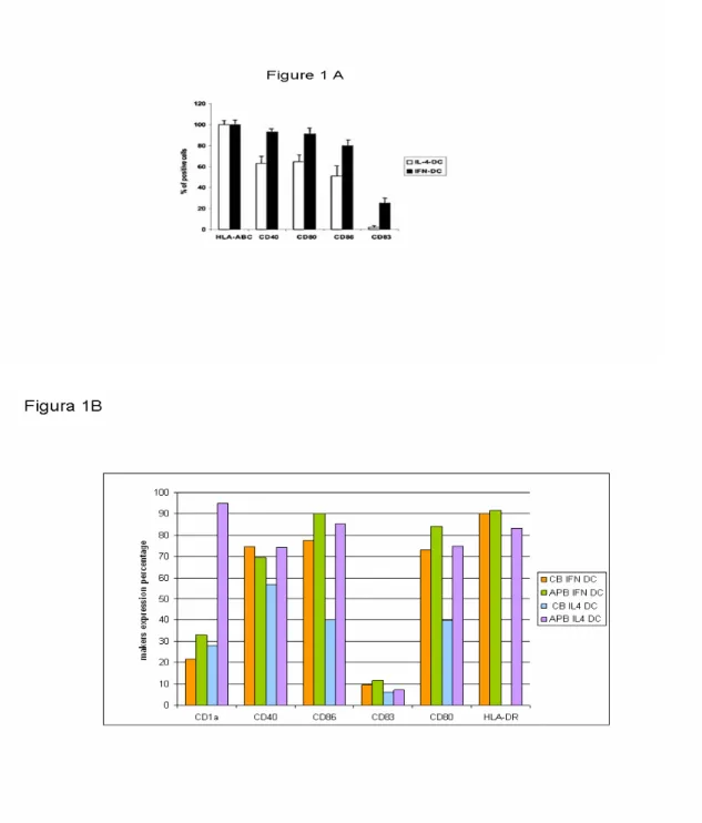
Figure 1A shows the phenotype of the two types of DCs used in these experiments derived from IFN treatment of peripheral blood and cord blood monocytes. Consistently with previously published results (Santini, Lapenta et al. 2000) peripheral blood IFN-DCs were characterized by a higher percentage of cells expressing CD40, CD80, CD86 (Figure 1A). The up-regulation of membrane expression of these markers can be also associated with the appearance of the DC maturation marker CD83
+
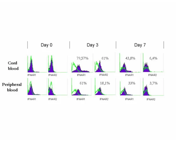
. Notably, APB IFN-DCs nearly exhibited a two-fold increase of HLA Class-I molecule expression intensity as compared to APB IL-4 DCs (data not shown). Cord blood IFN DCs were found to have a more immature phenotype as they express lower lewels of CD1a, CD80 and CD86 with respect to the IFN adult counterpart. Similar observations were made when cord blood monocytes were cultured in the presence of GM-CSF and IL4 as they displayed reduced HLADR, CD80 and CD40 expression compared to the adult IL4 DCs (Figure 1B). Of note, in cord blood monocytes/DCs, IFN induced a higher degree of differentiation with respect to IL4. To gain insight into the mechanism(s) involved in the weakened response to IFNα of neonatal DCs, time-course experiments were conducted to investigate the surface expression of type I IFN receptor chains, IFNAR1 and IFNAR2, during DC differentiation. As shown in Fig. 2, circulating cord blood and
peripheral blood monocytes expressed comparable low levels of both receptor components. However,at day 3, a marked increase inthe surface expression of both receptor chains was observed in APB as well as in CB coltures, concomitantlyto a significant increase in CD86 and CD83 expression in APB DCs. At latertime points, such as day 7 the surface expression of IFNAR1 andIFNAR2 was barely detectable. No significant differences between cord blood and peripheral blood monocytes were found in the IFN receptor expression during their in vitro differentiation into DCs.
Figure 1. Phenotype of PB and CB IFN-DCs and IL-4-DCs. (A) Percentage of APB DCs expressing a series of selected membrane markers when differentiated in the presence of IFN or IL4. (B) Percentage of selected membrane markers expressed on APB and CB DCs differentiated in the presence of IFN or IL4.
Figure 2.Time-course analysis of IFNAR1 and IFNAR2 subunit surface expression on APB and CB monocytes during the course of their in vitro differentiation. APB and CB DCs, generated in the presence of IL4, were labeled with the indicated Abs, fixed, and analyzed by flow cytometry.
IFN signalling activation in neonatal and adult monocyte-derived DCs
The signaling activity of type I IFN receptor during the course of in vitro DC differentiation was evaluated by monitoring the IFNα induced tyrosine
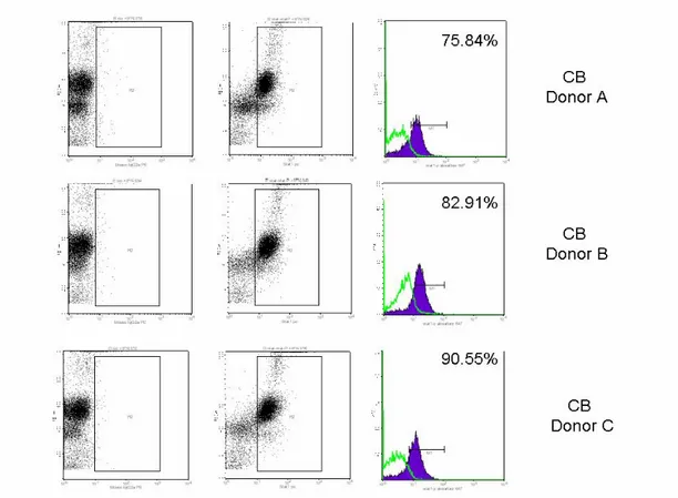
results of a representative experiment in which we analyzed the tyrosine phosphorylation ofSTAT-1 in peripheral blood and cord blood monocytes after IFNα stimulation (7500U/L dose). Only cord blood histograms are shown. After a 15 min stimulation a marked tyrosine phosphorylation of STAT1 was observed both in neonatal and adult monocytes at a comparable level ranging from 75% to 90%. Notably, when peripheral blood DCs were stimulated with LPS, a higher expression of STAT-1 was consistently detectedin LPS stimulated mature DCs with respect to immature DCs (data not shown).
Figure 3. Phosphorylation state of STAT-1 in CB monocytes. Monocytes were stimulated with IFNα (7500 U/L) for 15 min. Cells were fixed, labeled with phosphorylation-specific Abs and analyzed by flow cytometry. The percentage of STAT1 phosphorylated form with respect to the unphosphorilated is shown for each donor.
IFN-DCs and IL-4-DCs antigen-uptake and endosomal processing
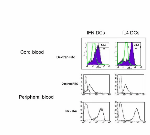
Antigen uptake by DCs is mediated predominantly by either mannose receptor-mediated endocytosis or macropinocytosis, which are modulated during DC differentiation. We have evaluated mannose receptor-mediated endocytosis by measuring the uptake of FITC-conjugated dextran or ovalbumin, while macropinocytosis and endosomal processing capacity has been evaluated by the uptake of DQ ovalbumin, which is a self-quenched conjugate of albumin exhibiting bright green fluorescence upon endo-lysosomal protease-dependent degradation, thus permitting the evaluation of both antigen uptake and processing by live DCs. As illustrated in Figure 4C, no major difference in the ovalbumin uptake capacity was detected between the two DC types (IFN-DCs and IL-4-DCs) from peripheral blood. Likewise, both DC types exhibited similar FACS profile after incubation with DQ ovalbumin (Figure 4D), suggesting that the majority of cells retained comparable phagocytic and processing activity. In particular, time-course analyses of antigen uptake and processing revealed similar kinetics for both DC types (data not shown). On the contrary cord blood IFN-DCs showed a less efficient uptake capability with respect to the CB IL4-DCs (Figure 4E). The higher degree of immaturity found in cord blood IFN-DCs phenotype is associated to reduced antigen uptake rates.
Figure 4. Antigen uptake and processing by the IFN-DCs and IL-4 DCs. Cells were incubated for 60 min at 37°C with 50 µg/ml of dextran-FITC conjugate (E and C) or 100 µg/ml of DQ-Ovalbumin. After 60 min, cells were washed and analysed by Flow cytometry. DQ ovalbumin is a self-quenched conjugate of albumin that exhibits bright green fluorescence only upon proteolityc degradation.
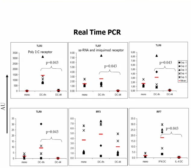
Different expression of TLRs between IFN-DCs and IL4-DCs
The expression pattern of TLRs in APB IFN-DCs and IL4-DCs have been compared by Real Time PCR analysis. The relevant results are shown in Figure 5, which shows differerential expression of some TLRs in this two classes of DCs. TLR3, known to be constitutively expressed in myeloid dendritic cells, appeared to be expressed at higher level in IFN-DCs as compared to IL4-DCs. TLR7 has been found to be expressed exclusively by IFN-DCs and not by IL4-DCs, as previously observed by Mohty and colleagues (Mohty M. et al., 2003). Interestingly, low levels of TLR9 transcription have been evidenced in IFN-DCs. Since TLRs downstream signalling involve the activation of several transcription factors including the Interferon Regulatory Factors (IRFs) some of which are specifically expressed or induced in different classes of DCs, we have comparatively analyzed IRF3 and IRF7 expression in APB IFN-DCs and IL4-DCs. No significant differences in the expression of IRF3 were foundbetween IFN-DC sand IL4-DCs (Figure 5). Interestingly, expression of IRF7, known to be transcribed in mature DCs, has been selectively detected IFN-DCs but not in immature IL4-DCs (Figure 5). TLRs expression in coord blood monocytes versus peripheral blood monocytes was assessed only by immunofluorescent staining, real time PCR is still ongoing. All monocytes from both APB and CB samples expressed TLR-2 and TLR-4 but not TLR-3. (Table 1) No significant differences were observed between CB and APB concerning the frequency of TLR-2 expressing cells or the amount of TLR-2 expressed per cell. Despite the comparable frequencies of monocytes expressing TLR-4, those from CB expressed lower amounts of TLR-4 than those from APB (p < 0.05).
Figure 5. Evaluation of the levels of mRNA expression of the TLRs and IRFs by TaqMan real-time RT-PCR. Quantitative RT-PCR for TLR3, TLR7, TLR8, TLR9, IRF3, IRF7 gene expression by DCs was performed. Specific mRNA transcript levels
Table 1.
TLRs expression monocytes from peripheral blood and cord blood assessed by cytofluorimetric analysis
Adult Peripheral blood Cord blood
CD14+ monocytes TLR-2 % 100 ± 0 (100) 100 ± 0 (100) MFI 118 ± 62.5 (105) 116 ± 41.2 (111) TLR-3 % 0 ± 0 (0) 0 ± 0 (0) MFI N.D. N.D. TLR-4 % 98.8 ± 1.5 (99.2) 91.8 ± 5.9 (94) MFI 79.9 ± 41.7 (72) 42.4 ± 21.1 (32.8)a
Results are expressed as Mean ± SD (median).
% = Percentage of positive cells; MFI = Mean Fluorescence Intensity of positive cells. Statistically significant differences were considered when p < 0.05 (Mann– Whitney test).
a
Functional properties of IFN-DCs
To investigate the function of IFN-DCs their ability to stimulate etrologous T lymphocytes in a MLR was compared with that of IL-4-DCs. At all stimulator:responderratios, APB IFN-DCs were found to be superior asstimulators to APB IL-4-DCs (figure 5). Conversely CB IFN DCs were poor stimulators both at day 3 and at day 5 with respect to IL4 cord blood DCs (data not shown). Moreover, as CB DCs are at a phenotypically more immature state when comparedwith PB-DCs, T cell proliferation responses induced by this cells are lower with respect to the peripheral blood.
Figure 6. We have assayed the proliferation activity of eterologous CD3+ T cells, with both types of DCs. The IFN-DCs from peripheral blood exhibited a significantly higher efficiency in inducing the proliferation of T cells as compared to IL4-DCs. Conversely this ability of IFN-DCs was absent in the proliferation assays from coord blood.
Neonatal bone marrow DCs: effects of IFN stimulation on costimulatory molecule expression and function
Bone marrow derived DCs precursors were cultured in vitro with GM-CSF and IFNα alone or in combination with the HBHA (heparin binding haemoagglutinin, a mycobacterial adhesin) antigen (10 µg/ml), wich is a strong maturative stimulus; LPS induction (0,5 µg/ml) was used as a control. Figure 7A shows the costimulatory molecule expression of CD11c low precursors after in vitro treatment. IFNα treatment proved to be similar to HBHA and LPS stimulation in terms of CD40, CD86 and Class II expression, only the combination of HBHA plus IFNα enhanced DC maturation expecially for Class I expression. The analysis of percentage of mature cells after the stimulation confirmed that IFN alone was not superior to the other stimuli, but it appeared to have an additive effect when used in combination with HBHA (figure 7B) and other antigens (data not shown). Neonatal bone marrow DCs treated with IFNα, LPS and HBHA were assayed for their ability to induce proliferation in CD4+ and CD8+ mouse lymphocytes (percentage of proliferating cells were calculated on total splenocytes). As illustrated in Figure 7C IFN treated
DCs were more efficient inducers, followed by LPS treated DCs. The synergic effect of IFN treatment with HBHA was also confirmed.
Figure 7.CD11c low neonatal bone marrow DCs: (A)costimulatory molecules expression is indicated as percent values in relative gates after in vitro treatment with IFNα, LPS and HBHA.(B) Effects of in vitro stimulation on cell maturation (SSC/FSC plots).Blue dots in plots represent CD11c low marker+ BM-DCs and blue numbers represent their relative percentage.(C) Proliferating CD4+ and CD8+ lymphocytes in MLR assays with treated neonatal bone marrow DCs. The DC/lymphocytes ratios were 1:1 and 1:2.
Discussion
In the present study, we provided evidence on the diverse effects of Type I IFN on differentiation/maturation of DCs from human umbilical cord blood or from mouse bone marrow precursors with respect to adult peripheral blood (APB) DCs. Whereas human APB monocytes cultured in the presence of IFNα and GM-CSF generate semi-mature DCs with potent functionalactivities both in vitro and in vivo (Lapenta, Santini et al. 2006), exogenous IFNα didn’t induce a rapid and stable differentiation of cord blood monocytes. Moreover moDCs obtained after the standard IL-4/GM-CSFtreatment retained an immature phenotype thus revealing that human neonatal moDCs exhibit severe defects in their capacity to mature in vitro. In particular, IFNα induces an early detachment of adult monocytes from culture plates, they appeare with a dendriform morphology earlier in the course of differentiation and they show high levels of CD40, CD54, CD80, CD86, and HLA-DR molecules within 3 d, whereas APB IL-4/GM-CSF–treated monocytes require atleast 6 d to fully acquire the immature DC phenotype. IFNα treated cord blood cells readily lose their adherence, CD40, CD80, CD86 and CD83 marker expression was increased as compared to IL4 treated neonatal DCs while these markers were decreased with respect to the adult counterpart. In particular CD1a didn’t increase in both IFNα and IL4 treated neonatal DCs as it happens in adult DCs, but IL4 treated monocytes downregulated the expression of CD14 earlier during their differentiation as it is described by (Ruppert, Friedrichs et al. 1991). Actually cultured neonatal monocytes,
early in the course of differentiation in the presence of IFNα (i.e. day 3) still contain a subpopulation of differentiating cells which is CD14 positive.
Surface expression of CD86 was detected for neonatal cord blood monocytes, ex vivo, on day 0, during the differentiation process this expression was GM-CSF induced as it has been previously demonstrated (Dilioglou, Cruse et al. 2003).
Notably, an upregulation of CD83 expression by type I IFN hasbeen reported, under very different experimental conditions, by Luft (Luft, Pang et al. 1998) using DCs generated from CD34+ progenitor cellswhich was absent in our cells. It appears that for neonatal cultured monocytes IFNα stimulation, also at high doses (up to 30.000 U/ml) was not yet sufficient to fully acquire an immature but committed DC phenotype (data not shown).Of note IFN induced differentiation is still superior to IL4 induced maturation in neonatal cells.
An outstanding question is when APC in vivo develop an adult like phenotype, Birle et al found that neonatal monocyte HLA-DR levels become equivalent to adult levels at 1 week of age, except in infants of less than 32 weeks gestation (Birle, Nebe et al. 2003). Other studies suggest prolonged kinetics of maturation of certain APC functions including the proportion of CD11c- DC.
Interferons exert their characteristic biological effects by binding to high-affinity receptors on the cell surface resulting in rapid tyrosine phosphorylation and activation of STAT proteins which regulate the transcription of IFN responsive genes resulting in the characteristic biological activities of the interferons. Thus, IFN sensitivity is determined by receptor expression, and the biological activity of IFN- is
freshly isolated CD14+ cells from human cord blood express detectable levels of IFN receptors, and if so whether such receptors are functional. In our time course experiments the receptor components expression was independent from costimulatory markers’appearance and seemed to be modulated during the in vitro differentiation with an analogous progression in cord blood as well as in adult peripheral blood DCs. The number of IFNα receptors seemed to increase during the differentiation of CD14+ cells at day 3 and then to decrease at day 7. Although the biological significance of these data can hardly be ascertained, these results are in keeping with previous reports that IFNα/β receptor expression augments during the course of monocyte and megakaryocyte differentiation in vitro (Eantuzzi, Eid et al. 1997) and an increase in type I IFNreceptor has been observed upon in vitro differentiation of CD34+ cells (Giron-Michel, Weill et al. 2002).
In common with numerous other cytokines binding of IFN- to its cell surface receptor is followed by rapid tyrosine phosphorylation and activation of specific STAT proteins by Janus kinases. Activated STAT proteins translocate to the nucleus leading to activated transcription of a specific set of IFN-responsive genes. Thus, treatment of purified CD14+ cells with IFN- was shown to result in rapid tyrosine phosphorylation of STAT1, indicating that freshly isolated cord blood (day 0) monocytes possess functional type I IFN receptors. The percentage of STAT1 phosphorylated molecules versus STAT1 were comparable in both adult and neonatal monocytes. It is worth speculating that even if IFN signalling, in its first step, seems to be functional in cord blood monocyte derived dendritic cells, this doesn’t imply that the IFN inducible genes or the IFN synthesis is not affected by other mechanisms that need to be further elucidated.
The observation that a modulation of receptor expressionoccurs in DCs during the course of their differentiation providesan additional evidence on the important role that typeI IFNs may play in DC biology. In this regard, it is temptingto speculate that a regulation of type I IFN receptor expression may contribute to protect DCs from IFN-induced apoptosis. The pro-apoptotic activity of type I IFNs on DCs has been independently described by different groups (McRae, Nagai et al. 2000; Santini, Lapenta et al. 2000) that observed a reduced cellrecovery after DC culture in the presence of type I IFNs. In addition, it has been reported that type I IFNs in combinationwith LPS induce apoptosis of monocyte-derived DCs (Lehner, Felzmann et al. 2001).
DCs in their immature form recognise conserved molecular motifs found in microrganisms by means of pattern-recognition receptors called toll-like receptors (TLRs). The activation of TLRs regulates the production of immunomodulatory cytokines, all of which have a profound effect on T-cell priming and differentiation. In this context, the functional maturation of TLR expression on neonatal monocytes and DCs has attained substantial interest. Cord blood monocytes of pre term humans have diminished basal TLR4 expression but normal basal TLR2 expression compared with adult peripheral blood monocytes (Forster-Waldl, Sadeghi et al. 2005). Several studies have established that coord blood monocytes from full term human newborns express comparable amounts of TLRs to adult monocytes (Levy, Zarember et al. 2004; Yan, Qing et al. 2004). In our study all monocytes from peripheral blood (data not shown) and from cord blood expressed TLR2 and TLR4 but not TLR3, with
TLR4, but higher levels of TLR2 in cord blood monocytes (Drohan, Harding et al. 2004). TLR2 and TLR4 are known to be involved in detection of endotoxin proteins and LPS respectively. Whether the basal TLRs expression of blood monocytes is similar to that of adults or not, the functional consequences of their activation are very different. Actually cord blood untreated monocytes and DCs show a deficiency in their ability to respond to LPS (Aksoy, Albarani et al. 2007). Similarly, IFN coord blood DCs as well as IL4 DCs also showed this impaired response (data not shown). As far as we can determine the failure of neonatal moDCs to mature was observed at LPS doses ranging from 10 ng/ml to 1000 ng/ml, highlighting that the abrogated response was not simply due to an increased LPS requirement for stimulation. The diminished upregulation of HLA-DR and CD40 costimulatory molecule and the slowed CD14 (LPS receptor) downmodulation that we observed argue for developmental limitations in LPS recognition per se. Several soluble factors may also influence the response to LPS including LPS-binding protein and soluble CD14 (Tobias, Soldau et al. 1995), wich has been shown to be present in reduced amounts in cord blood (Levy, Zarember et al. 2004). In fact LPS binding to cord blood monocytes was found to be reduced as compared with adult cells (Qing, Rajaraman et al. 1995).Unlikely, other studies (Drohan, Harding et al. 2004) reported parallel CD14 and TLR4 expression between APB and CB in freshly isolated monocytes. The expression of TLR2 and TLR4, upon in vitro differentiation of adult monocytes into DCs with GMCSF/IL4, has been proved to be dramatically downregulated (Kadowaki, Ho et al. 2001). Furthermore in vitro culture in the presence of IFNα resulted into a differential expression of some TLRs in peripheral blood DCs. In particular IFN DCs expressed TLR7, whose RNA was undetectable in IL4 DCs,
higher levels of TLR3 and low levels of TLR9 as compared to IL4 DCs. No significant differences were found in the IRF3 and IRF7 expression between the two classes of DCs. In this regard Aksoy et al reported that neonatal monocyte derived dendritic cells display a decreased interaction between IRF3 and its coactivator the CREB-binding protein that leads to the impaired expression of IFNβ and could play a role in the defective synthesis of IL12-p35 subunit in response to LPS (Aksoy, Albarani et al. 2007). This findining suggests that TRIF–dependent signalling events leading to IFNβ syntesis are preferentially affected in neonatal cells. This is consistent with the deficient up regulation of costimulatory molecules on LPS stimulated neonatal DCs as this process depends on the type I IFN autocrine loop. Stimulation of adult IFN DCs and IL4 DCs with TLR7 or TLR3 agonists and LPS revealed that IFN DCs produced higher quantities of TNFα, IL23 and IL12p70 respectively (unpublished observations and (Santini, Lapenta et al. 2000). From preliminary data our cord blood IFN DCs seem not to overcome the the bias of neonatal mononuclear cells towards the production of IL12, but they need to be confirmed. Indeed a polarization for neonatal TLR-mediated responses has been identified. Recent evidence indicates that distinct physiological state of neonatal mononuclear cells, including their exposure to distinct humoral factors inherent to gestation and or birth may substantially contribute to the polarization of their TLR mediated cytokine responses. Newborn monocytes cultured in whole blood or in autologous plasma showed a severe impairment in TNF production in response to agonists of TLR1-TLR7 (Levy, Zarember et al. 2004; Angelone, Wessels et al.
production was observed in response to LPS, Poli I:C and CD40 ligation (Goriely, Vincart et al. 2001; Langrish, Buddle et al. 2002). This impairment is due to a specific defect in the transcription of the IL12 p35 subunit while the IL12p40 subunit transcription is preserved. In LPS (TLR4 agonist) stimulated neonatal monocyte derived DCs expression of IFNβ was impaired (Aksoy, Albarani et al. 2007). Intrerestingly, addition of adult plasma did not restore the ability of neonatal blood cells to produce IL12p70, excluding the possibility that soluble factors such as IL10 or adenosine would be responsible for this effect, as observed for IL12 and TNF respectively (Levy, Zarember et al. 2004; Levy, Coughlin et al. 2006). In sharp contrast with what has been observed for IL12, TLR mediated production of IL23 by neonatal monocytes is actually enhanced relative to adult cells (Vanden Eijnden, Goriely et al. 2006). In particular cord blood DC at the end of their in vitro differentiation from monocytes (by IL4) produced very low or undetectable levels of IL23, while under LPS stimulation both mononuclear cells and dendritic cells showed an higher production of IL23 in comparison with adult PBMC and DC. Cord blood DC derived IL23 promoted the differentiation of CD8+ neonatal T cells into IL17 producing cells (Vanden Eijnden, Goriely et al. 2006). In human newborns where the IL12/IFNγ axis is likely to be suboptimal, IL23, wich can upregulate the IFNγ production in activated CD4+ and CD8+ cells, might also play a role in protective immune responses. Of note Lapenta et al recently reported that IFN DC, generated from adult monocytes, produce higher levels of TNF-α and IL-23 with respect to IL4-DCs even in the absence of exogenous stimuli as well as under TLR3 or TLR7 agonist stimulation (Lapenta, Santini et al. 2006). These data suggest that adult IFN-DCs can display an aptitude to induce TH1 polarization and that these cells further
increase this property in response to specific TLR agonists. To this regard, it would be of particular interest to compare the TNF and IL23 production of IFN treated and untreated cord blood dendritic cells and to investigate whether neonatal IFN-DCs can promote the differentiation and or expansion of autologous Th17 cells [Volpe E. et al., 2008]. Experiments aiming at answering these questions are currently in progress. Even though neonatal monocytes and monocytes derived DCs (moDC) exhibit severe defects in their capacity to mature and exhert their effects, certain aspects of their activity are functional. Mature cord blood moDC were found to be efficient to activate antigen-specific CD8+ T cells leading to cytolytic activity and IFN- production (Salio, Dulphy et al. 2003). Our experiments in mice revealed that bone marrow DCs progenitors differentiated in the presence of IFN were able to stimulate and support naïve T cell proliferation and activate CD4+ and CD8+ cells. However IFN alone proved not to be efficient in inducing costimulatory molecules expression, as compared with other stimuli (LPS or HBHA), while its combination with HBHA enhanced the percentage of mature dendritic cells.
Even though the majority of studies on the effects of Type I IFN on DC generation have emphasized a promoting effect on differentiation/activation, some studies have described IFN induced effects that may imply inhibitory activities on DC functions, including decreased IL12 production (Bartholome, Willems et al. 1999; McRae, Beilfuss et al. 2000; McRae, Nagai et al. 2000; Wiesemann, Sonmez et al. 2002). Some apparently contradictory results may be explained by the different response to IFNs by the various cell types generated under specific experimental conditions, wich
functional dormant haematopoietic stem cells in vivo. In particular short term IFN stimulation promotes the proliferation of dormant haematopoietic stem cells whereas chronic IFN treatment exert inhibitory effects on HSC self renewal. It is conceivable that IFN can exert opposite effects depending on the timing and the cells differentiation stage, for example by selectively enhancing functional activity at the stage of maturing DCs but exerting an inhibitory effect on DC functions when given at early stages of differentiation. Further studies are needed to understand the differential roles that Type I IFN may play in affecting DC function and shaping the immune response in the neonatal environment.
Aknowledgements
I’m very grateful to Dr. Filippo Belardelli and Dr. Enrico Proietti for the great scientific support and critically suggestions in this study. Thanks are given to all the staff of Clinical Application of Biological Therapies section for the constructive suggestions and useful pratical support.
References
Adkins, B. (1999). "T-cell function in newborn mice and humans." Immunol Today
20(7): 330-5.
Akira, S. (2006). "TLR signaling." Curr Top Microbiol Immunol 311: 1-16.
Aksoy, E., V. Albarani, et al. (2007). "Interferon regulatory factor 3-dependent responses to lipopolysaccharide are selectively blunted in cord blood cells." Blood
109(7): 2887-93.
Albert, M. L., B. Sauter, et al. (1998). "Dendritic cells acquire antigen from apoptotic cells and induce class I-restricted CTLs." Nature 392(6671): 86-9.
Angelone, D. F., M. R. Wessels, et al. (2006). "Innate immunity of the human newborn is polarized toward a high ratio of IL-6/TNF-alpha production in vitro and in vivo." Pediatr Res 60(2): 205-9.
Banchereau, J. and R. M. Steinman (1998). "Dendritic cells and the control of immunity." Nature 392(6673): 245-52.
Bartholome, E. J., F. Willems, et al. (1999). "IFN-beta interferes with the differentiation of dendritic cells from peripheral blood mononuclear cells: selective inhibition of CD40-dependent interleukin-12 secretion." J Interferon Cytokine Res
19(5): 471-8.
Belardelli, F. (1995). "Role of interferons and other cytokines in the regulation of the immune response." APMIS 103(3): 161-79.
Belardelli, F. and M. Ferrantini (2002). "Cytokines as a link between innate and adaptive antitumor immunity." Trends Immunol 23(4): 201-8.
Belardelli, F. and I. Gresser (1996). "The neglected role of type I interferon in the T-cell response: implications for its clinical use." Immunol Today 17(8): 369-72.
Belardelli, F., F. Vignaux, et al. (1984). "Injection of mice with antibody to interferon renders peritoneal macrophages permissive for vesicular stomatitis virus and encephalomyocarditis virus." Proc Natl Acad Sci U S A 81(2): 602-6.
Bell, D., J. W. Young, et al. (1999). "Dendritic cells." Adv Immunol 72: 255-324. Birle, A., C. T. Nebe, et al. (2003). "Age-related low expression of HLA-DR molecules on monocytes of term and preterm newborns with and without signs of infection." J Perinatol 23(4): 294-9.
Borras, F. E., N. C. Matthews, et al. (2001). "Identification of both myeloid CD11c+ and lymphoid CD11c- dendritic cell subsets in cord blood." Br J Haematol 113(4): 925-31.
Brassard, D. L., M. J. Grace, et al. (2002). "Interferon-alpha as an immunotherapeutic protein." J Leukoc Biol 71(4): 565-81.
Carbonneil, C., A. Aouba, et al. (2003). "Dendritic cells generated in the presence of granulocyte-macrophage colony-stimulating factor and IFN-alpha are potent inducers of HIV-specific CD8 T cells." AIDS 17(12): 1731-40.
Caux, C., C. Dezutter-Dambuyant, et al. (1992). "GM-CSF and TNF-alpha cooperate in the generation of dendritic Langerhans cells." Nature 360(6401): 258-61.
Caux, C., B. Vanbervliet, et al. (1996). "CD34+ hematopoietic progenitors from human cord blood differentiate along two independent dendritic cell pathways in response to GM-CSF+TNF alpha." J Exp Med 184(2): 695-706.
Cella, M., D. Jarrossay, et al. (1999). "Plasmacytoid monocytes migrate to inflamed lymph nodes and produce large amounts of type I interferon." Nat Med 5(8): 919-23. Cella, M., F. Sallusto, et al. (1997). "Origin, maturation and antigen presenting function of dendritic cells." Curr Opin Immunol 9(1): 10-6.
Coccia, E. M. (2008). "IFN regulation and functions in myeloid dendritic cells." Cytokine Growth Factor Rev 19(1): 21-32.
Dakic, A., Q. X. Shao, et al. (2004). "Development of the dendritic cell system during mouse ontogeny." J Immunol 172(2): 1018-27.
Danis, B., T. C. George, et al. (2008). "Interferon regulatory factor 7-mediated responses are defective in cord blood plasmacytoid dendritic cells." Eur J Immunol
38(2): 507-17.
De Wit, D., V. Olislagers, et al. (2004). "Blood plasmacytoid dendritic cell responses to CpG oligodeoxynucleotides are impaired in human newborns." Blood 103(3): 1030-2.
Dilioglou, S., J. M. Cruse, et al. (2003). "Costimulatory function of umbilical cord blood CD14+ and CD34+ derived dendritic cells." Exp Mol Pathol 75(1): 18-33. Drohan, L., J. J. Harding, et al. (2004). "Selective developmental defects of cord blood antigen-presenting cell subsets." Hum Immunol 65(11): 1356-69.
Eantuzzi, L., P. Eid, et al. (1997). "Post-translational up-regulation of the cell surface-associated alpha component of the human type I interferon receptor during differentiation of peripheral blood monocytes: role in the biological response to type I interferon." Eur J Immunol 27(5): 1075-81.
Encabo, A., P. Solves, et al. (2007). "The functional immaturity of dendritic cells can be relevant to increased tolerance associated with cord blood transplantation." Transfusion 47(2): 272-9.
Ferrantini, M., I. Capone, et al. (2007). "Interferon-alpha and cancer: mechanisms of action and new perspectives of clinical use." Biochimie 89(6-7): 884-93.
Fitzgerald-Bocarsly, P. and D. Feng (2007). "The role of type I interferon production by dendritic cells in host defense." Biochimie 89(6-7): 843-55.
Forster-Waldl, E., K. Sadeghi, et al. (2005). "Monocyte toll-like receptor 4 expression and LPS-induced cytokine production increase during gestational aging." Pediatr Res
58(1): 121-4.
Gabriele, L., P. Borghi, et al. (2004). "IFN-alpha promotes the rapid differentiation of monocytes from patients with chronic myeloid leukemia into activated dendritic cells tuned to undergo full maturation after LPS treatment." Blood 103(3): 980-7.
Gallucci, S. and P. Matzinger (2001). "Danger signals: SOS to the immune system." Curr Opin Immunol 13(1): 114-9.
Gasparoni, A., L. Ciardelli, et al. (2003). "Age-related changes in intracellular TH1/TH2 cytokine production, immunoproliferative T lymphocyte response and natural killer cell activity in newborns, children and adults." Biol Neonate 84(4): 297-303.
Giron-Michel, J., D. Weill, et al. (2002). "Direct signal transduction via functional interferon-alphabeta receptors in CD34+ hematopoietic stem cells." Leukemia 16(6): 1135-42.
Goriely, S., B. Vincart, et al. (2001). "Deficient IL-12(p35) gene expression by dendritic cells derived from neonatal monocytes." J Immunol 166(3): 2141-6.
Heath, W. R. and F. R. Carbone (2001). "Cross-presentation in viral immunity and self-tolerance." Nat Rev Immunol 1(2): 126-34.
Honda, K., S. Sakaguchi, et al. (2003). "Selective contribution of IFN-alpha/beta signaling to the maturation of dendritic cells induced by double-stranded RNA or viral infection." Proc Natl Acad Sci U S A 100(19): 10872-7.
Honda, K., H. Yanai, et al. (2005). "Regulation of the type I IFN induction: a current view." Int Immunol 17(11): 1367-78.
Hunt, D. W., H. I. Huppertz, et al. (1994). "Studies of human cord blood dendritic cells: evidence for functional immaturity." Blood 84(12): 4333-43.
Jonuleit, H., U. Kuhn, et al. (1997). "Pro-inflammatory cytokines and prostaglandins induce maturation of potent immunostimulatory dendritic cells under fetal calf serum-free conditions." Eur J Immunol 27(12): 3135-42.
Lambert, P. H., M. Liu, et al. (2005). "Can successful vaccines teach us how to induce efficient protective immune responses?" Nat Med 11(4 Suppl): S54-62.
Langrish, C. L., J. C. Buddle, et al. (2002). "Neonatal dendritic cells are intrinsically biased against Th-1 immune responses." Clin Exp Immunol 128(1): 118-23.
Lapenta, C., S. M. Santini, et al. (2003). "Potent immune response against HIV-1 and protection from virus challenge in hu-PBL-SCID mice immunized with inactivated virus-pulsed dendritic cells generated in the presence of IFN-alpha." J Exp Med
198(2): 361-7.
Lapenta, C., S. M. Santini, et al. (2006). "IFN-alpha-conditioned dendritic cells are highly efficient in inducing cross-priming CD8(+) T cells against exogenous viral antigens." Eur J Immunol 36(8): 2046-60.
Lehner, M., T. Felzmann, et al. (2001). "Type I interferons in combination with bacterial stimuli induce apoptosis of monocyte-derived dendritic cells." Blood 98(3): 736-42.
Levy, D. E., I. Marie, et al. (2003). "Ringing the interferon alarm: differential regulation of gene expression at the interface between innate and adaptive immunity." Curr Opin Immunol 15(1): 52-8.
Levy, O., M. Coughlin, et al. (2006). "The adenosine system selectively inhibits TLR-mediated TNF-alpha production in the human newborn." J Immunol 177(3): 1956-66.
Levy, O., K. A. Zarember, et al. (2004). "Selective impairment of TLR-mediated innate immunity in human newborns: neonatal blood plasma reduces monocyte TNF-alpha induction by bacterial lipopeptides, lipopolysaccharide, and imiquimod, but preserves the response to R-848." J Immunol 173(7): 4627-34.
Liu, Y. J. (2001). "Dendritic cell subsets and lineages, and their functions in innate and adaptive immunity." Cell 106(3): 259-62.
Luft, T., K. C. Pang, et al. (1998). "A serum-free culture model for studying the differentiation of human dendritic cells from adult CD34+ progenitor cells." Exp Hematol 26(6): 489-500.
Luft, T., K. C. Pang, et al. (1998). "Type I IFNs enhance the terminal differentiation of dendritic cells." J Immunol 161(4): 1947-53.
Marchant, A., T. Goetghebuer, et al. (1999). "Newborns develop a Th1-type immune response to Mycobacterium bovis bacillus Calmette-Guerin vaccination." J Immunol
163(4): 2249-55.
Martinez, X., C. Brandt, et al. (1997). "DNA immunization circumvents deficient induction of T helper type 1 and cytotoxic T lymphocyte responses in neonates and during early life." Proc Natl Acad Sci U S A 94(16): 8726-31.
McRae, B. L., B. A. Beilfuss, et al. (2000). "IFN-beta differentially regulates CD40-induced cytokine secretion by human dendritic cells." J Immunol 164(1): 23-8. McRae, B. L., T. Nagai, et al. (2000). "Interferon-alpha and -beta inhibit the in vitro differentiation of immunocompetent human dendritic cells from CD14(+) precursors." Blood 96(1): 210-7.
Mizukoshi, E., S. Kaneko, et al. (1998). "Expression of interferon alpha/beta receptor in the liver of chronic hepatitis C patients." J Med Virol 56(3): 217-23.
Mohty, M., A. Vialle-Castellano, et al. (2003). "IFN-alpha skews monocyte differentiation into Toll-like receptor 7-expressing dendritic cells with potent